
The Identification and Characterization of a Novel Human
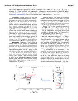
From www.bloodjournal.org by guest on February 6, 2015. For personal use only.
The Identification and Characterization of a Novel Human
Differentiation-Inhibiting Protein That Selectively Blocks Erythroid
Differentiation
By J.P. Durkin, J.M. Biquard, J.F. Whitfield, N. Morardet, J. Royer, P. Macdonald, R. Tremblay, J.D. Legal, R. Doyonnas,
J.P. Blanchet, and V. Krsmanovic
We have isolated a novel inhibitor of erythropoieticdifferentiation from the plasma of a patient suffering from idiopathic
pure red cell aplasia. This differentiation-inhibitingprotein
(DIP) specifically blocked the differentiation of human burstforming unit-erythroid (BFU-E), but not colony-forming uniterythroid (CFU-E) cells. DIP also blocked the maturation of
murine BFU-E cells, but not CFU-E or CFU-granulocytemacrophage cells, and it inhibited the dimethyl sulfoxide
(DMSO)-induceddifferentiation of Friend murine erythroleukemia cells (FLC) at levels between lO-’’and IO-’’ mol/L. DIP
activity was not detectable in the plasma of normal, healthy
subjects. Unlike other known inhibitors of hematopoiesis,
DIP appears to directly inhibit erythropoietic differentiation,
because it did not affect the proliferation of untreated FLC
and it effectively blocked FLC hemoglobinization without
affecting the ability of the blocked cells to proliferate. DIP
blocked FLC differentiation only when added to the culture
medium within 1 hour of inducing the cells with DMSO,
suggesting that the protein inhibited an early, but critical,
DMSO-induced cellular procesd. DIP appears to be at least
partially responsible for the patient’s anemia, and its unique
activity suggests a role in the early development of some
erythroleukemias.
o 1992 by The American Society of Hematology.
T
precursor cells suggested the possible existence of a human
homolog that could potentially play a role in normal
erythropoiesis as well as aplastic anemias and erythroleukemia. The strategy used to identify such a factor was based
on the proposition that its overproduction would specifically block erythrocytk development in otherwise normal
bone marrow, a condition found in the blood disorder
known as pure red cell aplasia (PRCA).16Therefore, blood
samples from patients suffering from PRCA of unknown
cause were screened for a novel erythroid differentiationinhibiting activity. We report here the isolation of the first
human erythroid differentiation-inhibiting protein (DIP)
from the blood of a 60-year-old woman suffering from
PRCA. The isolation, purification, and inhibitory activity of
this novel protein are described.
HE PROLIFERATION and differentiation of cells of
the different hemopoietic lineages is driven by a
variety of factors such as erythropoietin (EPO), granulocytemacrophage colony-stimulating factor (GM-CSF), interleukin-3 (IL-3), and macrophage colony-stimulating factor
(M-CSF).’ On the other hand, the proliferation and differentiation of a wide variety of hematopoietic cells can be
inhibited by agents that primarily block cell cycle progression, such as transforming growth factor-p (TGF-p): tumor
necrosis factor-a (TNF-CX),~.~
and several other less welldefined
A different type of highly selective hematopoietic inhibitor was found by Fasciotto et all4 in the conditioned
medium of tsAEV-LSCCHD3 chicken erythroleukemia
cells, which had been transformed by the erb-B gene of the
avian erythroblastosis virus (AEV). This 45-Kd protein,
subsequently named autocrine differentiation-inhibiting factor (ADIF), inhibited the EPO-induced differentiation of
the producer chicken cells and the dimethyl sulfoxide
(DMSO)-induced differentiation of Friend murine erythroleukemia cells (FLC).14,15
The unique characteristic of this
protein was its ability to block differentiation without
directly affecting cell cycle progression.” Moreover, chicken
ADIF selectively blocked the maturation of burst-forming
unit-erythroid (BFU-E) erythroid progenitor cells without
affecting the more advanced colony-forming unit-erythroid
(CFU-E) cells, or cells of the granulocyte-macrophage
lineage in human and murine bone marrow culture^.'^^^^
Thus, with respect to their sensitivity to ADIF, both the
tsAEV-LSCCHD3 chicken cells and the clone of FLC cells
used in the present study have at least a partial BFU-E
phenotype. Another ADIF-related protein has recently
been identified that inhibits both tlie macrophage differentiation of the murine myeloid leukemia cells that secrete it
and blocks the maturation of other murine (but not human)
myeloid cells without affecting cell proliferation.’ In principle, agents that block the differentiation of hemopoietic
cells without affecting the ability of the blocked cells to
proliferate can be seen as contributing to the first of a series
of steps leading towards leukemia.
The ability of the chicken erythroid ADIF to selectively
inhibit the differentiation of human BFU-E erythroid
Blood, Vol79, No 5 (March I),
1992: pp 1161-1171
MATERIALS AND METHODS
Leukemia Cell Lines
FLC, a clone of Friend C19X10 cells,14 were cultivated in a
complete medium consisting of 90% RPMI (Flow Laboratories,
McLean, VA), 10% decomplemented fetal bovine serum (FBS;
GIBCO BRL, Grand Island, NY), and penicillin (100 U/mL)/
streptomycin (0.1 mg/mL). The human promyelocytic cell line,
HL-60, was grown in the same medium except that 20% decomplemented FBS was used.
From the Cell Signals Group, Institute for Biological Sciences,
National Research Council of Canada, Ottawa, Ontario, Canada, the
Unite de Recherches en Imagerie Medicale Morphologique et Fonctionnelle, Institut Gustave Roussy (INSERM U66), villejuij France; and
the Centre de Genetique Moleculaire et Cellulaire, Universite de Lyon
1, Villeurbanne C e d e France.
Submitted July 22,1991;accepted October 24,1991.
Address reprint requests to J.P. Durkin, PhD, Cell Signals Group,
Institute for Biological Sciences, National Research Council of Canada, Ottawa, Ontario, Canada KIA OR6.
The publication costs of this article were defiayed in part by page
charge payment. This article must therefore be hereby marked
“advertisement” in accordance with 18 U.S.C.section 1734 solely to
indicate this fact.
0 1992 by The American Sociev of Hematology.
0006-497ild2/7905-OOO5$3.OO/O
1161
From www.bloodjournal.org by guest on February 6, 2015. For personal use only.
1162
FLC dijierenfiafionassay. FLC cells, taken from stock cultures
at 1.5 to 2.0 x lo6cells/mL, were plated at 2.0 x lo5cells/mL in 1.5
mL of complete medium in 24-well dishes (Falcon 3047; Becton
Dickinson, New Jersey). In some cultures, the cells were induced to
differentiate into hemoglobin-synthesizing erythroid cells by adding 2% DMSO to the culture medium. Samples (15 pL) to be
tested for differentiation-inhibiting activity were added to the
cultures 5 minutes before the DMSO. The proportion of hemoglobin-synthesizing cells was determined by the benzidine staining
technique” on day 4, or when the percentage of hemoglobinized
cells reached 70% to 85% in control (DMSO only) cultures.
Typically, the percentage of benzidine-positive (b+) cells in uninduced FLC cultures was less than 2%.
Human eiytkroid and myeloid cells. Human erythroid precursor
cells from bone marrow were enriched by Ficoll density centrifugation and washed twice with Iscove’s modified Dulbecco medium
(IMDM). The cells were allowed to adhere to the bottom of T-25
tissue culture flasks for 1.5 hours at 37°C in an atmosphere
consisting of 95% air and 5% CO,. Cells adhering to the flasks were
discarded and only the nonadherent cells were used. Where
required, DIP was added at the indicated concentrations to lo6
nonadherent bone marrow or peripheral blood cells that were
preincubated for 1.5 hours. For BFU-E and CFU-E progenitor cell
assays, lo6 DIP-treated or untreated cells were seeded in 35-mm
Petri dishes containing 1 mL of a semi-solid medium consisting of
85% IMDM, 15% FBS, 1% deionized bovine serum albumin, 2
U/mL human recombinant EPO, 0.075 mmol/L a-thioglycerol,
and incubated at 37°C in a humidiand 0.8% methylcellulose,’8~~a
fied atmosphere of 95% air and 5% CO,. The FBS provided
enough IL-3 for BFU-E and CFU-E growth and differentiation.
The numbers and appearance of CFU-E colonies were determined
on day 7, and BFU-E colonies on days 14 and 18. The cells were
examined unstained with an inverted microscope. All orange-red
aggregates having more than 50 cells were counted as a burst.
Colonies were removed and stained with the May-GriinwaldGiemsa stain for further examination.
Murine eiytkroid and myeloid cells. Freshly isolated mouse bone
marrow from 6- to 8-week-old C57 Black/6 mice was used as the
source of BFU-E, CFU-E, and CFU-granulocyte-macrophage
(CFU-GM) progenitors. Semi-solid cultures of CFU-E were established in IMDM supplemented with 30% FBS, 1% deionized
bovine serum albumin, 75 pmol/L P-mercaptoethanol, 0.2 U/mL
porcine EPO (Centre National de Transfusion Sanguine, Paris,
France), and 0.8% methylcellulose?0 Cells were plated at a final
concentration of 2 X 10⁴/mL in a volume of 100 pL in 16-mm
diameter wells and incubated for 2 days at 37°C in a watersaturated atmosphere consisting of 97.5% air and 2.5% CO,.
Colonies of more than 8 cells derived from CFU-E were enumerated after benzidine staining using an inverted microscope.
BFU-E and CFU-GM cells were grown in IMDM supplemented
with 20% FBS, 1% deionized bovine serum albumin, 75 pmol/L
P-mercaptoethanol, 45 pmol/L hemin, 0.5 U/mL porcine EPO
(specific activity, 1,030 U/mg), 50 U/mL purified mlL-3 (lo6U/mg;
Genzyme Corp, Boston, MA), and 0.8% methylcellulose. Cells
were plated at a final concentration of 2 x 10‘/mL in a volume of
500 pL in microtitration plates and incubated at 37°C. Colonies
derived from BFU-E precursor cells, and granulocyte-macrophage
colonies produced by CFU-GM cells were counted at 7 days
without staining?’
Fractionation of plasma. One hundred milliliters of PRCA or
normal plasma was dialyzed overnight against 1 mol/L acetic acid
and the resulting precipitate was removed by centrifugation at
10,OoOg for 5 minutes. The supernatant was dialyzed extensively
against 0.15 mol/L NaCI, 10 mmol/L HEPES, pH 9.0, and the
dialyzed protein extract (1.5 g) was applied to a Chelating
DURKIN ET AL
Sepharose 6B column (1.2 x 15 cm; Pharmacia LKB, Uppsala,
Sweden), charged with 100 mmol/L ZnSO,, and equilibrated with
150 mmol/L NaC1-10 mmol/L HEPES (pH 9.0) buffer solution, at
a flow rate of 50 mL/min. The unadsorbed pH 9.0 flow-through
fraction (100 mL) was collected, and the material adsorbed to the
column was eluted using a stepwise pH gradient from pH 8.0 to 4.0.
Each of the eluted fractions (100 mL) was dialyzed against
phosphate-buffered saline (PBS), and 20 pL samples of each
fraction were then tested for their abilities to inhibit the differentiation of human BFU-E colony development or FLC hemoglobinization. The pH 9.0 fraction, containing most of the inhibitory activity,
was dialyzed against 1 mol/L acetic acid and lyophilized. Samples
of this material (8 to 10 mg) were redissolved in 1 mL of 1 mol/L
acetic acid and applied to a P-60 gel filtration column (20 X 100
cm; Bio-Rad Laboratories, Richmount, CA) equilibrated in 1
mol/L acetic acid. Samples (50 pL) of each eluted fraction (1.5
mL) were pooled into groups, lyophilized, resuspended in an equal
volume of PBS, and tested for differentiation-inhibiting activity.
The most active fractions (fractions 39 to 41) were lyophilized,
resuspended in 1mL of PBS, and rechromatographed on a P-60 gel
filtration column (20 X 100 cm) equilibrated in PBS. The inhibitory activity of each fraction (1.5 mL) was measured by the FLC
differentiation assay. The fraction of peak activity (fraction 33) was
dialyzed against 1 mol/L acetic acid, lyophilized, resuspended in
100 pL solubilization buffer (20% glycerol, 6% sodium dodecyl
sulfate [SDS], 10% P-mercaptoethanol, 100 mmol/L Tris-HC1, pH
6.8) and heated to 80°C for 5 minutes. The sample was applied to
several wells of a reducing SDS-polyacrylamide (10%) gel and
electrophoresed under standard Laemmli conditions.2z Rainbow
protein molecular weight markers (Amersham Corporation, Arlington Heights, IL) served as visual landmarks on unstained gels.
After electrophoresis, the gel was halved and the protein bands on
one-half of the gel were visualized by silver stain (Bio-Rad). The
regions of interest were cut from the unstained portion of the gel,
divided into 1-mm slices, and each gel slice was eluted overnight at
10 mA (Bio-Rad Model 422 Electo-eluter) into 50 mmol/L
ammonium bicarbonate-0.1% SDS at 4°C. The electroeluted proteins were dialyzed for 4 hours against water, lyophilized, resuspended in RPMI medium, and then tested for differentiationinhibiting activity.
RESULTS
Purification of the Differentiation-ZnhibitingActivity
The anemia of the 60-year-old woman suffering from
idiopathic PRCA was not caused by an EPO deficiency; her
serum EPO levels were actually 8 to 9 times above normal,
as measured by standard bioassay. Despite the high concentration of EPO, the patient’s serum only feebly stimulated
fetal liver erythroblast proliferation, which suggested the
presence of a circulating inhibitor of erythropoiesis. As
shall be shown, there was no evidence that this inhibitor was
a circulating autoantibody (the most common cause of
PRCA23,24),
nor was the inhibition linked to a detectable
thymoma or other malignancy that often accompanies
PRCA.16 Finally, physical examination of the patient and
electron microscopic examination of the particulate and
soluble fractions of her plasma did not show an underlying
viral infection. Collectively, the clinical evidence pointed to
the presence of a novel circulating inhibitor of erythropoiesis that we proceeded to isolate.
Because of the chicken ADIF’s proven stability in acid
conditions, the patient’s plasma was initially treated with 1
From www.bloodjournal.org by guest on February 6, 2015. For personal use only.
1163
INHIBITORS OF ERYTHROID DIFFERENTIATION
mol/L acetic acid, a step that precipitated the majority
(-80%) of the plasma protein. The supernatant was
clarified by centrifugation, and small samples (20 FL) were
lyophilized to remove acetic acid and then tested for
biologic activity. The samples inhibited the DMSO-induced
hemoglobinization of FLC (Fig 1A) but, in this crude state,
they could not block the development of human BFU-E
erythroid cells in soft-agar assay (Fig 1B). By contrast,
501A7
200
.
'
-
40
-
150
30
-
F
100-
-
El
0
Z
20
+
0
U
n
10
50
-
0 -
0
B
100
1
80
C
r
AP
9.0 8.0
6.5
5.0
4.0
pH OF ELUANT
Fig 1. Metal chelate affinity chromatography of PRCA plasma.
was
Acidified PRCA plasma (a)(1.5 g protein) or normal plasma (0)
applied t o a Zn*+-chargedchelating Sepharose 68 column equilibrated
with 150 mmol/L NaCI-10 mmol/L HEPES, pH 9.0. The eluate (100 mL)
was collected and the proteins bound t o the column were eluted with
a step-wise pH gradient from pH 8.0 t o 4.0, as described in Materials
and Methods. Each fraction, along with the unfractionated acidified
plasma (AP), was dialyzed against PBS and samples (20 pL) were
tested for their ability (A) t o inhibit the hemoglobinization of DMSOinduced FLC. The percentage of b+cells was determined 5 days after
adding 2% DMSO t o the culture medium as described in Materials and
Methods. The percentage of b' cells in DMSO-treated and untreated
control cultures was 75.3 f 1.6 and 2.4 f 1.0, respectively. Each point
is the mean f SEM of four cultures. The fractions and AP were also
tested for their ability (B) t o inhibit the development of human
BFU-E-derived colonies in soft-agar assay. Human bone marrow cells
were cultured as described in Materials and Methods and the number
of BFU-E-derived bursts containing greater than 50 cells was determined on days 14 and 18. The number of colonies in untreated, control
cultures averaged 69 per well. The data are representative of three
separate determinations performed in duplicate.
identically processed plasma from normal subjects with the
same type-A blood as the PRCA patient did not affect FLC
differentiation. The PRCA and normal acidified plasmas
were dialyzed against a solution containing 150 mmol/L
NaCl and 10 mmol/L HEPES (pH 9.0) and lyophilized. The
resuspended material was then subjected to chelating
SepharosedB chromatography, on a column (1.2 x 15 cm)
charged with 100 m ZnSO, and equilibrated with the 150
mmol/L NaCl-10 mmol/L HEPES (pH 9.0) solution. The
flow-through fraction (100 mL) was collected, and the
protein that bound to the column was eluted by a pH
step-gradient (ranging from pH 9.0 to 4.0). The pH 9.0
flow-through fraction was the only one that inhibited
significantly the DMSO-induced differentiation of FLC
(Fig 1A). Unlike the unfractionated material, this pH 9.0
fraction inhibited by about 75% the development of bursts
derived from human BFU-E progenitors (Fig lB), and the
bursts that did develop were smaller and, on average, were
about 75% less hemoglobinized than bursts from control
cultures (data not shown). Acidified normal plasma treated
in the same way only marginally inhibited BFU-E and FLC
differentiation (Fig 1). As shown in Fig lB, other fractions
eluting from the chelating SepharosedB exhibited relatively low levels of BFU-E-inhibiting activity, which did not
inhibit FLC differentiation. For these reasons, only the pH
9.0 fractions were subjected to further purification.
Although human BFU-E erythroid cells were more
responsive than FLC to the differentiation-inhibiting activity of the PRCA factor, a 70% to 80% reduction of human
BFU-E differentiation translated consistently into a 35% to
40% reduction in FLC differentiation. Cells transformed by
the Friend virus (a complex of spleen focus forming virus
and helper murine leukemia virus) are thought to exhibit a
phenotype that is a mixture of late BFU-E and early
CFU-E.Z5,26
The specific FLC clone used in these studies
most certainly manifests the BFU-E property of being
inhibitable by chicken ADIF, a protein that blocks the
differentiation of chicken and murine BFU-E, but not
CFU-E.14,15
Because of its greater speed and convenience,
and its reliable reflection of a larger effectiveness on human
BFU-E cells, the FLC differentiation assay was mainly used
to measure the differentiation-inhibiting activity during the
subsequent purification steps.
The differentiation-inhibiting activity in the pH 9.0 fraction was dialyzed against 1 mol/L acetic acid, lyophilized,
resuspended in a minimum volume of 1 mol/L acetic acid
and applied to a Bio-Gel P-60 column (20 x 100 cm) that
had been equilibrated in 1 mol/L acetic acid. Samples of
each eluted fraction (1.5 mL) were pooled into a series of
groups, and each group tested for its ability to inhibit
DMSO-induced FLC differentiation. When tested at a
1:lo4dilution, most of the differentiation-inhibiting activity
eluted in two peaks with mean apparent molecular weights
of approximately 45 Kd and 66 Kd (Fig 2A). By comparison,
fractionated normal plasma contained virtually no differentiation-inhibiting activity when tested at the same dilution
(Fig 2A), or even at levels 100 times that shown. Although
not shown in Fig 2, low molecular weight material ( < 15
Kd) eluting in fractions 55 to 70 had no more inhibitory
From www.bloodjournal.org by guest on February 6, 2015. For personal use only.
1164
DURKIN ET AL
'
i
E
40
tA
0.6
45kDa
66kDa
I
W
LL
4
0
LL
0
0.4
30
W
V
Z
Z
2
DL
0
t- 20
m
I
-z
g
W
0
LL
2
0
VI
0.2
t
m
I
z
W
0
10
z
P
10
u
DL
a
-I
20
30
40
50
0.0
60
ap
FRACTION
Fig 2. P-60 (acid) gel filtration chromatography of the differentiation-inhibitingactivity. (A) The pH 9.0 fraction of the PRCA (EA) and control (0)
plasmas was dialyzed, lyophilized, and a portion (8 to 10 mg protein) applied to a P-60 gel filtration column equilibratedin 1 mol/L acetic acid as
described in Materials and Methods. The eluted fractions (1.5 mL) were pooled into groups and tested at a 1:1O,OOO dilution for their ability to
inhibitthe hemoglobinizationof DMSO-induced FLC as described in Materials and Methods and the legend to Fig 1. The percentage of b+cells in
DMSO-treated control cultures was 76.3 f 2.5. Each point is the mean f SEM of four cultures. (6)The 45-Kd regions (pooled fractions 37 to 42) of
both PRCA and control plasma were tested at a 1:lO.OOO dilution for their ability to inhibit the development of human BFU-E-derivedcolonies in
soft-agar assay as described in Materials and Methods and the legend to Fig 1. The number of colonies in untreated,control cultures averaged 58
per well. The data are representativeof five separate determinations performed in duplicate.
activity than the corresponding fractions of control serum.
Because the active 66-Kd fractions coeluted with a major
contaminating protein peak (Fig 2A), we focused on the
45-Kd peak, which clearly had the higher specific activity.
This 45-Kd activity strongly inhibited the differentiation of
human BFU-E progenitor cells in soft-agar assay by greater
than 80% (Fig 2B), and the few bursts that did develop
were both smaller and much less hemoglobinized. The
same fractions from the control plasma had no differentiation-inhibiting activity (Fig 2B). The separate fractions that
made up the 45-Kd peak were tested for differentiationinhibiting activity and also subjected to SDS-polyacrylamide gel electrophoresis (SDS-PAGE). The peak activity was
restricted to those fractions (fractions 39 to 41) in which a
protein with an apparent molecular weight of 65 Kd
appeared on the corresponding SDS-PAGE gels (Fig 3).
This protein, which was detectable in all three fractions,
was most intensely stained in that fraction (fraction 40)
which contained the maximum differentiation-inhibiting
activity. The appearance of the 65-Kd protein was unexpected, because proteins of this size would normally have
eluted much earlier from the P-60 column in fractions 26
to 30. It may well be that the 65-Kd band reflects the
aberrant mobility of the protein under the conditions of
either SDS-PAGE or gel filtration. Fractions 39 to 41 were
pooled, lyophilized, and subjected to further purification.
Attempts to purify the protein further by ion-exchange
chromatography resulted in unacceptable losses of differentiation-inhibiting activity. Therefore, the pooled active
fractions were rechromatographed on a Bio-Gel P-60 column (20 x 100 cm) that had been equilibrated with PBS
instead of 1 mol/L acetic acid. The rationale for this
-
-
approach was that the migration pattern of the protein
could vary if eluted from the column under neutral, instead
of acidic, conditions. Indeed, most of the FLC differentiation-inhibiting activity eluted from the P-60 (PBS) column
with an apparent molecular weight of 58 Kd (fractions 31 to
37), which coincided with the major protein peak (Fig 4A).
The material purified in this way inhibited the formation of
bursts from human BFU-E erythroid progenitor cells by
greater than 85% (data not shown). The fraction with peak
differentiation-inhibiting activity (fraction 33), when subjected to SDS-PAGE, was found to be greatly enriched in
the 65-Kd protein with a minor protein appearing at about
80 Kd (Fig 4B).
We had previously shown that the differentiationinhibiting activity of the PRCA plasma was stable to
SDS-PAGE conditions and that it retained its biologic
activity after electroelution from unstained gels. Thus, the
region on unstained gels containing both the 65-Kd and
80-Kd bands was cut into 1-mm slices, and the protein in
each slice was electroeluted from the gel into 50 mmol/L
NH,HCO,-0.1% SDS solution. The eluted samples were
dialyzed against water and tested for differentiationinhibiting activity. As shown in Fig 5, both the 65-Kd and
80-Kd bands had moderate differentiation-inhibiting activity when tested at a 1:104 dilution against DMSO-induced
FLC. At a 1:105 dilution the activity associated with the
80-Kd, but not the 65-Kd, band was lost (data not shown),
suggestingthat the majority of the differentiation-inhibiting
activity was associated with the 65-Kd protein. Several
other regions of the gel that were similarly excised and
electroeluted had no differentiation-inhibiting activity. The
protein content of the gel slice containing the greatest
From www.bloodjournal.org by guest on February 6, 2015. For personal use only.
INHIBITORS OF ERYTHROID DIFFERENTIATION
1165
Fig 3. A comparison of the differentiation-inhibking activity, and the SDS-PAGE profile, of the individual fractions of the 45-Kd peak area. The individual
fractions surrounding the region of maximum inhibitory activity (fractions 34 t o 44; Fig 2) were tested at
a 1:lO.OOO dilution for their ability t o inhibit the
hemoglobinization of DMSO-induced FLC as described in Materials and Methods and the legend t o
Fig 1. The percentage of benzidine-positive cells in
DMSO-treated control cultures was 85.5 2 4.2. Samples (50 pL) of each fraction were also analyzed by
SDS-10% PAGE and the proteins visualized on the
reducing gel by silver stain. The arrow indicates the
62-Kd protein whose appearance coincides with the
fraction of peak inhibitory activity. A weak band
corresponding t o a 80-Kd protein was also present.
amount of differentiation-inhibiting activity in the 65-Kd
region (ic, slice 10, Fig 5) migrated as a single band of the
same molecular weight when reanalyzed by SDS-PAGE
(Fig SB), indicating that the inhibitory activity was attributable to a single protein identified as a 65-Kd band on
SDS-PAGE gels.
DIP Is a Unique Differentiation-InhibitingProtein
DIP activity was lost when the protein was treated with
low levels of trypsin, but its activity was unaffected by
heating to 100°C for 5 minutes even in the presence of the
levels of SDS and p-mecaptoethanol found in electrophoretic sample buffer. The protein’s migration rate on
reducing gels was clearly differentfrom other differentiationinhibiting activities such as the homodimeric TGF-P (molecular weight 25 Kd) or TNF-a (molecular weight 17 Kd).
Unlike DIP, TGF-P cannot be extracted from rcducing gels
in an active state.” Moreover, DIP was unable to inhibit the
proliferation of human K562, human erythroleukemia
(HEL) and HL-60 cells, as does TNF-CX.~
DIP‘S unique
electrophoretic pattern and stability in both acid and
reducing SDS-PAGE conditions indicated that the protein
was not a differentiation-inhibiting autoimmune antibody
(the most common cause of PRCA). In addition, while the
main peak of differentiation-inhibiting activity eluted from
the P-60 column at 45 Kd, there was no inhibitory activity in
the k-rich void volume (Fig 2A), and the k G fractions of
the PRCA patient’s blood had no detectable differentiationinhibiting activity (data not shown). AS will be shown, DIP
is structurally and functionally distinct from inhibitory
activities that have been identified in lcukemia Cells and in
the conditioned medium of leukemia cell lines.
DIP Is a Potent Inhibitor of Etythroid Differentiation
P-60 (PBS)-purified DIP maximally suppressed the hemoglobinization of DMSO-induced FLC at concentrations of
From www.bloodjournal.org by guest on February 6, 2015. For personal use only.
DURKIN ET AL
1166
z
0
I-
.010
9
I-
z
W
LT
66KdA
4
I
W
LL
i
45KdA
D
IL
W
.008
(v
2
W
0
0
.004
m
I
kDo
C
Z
k
-
E
0
03
.006
0
f
-
-97
-66
Z
U
m
K
z
0
in
t-
.002 m
U
Z
w
0
LT
W
a
0 -45
0
20
40
60
80
FRACTION
Fig 4. P-60 (PES) gel filtration chromatography of the fractions of peak differentiation-inhibitingactivity (fractions 39 to 41). (A) Fractions39 to
41 (Fig 3) were pooled (4.5 mL), dialyzed against PES, lypholized, and applied to a P-60 gel filtration column equilibrated in PES as described in
Materials and Methods. The eluted fractions (1.5 mL) were pooled into groups and tested at a 1:10,000 dilution for their ability to inhibit the
hemoglobinization of DMSO-induced FLC as described in Materials and Methods and the legend to Fig 1. The percentage of b' cells in
DMSO-treated control cultures was 76.3 2 2.5. Each point is the mean f SEM of four cultures. (E) ReducingSDS-10% PAGE of a sample (50 ILL)of
the fraction of peak differentiation-inhibiting activity (fraction33) from the P-60 (PES) column showing a 62-Kd and 80-Kd protein.
500 pg/mL ( < lo-'' mol/L) (Fig 6). In addition to its effects
on thc maturation of human BFU-E cells, DIP effectively
blocked the diffcrentiation of BFU-E cells in murine bone
marrow cultures (Fig 7A). DIP inhibited the development
of murine BFU-E-dcrived bursts by 57% when added as a
single dose to mouse bone marrow cultures at 100 ng/mL
(Fig 7A). By comparison, the inhibitor maximally blocked
by more than 80% the development of human BFU-Ederived bursts at concentrations as low as 25 ng/mL (Fig
7B). An interesting finding was that higher concentrations
of P-60 (PBS)-purified DIP were consistently less inhibitory
for FLC (Fig 6), mouse BFU-E (Fig 7), and human BFU-E
differentiation. It is possible that a high-affinity DIP receptor mediatcs the protcin's inhibitory activity, while the
activation of a low-affinityreceptor overrides this inhibitory
effect. Such a mechanism would, in principal, allow the cell
to restrict the inhibitory cffects of DIP to a very narrow
concentration range.
human BFU-E burst formation, it did not affect human
CFU-E progenitor cell development in colony assays (Fig
7B). Further support for DIP being an erythroid-specific
factor was providcd by its inability to block the differentiation of HL-60 human promyelocytie leukemia cells induced
by either DMSO or 12-0-tetradecanoylphorbol-13-acetate
(TPA) (data not shown).
IL-3, GM-CSF, and EPO stimulate the proliferation of
early and/or late BFU-E progenitor cells.s33 The presence
of IL-3, GM-CSF, and EPO (all from Genzyme Corp)
added singularly or in combination at levels 100 to 1,000
times above saturation, did not affect the ability of DIP to
block the differentiation of human BFU-E cells or DMSOinduced FLC (data not shown). Thus, DIP does not
function by interfering with the binding of these hematopoietic growth factors to their respective receptors.
Specificity of DIP Inhibition
Most of the known hematopoietic inhibitors affect precursor cell maturation by primarily blocking cell-cycle processes needed for the initiation of DNA synthesis and
proliferation. Included in this list of growth-inhibiting
factors are TGF-P,' TNF-a,'.J superoxide dismutase;' and
inhibin." These inhibitory proteins all block the cell-cycle
progression of a variety of myeloid progenitor cells and
lcukcmia cell lines. A second, and very select, group of
hematopoietic inhibitors appears to block cell differentiation
directly, not as a secondary response to the inhibition of
Human DIP is not species-specific because it effectively
blocks the differentiation of mouse as well as human
BFU-E crythroid precursor cells. However, the inhibitor
appears to act selectively on BFU-E precursor cells. While
DIP blocked mouse BFU-E burst development, it did not
significantly affcct the development of colonies from the
morc advanced CFU-E erythroid precursors nor the development of granulocyte-macrophage (CFU-GM) colonies
(Fig 7A). Similarly, whilc DIP was a potent inhibitor of
DIP Directly Blocks Cell Differentiation and not Prolifeation
From www.bloodjournal.org by guest on February 6, 2015. For personal use only.
INHIBITORS OF ERYTHROID DIFFERENTIATION
1167
I
40tA
t
30
-
80kDa
kDa
-
65kDa
I
-97
-66
20
-45
10
0
2
4
6
8
10
12
FRACTION NUMBER
, The differentiation-inhibiting activity of the 62-Kd and 80-Kd protein bands electroeluted from a reducing SDS-10% PAGE gel. (A) The
fraction of peak activity (fraction 33) was dialyzed against 1 mol/L acetic acid, lyophilized, resuspended in 100 pL solubilization buffer (20%
glycerol, 6% SDS, 10% p-mercaptoethanol, 100 mmol/ L Tris-HCI, pH 6.8) and heated t o 80°C for 5 minutes. The sample was run on several lanes of
a reducing SDS-lO% PAGE gel, and the region containing the 80-Kd and 62-Kd bands was c u t from the unstained gel as described in Materials and
Methods. This section of gel was cut into 12 1-mm slices, and the protein present in each gel slice was electroeluted as described in Materials and
Methods. The eluted proteins were dialyzed against water, lyophilized, resuspended in RPMl medium, and tested at a 1:lO.OOO dilution for their
ability t o inhibit the hemoglobinizationof DMSO-induced FLC as described in Materials and Methods and the legend t o Fig 1. The percentage of b’
cells in DMSO-treated control cultures was 90.1 2 4.5. Each point is the mean 2 SEM of four cultures. (B) The eluted material from the gel slice
containing the 62-Kd protein (slice no. 4) was rerun on a reducing SDS-10% PAGE gel, and the protein visualized by silver stain.
proliferation.'."^" As shown in Fig 8, adding DIP to the
medium of FLC cultures at the time of DMSO induction
effectively blocked the hemoglobinization of the cells from
day 4 onwards. However, DIP did not inhibit the proliferation of uninduccd FLC, nor did it affect the proliferation of
the more slowly growing DMSO-treated cells (which ceased
proliferating on day 8 [data not shown]) during the period
that differentiation was affected (Fig 8). Similarly, DIP did
not affect the prolifcration of HL-60 cells, or tsAEV-LSCC
HD3 chicken erythroleukemia cells. To our knowledge,
DIP is the first example of a human hematopoietic inhibitor
protein that directly blocks cell differentiation rather than
the cell cycle.
DIP R I Q c Ear(v
~
Events in DMSO-Induced Differentiation
It took approximately 3 days for DMSO to induce
hemoglobin production in 50% of the cells of the FLC line
used in this study (Fig 9, insert). The continuous presence
of DMSO was ncedcd in the culture during the first 48 to 72
hours to fully commit the cells to hcmoglobinization (Fig 9,
insert). The question arose as to whcrc in this commitment
phase DIP acted in order to block differentiation. DIP
inhibition was maximally effective when added to the
culture shortly before (data not shown) or at the same time
as DMSO (Fig 9A). As indicated in Fig 9A, the “window”
of DIP action was extremely narrow; the inhibitor was
effective only when added to cultures within 1 hour after
adding DMSO. DIP did not block the differentiation of
FLC when added later than 1 hour after DMSO (Fig 9A).
These results were confirmed by washout experiments that
showed that DIP had to be in the culture medium only
during the first hour in order to inhibit the hemoglobinization of DMSO-induced FLC 5 days later (Fig 9B). Thus,
DIP appears to function by inhibiting an early and crucial
From www.bloodjournal.org by guest on February 6, 2015. For personal use only.
DURKIN ET AL
1168
step in the process by which DMSO stimulates FLC
differcntiation.
50
z
0
+
4I-
z
DISCUSSION
40
W
IY
L
W
k
0
LL
30
0
z
0
k
m
I
z
5
W
V
20
10
a
W
a
0
I
0.001
. .......I .
0.01
.
.......I
.......I
.
1
0.1
.......I
10
. .......I
100
LOG PROTEIN (ng/ml)
Fig 6. The effect of Increasing concentrations of DIP on DMSOinduced FLC differentiation. Various concentrations of P-60 (PES).
purified DIP (V),or identicallytreated normal plasma (L)were tested
for their ability to inhibit the hemoglobinization of DMSO-induced
FLC as described in Materials and Methods and the legend to Fig 1.
The percentage of b‘ cells in DMSO-treated control cultures was
82.9 4.1. Each point is the mean 2 SEM of four cultures.
+
BI
60
A 65-Kd protein isolated from the plasma of a PRCA
patient was found to bc a potent and specific inhibitor of
erythroid differentiation. It was able to block the maturation of human and mousc BFU-E crythroid cclls, but not
human or mouse CFU-E cclls or mouse CFU-GM cells.
Consistent with these findings, DIP inhibited the DMSOinduced differentiation of Friend erythroleukemia cclls
(arrested at the late BFU-E/early CFU-E stagc of dcvclopmerit"." and exhibiting at least a partial BFU-E phenotype“.”), but not the promyelocytic cell line, HL-60. Collcctively, these data indicate that DIP is a novcl inhibitor of
differentiation, which is likely to be responsiblc for the
patient’s PRCA. This cortclusion is strongly supportcd by
preliminary experiments that indicatc that injccting DIP
into normal mice selcctivcly induccs a profound crythroblastopenic condition in which the maturation of othcr hematopoietic lineages is not significantly affcctcd (manuscript in
preparation). To our knowlcdgc, this is thc first diffcrentiation-inhibiting activity to bc purificd from human plasma
with the potcntial to cause hcmatopoictic discasc. The
involvement of this protein in othcr ancmias remains to bc
determined.
c
B
6
I 100 -
A MOUSE
a
s
a
E
50
5
40
0
30
n
s
0
LL
0
Z
HUMAN
T
20
80
-
60
-
40
-
20
-
0
t
m
I
z
10
M
0
L
10
L
100
1000
LOG PROTEIN (ng/ml)
Fig 7. The effects of DIP on the development of IA) m(8)BFU-E, (0)
CN-E, and (0)CFU-OM, and (B) human BFU-E and CFU-E progenitor
cells. Various concentrations of P.80 (PBS)-purified DIP were tested for their ability to inhibit the development of mouse hematopoietic
progenitorsin colony assays as described in Materials and Methods. The number of EFU-E bunts and CFU-E and CFU-GMcolonies in untreated,
control mouse cultures averaged 24, 200, and 37 per well, respecthrely. DIP was tested on human EFU-E and CFU-E cells at 0.25 nglmL, the
optimal concentration for human EFU-E inhibition (data not shown). The data are representative of three separate determinations each of which
was performed in triplicate.
From www.bloodjournal.org by guest on February 6, 2015. For personal use only.
INHIBITORS OF ERYTHROID DIFFERENTIATION
40
1169
A
T
CI
VI
?
30
X
v
IL:
W
m
20
25
3
z
-I
-I
W
0
10
0
1
2
3
4
5
0
6
1
2
3
4
5
6
DAYS
DAYS
Fig 8. A comparison of the effects of DIP on the (A) proliferation and the (B) differentiation of FLC cells. FLC cells were plated at 70,000
cells/mL (in 24-well dishes) in RPMl medium containing 10% FBS. DIP (0.5 ng/mL) was added to the medium of some (V, V),but'not other (0.0)
FLC cukyres at t = 0. DMSO was then added to a final concentration of 2% to some (V,0).but not other (7,0 )dishes and the cultures were
days, small gamples were removed from each well and (A) stained with trypan blde and the number of viable
incubated at 37"E On the in@icated
cells determined, and (B) the proportion of hemoglobin (b+)-producingcells was determined as described in Materials and Methods. The
proliferation of DMSO-treated, but not untreated, cells ceased on days. The plues are the mean f SEM from triplicate cultures.
such as TNF-a and TGF-f3 are potent inhibitors of human
and mouse CFU-E cell differentiation but they rapidly
inhibit the proliferation of a wide variety of cell lines.24
$imilarly, the erythroid inhibitory activity (EIA) isolated
DIP is a novel differgntiation-inhibiting factor because of
its apparent specificity for BFU-E progenitorsand its ability
to inhibit the differenfiation of DMSO-induced FLC without being a cell cycle blocker. Hematopoietic inhibitors
I-
B
60
30
z
P
$
z
e
$
5O
z
w
I):
f
z
w
E
E
40
LL
8
z
P
L
30
0
z
o_
c-
y
20
E
0
t
m
I
20
E
z
-
be
w
10
0.
l o0
0
1
2
TIME OF DIP ADDITION (HRS)
3
Il
p
1
2
TIME OF DIP REMOVAL (HRS)
Fig 9. DIP blocks an early and crucial step in the process by which DMSO stimulates FLC differsntiation. (A) FLC cells were induced to
differentiate into hemoglobin-producing cells by the addition of 2% DMSO as described in Materials and Methods. DIP (0.5 ng/mL) was added tq
the culture medium at the indicatedtimes after OMSO aadition, and its effect on hemoglobinization was 9Pasured 4 days later. The percentage of
b+ cells in DMSO-treated control cultures was 72.3 k 3.0. Each point is the mean f SEM of four cultures. (Inrert) The reqirdmgnt for DMSd
throughout the c9urse of hemoglobin production in differentiating FLC. Cells were plated in complete medium containing $% DMSO and th,e tells
were (0)maintained in this medium, or the medium was replaced with fresh, DMSO-free medium (0)
24, (0)48, or 72 hburs (0)
later. The
percentage of b+cells was determined at the indicattd times. (B) FLC were plated in complete medium containing DIP (0.5 ng/mL) and 2% DMSO.
At the indicated times, excess DIP was removed by washing the cells with complete medium, and fresh, complete medium containing 2% DMSO
was added. The effect of these treatments an FLC hemoglobinization was determined 4 days later as described in Materials and Methods. The
percentage of b' cells in control cultures not exposed to DIP was 78.3 f 3.7.
From www.bloodjournal.org by guest on February 6, 2015. For personal use only.
DURKIN ET AL
1170
from murine macrophage cell lines is a potent inhibitor of
FLC pr~liferation.~~
DIP is functionally different from
inhibitory proteins such as leukemia inhibitory activity
(LIA) and leukemia-associated inhibitor (LAI), both of
which mainly affect granulopoie~is.'~~'~
LAI, originally isolated from the conditioned medium of promyelocytic HL-60
cell cultures,'* migrates as a 125-Kd protein band on
SDS-PAGE gels, cannot be eluted from gels in an active
state, and inhibits CFU-GM but not BFU-E differentiation.I3 LIA is an acidic isoferritin, a metal binding protein
complex of 24 subunits (-500 Kd) composed of a higher
proportion of heavy ( 21 Kd) to light ( 19 Kd) subunits,
which suppresses colony formation by human and mouse
CFU-GM and BFU-E progenitor cells in vitro.'",'' LIA
migrates as a pair of 20-Kd bands on reducing gels that are
not biologically active after elution from the gel.'" Finally,
DIP is different from the inhibitor isolated from C57
BL/BG mouse bone marrow cultures that reversibly inhibits the initiation of DNA replication in BFU-E precursor
The inhibitory activity of
cells and a variety of cell
this 16-Kd protein, recently identified as superoxide dismutase? is readily suppressed by excess IL-3. Clearly, the
physical and functional characteristics of these, and other,
hematopoietic inhibitors are clearly distinct from those of
DIP.
DIP blocks the hemoglobinization of FLC, which occurs
4 to 5 days after the cells are exposed to DMSO. The
protein appears to do this within the first hour after DMSO
addition by blocking a crucial, transient early step in the
long process leading to hemoglobinization. The inhibitory
effect of DIP persists even when the inhibitor is removed
from the medium after the first hour, suggesting that the
DIP-sensitive step(s) must occur in a coordinated and
timed manner for hemoglobinization to normally occur
several days later.
DIP is a third (and the first human-derived) member of a
group of proteins able to directly inhibit hematopoietic cell
differentiation. These proteins are distinguished by their
ability to inhibit the differentiation of leukemia cell lines in
culture without directly affecting cell proliferation. The first
member of this group to be identified was chicken ADIF,
which blocks the differentiation of both chicken and murine
erythroleukemia cells into hemoglobin-producing cells without inhibiting pr~liferation.'~.'~.~~
The other member of this
group was isolated from the conditioned medium of R-1
mouse myeloid leukemia cells and inhibits the differentiation and increases the oncogenicity of murine myeloid cell
-
-
cultures without directly blocking cell pr~liferation.~
Unlike
DIP, this protein is species-specific and does not affect
human cells.' While DIP is functionally similar to ADIF,
the two proteins are not the same because human DIP does
not affect the differentiation of the ADIF-producing chicken
tsAEV-LSCCHD3 cells.
If DIP acts primarily by inhibiting cell differentiation,
why does it not affect the proliferation of FLC but does
block the growth of bursts derived from BFU-E progenitor
cells in vitro? The answer probably lies in the bidirectional
coupling of differentiation and proliferation in normal
hematopoietic cells such as BFU-E. It is well understood
that, in most cases, the differentiated phenotype develops
only in those cells that are proliferating. What is less clearly
understood is the reverse scenario-the dependence of
proliferation/viability on normal cell differentiation. This
dependence, lost in erythroleukemia cell lines that proliferate persistently without differentiating, may operate to
safeguard normal hematopoiesis. By this mechanism, the
proliferation of BFU-E cells would be halted (perhaps by
apoptosis) if the cells were prevented from differentiating
into CFU-E within a prescribed number of cell divisions.
This would safeguard the organism from the disastrous
consequences of allowing uncontrolled proliferation of
undifferentiated cells to occur simply because of a block in
BFU-E maturation. If this is true, then the in vivo effects of
DIP would be expected to cause erythroblastopenia, the
condition found in the PRCA patient. Thus, a block in
erythroid differentiation is in itself unable to cause erythroleukemia. In principle, these cells could become erythroleukemic only if they acquire the ability to proliferate despite
the block in differentiation. This uncoupling of proliferation from differentiation is characteristic of erythroleukemia cell lines, such as FLC, that proliferate continuously as
immature precursor cells. It follows, therefore, that DIP
can inhibit FLC differentiation without affecting proliferation, but that proliferation of normal BFU-E is halted as a
secondary response to the inhibitor. Finally, it is of interest
that a significant proportion of patients suffering from
prolonged idiopathic PRCA go on to develop erythroleukemia.I6 If DIP-like proteins are responsible for a proportion
of idiopathic PRCA, then their inhibition of erythropoiesis
may be an early step in the process leading to leukemia.
Studies are in progress to determine the frequency at which
DIP occurs in patients suffering from PRCA and other
hematopoietic disorders, such as leukemias.
REFERE:NCES
1. Dexter TM, Garland JM, Testa NG (eds): Colony-Stimulating Factors. Molecular and Cellular Biology. New York, NY,
Dekker, 1989
2. Lawrence D A Transforming growth factor+: A multifunctional growth factor, in Krsmanovic V, Whitfield JF (eds): Malignant Cell Secretion. Boca Raton, FL, CRC, 1990, p 234
3. Munker R, Koeffler HP: In vitro action of tumor necrosis
factor on myeloid leukemia cells. Blood 69:1102,1987
4. Roodman GD, Bird A, Hutzler D, Montgomery W Tumor
necrosis factor-a and hematopoietic progenitors: Effects of tumor
necrosis factor on the growth of erythroid progenitors CFU-E and
BFU-E and the hematopoietic cell lines. Exp Hematol15:928,1987
5. Del Rizzo DF, Eskinazi D, Axelrad AA: Negative regulation
of DNA synthesis in early erythropoieticprogenitor cells (BFU-E)
by a protein purified from the medium of C57BL/6 mouse marrow
cells. Proc Natl Acad Sci USA 85:4320, 1988
6. Metcalf D, Hilton DJ, Nicola N A Clonal analysis of the
actions of the murine leukemia inhibitory factor on leukemic and
normal murine hemopoietic cells. Leukemia 2216,1988
7. Okabe-Kado J, Kasukabe T, Honma Y, Hayashi M, Hozumi
M: Purification of a factor inhibiting differentiation from condi-
From www.bloodjournal.org by guest on February 6, 2015. For personal use only.
INHIBITORS OF ERYTHROID DIFFERENTIATION
tioned medium of nondaerentiating mouse myeloid leukemia
cells. J Biol Chem 263:10994,10999,1988
8. Olsson I, Arnljots K, Gullberq U, Lantz M, Peetre C, Richter
J: Myeloid cell differentiation:The differentiation inducing factors
of myeloid leukemia cells. Leukemia 2165,1988
9. Smith AG, Heth JK, Donaldson DD, Wong GG, Moreau J,
Stahl M, Rogers D: Inhibition of pluripotential embryonic stem
cell differentiation by purified polypeptides. Nature 336688, 1988
10. Broxmeyer HE, Bognacki J, Dorner MH, Sousa M: Identification of leukemia associated inhibitory activity as acidic isoferritins. J Exp Med 153:1426, 1981
11. Broxmeyer HE, Lu L, Bicknell DC, Williams DE, Cooper S,
Levi S, Salfeld J, Arosio P: The influence of purified recombinant
human heavy-subunit ferritins on colony formation in vitro by
granulocyte-macrophage and erythroid progenitor cells. Blood
68:1257,1986
12. Olofsson T, Olsson I: Suppression of normal granulopoiesis
in vitro by a leukemia associated inhibitor (LAI) derived from a
human promyelocytic cell line (HL-60). Leuk Res 4:437,1980
13. Olofsson T Leukemia associated inhibitor: Biological characterization and purification of the active subunit, Najman A,
Guigon M, Gorin NC, Mary JY (eds): The Inhibitorsof Hematopoiesis. Paris, France, John Libbey Eurotext Ltd, 1987,p 177
14. Fasciotto B, Kanazir D, Durkin JP, Whitfield JF, Krsmanovic
V: AEV-transformed chicken erythroid cells secrete autocrine
factors which promote soft agar growth and block erythroleukemia
cell differentiation. Biochem Biophys Res Commun 143:775, 1987
15. Krsmanovic V, Morardet N, Biquard JM, Mouchiroud G,
Fasciotto B, Ristic A, Parmentier C, Blanchet JP, Kanazir D,
Durkin JP, Whitfield JF: Autocrine differentiation-inhibitingfactor (ADIF) from chicken erythroleukemia cells acts on human and
mouse early BFU-E erythroid precursors. Biochem Biophys Res
Commun 157:762,1988
16. Dessypris E N Pure Red Cell Aplasia. Baltimore, MD,
Johns Hopkins University, 1988
17. Orkin SH, Harosi FI, Leder P: Differentiation in erythrleukemia cells and their somatic hybrids. Proc Natl Acad Sci USA
72:98,1975
18. Dainiak N, Kreczko S, Cohen A, Pannel R, Lawler L
Primary human marrow cultures for erythroid bursts in a serumsubstituted system. Exp Hematol13:1073,1985
19. Kobari L, Dainiak N, Najman A, Kreczko S, Gorin NC,
Duhamel G, Frindel E Stimulatory effects of plasma from patients
with acute nonlymphoblasticleukemia on early erythroid progenitors and pluripotent stem cells. Exp Hematol15:838,1987
20. Iscove NN, Sieber F: Erythroid progenitors in mouse bone
marrow detected by macroscopic colony formation in culture. Exp
Hematol3:32, 1975
21. Blanchet JP, Arnaud S, Samarut J, Bouabdelli M: A factor
present in normal mouse serum stimulates later erythroid precursor proliferation. Exp Hematol 12595,1984
1171
22. Laemmli U K Cleavage of structural proteins during the
assembly of the head of bacteriophage T4. Nature 227680,1970
23. Koenig HM, Lightsey AL, Nelson DP, Diamond LK Immune suppression of erythropoiesisin transient erythroblastopenia
of children. Blood 53:742,1979
24. Dessypris EN, Krantz SB, Roloff JS, Lukens JN: Mode of
action of the IgG inhibitor of erythropoiesis in transient erythroblastopenia of children. Blood 59114,1982
25. Kabat D: Molecular biology of friend viral erythroleukemia.
Curr Top Microbiol Immunol148:1,1989
26. Ostertag W, Stocking C, Johnson GR, Kluge N, Kollek R,
Franz T, Hess N Transforming genes and target cells of murine
spleen focus-formingviruses. Adv Cancer Res 48:193,1987
27. Roberts AB, Anzano MA, Meyers CA, Wideman J, Blacher
R, Yu-Ching E, Stein S, Lehrman SR, Smith JM, Lamb LC, Sporn
MB: Purification and properties of a type transforming growth
factor from bovine kidney. Biochemistry225692,1983
28. Iscove NN, Sieber F, Winterhalter KH: Erythroid colony
formation in cultures of mouse and human bone marrow: Analysis
of the requirement for erythropoietin by gel filtration and affinity
chromatography on agarose con A. J Cell Physiol30983,1974
29. Tojo A, Fukamachi H, Saito T, Kasuga M, Urabe A, Takaku
F Induction of the receptor for erythropoietin in murine erythroleukemia cells after dimethyl sulfoxide treatment. Cancer Res
48:1818,1988
30. Sawyer ST, Krantz SB, Luna J: Identificationof the receptor
of erythropoietin by cross-linking to Friend virus-infected erythroid cells. Proc Natl Acad Sci USA 84:3690,1987
31. Donahue RE, Emerson SG, Wang EA, Wong GG, Clark SE,
Nathan DG: Demonstration of burst promoting activity of recombinant human GM-CSF on circulating erythroid progenitors using an
assay involving the delayed addition of erythropoietin. Blood
66:1479,1985
32. Ihle JN, Keller J, Greenberger JS, Henderson L, Yetter RA,
Morse H C Phenotypic characteristics of cell lines requering
interleukin 3 for growth. J Immunol129:1377,1982
33. Westbrook CA, Gasson JC, Gerber SE, Selsted ME, Golde
DW: Purification and characterization of human T-lymphocytederived erythroid-potentiatingactivity. J Biol Chem 259:9992,1982
34. Pluthero FG, Shreeve M, Eskinazi D, van der Gaag H,
Huang KS, Hulmes JD, Blum M, Axelrad A Purification of an
inhibitor of erythroid progenitor cell cycling and antagonist to
interleukin 3 from mouse marrow cell supernatants and its identification as cytosolic superoxide dismutase. J Cell Biol111:1217,1990
35. De Jong FH: Inhibin. Physiol Rev 68:555, 1988
36. Sassa S, Wolpe S, Cerami A Inhibition of erythroid differentiation of mouse erythroleukemia cells by a macrophage product(s). Blood Cells 13:161,1987
37. Krsmanovic V, Biquard J M Erythroleukemia cell secretion
and erythroid cell differentiation-inhibitingfactors, in Krsmanovic
V, Whitfield JF (eds): Malignant Cell Secretion. 1990, p 151
From www.bloodjournal.org by guest on February 6, 2015. For personal use only.
1992 79: 1161-1171
The identification and characterization of a novel human
differentiation-inhibiting protein that selectively blocks erythroid
differentiation
JP Durkin, JM Biquard, JF Whitfield, N Morardet, J Royer, P Macdonald, R Tremblay, JD Legal, R
Doyonnas and JP Blanchet
Updated information and services can be found at:
http://www.bloodjournal.org/content/79/5/1161.full.html
Articles on similar topics can be found in the following Blood collections
Information about reproducing this article in parts or in its entirety may be found online at:
http://www.bloodjournal.org/site/misc/rights.xhtml#repub_requests
Information about ordering reprints may be found online at:
http://www.bloodjournal.org/site/misc/rights.xhtml#reprints
Information about subscriptions and ASH membership may be found online at:
http://www.bloodjournal.org/site/subscriptions/index.xhtml
Blood (print ISSN 0006-4971, online ISSN 1528-0020), is published weekly by the American
Society of Hematology, 2021 L St, NW, Suite 900, Washington DC 20036.
Copyright 2011 by The American Society of Hematology; all rights reserved.


© Copyright 2026