
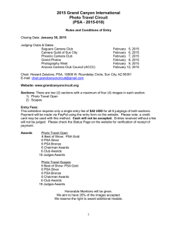
Download Full Text - Dash
An intestinal commensal symbiosis factor controls
neuroinflammation via TLR2-mediated CD39 signaling
The Harvard community has made this article openly available.
Please share how this access benefits you. Your story matters.
Citation
Wang, Y., K. M. Telesford, J. Ochoa-Repáraz, S. Haque-Begum,
M. Christy, E. J. Kasper, L. Wang, et al. 2014. “An intestinal
commensal symbiosis factor controls neuroinflammation via
TLR2-mediated CD39 signaling.” Nature communications 5 (1):
4432. doi:10.1038/ncomms5432.
http://dx.doi.org/10.1038/ncomms5432.
Published Version
doi:10.1038/ncomms5432
Accessed
February 6, 2015 10:58:22 AM EST
Citable Link
http://nrs.harvard.edu/urn-3:HUL.InstRepos:13890745
Terms of Use
This article was downloaded from Harvard University's DASH
repository, and is made available under the terms and conditions
applicable to Other Posted Material, as set forth at
http://nrs.harvard.edu/urn-3:HUL.InstRepos:dash.current.terms-ofuse#LAA
(Article begins on next page)
NIH Public Access
Author Manuscript
Nat Commun. Author manuscript; available in PMC 2015 January 21.
NIH-PA Author Manuscript
Published in final edited form as:
Nat Commun. ; 5: 4432. doi:10.1038/ncomms5432.
An intestinal commensal symbiosis factor controls
neuroinflammation via TLR2-mediated CD39 signaling
Yan Wang1, Kiel M. Telesford1, Javier Ochoa-Repáraz1, Sakhina Haque-Begum1, Marc
Christy1, Eli J. Kasper1, Li Wang2, Yan Wu3, Simon C. Robson3, Dennis L. Kasper4, and
Lloyd H. Kasper1,*
1Department
of Microbiology and Immunology, Geisel School of Medicine, Dartmouth College,
Lebanon, New Hampshire 03755, USA
2Department
of Microbiology and Molecular Genetics, Medical College of Wisconsin, Milwaukee,
Wisconsin 53226, USA
NIH-PA Author Manuscript
3Division
of Gastroenterology, Department of Medicine, Beth Israel Deaconess Medical Center,
Harvard Medical School, Boston, Massachusetts 02115, USA
4Department
of Microbiology and Immunobiology, Harvard Medical School, Boston,
Massachusetts 02115, USA
Abstract
NIH-PA Author Manuscript
The mammalian immune system constitutively senses vast quantities of commensal bacteria and
their products through pattern recognition receptors, yet excessive immune reactivity is prevented
under homeostasis. Intestinal microbiome can influence host susceptibility to extra-intestine
autoimmune disorders. Here we report that polysaccharide A (PSA), a symbiosis factor for human
intestinal commensal Bacteroides fragilis, protects against central nervous system demyelination
and inflammation during experimental autoimmune encephalomyelitis (EAE), an animal model
for multiple sclerosis, through toll-like receptor 2 (TLR2). TLR2 mediates tissue-specific
expansion of a critical regulatory CD39+ CD4 T cell subset by PSA. Ablation of CD39 signaling
abrogates PSA control of EAE manifestations and inflammatory cytokine responses. Further,
CD39 confers immune-regulatory phenotypes to total CD4 T cells and Foxp3+ CD4 Tregs.
Importantly, CD39-deficient CD4 T cells show an enhanced capability to drive EAE progression.
Our results demonstrate the therapeutic potential and underlying mechanism by which an
intestinal symbiont product modulates CNS-targeted demyelination.
*
Correspondence and requests for materials should be addressed to L.H.K. ([email protected]).
DISCLOSURES
The authors have no conflicting financial interests.
AUTHOR CONTRIBUTIONS
Y.W. and K.M.T. performed experiments and did the research. Y.W. designed research, prepared the figures and wrote the
manuscript. J.O-R. contributed research design and data. S.H-B., M.C. and E.J.K. contributed experiments. L.W. contributed reagents
and research support. S.C.R. edited the manuscript and contributed key mouse strain. Y.W. provided technical support to key mouse
strain. D.L.K. graciously provided PSA and critical comments on the research. L.H.K. supervised the research and reviewed the
manuscript.
Wang et al.
Page 2
INTRODUCTION
NIH-PA Author Manuscript
The skin and mucosal surfaces of humans are colonized by a wide variety of commensal
microorganisms that outnumber human somatic cells1. The GI tract harbors the greatest
quantity and diversity of microorganisms. Traditionally appreciated for its function to
facilitate metabolism and nutrient intake, intestinal microbiome has now been intensively
studied for its role in maintaining host immune homeostasis2–5. The composition of intestine
microbiota, or enterotype, influences host predisposition to autoimmune disorders affecting
tissue sites proximal and distal to the intestine6–11. EAE is an animal model of human
multiple sclerosis where self-reactive immune-pathology targets the central nervous system
(CNS). While colonization of germ-free mice with segmented filamentous bacteria (SFB)
increases susceptibility to EAE, oral administration with Bacteroides fragilis,
Bifidobacterium animalis and other probiotic mixtures have been shown to prevent EAE
development12–16. These studies associated different commensal strains with opposite
outcomes of CNS autoimmunity.
NIH-PA Author Manuscript
Commensal bacteria can be characterized as symbionts or pathobionts, depending on the
survival strategies employed. Symbionts live in a lifelong mutualistic pattern with the host
whereas pathobionts are often opportunistic pathogens5. To facilitate co-existence,
symbionts must limit aggressive immune attacks or induce immune tolerance by utilizing
some microbial products as symbiosis factors. In this context, polysaccharide A (PSA)
produced by the human intestinal symbiont Bacteroides fragilis has been identified as a
model molecule that elicits beneficial immune responses for both commensals and host. B.
fragilis lacking PSA have defective mucosal colonization due to inability to restrain Th17
responses17. Initially found to direct host adaptive immunity maturation, PSA then reveals
its capability of resolving autoimmunity18,19. Oral treatment with PSA prevents murine
experimental colitis by inducing IL-10+Foxp3+ Tregs20. Notably, PSA also suppresses CNS
inflammation during murine EAE, operational through IL-10 and CD103+ DCs21. The
precise signal by which PSA imparts on the adaptive immune cells, particularly the CD4+ T
cells, to prevent disease progression remains uncertain.
NIH-PA Author Manuscript
Microbial ligands are detected by host innate immunity as microbe-associated molecular
patterns (MAMPs). Sensing of microbial ligands by toll-like receptors (TLRs) can mobilize
innate immunity and subsequently activate adaptive immunity, leading to the host exclusion
of microbes22. PSA, however, represents a reverse example where TLR detection of
microbial ligand promotes the co-existence of symbionts with the host. TLR2 signals on
CD4+ T cells are crucial for PSA elicitation of immunologic tolerance toward B. fragilis
intestinal colonization17. Further, while many other microbial ligands sensed by TLRs act as
adjuvants to exacerbate autoimmunity, detection of PSA by TLR2 resolves intestinal
inflammation20,23,24. Our previous study has shown that oral treatment with B. fragilis PSA
prevents murine EAE both prophylactically and therapeutically21. In this study, we
demonstrate thatTLR2 signaling is essential for PSA-mediated modulation of CNS immunepathology. The expansion of CD39+CD4+ T cells downstream TLR2 is responsible for PSAmediated immune-regulation. CD39 defines regulatory phenotypes in CD4 T cells
irrespective of Foxp3 co-expression and is essential for the protective function of PSA
during CD4+ T cell-driven neuroinflammation.
Nat Commun. Author manuscript; available in PMC 2015 January 21.
Wang et al.
Page 3
RESULTS
NIH-PA Author Manuscript
Bacteroides fragilis PSA controls CNS inflammation via TLR2
To evaluate whether TLR2 is required for PSA regulation of CNS inflammatory disease,
WT or TLR2KO mice were given oral prophylactic treatment with PSA or PBS during EAE.
Consistent with previous results, treatment with PSA in WT mice delayed the clinical onset
and progression of EAE, as reflected by the clinic curve and cumulative score. The
protective effects were abrogated in TLR2KO mice. At basal level, PBS-treated WT and
TLR2KO mice developed comparable severity of EAE (Fig. 1A, B). Histological assays
confirmed the clinical results. H&E staining (Fig. 1C first and third columns) revealed that
PSA inhibited the robust lymphocyte infiltration into the CNS (spinal cord and brain) in WT
mice. This inhibitory function of PSA was abrogated in TLR2KO mice. Lymphocytic influx
leads to demyelination in EAE. As LFB staining (Fig. 1C second column) showed, whilst
PSA reduced demyelination lesions of the spinal cord in WT mice, this effect was not
observed in TLR2KO mice. Oral therapeutic treatment with PSA also protects against EAE
via TLR2 (Supplementary Fig. 1).
NIH-PA Author Manuscript
CNS inflammation is the hallmark of both human multiple sclerosis and murine EAE25,26.
To investigate whether PSA restricts CNS inflammation via TLR2, transcriptional levels of
genes in the brain were compared for PSA versus PBS-treated WT mice and PSA-treated
WT versus TLR2KO mice (Fig. 1D). PSA down-regulated multiple genes through TLR2 that
encode for (1) transcription factors Tbx21 (T-bet) and Stat4, that govern Th1 cell
development; (2) pro-inflammatory cytokines IL-17, IL-6 and IL-12p40, that mediate Th1/
Th17 as well as TNFα; (3) chemokines IP-10 (CXCL10), MCP-1 (CCL2), Rantes (CCL5),
MIP-1α (CCL3) and MIP-1β (CCL4), which are attractants for T cell, macrophages and
monocytes. In TLR2KO mice, PSA failed to restrict or might exacerbate the expression of
inflammation-related genes. The expression trend of chemokines in the CNS corroborated
the histological visualization by H&E staining. Taken together, these results demonstrated
that TLR2 is required for B. fragilis PSA control of CNS inflammation during EAE.
PSA triggers specific signal on CD4 T cells via TLR2
NIH-PA Author Manuscript
CD4+ T cells are the primary immune population that drives EAE pathogenesis25. Cervical
lymph nodes (CLNs) are important anatomical sites for immune cells draining to the CNS
during autoimmunity27. Flow cytometric profiles of CLN CD4+ T cells were compared for
PSA-or PBS-treated WT or TLR2KO mice by mean fluorescence intensity (MFI)(Fig. 2). A
pool of CD4+ T cell lineage and functional markers was included. CD39 was identified as a
significant PSA- and TLR2-specific signal, which is correlated by comparing positive subset
frequencies in Supplementary Fig. 2. Both CD39 and CD73 are classified as
ectonucleotidase, whose main function is to coordinate the conversion of extracellular
ATP/ADP into adenosine28,29. CD73 signal on CLN CD4+ T cells, however, did not appear
to be regulated through PSA/TLR2 axis (Supplementary Fig. 3).
PSA expands CD39+ CD4 T cells via TLR2 at specific tissues
To further characterize in vivo regulation of CD39 signal on CD4 T cells, we examined the
frequencies of CD39+ subset within CD4 T cells in EAE-induced WT or TLR2KO mice
Nat Commun. Author manuscript; available in PMC 2015 January 21.
Wang et al.
Page 4
NIH-PA Author Manuscript
NIH-PA Author Manuscript
orally treated with PBS or PSA. Consistent with MFI trends in Fig. 2, representative
histograms showed that PSA stimulated CD39 signal on CD4 T cells via TLR2 at both
CLNs (Fig. 3A) and mesenteric lymph nodes (MLNs) (Fig. 3B). To determine whether the
TLR2-dependent PSA expansion of CD39+ CD4 T cell subsets is local or systemic,
frequencies of CD39+ subsets among CD4 T cells were quantified for CLNs, MLNs, spleen,
whole blood (Fig. 3C) and immunization DLNs (Supplementary Fig. 4). Of these tissue
sites, only CLNs and MLNs displayed TLR2-dependent PSA up-regulation of CD39+ CD4
T cells. CD39+ frequencies within splenic CD4 T cells were not affected. CD39+
frequencies within blood and DLN CD4 T cells were down-regulated with PSA treatment.
PSA does not significantly alter CD4 T cell-derived CD39 signals via TLR2 in naïve mice
(Supplementary Fig. 5). Thus, the TLR2-dependent PSA expansion of CD39+ CD4 T cells
during EAE is localized. Amongst lymphocytes, CD39 is expressed on both CD4+ T cells
and B cells29. To investigate whether B cell-derived CD39 signal is concomitantly regulated
by PSA/TLR2 axis at the affected sites, CD39+ frequencies within CD19+ B cells were
quantified for CLNs and MLNs (Fig. 3D). Though CD19+ B cells showed higher basal level
CD39+ frequencies compared to CD4+ T cells, PSA treatment did not significantly alter
CD39+ frequencies within B cells. In vitro, PSA stimulated CD39 signals on CD4 T cells
through DCs (Supplementary Fig. 6A–C). In summary, these results show that TLR2dependent PSA-mediated enhancement of CD39 signal is both CD4 T cell-specific and
tissue site-specific.
Phenotyping CLN-sourced CD39+ versus CD39− CD4 T cells
NIH-PA Author Manuscript
The phenotype of CD39+ CD4 T cells at inflammatory sites is not well understood. As PSA
robustly stimulates CD39+CD4 T cells at specific tissue sites during EAE, our study offers
the opportunity to examine differences between inflammatory-state CD39+ andCD39− CD4
T cells. CLNs were harvested from PSA-treated EAE-affected mice. Total CD4+ T cells
were gated into CD39+ and CD39− subsets and phenotyped by flow cytometry. The
expression level of each individual marker was described by both histograms (Fig. 4A) and
MFI (Fig. 4B). We found that the CD39+ CD4 T cell subset, as compared to the CD39− CD4
T cell subset, expressed remarkably higher levels of IL-10 and significantly higher levels of
latency-associate peptide (LAP), denoting TGF-β production. As for surface immuneregulatory markers, the CD39+ CD4 T cell subset exhibited significantly higher levels of
programmed death-1 (PD-1), glucocorticoid-induced TNFR-related gene (GITR), cytotoxic
T-lymphocyte antigen 4(CTLA-4), inducible T-cell costimulator (ICOS) and relatively
higher expression of lymphocyte activation gene-3 (LAG-3) and V-domain Ig suppressor of
T cell activation (VISTA). PD-1, GITR, CTLA-4, ICOS and LAG-3 are all co-inhibitory
molecules with known ligands30–32; VISTA, is a co-inhibitory molecule with as yet no
recognized ligand(s)33. Conversely, 4-1BB (CDw137), a co-stimulatory molecule, was
noted at lower levels on the CD39+ CD4 T cell subset34.
The expression of CD25 and CD44 positively correlated with CD39 in CD4 T cells, whereas
CD62L and CD69 did not. Moreover, CD40L (CD154) expression was enriched within
CD39+ CD4 T cells. These in general indicate that CD39+ CD4 T cell is an activated subset
responsive to PSA exposure. CD39+ CD4 T cell subset expressed remarkably higher CD49d
and CD29 compared to CD39− CD4 T cell subset. CD49d (α4 integrin) and CD29 (β1
Nat Commun. Author manuscript; available in PMC 2015 January 21.
Wang et al.
Page 5
NIH-PA Author Manuscript
integrin) form very late antigen-4 (VLA-4), a vital CNS-homing marker35. Increased level
of CD73 was seen in CD39+ as opposed to CD39− CD4 T cell subset. CD39+ CD4 T cell
subset expressed relatively lower CD127 but remarkably higher CD103. CD127 (IL-7Rα) is
down-regulated on Treg cells36. CD103 (αE integrin) is mostly expressed by IELs but also
by a subset of Tregs37. Collectively, CD39+ CD4 T cell is phenotyped as a regulatory subset
with abundant expression of multiple immune-suppressive molecules. The regulatory
phenotype of CD39+ CD4 T cell subset was not confined to either tissue sites or treatment,
as CD39+ CD4 T cell derived from other tissues or treatment groups displayed similar
phenotypic pattern (Supplementary Fig. 7).
CD39 defines regulatory phenotypes regardless of Foxp3
NIH-PA Author Manuscript
Delineation of murine CD4 T cells by CD39 and Foxp3 revealed four populations:
CD39+Foxp3+, CD39−Foxp3+, CD39+Foxp3−, and CD39−Foxp3− (Supplementary Fig. 8A).
To determine whether CD39 could confer regulatory phenotypes on its own, we further
profiled CD39+ versus CD39− Foxp3+ CD4 Treg (Supplementary Fig. 8B) and Foxp3− CD4
Teff (Supplementary Fig. 8C) subsets sourced from CLNs of PSA-treated EAE-affected
mice. The same markers as in Fig. 4 were used and quantified by MFI values. CD39 defined
a similar expression profile of markers in Foxp3+ CD4 Tregs as in total CD4 T cells with
exception to VISTA, CD40L and CD127 (Supplementary Fig. 8B). Further, CD39 defined a
similar expression profile in Foxp3− CD4 Teffs excluding VISTA, CD73, CD127 and
CD40L (Supplementary Fig. 8C). Collectively these data demonstrate that CD39 expression
in response to PSA treatment is associated with multiple immune-suppressive features
independent of Foxp3 co-expression.
CD39 is essential for PSA-mediated immuno-regulation in EAE
NIH-PA Author Manuscript
To investigate the requirement for CD39 signaling in PSA-mediated suppression of CNS
inflammation, WT or CD39KO mice were orally gavaged with PSA before and after the
induction of sub-optimal active EAE. CD39 deficiency abrogated the immune-regulatory
function of PSA (Fig. 5A–B). WT and CD39KO mice developed similar basal levels of EAE
(Supplementary Fig. 9). The similar clinic trend was observed in optimal EAE
(Supplementary Fig. 10). The clinic results in Fig. 5A–B were confirmed by histological
assays. PSA-mediated suppression of lymphocyte influx and demyelinating lesions in the
CNS was prominent in WT mice but diminished in CD39KO mice (Fig. 5C). CD39 is
required for PSA control of both neural and systemic inflammation during EAE
(Supplementary Fig. 11). PSA-treated WT mice exhibited lower T cell activation levels than
did PSA-treated CD39KO mice. The trend was evident at CLN and also seen at MLN, but
not at spleen or whole blood (Fig. 5D), correlating with the PSA-mediated site-specific
expansion of CD39+ CD4 T cells.
To test whether CD39 differentially regulates pro- versus anti-inflammation cytokine
responses to PSA or neural auto-antigen MOG35–55, splenocytes were isolated from PSAtreated, EAE-affected WT and CD39KO mice and recalled with dose-escalating PSA,
MOG35–55 peptide or combined in vitro for 48 hrs. IL-10 and IL-17A production was
measured by ELISA. Although basal IL-10 release was comparable between WT and
CD39KO splenocytes, PSA dosage-dependently increased IL-10 release from WT but not
Nat Commun. Author manuscript; available in PMC 2015 January 21.
Wang et al.
Page 6
NIH-PA Author Manuscript
CD39KO splenocytes. MOG35–55 peptide suppressed IL-10 release from WT and CD39KO
splenocytes. Adding PSA to MOG35–55 peptide restored dosage-dependent IL-10 release
from WT splenocytes; IL-10 response of CD39KO splenocytes remained compromised (Fig.
5E). In contrast, PSA decreased IL-17A release from WT splenocytes, whereas CD39KO
splenocytes displayed elevated IL-17A response when contrasted to WT splenocytes restimulated with dose-escalating PSA, MOG35–55 peptide or combined in vitro (Fig. 5F). The
requirement of CD39 in IL-10 responsiveness is antigen (PSA)-specific, as splenocytes from
the same mice groups showed comparable in vitro IL-10 response to anti-CD3/CD28 beads
(Supplementary Fig. 12). IFNγ release, however, was only slightly enhanced in CD39KOas
opposed to WT splenocytes (Supplementary Fig. 13). CD39 is also crucial for PSA-induced
IL-10 release from CD4 T cells in vitro (Supplementary Fig. 6D). Thus, PSA-mediated
reciprocal regulation of anti- versus pro-inflammatory cytokines is CD39-dependent.
NIH-PA Author Manuscript
To determine whether CD4 T cell-restricted CD39 signaling is required for PSA’s
regulatory function during EAE, splenic CD4 T cells isolated from EAE-induced PSA- or
PBS-treated WT or CD39KO donor mice were introduced into RagKO recipients, which lack
mature B and T cells. Optimal active EAE was induced following reconstitution. As clinic
curve revealed (Fig. 5G), the recipient group given PSA-modified WT CD4 T cells showed
slower EAE progression. All groups culminated in comparable severity of EAE by day 21.
Cumulative score quantified clinical differences (Fig. 5H). RagKO recipients receiving naïve
WT or CD39KO CD4 T cells were given the same regimen (Supplementary Fig. 14). In both
settings, CD4 T cell-specific CD39 signaling is required for PSA-mediated control of EAE.
In conclusion, TLR2 signaling is essential for PSA-mediated protection against CNS
autoimmunity and localized expansion of CD39+ CD4 T cells defined as an indispensable
regulatory subset both phenotypically and functionally.
DISCUSSION
NIH-PA Author Manuscript
There exists a dynamic equilibrium in host-microbial mutualism. From the co-evolutionary
perspective, both hosts and commensals must evolve regulatory mechanisms to quench
deleterious mucosal and subsequent systemic inflammation. On the host side, several
molecules have been discovered as the negative regulators of TLR-mediated inflammation
such as the single immunoglobulin IL-1 receptor related molecule (SIGIRR), peroxisome
proliferators-activated receptor-γ (PPARγ), non-specific IgAs and inhibitory cytokines, for
instance IL-1038,39. On the commensal side, B. fragilis capsular polysaccharide PSA typifies
a molecular pattern that exploits TLR2 to deliver negative immune signals. The regulatory
capacity of PSA could be due to its zwitterionic structure, as previous studies indicated that
disruption of this structure inhibited PSA functions in vitro40.
MS is a CNS-targeting immune-mediated disease affected by both genetic and
environmental factors. Murine EAE studies suggest that modification of intestinal
commensals can alter disease outcomes41,42. The intestine is an important site for peripheral
education of immune cells. Exposure to different commensal products may differentiate the
CNS-antigen-reactive lymphocytes into an auto-aggressive or self-restrictive phenotype. Our
previous studies identified that B. fragilis PSA could alleviate CNS inflammation21. We
herein report that TLR2 is critical for PSA-mediated prevention of clinic progression, CNS
Nat Commun. Author manuscript; available in PMC 2015 January 21.
Wang et al.
Page 7
demyelination and inflammation during EAE. TLR2 induced the tissue-specific expansion
of a key regulatory CD39+ CD4 T cell subset.
NIH-PA Author Manuscript
Whether detection of PSA via APC-restricted TLR2 is critical for CD4+ T cell acquisition of
CD39 signal remains uncertain. It has been found that vesicle-associated PSA imposes
specific genetic changes on DCs through TLR224. Importantly, our previous studies
indicated that CD103+ DCs accumulated at CLNs during PSA treatment of EAE and
displayed elevated expression of TLR2 and MHC II21. Concomitant local expansion of both
DC subsets and CD39+ CD4 T cells suggests that more than one pathway besides TLR2 may
be involved in the process, which is currently under investigation.
NIH-PA Author Manuscript
In this study, TLR2 liaised the connection between the commensal symbiosis factor PSA
and CD39, a cell surface ectonucleotidase member of the ecto-nucleoside triphosphate
diphosphohydrolase (E-NTPDase) family. Extracellular ATP activates multiple immune
functions that lead to inflammation43. Intestinal luminal contents comprise high
concentrations of ATP produced by commensal bacteria. It has been reported that
commensal-derived ATP is capable of inducing IL-6 and IL-23 production by lamina
propria APCs and expands intestinal Th17 cells44. In this sense, B. fragilis PSA stimulation
of CD39 signaling may represent a symbiont-evolved strategy to limit the excessive
inflammatory response to commensal release of ATP. Indeed, we found that the selective
induction of CD39 signaling on CD4+ T cell is required for PSA immune-regulation at the
systemic level. PSA seems more optimal in controlling Th17 than Th1 response, which is
dependent on CD39 signaling. CD39 and CD73 coordinate hydrolysis of extracellular ATP
to adenosine. The generated extracellular adenosine further inhibits TCR-activated effector
T cells via cognate A2a receptors28. Consistently, a reciprocal decrease of activated CD4+ T
cells seems to accompany the increase of CD39+ CD4 T cells at CLNs during PSA treatment
of EAE.
NIH-PA Author Manuscript
CD39 is essential for the regulatory function of Foxp3+ CD4 Tregs29. It has been reported
that CD39+Foxp3+ CD4 Tregs but not CD39−Foxp3+ CD4 Tregs can effectively suppress
Th1745. However, several studies indicate that CD39+ CD4 T cell is in itself a regulatory
subset regardless of Foxp346,47. It is speculated that CD39+ CD4 T cell consists of both
Foxp3+ Tregs and Tr1 cells. Here we show that CD39+ CD4 T cell expressed remarkably
higher IL-10 than CD39− CD4 T cell. Moreover, CD39 is required for PSA-triggered IL-10
response. It is feasible that CD39 defines immuno-regulatory phenotype in both CD4 T cells
and Foxp3+ CD4 Tregs. Previous studies showed that whilst PSA induced a limited
quantitative increase of Foxp3+ CD4 Tregs, it robustly imprinted a functional suppressive
genetic profile on Foxp3+ CD4 Tregs20. Therefore, CD39 is possibly the key signal that
directs CD4 Tregs functional maturation in response to commensal antigen exposure.
Collectively, this study on B. fragilis PSA deepens our understanding of the therapeutic
potential and mechanism of commensal products in modulating CNS autoimmunity.
Nat Commun. Author manuscript; available in PMC 2015 January 21.
Wang et al.
Page 8
METHODS
NIH-PA Author Manuscript
Mice and treatment
Eight-week old femaleC57BL/6 mice were obtained from the Jackson Laboratories (Bar
Harbor, ME). TLR2KO mice were obtained from Dr. Brent L. Berwin (Dartmouth College).
IL-10 Thy1.1 reporter mice (10BiT) were obtained from Dr. Casey T. Weaver (University of
Alabama at Birmingham) and crossed with TLR2KO strain by Yan Wang. CD39KO mice
were obtained from Dr. Simon C. Robson (Harvard University). Rag-1, 2 DKO mice
(RagKO) were obtained from Dr. Mary Jo Turk (Dartmouth College). Mice were treated with
100 μg of purified B. fragilis PSA or PBS control by oral gavage every 3 days. Treatment
begins 6 days before EAE induction and terminates 9 days after disease induction. All
animal care and procedures were in accordance with protocols approved by the Institutional
Animal Care and Use Committee of Dartmouth College.
EAE induction
NIH-PA Author Manuscript
Active optimal or sub-optimal EAE was induced with s.c. injection of 200 or 160 μg
MOG35–55 peptide (Peptides International; Louisville, KY) respectively in 150 μl of
Complete Freund’s Adjuvant (Sigma-Aldrich, St. Louis, MO). On day 0 and 2 postimmunization, i.p. injection of 400 or 300 ng of Bordetella pertussis toxin (PT; List
Biological Laboratories, Campbell, CA) was respectively performed for optimal or
suboptimal EAE induction. For passive EAE in this study, RagKO mice received i.v.
injection of 3 × 10⁶ purified splenic CD4+ T cells from indicated donors 2 weeks before
active optimal EAE induction. EAE severity was scored daily using the standard scale.
Histological analysis
Transversal sections of spinal cords or brain stems were dissected from diseased animals and
fixated with 10% formalin overnight. Paraffin-embedded samples were stained with
hematoxylin and eosin (H&E) method for visualizing leukocyte infiltration or with luxol fast
blue (LFB) method for assessing demyelination. Pathology Translational Research Division
of Dartmouth-Hitchcock Medical Center (DHMC) performed all histology staining.
Quantitative real-time PCR
NIH-PA Author Manuscript
1 μg of mRNA was purified by QIAgen RNeasy kit (Qiagen, Germantown, MD) and reverse
transcribed by MultiScribe Reverse Transcriptase (Invitrogen, Carlsbad, CA). cDNA(200
ng) was amplified using SYBR Green PCR Master Mix (Applied Biosystems, Fostor City,
CA) on Bio-Rad CFX96 touch systems. Relative gene expression (duplicates) were
normalized to β-actin and calculated using the CT method, where relative expression =
2^(exp –actin) *1000.
Enzyme-linked immunosorbent assay
Specific ELISA kits (Biolegend, San Diego, CA) were used to quantify cytokine levels in
supernatants. Cells were cultured in 96-well or 48-well tissue plates at 2 × 10⁶ cells/ml
concentration in the presence of indicated soluble reagents or anti-CD3/CD28 Dynabeads
(Invitrogen, Carlsbad, CA) for 48 hrs and supernatants were collected.
Nat Commun. Author manuscript; available in PMC 2015 January 21.
Wang et al.
Page 9
Flow cytometric analysis
NIH-PA Author Manuscript
Singe cell preparations were isolated from indicated tissue sites and flow stained.
Fluorochrome-conjugated mAbs specific to mouse CD4 (clone GK1.5), CD19 (clone 6D5),
CD25 (clone PC61.5), CD29 (clone HMβ1-1), CD39 (clone 24DMS1), CD40L (clone
MR1), CD44 (clone IM7), CD49d (clone R1-2), CD62L (clone MEL-14), CD69 (clone
H1.2F3), CD73 (Clone eBioTY/11.8), CD103 (clone 2E7), CD127 (clone A7R34), 4-1BB
(clone 17B5), GITR (clone DTA-1), ICOS (clone 7E.17G9), LAG-3 (clone eBioC9B7W),
LAP (clone TW7-16B4), PD-1 (clone 29F.1A12) and Thy1.1 (clone OX-7) (1:400 dilution,
Biolegend or eBioscience) were used in various combinations for cell surface staining.
Biotin-labeled anti-VISTA mAb was provided by Dr. Li Wang (Medical College of
Wisconsin). The Fc receptor was blocked by CD16/32 antibody (Biolegend). For Foxp3
(1:400 dilution; clone FJK-16s, eBioscience) and CTLA-4 (1:400 dilution; clone UC10-4B9,
Biolegend), cells were treated by eBioscience intracellular fixation/permeabilization buffer
set and intracellular staining was performed. Stained cells were assayed by MACSQuant
Analyzers (Miltenyi, Bergisch Gladbach, Germany).
Cell purifications
NIH-PA Author Manuscript
Splenic CD4+ T cells were enriched from with mouse CD4+ T cell isolation kit (StemCell
Technologies, Vancouver, Canada) according to provided protocols.
Statistical analysis
Two-tailed Student’s t test was used to show statistical differences of mRNA relative
expression in qRT-PCR, fluorescence intensity or cell frequencies in FACS and cytokine
levels in ELISA. ANOVA followed by post-hoc t test was used where applicable. MannWhitney U-test was applied to cumulative clinic scores. Kruskal-Wallis test was applied
when multiple groups of clinic curves were compared. Data are shown as means ± SEM. P
values <0.05, <0.01 and <0.001 were indicated.
Supplementary Material
Refer to Web version on PubMed Central for supplementary material.
NIH-PA Author Manuscript
Acknowledgments
We thank Dr. Randolph J. Noelle (Dartmouth College; King’s College London, UK) for research guidance. We
thank DartLab (Dartmouth College) members Dr. Jacqueline Y. Smith, Dr. Daniel W. Mielcarz, John DeLong,
Gary A. Ward, Alan J. Bergeron, Christopher Sears, Robert Grady for technical support of flow cytometry and
critical guidance of research. We thank Kathryn A. Bennett (Dartmouth College) for helping animal care. We thank
Dr. William F. Hickey and Dr. Kenneth H. Ely (Dartmouth College) for guiding histological assays. We thank Dr.
Azizul Haque (Dartmouth College) for critical review.
References
1. Turnbaugh PJ, et al. The human microbiome project. Nature. 2007; 449:804–810. [PubMed:
17943116]
2. Abt MC, Artis D. The intestinal microbiota in health and disease: the influence of microbial
products on immune cell homeostasis. Curr Opin Gastroenterol. 2009; 25:496–502. [PubMed:
19770652]
Nat Commun. Author manuscript; available in PMC 2015 January 21.
Wang et al.
Page 10
NIH-PA Author Manuscript
NIH-PA Author Manuscript
NIH-PA Author Manuscript
3. Chow J, Lee SM, Shen Y, Khosravi A, Mazmanian SK. Host-bacterial symbiosis in health and
disease. Adv Immunol. 2010; 107:243–274. [PubMed: 21034976]
4. Elson CO, Cong Y. Understanding immune-microbial homeostasis in intestine. Immunol Res. 2002;
26:87–94. [PubMed: 12403348]
5. Round JL, Mazmanian SK. The gut microbiota shapes intestinal immune responses during health
and disease. Nat Rev Immunol. 2009; 9:313–323. [PubMed: 19343057]
6. Berer K, Krishnamoorthy G. Commensal gut flora and brain autoimmunity: a love or hate affair?
Acta Neuropathol. 2012; 123:639–651. [PubMed: 22322994]
7. Chervonsky AV. Intestinal commensals: influence on immune system and tolerance to pathogens.
Curr Opin Immunol. 2012; 24:255–260. [PubMed: 22445718]
8. Kranich J, Maslowski KM, Mackay CR. Commensal flora and the regulation of inflammatory and
autoimmune responses. Semin Immunol. 2011; 23:139–145. [PubMed: 21292499]
9. Ochoa-Reparaz J, Mielcarz DW, Begum-Haque S, Kasper LH. Gut, bugs, and brain: role of
commensal bacteria in the control of central nervous system disease. Ann Neurol. 2011; 69:240–
247. [PubMed: 21387369]
10. Wang Y, Kasper LH. The role of microbiome in central nervous system disorders. Brain Behav
Immun. 2013
11. Wu HJ, Wu E. The role of gut microbiota in immune homeostasis and autoimmunity. Gut
Microbes. 2012; 3:4–14. [PubMed: 22356853]
12. Lee YK, Menezes JS, Umesaki Y, Mazmanian SK. Proinflammatory T-cell responses to gut
microbiota promote experimental autoimmune encephalomyelitis. Proc Natl Acad Sci U S A.
2011; 108 (Suppl 1):4615–4622. [PubMed: 20660719]
13. Ochoa-Reparaz J, et al. Central nervous system demyelinating disease protection by the human
commensal Bacteroides fragilis depends on polysaccharide A expression. J Immunol. 2010;
185:4101–4108. [PubMed: 20817872]
14. Ezendam J, de Klerk A, Gremmer ER, van Loveren H. Effects of Bifidobacterium animalis
administered during lactation on allergic and autoimmune responses in rodents. Clin Exp
Immunol. 2008; 154:424–431. [PubMed: 19037925]
15. Kwon HK, et al. Amelioration of experimental autoimmune encephalomyelitis by probiotic
mixture is mediated by a shift in T helper cell immune response. Clin Immunol. 2013; 146:217–
227. [PubMed: 23416238]
16. Lavasani S, et al. A novel probiotic mixture exerts a therapeutic effect on experimental
autoimmune encephalomyelitis mediated by IL-10 producing regulatory T cells. PLoS One. 2010;
5:e9009. [PubMed: 20126401]
17. Round JL, et al. The Toll-like receptor 2 pathway establishes colonization by a commensal of the
human microbiota. Science. 2011; 332:974–977. [PubMed: 21512004]
18. Mazmanian SK, Liu CH, Tzianabos AO, Kasper DL. An immunomodulatory molecule of
symbiotic bacteria directs maturation of the host immune system. Cell. 2005; 122:107–118.
[PubMed: 16009137]
19. Mazmanian SK, Round JL, Kasper DL. A microbial symbiosis factor prevents intestinal
inflammatory disease. Nature. 2008; 453:620–625. [PubMed: 18509436]
20. Round JL, Mazmanian SK. Inducible Foxp3+ regulatory T-cell development by a commensal
bacterium of the intestinal microbiota. Proc Natl Acad Sci U S A. 2010; 107:12204–12209.
[PubMed: 20566854]
21. Ochoa-Reparaz J, et al. A polysaccharide from the human commensal Bacteroides fragilis protects
against CNS demyelinating disease. Mucosal Immunol. 2010; 3:487–495. [PubMed: 20531465]
22. Chervonsky A. Innate receptors and microbes in induction of autoimmunity. Curr Opin Immunol.
2009; 21:641–647. [PubMed: 19747810]
23. Marshak-Rothstein A. Toll-like receptors in systemic autoimmune disease. Nat Rev Immunol.
2006; 6:823–835. [PubMed: 17063184]
24. Shen Y, et al. Outer membrane vesicles of a human commensal mediate immune regulation and
disease protection. Cell Host Microbe. 2012; 12:509–520. [PubMed: 22999859]
Nat Commun. Author manuscript; available in PMC 2015 January 21.
Wang et al.
Page 11
NIH-PA Author Manuscript
NIH-PA Author Manuscript
NIH-PA Author Manuscript
25. McFarland HF, Martin R. Multiple sclerosis: a complicated picture of autoimmunity. Nat
Immunol. 2007; 8:913–919. [PubMed: 17712344]
26. Baxter AG. The origin and application of experimental autoimmune encephalomyelitis. Nat Rev
Immunol. 2007; 7:904–912. [PubMed: 17917672]
27. van Zwam M, et al. Surgical excision of CNS-draining lymph nodes reduces relapse severity in
chronic-relapsing experimental autoimmune encephalomyelitis. J Pathol. 2009; 217:543–551.
[PubMed: 19023878]
28. Sitkovsky M, et al. Adenosine A2A receptor antagonists: blockade of adenosinergic effects and T
regulatory cells. Br J Pharmacol. 2008; 153 (Suppl 1):S457–464. [PubMed: 18311159]
29. Deaglio S, et al. Adenosine generation catalyzed by CD39 and CD73 expressed on regulatory T
cells mediates immune suppression. J Exp Med. 2007; 204:1257–1265. [PubMed: 17502665]
30. Kinnear G, Jones ND, Wood KJ. Costimulation blockade: current perspectives and implications for
therapy. Transplantation. 2013; 95:527–535. [PubMed: 23129000]
31. Ermann J, Fathman CG. Costimulatory signals controlling regulatory T cells. Proc Natl Acad Sci U
S A. 2003; 100:15292–15293. [PubMed: 14676329]
32. Baixeras E, et al. Characterization of the lymphocyte activation gene 3-encoded protein. A new
ligand for human leukocyte antigen class II antigens. J Exp Med. 1992; 176:327–337. [PubMed:
1380059]
33. Wang L, et al. VISTA, a novel mouse Ig superfamily ligand that negatively regulates T cell
responses. J Exp Med. 2011; 208:577–592. [PubMed: 21383057]
34. Chacon JA, et al. Co-stimulation through 4-1BB/CD137 improves the expansion and function of
CD8(+) melanoma tumor-infiltrating lymphocytes for adoptive T-cell therapy. PLoS One. 2013;
8:e60031. [PubMed: 23560068]
35. Engelhardt B, Martin-Simonet MT, Rott LS, Butcher EC, Michie SA. Adhesion molecule
phenotype of T lymphocytes in inflamed CNS. J Neuroimmunol. 1998; 84:92–104. [PubMed:
9600713]
36. Liu W, et al. CD127 expression inversely correlates with FoxP3 and suppressive function of
human CD4+ T reg cells. J Exp Med. 2006; 203:1701–1711. [PubMed: 16818678]
37. Lehmann J, et al. Expression of the integrin alpha Ebeta 7 identifies unique subsets of CD25+ as
well as CD25− regulatory T cells. Proc Natl Acad Sci U S A. 2002; 99:13031–13036. [PubMed:
12242333]
38. Honda K, Takeda K. Regulatory mechanisms of immune responses to intestinal bacteria. Mucosal
Immunol. 2009; 2:187–196. [PubMed: 19262502]
39. Tanoue T, Umesaki Y, Honda K. Immune responses to gut microbiota-commensals and pathogens.
Gut Microbes. 2010; 1:224–233. [PubMed: 21327029]
40. Wang Q, et al. A bacterial carbohydrate links innate and adaptive responses through Toll-like
receptor 2. J Exp Med. 2006; 203:2853–2863. [PubMed: 17178920]
41. Ochoa-Reparaz J, et al. Role of gut commensal microflora in the development of experimental
autoimmune encephalomyelitis. J Immunol. 2009; 183:6041–6050. [PubMed: 19841183]
42. Berer K, et al. Commensal microbiota and myelin autoantigen cooperate to trigger autoimmune
demyelination. Nature. 2011; 479:538–541. [PubMed: 22031325]
43. Robson SC, Sevigny J, Zimmermann H. The E-NTPDase family of ectonucleotidases: Structure
function relationships and pathophysiological significance. Purinergic Signal. 2006; 2:409–430.
[PubMed: 18404480]
44. Atarashi K, et al. ATP drives lamina propria T(H)17 cell differentiation. Nature. 2008; 455:808–
812. [PubMed: 18716618]
45. Fletcher JM, et al. CD39+Foxp3+ regulatory T Cells suppress pathogenic Th17 cells and are
impaired in multiple sclerosis. J Immunol. 2009; 183:7602–7610. [PubMed: 19917691]
46. Kochetkova I, Golden S, Holderness K, Callis G, Pascual DW. IL-35 stimulation of CD39+
regulatory T cells confers protection against collagen II-induced arthritis via the production of
IL-10. J Immunol. 2010; 184:7144–7153. [PubMed: 20483737]
47. Kochetkova I, Thornburg T, Callis G, Pascual DW. Segregated regulatory CD39+CD4+ T cell
function: TGF-beta-producing Foxp3− and IL-10-producing Foxp3+ cells are interdependent for
Nat Commun. Author manuscript; available in PMC 2015 January 21.
Wang et al.
Page 12
protection against collagen-induced arthritis. J Immunol. 2011; 187:4654–4666. [PubMed:
21967895]
NIH-PA Author Manuscript
NIH-PA Author Manuscript
NIH-PA Author Manuscript
Nat Commun. Author manuscript; available in PMC 2015 January 21.
Wang et al.
Page 13
NIH-PA Author Manuscript
NIH-PA Author Manuscript
Figure 1.
NIH-PA Author Manuscript
Oral treatment with Bacteroides fragilis PSA protects against murine EAE in a TLR2dependent manner. Wild-type and TLR2KO mice on C57BL/6 background were
prophylactically treated with 100 μg of purified B. fragilis PSA or PBS as control by oral
gavage every 3 days. Treatment was initiated 6 days before optimal active EAE induction
(s.c. injection with MOG35–55 peptide plus two doses of i.p. injection with pertussis toxin)
and terminated 9 days after disease induction. (A) EAE clinic scores were monitored till day
25. Depicted are the combined results of two independent experiments (n = 8, per group).
Mean ± SEM scores are shown. *, P< 0.05; **, P< 0.01; ***, P< 0.001 (Kruskal-Wallis test
of all groups). (B) Cumulative scores were derived from (A) as the sum of clinic scores
divided by the number of mice per group. Mean ± SEM scores are shown. P value was
calculated by Mann-Whitney U-test. (C) Histological analyses show transversal sections of
spinal cords stained with haematoxylin and eosin (H&E) and Luxol fast blue (LFB), and
sections of brain stems stained with H&E. H&E stain targeted nucleated cells (red arrows
point at the clusters of infiltrating lymphocytes); LFB stain targeted myelin (red arrows
point at the demyelination spots). Depicted are representative pictures (magnification 40x)
from the indicated EAE-induced groups: PBS-treated WT mice, PSA-treated WT mice, and
PSA-treated TLR2KO mice. All samples were collected (n = 4 sections per mouse, n = 3
mice per group) at the peak of disease. Images in each row are the same treatment group,
Nat Commun. Author manuscript; available in PMC 2015 January 21.
Wang et al.
Page 14
NIH-PA Author Manuscript
and in each column are the same staining method. Scale bars:100 μm. (D) Brains of PBS or
PSA-treated EAE-induced WT mice or PSA-treated EAE-induced TLR2KO mice were
harvested at the onset stage of the disease. Relative expression of transcription factors
(Tbx21, Stat4), cytokines (IFNγ, IL-17, IL-6, TNFα, IL-12p40) and chemokines (IP-10,
MCP-1, Rantes, MIP-1α, MIP-1β) were measured by qRT-PCR. Mean ± SEM mRNA
relative expression from two independent experiments is shown. P value was calculated by
two-tailed Student’s t test (n = 5–12 for each group).
NIH-PA Author Manuscript
NIH-PA Author Manuscript
Nat Commun. Author manuscript; available in PMC 2015 January 21.
Wang et al.
Page 15
NIH-PA Author Manuscript
NIH-PA Author Manuscript
Figure 2.
NIH-PA Author Manuscript
PSA triggers specific signal on CD4 T cells via TLR2 under inflammatory conditions. Wildtype and TLR2KO mice on IL-10 Thy1.1 reporter strain (10BiT) background were orally
gavaged with PSA or PBS and induced with optimal active EAE as in Fig 1A setting. At the
peak of disease, total leukocytes were isolated from cervical lymph nodes. Flow cytometry
analysis was performed to identify the specific CD4 T cell marker(s) triggered by PSA in a
TLR2 dependent manner. CD4 T cells were pre-gated and mean fluorescence intensity
(MFI) was used to measure the expression level of the indicated markers. Mean ± SEM MFI
values were compared between PSA- or PBS-treated WT or TLR2KO mice. Depicted are
representative results from two independent experiments (n = 4 for each group). P value was
calculated by one-way ANOVA test.
Nat Commun. Author manuscript; available in PMC 2015 January 21.
Wang et al.
Page 16
NIH-PA Author Manuscript
NIH-PA Author Manuscript
Figure 3.
NIH-PA Author Manuscript
TLR2-dependentPSA expansion of CD39+ CD4 T cells is tissue site-specific. Wild-type and
TLR2KO mice were treated with the same regimen as described in Fig 1A setting. At the
peak of disease, total leukocytes were isolated from cervical lymph nodes (CLNs),
mesenteric lymph nodes (MLNs), spleen and whole blood and flow cytometry analysis was
performed. Oral PSA administration stimulates CD39 expression on CD4 T cells via TLR2
at both (A) CLNs and (B) MLNs. CD4 T cells were pre-gated and CD39 expression level
was measured in (A) and (B). Depicted are the typical histograms for each indicated
treatment group from two independent experiments (n = 3–10, per group). (C) TLR2dependent PSA induction of CD39 signal on CD4 T cells is restricted to CLNs and MLNs.
Frequencies of CD39+ subsets among CD4 T cells were quantified for the indicated tissue
sites. Depicted are combined representative results of two independent experiments (n = 6–
10, per group). P value was calculated by two-tailed Student’s t test. (D) PSA does not
influence CD39 signal on CD19 B cells. Frequencies of CD39+ subsets among CD19 B cells
were quantified for both CLNs and MLNs. Combined representative results of two
experiments are shown (n = 6–10, per group).
Nat Commun. Author manuscript; available in PMC 2015 January 21.
Wang et al.
Page 17
NIH-PA Author Manuscript
Figure 4.
NIH-PA Author Manuscript
Phenotyping cervical lymph node-sourced CD39+ versus CD39− CD4 T cells. CLNs were
harvested from PSA-gavaged EAE-induced mice (day 18 post EAE induction). Total
leukocytes were isolated and analyzed by flow cytometry. (A) CD39+ CD4 T cells display
elevated regulatory signatures as compared to CD39−CD4 T cells. CD39+ and CD39− CD4
T cell populations were analyzed for the expression of each indicated marker. Depicted are
the representative histograms for individual analysis from two independent experiments (n =
4–5, per group). Red line, CD39+CD4 T cells; blue line, CD39−CD4 T cells; grey shade,
isotype. (B) For each analysis in (A), Mean ± SEM MFI values were quantified. Black,
CD39+CD4 T cells; white, CD39−CD4 T cells; grey, corresponding isotype for each
indicated marker. P value was calculated by two-tailed Student’s t test (n = 4–5 for each
group).
NIH-PA Author Manuscript
Nat Commun. Author manuscript; available in PMC 2015 January 21.
Wang et al.
Page 18
NIH-PA Author Manuscript
NIH-PA Author Manuscript
Figure 5.
NIH-PA Author Manuscript
CD39 signaling is essential for PSA-mediated immuno-regulation during murine EAE.
Wild-type and CD39KO C57BL/6 mice were treated with PSA as in Fig 1A and induced
sub-optimal active EAE. (A) EAE was monitored till day 25. Mean ± SEM scores typical of
two independent experiments are shown (n = 8, per group). *, P< 0.05; **, P< 0.01; ***, P<
0.001. (B) Mean ± SEM cumulative scores were calculated from (A). (C) H&E and LFB
staining was performed same as in Fig 1C. Depicted are typical pictures (magnification 40x)
from EAE-induced PSA-treated WT or CD39KOmice. All samples were collected (n = 4
sections per mouse, n = 3 mice per group) at the peak of disease. Scale bars: 100 μm. (D)
Tissues were collected at the onset of EAE and flow cytometric analysis of cells was
performed. Mean ± SEM frequencies of activated subsets within total CD4 T cells were
compared for the indicated sites (n = 4). (E, F) Total splenocytes were obtained from wildtype or CD39KO mice treated as in (A) on day 14 of EAE induction and cultured in vitro
with PSA, MOG35–55 peptide, or a combination of both at escalating doses (10, 100, or 200
μg/ml of each). Supernatants were collected 48 hr after culture. IL-10 (E) and IL-17A (F)
were measured by ELISA. Mean ± SEM results are combined from two experiments (n = 3–
4). (G) RagKO recipient mice were reconstituted with splenic CD4 T cells from day 14
Nat Commun. Author manuscript; available in PMC 2015 January 21.
Wang et al.
Page 19
NIH-PA Author Manuscript
EAE-induced wild-type and CD39KO mice treated with PSA as in (A) or PBS. Two weeks
later, optimal active EAE was introduced into recipients. EAE was monitored till day 21.
Mean ± SEM scores representative of two experiments are shown (n = 6–8, per group). *,
P< 0.05; **, P< 0.01. (H) Mean ± SEM cumulative scores were derived from (G). P value
was calculated by Mann-Whitney U-test in (A), (B) and (H), by two-tailed Student’s t test in
(D), (E) and (F) and by Kruskal-Wallis test in (G).
NIH-PA Author Manuscript
NIH-PA Author Manuscript
Nat Commun. Author manuscript; available in PMC 2015 January 21.

© Copyright 2026