
Receptor Knockout Mice - International Journal of
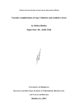
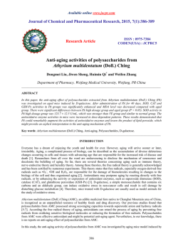
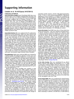
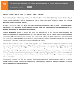
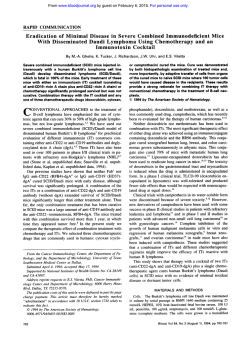
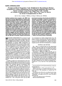
International Journal of Neuropsychopharmacology Advance Access published January 31, 2015
International Journal of Neuropsychopharmacology, 2015, 1–10
doi:10.1093/ijnp/pyu075
Research Article
research article
Melancholic-Like Behaviors and Circadian
Neurobiological Abnormalities in Melatonin MT1
Receptor Knockout Mice
Stefano Comai, PhD; Rafael Ochoa-Sanchez, PhD; Sergio Dominguez-Lopez, PhD;
Francis Rodriguez Bambico, PhD; Gabriella Gobbi, MD, PhD
Neurobiological Psychiatry Unit, Department of Psychiatry, McGill University and McGill University Health
Center, Montréal, QC, Canada (Drs Comai, Ochoa-Sanchez, Dominguez-Lopez, Bambico, and Gobbi).
Correspondence: Gabriella Gobbi, MD, PhD, Neurobiological Psychiatry Unit, Room 220, Irving Ludmer Psychiatry Research and Training Building, 1033
Pine Avenue West, McGill University, Montreal, PQ, Canada H3A 1A1 ([email protected]).
Abstract
Background: Melancholic depression, described also as endogenous depression, is a mood disorder with distinctive specific
psychopathological features and biological homogeneity, including anhedonia, circadian variation of mood, psychomotor
activation, weight loss, diurnal cortisol changes, and sleep disturbances. Although several hypotheses have been proposed,
the etiology of this disorder is still unknown.
Methods: Behavioral, electrophysiological and biochemical approaches were used to characterize the emotional phenotype,
serotonergic and noradrenergic electrical activity, and corticosterone in melatonin MT1 receptor knockout mice and their
wild type counterparts, during both light and dark phases.
Results: Melatonin MT1 receptor knockout mice have decreased mobility in the forced swim and tail suspension tests as well
as decreased sucrose consumption, mostly during the dark/inactive phase. These mood variations are reversed by chronic
treatment with the tricyclic antidepressant desipramine. In addition, MT1 receptor knockout mice exhibit psychomotor
disturbances, higher serum levels of corticosterone the dark phase, and a blunted circadian variation of corticosterone levels.
In vivo electrophysiological recordings show a decreased burst-firing activity of locus coeruleus norepinephrine neurons
during the dark phase. The circadian physiological variation in the spontaneous firing activity of high-firing neuronal
subpopulations of both norepinephrine neurons and dorsal raphe serotonin neurons are abolished in MT1 knockout mice.
Conclusions: These data demonstrate that melatonin MT1 receptor knockout mice recapitulate several behavioral and
neurobiological circadian changes of human melancholic depression and, for the first time, suggest that the MT1 receptor may
be implicated in the pathogenesis of melancholic depression and is a potential pharmacological target for this mental condition.
Keywords: corticosterone, daily mood variations, melancholic depression, monoamines, MT1 receptors, norepinephrine,
serotonin, circadian rhythm
Introduction
Melancholia, also described as endogenous, endogenomorphic,
autonomous, type A, psychotic, and typical depression (Parker
et al., 2010), is a mood disorder with specific psychopathological
symptoms, including disturbances in affect, diurnal variation with mood generally worse in the morning, anhedonia,
psychomotor retardation or agitation, cognitive impairment,
Received: May 24, 2014; Revised: July 15, 2014; Accepted: July 30, 2014
© The Author 2015. Published by Oxford University Press on behalf of CINP.
This is an Open Access article distributed under the terms of the Creative Commons Attribution License
(http://creativecommons.org/licenses/by/4.0/), which permits unrestricted reuse, distribution, and
reproduction in any medium, provided the original work is properly cited.
1
2 | International Journal of Neuropsychopharmacology, 2015
and neurobiological/somatic impairments manifested as loss
of weight, hypercortisolemia, and sleep disturbances, mainly
at the level of rapid eye movement sleep (REMS; Rush and
Weissenburger, 1994; Armitage, 2007). Melancholic patients
respond better to broad-action tricyclic antidepressants than to
narrow-action antidepressants (Perry, 1996), and their response
to placebo, psychotherapy, or even social intervention is very
poor (Brown, 2007).
Few animal models of melancholic depression have been
proposed (Hill and Gorzalka, 2005; Kato et al., 2008; Shen et al.,
2010), but their attempts to reproduce the whole complex symptomatology of the disease, in particular the diurnal variations of
mood, failed.
Diurnal mood variations are indeed the hallmark of melancholic depression (Parker et al., 2010), and differentiate
melancholic versus non-melancholic depression (Duncan,
1996). They have been linked to a dysfunction in circadian
rhythms that are driven by the suprachiasmatic nucleus
(SCN). The activity of the SCN is regulated by the neurohormone melatonin (MLT), which acts through its two G protein–
coupled receptors, MT1 and MT2 (Liu et al., 1997; Jin et al., 2003;
Dubocovich, 2007). Previous literature has suggested a link
between MLT and melancholic depression. Brown et al. (1985)
reported lower nocturnal MLT levels in melancholic patients
than in controls, and Fountoulakis et al. (2001) found that melancholic patients had lower 23.00 h MLT blood levels in comparison to atypical, somatic syndrome, or “undifferentiated”
depressed patients. Melatonin MT1 receptor knockout (MT1-/-)
mice showed altered REMS and sleep architecture (Comai
et al., 2013), increased immobility in the forced swim test, and
deficits in sensorimotor gating (Weil et al., 2006). Altogether,
these findings led us to extensively investigate the psychobiological phenotype of MT1-/- mice as a possible model of melancholic depression using multiple behavioral and biological
tests and in vivo electrophysiology recording of serotonin
(5-HT) and norepinephrine (NE) neurons, whose neurotransmission was impaired in melancholic depression (Pier et al.,
2004; Malhi et al., 2005). Nonetheless, we tried to reverse their
depressive-like phenotype using the tricyclic antidepressant
desipramine. To detect diurnal changes observed in melancholic patients, all experiments were performed during both
the light and dark phases.
Materials and Methods
Animals
Adult (2–4 month-old) male C3H/HeN mice were purchased from
Charles River. C3H/HeN MT1-/- mice (Liu et al., 1997) were purchased from David Weaver (University of Massachusetts Medical
School), and age-matched mice born in our facility were also
used. Mice were kept under standard laboratory conditions (12 h
light/dark cycle, lights on at 07:30 h; temperature at 20 ± 2°C) with
food and water ad libitum, and were used and handled in accordance with the guidelines of McGill University and the Canadian
Institutes of Health Research for animal care and scientific use.
Light phase experiments were conducted between 14:00 and
17:00 h, whereas dark phase experiments between 2:00 and 5:00 h.
Behavior
We used 7–11 mice per genotype and phase of the day. Given the
high number of behavioral tests to be performed, we employed
three different groups of animals per genotype and phase of
the day: one group underwent the open field, forced swim, and
tail suspension tests; a second group was used in the elevated
plus maze and novelty suppressed feeding tests; and the third
group was evaluated in the sucrose preference test only. For
animals of groups 1 and 2, a 7-day interim period was interlaced between tests to minimize the stress and mood effects
of one experiment carrying over to the next. For habituation,
mice were placed in the behavioral room 1 h prior to the experimental session. The apparatus was cleaned before each session
using a 70% alcohol solution and paper towel. Behaviors were
recorded, stored, and analyzed using an automated behavioral
tracking system (Videotrack, View Point Life Science) equipped
with infrared light-sensitive CCD cameras. Light phase experiments were conducted using standard room lighting (350 lx)
and a white lamp (100 W), and dark phase experiments using
infrared light-emitting diodes and a lamp with a red light bulb
(8 lux).
Open Field Test
Mice were singly placed at the corner of a white-painted open
field arena (40 × 40 × 30 cm) and their behavior was recorded for
20 min. Frequency and total duration of central zone (20 × 20 cm)
visits and total locomotor activity were analyzed (Bambico et al.,
2010).
Novelty-Suppressed Feeding Test
Mice were food-deprived for 48 h and then individually placed
in the corner of an open arena (40 × 40 × 30 cm) containing 3 pellets of lab chow in the middle. The latency to initiate feeding
was measured. Animals were then returned to the home cage,
in which 3 pellets of food where placed in the center. The home
cage feeding latency was noted (Bambico et al., 2010).
Elevated Plus Maze Test
The plus maze (50 cm off the floor) was made of white
Plexiglass with two open arms (30 × 5 cm) and two arms of the
same size enclosed by walls (15 cm) which converge perpendicularly into a central platform (5 × 5 cm). Mice were singly
placed in the central platform facing the open arm, and their
behavior was recorded for 5 min. The following measures were
collected: time spent in the open and closed arms, frequency
of open and closed arm entries, and frequency and total
duration of head dips beyond the borders of the open arms
(Bambico et al., 2010).
Forced Swim Test
The test was conducted according to Porsolt et al. (1977). Mice
were individually placed in a Plexiglas cylinder (20 cm diameter,
50 cm high) containing 20 cm water (25°C), from which they could
not escape. The experiment lasted 6 min and the duration and
frequency of immobility during the last 4 min were analyzed.
Treatment
Desipramine (10 mg/kg, Sigma-Aldrich) was dissolved in 0.9%
NaCl and injected intraperitoneally (0.1 ml) once daily for
20 days.
Tail Suspension Test
Mice were individually suspended from a lever by adhesive tape
placed approximately 1 cm from the tip of the tail. Duration and
Comai et al. | 3
frequency of immobility were determined for 6 min (Bambico
et al., 2010).
Sucrose Preference Test
Mice were individually housed 3 days before the beginning of
the test. They were then trained for 3 days to consume water
from two bottles. During these 3 days, the two bottles containing water were replaced for 1 h a day with two bottles filled with
a 2% (w/v) sucrose solution. Next, mice were subjected to a 48 h
procedure during which they were allowed to discriminate and
select between 2 drinking bottles, one containing water and the
other the sucrose solution. To avoid conditioned place preference learning, the position of the bottles was switched at the
middle of the light and dark phases. Bottles were weighed at
the onset of the light and dark phases in order to measure separately the sucrose preference for each phase of the day. The
sucrose preference (%) was determined as follows: sucrose solution intake (g)/total fluid intake (g) × 100.
Electrophysiology
In vivo single-unit extracellular recordings of dorsal raphe
nucleus (DRN) 5-HT and locus coeruleus (LC) NE neurons were
performed following well-validated procedures (Gobbi et al.,
2005; Bambico et al., 2010; Bambico, 2010) in our lab and are
detailed in Supplementary Figure 1. Briefly, mice were anesthetized and placed in a stereotaxic frame. The stereotaxic
brain coordinates of the DRN and LC were in agreement with
the Paxinos and Franklin (2001) atlas. Spontaneous electrical
activity of single cells was recorded using single-barreled glass
micropipettes. The analog signal was converted into a digital
signal via a 1401 Plus interface (CED) and analyzed off-line using
Spike2 version 5.20 (CED). The recording site was marked for
later histological verification.
Determination of Corticosterone Serum Levels
All animals were sacrificed by decapitation at the same time
behavioral and electrophysiological experiments were conducted (light phase, 14:00 h; dark phase, 02:00 h). Trunk blood
was collected within 30 s after the animal’s removal from the
cage. Corticosterone serum levels were analyzed using the
DetectX Corticosterone Enzyme Immunoassay kit (K-014-H1,
Arbor Assays).
Statistical Analysis
Data are reported as mean ± standard error of the mean. Data
analysis was performed using SigmaPlot 11 and SPSS 17. When
normality and variance homogeneity were satisfied, two-way
analyses of variance (ANOVA) or two-way ANOVA for repeated
measures followed by Student-Newman-Keuls post hoc comparisons were used, employing the factors genotype and phase
of the day. Three-way ANOVA was used to assess the effects of
desipramine. Student’s t-test was used to compare weight differences between genotypes. Differences between subgroups of
5-HT and NE firing neurons were measured using the Kruskal–
Wallis ANOVA on ranks followed by Dunn’s method. Genotype
differences in the open field test (OFT) parameters were analyzed using a regression analysis controlling for the factor time.
Fisher’s exact test was employed to determine genotype differences between bursting and non-bursting LC-NE neurons.
A p-value < 0.05 was considered significant.
Results
MT1-/- Mice Display a Depressive–Like Phenotype and
Anhedonia
In the forced swim test (FST) and tail suspension test (TST;
Figure 1), MT1-/- mice showed a depressive-like phenotype when
compared to wild-type controls (WT). In the FST (Figure 1A), MT1/mice spent more time immobile than WT (effect of genotype:
F1,38 = 12.46, p = 0.001; phase of the day: F1,38 = 7.74, p = 0.008; no
interaction genotype x phase of the day). In the TST (Figure 1B),
the duration of immobility was longer in MT1-/- than in WT mice
during the dark phase only (p = 0.006; interaction: F1,38 = 5.36,
p = 0.026). The sucrose preference (Figure 1C), a measure of
anhedonia, was reduced in MT1-/- compared to WT mice during
the dark phase only (p = 0.017, interaction: F1,38 = 6.37, p = 0.021).
Interestingly, while no effect due to the phase of the day was
observed in WT, in MT1-/- mice the sucrose preference was lower
during the dark than during the light phase (p < 0.002). In the
novelty-suppressed feeding test (NSFT; Figure 1H), the latency
to eat in a new environment was longer in MT1-/- than in WT
mice (genotype: F1,38 = 6.07, p = 0.018; phase of the day: F1,38 = 8.95,
p = 0.005; no interaction). No differences were observed for the
latency to eat in the home cage.
MT1-/- Mice Exhibit Hyperactivity and Increased Time
in the Center of OFT and in the Open Arm of EPMT
Regression analysis, testing for genotype and controlling for
the factor time, showed that in the OFT (Figure 1G) MT1-/- mice
covered a longer distance than WT during the light phase
(p = 0.005). While the number of entries in the center of the OF
was not affected by genotype, the time spent in the center of
the open field was higher in MT1-/- than in WT mice during both
light (p = 0.015) and dark (p < 0.001) phases. In the elevated plus
maze test (EPMT; Figure 1D–F), the distance traveled during the
5 min session by MT1-/- mice was longer than that covered by WT
(genotype: F1,38 = 419, p = 0.047; Figure 1D). The distance traveled
was longer during the dark phase compared to the light phase
(phase of the day: F1,38 = 23.50, p < 0.001). The percentage of time
spent in the open arm was greater in MT1-/- compared to WT
mice (p = 0.006) and longer during the dark phase (genotype:
F1,38 = 8.64, p = 0.006; phase of the day: F1,38 = 32.21, p < 0.001; no
interaction). The total duration of head dips was increased in
MT1-/- compared to WT mice (genotype: F1,38 = 4.19, p = 0.047) and
longer during the light than during the dark phase (phase of the
day: F1,38 = 20.78, p < 0.001).
Chronic Treatment with Desipramine Reverses the
Depressive-Like Phenotype of MT1-/- Mice
After 20 days of treatment with desipramine, WT and MT1-/- mice
underwent the FST and the TST (Figure 2). In the FST (Figure 2A),
desipramine reversed the differences of the duration of immobility between WT and MT1-/- that were observed in animals receiving vehicle. Three-way ANOVA (genotype x treatment x phase of
the day) indicated an effect of treatment (F1,66 = 10.36, p = 0.002)
and an effect of genotype (F1,66 = 8.93, p = 0.004), with no interaction. Similarly, in the TST (Figure 2B) desipramine produced a
significant decrease of immobility in both WT and MT1-/- mice
(treatment: F1,66 = 18.74, p < 0.001; phase of the day: F1,66 = 33.28,
p < 0.001; no interaction), reinstating a normal behavioral phenotype in MT1-/- mice. In order to rule out possible false-negative
or false-positive results in the FST and TST due to the effect of
4 | International Journal of Neuropsychopharmacology, 2015
Figure 1. MT1-/- mice displayed depressive-like behavior and psychomotor disturbances. MT1-/- mice showed increased immobility time in the forced swim test (A) and
in the tail suspension test (B). (C) The sucrose preference was decreased during the dark phase and was also affected by the phase of the day in MT1-/- mice. MT1-/- mice
exhibited increased locomotion (D), greater % of time spent in the open arm (E), and longer time spent head dipping (F) in the elevated plus maze test. (G) Locomotor
activity in the open field test is increased in MT1-/- mice during the light phase. The time spent in the center of the open field is higher in MT1-/- mice during both the
light and the dark phases. (H) MT1-/- mice showed increased latency to eat in a new environment but not in the home cage in the novelty suppressed feeding test. Results
are given as mean ± standard error of the mean. n = 10 per genotype in the light phase and n = 11 per genotype in the dark phase. *p < 0.05 and ***p < 0.001 MT1-/- vs.
WT mice; ##p < 0.01 light vs. dark phase, two-way ANOVA, followed by Student-Newman-Keuls post hoc test. In the open field test (G), *p < 0.05, **p < 0.01, ***p < 0.001
MT1-/- vs. WT mice, with regression analysis controlling for the factor time.
Comai et al. | 5
Figure 2. Chronic antidepressant treatment with desipramine reverses depressive-like behavior of MT1-/- mice. (A and B) Desipramine reduced the immobility time in
the forced swim test (A) and in the tail suspension test (B) in WT and MT1-/- mice. Results are given as mean ± standard error of the mean. The number of animals per
genotype and phase of the day is reported within the bar. *p < 0.01 and **p < 0.001 desipramine vs. vehicle, three-way ANOVA followed by Student-Newman-Keuls post
hoc test.
desipramine on motor activity, mice were tested for 10 min in
the OFT prior to the FST (Figure S1 in Supplementary Material).
Desipramine significantly reduced the locomotor activity of
both WT and MT1-/- mice, thus confirming its antidepressant-like
properties (increased swimming, decreased immobility) in both
behavioral paradigms of depression (treatment: F1,66 = 11.27, p <
0.001; phase of the day: F1,66 = 80.98, p < 0.001; no interactions).
LC NE Neural Activity is Reduced in MT1-/- Mice
No difference between genotypes was found in the spontaneous firing rate of LC NE neurons (Figure 3A). Conversely, important changes were detected regarding the neural burst activity.
Fisher’s exact test showed that during the dark phase, the percentage of bursting-firing neurons is lower in MT1-/- than in
WT mice (p = 0.015, Figure 3B). An example of a LC NE neural
recording from WT and MT1-/- mice is reported in Figure S2 in
Supplementary Material.
According to our previous study (Bambico et al., 2010), a
K-means cluster analysis allowed us to identify the clusters of
NE neurons with low and high firing activities for each genotype and phase of the day (Figure 3C and D). No differences were
observed regarding the subgroup of LC neurons with low firing
activity (Figure 3C) but, interestingly, Kruskal-Wallis one-way
ANOVA on ranks [H(3) = 10.33; p = 0.016] showed that the neural activity of LC NE neurons belonging to the high firing subgroup was higher during the dark than during the light phase
in WT mice (8.22 Hz [25th/75th percentile, 4.76/6.51 Hz] vs. 5.48
Hz [7.86/10.35]; p < 0.05, q = 3.00; Figure 3D). Importantly, the
physiological light/dark difference was abolished in MT1-/- mice.
Figure 3E–H reports the analysis of the LC NE neuronal burst
activity. The percentage of spikes in burst (Figure 3E) was lower in
MT1-/- than in WT mice (p = 0.001) and changed according to the
phase of the day in MT1-/- mice only (interaction: F1,42 = 6.78, p = 0.013;
genotype: F1,42 = 7.45, p = 0.009; phase of the day: F1,42 = 3.94, p = 0.05).
A tendency to fewer spikes per burst during the dark phase
was found in MT1-/- compared to WT mice (p = 0.067; interaction: F1,42 = 5.55, p = 0.023; Figure 3F). No genotype differences
and an interaction genotype per phase of the day were found
for the mean burst interspike (interaction: F1,42 = 3.71, p = 0.060)
and the mean burst length (interaction: F1,42 = 4.64, p = 0.037).
Very importantly, while the two parameters did not vary
according to the phase of the day in WT animals, in MT1-/- mice
they were both higher during the light than during the dark
phase (p = 0.002 and p = 0.007, respectively; Figure 3G and H).
DRN 5-HT Neural Activity is Altered in MT1-/- Mice
No differences in the DRN 5-HT firing rate between WT and
MT1-/- mice were observed (Figure 4A). Importantly, DRN 5-HT
firing activity was higher during the dark than during the light
phase (phase of the day: F1,208 = 6.18, p = 0.014). Among all 5-HT
neurons recorded in MT1-/- mice, only one per phase of the day
was found to discharge in bursts. On the contrary, in WT mice
we found that 8.8% and 12.5% of the neurons were discharging in bursts during the light and the dark phase, respectively
(Figure 4B). Due to the limited number of 5-HT bursting neurons in MT1-/- mice, statistical comparisons versus WT could not
be performed. However, a clear reduction in 5-HT neurons discharging in bursts was observed in MT1-/- mice. An example of a
DRN 5-HT neural recording from WT and MT1-/- mice is reported
in Figure S3 in Supplementary Material.
Two subpopulations of 5-HT neurons, one with low and
another with high firing activity, could be identified using
K-means cluster analysis (Bambico, Cassano, et al., 2010;
Figure 4C and D). No difference in the firing rate between the
subgroups with low 5-HT firing neurons was found (Figure 4C).
Conversely, in analyzing the subgroups with high firing 5-HT
neurons (Figure 4D), the median value during the dark phase
was higher than during the light phase in WT mice (KruskalWallis one-way ANOVA on ranks: H(3) = 11.03, p = 0.012; 1.91
Hz [25th/75th percentile, 1.51/2.62 Hz] vs. 1.36 Hz [1.04/1.87];
p < 0.05, q = 4.06). Remarkably, such diurnal difference was abolished in MT1-/- mice (Figure 4D).
Corticosterone Serum Levels are Higher in MT1-/Mice During the Dark Phase
The serum levels of corticosterone (Figure 5A) were found to
be higher in MT1-/- than in WT mice during the dark phase (p =
0.003; interaction: F1,35 = 4.14, p = 0.049; genotype: F1,35 = 5.68, p =
0.023; phase of the day: F1,35 = 3.57, p = 0.067), and more importantly, the physiological diurnal variation of cortisol was disrupted in MT1-/- mice.
6 | International Journal of Neuropsychopharmacology, 2015
Figure 3. Locus coeruleus norepinephrine (NE) burst activity is reduced in MT1-/- mice. (A) Mean NE firing activity in WT and MT1-/- mice during the light and the dark
phases. (B) The percentage of locus coeruleus NE neurons discharging in bursts is lower in MT1-/- mice during the dark phase. Boxplot representation of the low (C) and high
(D) NE single-spike firing subgroups. Neural activity of the NE high-firing subgroup does not increase during the dark/active phase in MT1-/- mice (D). Horizontal lines within
boxes, boxes, and error bars respectively represent the median, the 25th, and 75th percentiles and the 10th and 90th percentiles. The outliers are displayed as individual points.
#p < 0.05, Kruskal–Wallis analyses of variance on ranks followed by Dunn’s method. (E–H) NE neuronal firing pattern in WT and MT1-/- mice during the light and dark phases.
The percentage of spikes in burst (E), the mean burst interspike (G), and the mean burst length (H) are lower during the dark compared to the light phase in MT1-/- mice
only. Results are given as mean ± standard error of the mean and the number of recorded neurons is indicated at the bottom of each column. †p < 0.05 MT1-/- vs. WT mice,
Fisher’s exact test. *p < 0.001 MT1-/- vs. WT mice; ##p < 0.01 light vs. dark phase, two-way ANOVA, followed by Student-Newman-Keuls post hoc test.
MT1-/- Mice Weigh Less than WT
5 month-old MT1-/- mice displayed a reduced body weight when
compared to similar age WT (-9.07%; student’s t-test, t = 2.50, 18
df, p = 0.022; Figure 5B).
Discussion
We have examined the behavioral characteristics, the serotonergic
and norepinephrine neural activities, and the corticosterone levels
of MT1-/- mice. Our results suggest that MT1-/- mice may be considered
an animal model of human melancholic depression. Indeed, they
display (1) anhedonia, (2) depressive-like phenotype with diurnal
variations, (3) reduced body weight, (4) hyperlocomotion, (5) changes
in the monoaminergic neural activity, and (6) altered serum levels
of corticosterone with disrupted circadian variation, all features
that are core symptoms of melancholia (Table 1). Remarkably, the
depressive-like symptoms manifested by MT1-/- mice were reversed
by chronic treatment with desipramine. Similar to melancholic
patients (Parker et al., 2010), MT1-/- mice also exhibited REMS disturbances and reduced power of the delta band of non-REMS, a measure linked to the depth of sleep (Comai et al., 2013).
Comai et al. | 7
Figure 4. Dorsal raphe serotonergic high-firing activity in MT1-/- mice does not change according to the phase of the day. (A) Mean 5-HT firing activity in WT and MT1-/mice during the light and the dark phases. (B) Percentage of dorsal raphe nucleus 5-HT neurons discharging in burst. Results are given as mean ± standard error of the
mean. The total number of neurons per genotype and phase of the day is reported within the bar. Boxplot representation of the low (C) and high (D) 5-HT single-spike
firing subgroups. Neural activity of the 5-HT high firing subgroup does not increase during the dark/active phase in MT1-/- mice (D). Horizontal lines within boxes, boxes,
and error bars respectively represent the median, the 25th, and 75th percentiles and the 10th and 90th percentiles. The outliers are displayed as individual points. #p < 0.05,
Kruskal–Wallis analysis of variance on ranks followed by Dunn’s method.
Figure 5. MT1-/- mice displayed altered serum corticosterone levels and reduced
body weight. (A) Serum corticosterone levels are higher during the dark phase
and do not change according to the phase of the day in MT1-/- mice. (B) 5 monthold MT1-/- mice showed decreased body weight. **p < 0.01 MT1-/- vs. WT mice;
##p < 0.01 light vs. dark phase, two-way ANOVA, followed by Student-NewmanKeuls post hoc test. *p < 0.05 MT1-/- vs. WT mice, student t-test.
Weil et al. (2006) confirmed that MT1-/- mice had increased
immobility in the FST and an impaired prepulse inhibition
response, a neurobiological sign often correlated to psychoticlike symptoms. Notably, melancholic depression can also
include psychotic symptoms (Caldieraro et al., 2013).
The results obtained in the OFT (decreased thigmotaxis) and
in the EPMT (increased % of time spent in the open arm) suggest an “anxiety-resistant” phenotype of MT1-/- mice, while the
increased latency to feed in the NSFT may suggest an anxiogeniclike phenotype. Nevertheless, in the OFT and EPMT, MT1-/- mice
showed increased locomotor activity, which may be a bias when
evaluating anxiety-related parameters such as tigmotaxis and
percentage of entries into the open arms, respectively (Dawson
and Tricklebank, 1995). Therefore, the apparent “anxiety-resistant” phenotype of MT1-/- mice could instead be the result of a
disinhibitory/hyperactive behavior rather than a state of hypoanxiety. For this reason, the NSFT represents, in this case, the
most reliable test for anxiety in MT1-/- mice. Indeed, even though
in the novel arena knockout mice showed increased locomotor
activity and decreased thigmotaxis, the latency to feed was significantly longer, further validating their anxiogenic-like phenotype. In keeping with the anxiogenic behavior in MT1-/- mice,
melancholic patients usually display increased levels of anxiety
(Day and Williams, 2012). However, Parker et al. (2013) found a
lower prevalence of anxiety disorders in melancholic than in
non-melancholic patients, and therefore, the issue of anxiety in
melancholic depression is complex.
Finally, even if the interpretation of the results obtained in
the tests of anxiety may be arguable, the complex phenotype
emerging from the EPMT, OFT, and NSFT perfectly reflect the
multifaceted manifestations of anxiety observed in melancholic patients (Day and Williams, 2012; Parker et al., 2013). MLT,
through MT1 and MT2 receptors, controls the activity of the SCN;
in particular, MT1 receptors are involved in the acute inhibitory
effect of MLT on the SCN firing rate (Liu et al., 1997), contributing to the control of circadian rhythms. Importantly, SCN projects to the locus coeruleus (Aston-Jones et al., 2001) and dorsal
raphe (Deurveilher and Semba, 2005), influencing the rhythms
of noradrenaline and serotonin, respectively.
NE and 5-HT are involved in the pathophysiology of depression (Gobbi and Blier, 2005), and our results show that the two
monoaminergic neurotransmissions are significantly altered
after genetic inactivation of MT1 receptors. In keeping with
increased LC NE and DRN 5-HT firing activity during the dark/
8 | International Journal of Neuropsychopharmacology, 2015
Table 1. A Comparison Between the Psychological and Neurobiological Symptoms of Melancholic Depression and the Phenotype of MT1
Receptors Knockout Mice.
Symptoms of melancholic depression
(Parker et al., 2010)
Anhedonia
Depression
Weight loss
Psychomotor disturbances
(agitation or retardation)
Circadian variation
of mood
Hypercortisolemia
Disturbance in
sleep architecture
especially
at the level of REMS
Monoamine activity
alterations
Genetic causes
MT1 receptors knockout mice
Anhedonia (↓ sucrose preference)
Depression-like behavior
(↑ immobility in FST and TST;
↑ latency to eat in NSFT)
↓ Weight
Hyperlocomotion (↑ locomotion in OFT),
disinhibition (↑ open arm time and
entries in EPMT, ↑ head dips in EPMT)
behavioral light/dark differences
↑ corticosterone serum levels during
the dark phase, no light/dark
differences in corticosterone levels
↓ REMS duration, ↓ NREMS EEG delta
power and REMS EEG theta power (36)
↓ DRN 5-HT and LC NE neuronal
bursts activity; altered light/dark
firing pattern of DRN 5-HT and LC
NE neurons
Genetic inactivation of MT1 receptors
↑ = increase; ↓ = decrease. DRN, dorsal raphe nucleus; EEG, electroencephalographic; EPMT, elevated plus maze test; FST, forced swim test; LC, locus coeruleus; NE,
norepinephrine; NREMS, non-REMS; NSFT, novelty-suppressed feeding test; OFT,
open field test; REMS, rapid eye movement sleep; TST, tail suspension test.
active phase (Aston-Jones et al., 2001; Ursin, 2002), the spontaneous firing rate of the high-firing subgroup of both monoamines was higher during the dark than during the light phase
in control animals. Remarkably, this light/dark difference was
abolished in MT1-/- mice, suggesting a disruption of the diurnal
pattern of monoaminergic electrical activity in these animals.
Moreover, MT1-/- mice showed LC NE decreases in bursts-firing parameters during the dark/active phase and, notably, the
increased LC NE burst firing activity is related to the release of
the neurotransmitter in the terminal area (Florin-Lechner et al.,
1996) and to antidepressant-like activity (Gobbi et al., 2007). One
may hypothesize that the decrease in NE burst activity can be
related to a depressive-like behavior.
Nonetheless, DRN 5-HT burst activity was also presumably
reduced, even if the low number of bursting neurons found in
this sample did not allow us to reach a significant statistical difference; a correlation has been well established between 5-HT
burst activity, 5-HT release, and antidepressant-like activity
(Gartside et al., 2000; Gobbi et al., 2005). NE and 5-HT neurotransmissions are also important regulators of the sleep/wake cycle
and behavior (Aston-Jones et al., 2001; Ursin, 2002), and the NE
and 5-HT neural impairments observed in knockout mice were
paralleled by several behavioral and circadian impairments.
Remarkably, the greater behavioral differences between WT and
MT1-/- mice were found during the active/dark phase, when the
burst-firing activity of both monoamines was also reduced.
Most of the available treatments for depression target the
monoaminergic systems, and melancholic patients have a
greater response rate to old tricyclic antidepressants or monoamine oxidase inhibitors than to selective serotonin reuptake
inhibitors (Perry, 1996). In agreement, a chronic treatment with
desipramine was able to reverse the depressive-like phenotype
of MT1-/- mice, most likely (1) decreasing beta adrenergic neurotransmission in the hippocampus and affecting differentially
the sensitivity and the response of somatodendritic and terminal α2-autoreceptors (Lacroix et al., 1991); and (2) increasing the
responsiveness of postsynaptic hippocampus and ventral lateral geniculate neurons to 5-HT (Blier and de Montigny, 1994).
Even though desipramine is more selective for NE than for
5-HT (Frazer, 2001), it is not possible to disentangle whether the
behavioral recovery observed in these knockout mice was due
to NE, 5-HT, or both of them. Further experiments employing
pharmacological agents selective for each monoamine (e.g., one
selective serotonin reuptake inhibitor and one norepinephrine
reuptake inhibitor) may help to address this question.
The other important biological feature that distinguishes melancholic versus non-melancholic depression is the sustained activation of the hypothalamic–pituitary–adrenal axis, which results
in hypercortisolism and altered response to the dexamethasone
suppression test (Roy et al., 1985; Parker et al., 2010). In keeping
with this clinical evidence, MT1-/- mice showed both increased
serum corticosterone levels during the dark/active phase and
blunted physiological circadian diurnal fluctuation, confirming
the disturbances of the hypothalamic–pituitary–adrenal activity.
The role of MLT and its receptors in mood is not yet elucidated, and treatment with MLT alone does not seem to be an
effective antidepressant strategy in humans (Carman et al.,
1976; Quera Salva et al., 2011). Although few studies have associated MT2 receptors to depression (for review see Comai and
Gobbi, 2014), the present data strongly underlines a role for MT1
receptors in the etiology of depression, in particular of melancholic depression. In agreement with this hypothesis, a specific
increase of MT1 receptors in the SCN of depressive patients
was recently found in a post-mortem study (Wu et al., 2013).
Remarkably, the antidepressant agomelatine has a higher affinity for MT1 (Ki = 6.15x10⁻¹¹ M) than for MT2 (2.68x10⁻¹⁰ M) or 5-HT2c
(IC50 = 2.7x10⁻⁷ M) receptors (de Bodinat et al., 2010), even though
its antidepressant activity seems related to its multi-activity on
MT1, MT2, 5-HT2c, and 5-HT2b receptors (Chenu et al., 2014).
Here, we have reported for the first time that genetic inactivation of MT1 receptors in mice yield a phenotype that mimics several of the features of melancholic depression. Animal
models of depression have proven to be of considerable value in
elucidating pathophysiological mechanisms of disease and for
developing novel treatments, but very few were able to reproduce the whole human symptomatology, especially regarding
melancholic depression and its diurnal variations. Similarities
between the behavioral responses of gamma-aminobutyric
acid-type A receptor (Shen et al., 2010), CB1 receptor (Hill and
Gorzalka, 2005), or Wfs1 (Kato et al., 2008) knockout mice and
symptoms of melancholic depression have been reported.
Unfortunately, none of these animal models were able to mimic
the light/dark variation of symptoms observed in melancholic
patients, and furthermore, they only displayed a few of the
core symptoms of the disease. Several studies suggested that
an impaired circadian system, regulated by a network of “clock
genes,” contributes to the etiology and symptomatology of
mood disorders, highlighting an association between mutations
at the level of clock genes such us Clock or Per1-3 and variations
in mood (McCarthy and Welsh, 2012; Bunney and Bunney, 2013).
But clock gene mutant animals also failed in capturing the full
spectrum of symptoms shown by patients with mood disorders.
Although more research is needed to further explore the
role of the MT1 receptor in melancholic depression, our findings
Comai et al. | 9
suggest that the MT1 receptor may become a potential novel target for the therapeutics of endogenous depression, and selective
MT1 agonists deserve to be tested as antidepressant drugs with
chronobiotic effects.
Supplementary Material
For supplementary material accompanying this paper, visit
http://www.ijnp.oxfordjournals.org/
Acknowledgements
Dr Gobbi was supported by FRSQ (clinical scientist salary program), CIHR (MOP130285), CFI (23381), and MERST (PSR-SIIRI-855).
Dr Comai received a fellowship from MUHC; Dr Ochoa-Sanchez
from FQRNT, McGill Faculty of Medicine, and CONACYT (Mexico);
and Dr Dominguez-Lopez from CONACYT. Authors thank Ms
Rebecca Howell and Mr Michael Tau for technical help.
Statement of Interest
The authors declare no conflicts of interest.
References
Armitage R (2007) Sleep and circadian rhythms in mood disorders. Acta Psychiatr Scand Suppl 115 (s433):104–115.
Aston-Jones G, Chen S, Zhu Y, Oshinsky ML (2001) A neural circuit
for circadian regulation of arousal. Nat Neurosci 4:732–738.
Bambico FR, Cassano T, Dominguez-Lopez S, Katz N, Walker
CD, Piomelli D, Gobbi G (2010) Genetic deletion of fatty acid
amide hydrolase alters emotional behavior and serotonergic
transmission in the dorsal raphe, prefrontal cortex, and hippocampus. Neuropsychopharmacology 35:2083–2100.
Bambico FR, Nguyen NT, Katz N, Gobbi G (2010) Chronic exposure to cannabinoids during adolescence but not during
adulthood impairs emotional behaviour and monoaminergic
neurotransmission. Neurobiol Dis 37:641–655.
Blier P, de Montigny C (1994) Current advances and trends in the
treatment of depression. Trends Pharmacol Sci 15:220–226.
Brown R, Kocsis JH, Caroff S, Amsterdam J, Winokur A, Stokes PE,
Frazer A (1985) Differences in nocturnal melatonin secretion
between melancholic depressed patients and control subjects. Am J Psych 142:811–816.
Brown WA (2007) Treatment response in melancholia. Acta Psychiatr Scand Suppl 115(s433):125–129.
Bunney BG, Bunney WE (2013) Mechanisms of rapid antidepressant effects of sleep deprivation therapy: clock genes and circadian rhythms. Biol Psychiatry 73:1164–1171.
Caldieraro MA, Baeza FL, Pinheiro DO, Ribeiro MR, Parker G, Fleck
MP (2013) Prevalence of psychotic symptoms in those with
melancholic and nonmelancholic depression. J Nerv Ment
Dis 201:855–859.
Carman JS, Post RM, Buswell R, Goodwin FK (1976) Negative
effects of melatonin on depression. Am J Psych 133:1181–1186.
Chenu F, Shim S, El Mansari M, Blier P (2014) Role of melatonin, serotonin 2B, and serotonin 2C receptors in modulating the firing
activity of rat dopamine neurons. J Psychopharmacol 28:162–167.
Comai S, Gobbi G (2014) Unveiling the role of melatonin MT2
receptors in sleep, anxiety and other neuropsychiatric diseases: a novel target in psychopharmacology. J Psychiatry
Neurosci 39:6–21.
Comai S, Ochoa-Sanchez R, Gobbi G (2013) Sleep–wake characterization of double MT1/MT2 receptor knockout mice and
comparison with MT1 and MT2 receptor knockout mice.
Behav Brain Res 243:231–238.
Dawson GR, Tricklebank MD (1995) Use of the elevated plus
maze in the search for novel anxiolytic agents. Trends Pharmacol Sci 16:33–36.
Day CV, Williams LM (2012) Finding a biosignature for melancholic depression. Expert Rev Neurother 12:835–847.
de Bodinat C, Guardiola-Lemaitre B, Mocaer E, Renard P, Munoz
C, Millan MJ (2010) Agomelatine, the first melatonergic antidepressant: discovery, characterization and development.
Nat Rev Drug Discov 9:743–743.
Deurveilher S, Semba K (2005) Indirect projections from the
suprachiasmatic nucleus to major arousal-promoting cell
groups in rat: Implications for the circadian control of behavioural state. Neuroscience 130:165–183.
Dubocovich ML (2007) Melatonin receptors: Role on sleep and
circadian rhythm regulation. Sleep Med 8:34–42.
Duncan WC, Jr. (1996) Circadian rhythms and the pharmacology
of affective illness. Pharmacol Ther 71:253–312.
Florin-Lechner SM, Druhan JP, Aston-Jones G, Valentino RJ
(1996) Enhanced norepinephrine release in prefrontal cortex with burst stimulation of the locus coeruleus. Brain Res
742:89–97.
Fountoulakis KN, Karamouzis M, Iacovides A, Nimatoudis J, Diakogiannis J, Kaprinis G, Demitriadou A, Bech P (2001) Morning
and evening plasma melatonin and dexamethasone suppression test in patients with nonseasonal major depressive
disorder from northern Greece (latitude 40–41.5 degrees).
Neuropsychobiology 44:113–117.
Frazer A (2001) Serotonergic and noradrenergic reuptake inhibitors: prediction of clinical effects from in vitro potencies. J
Clin Psychiatry 62(Suppl 12):16–23.
Gartside SE, Hajos-Korcsok E, Bagdy E, Harsing LG, Jr., Sharp T,
Hajos M (2000) Neurochemical and electrophysiological studies on the functional significance of burst firing in serotonergic neurons. Neuroscience 98:295–300.
Gobbi G, Blier P (2005) Effect of neurokinin-1 receptor antagonists on serotoninergic, noradrenergic and hippocampal
neurons: comparison with antidepressant drugs. Peptides
26:1383–1393.
Gobbi G, Bambico FR, Mangieri R, Bortolato M, Campolongo P,
Solinas M, Cassano T, Morgese MG, Debonnel G, Duranti A,
Tontini A, Tarzia G, Mor M, Trezza V, Goldberg SR, Cuomo V,
Piomelli D (2005) Antidepressant-like activity and modulation of brain monoaminergic transmission by blockade of
anandamide hydrolysis. Proc Natl Acad Sci USA 102:18620–
18625.
Gobbi G, Cassano T, Radja F, Morgese MG, Cuomo V, Santarelli
L, Hen R, Blier P (2007) Neurokinin 1 receptor antagonism
requires norepinephrine to increase serotonin function. Eur
Neuropsychopharmacol 17:328–338.
Hill MN, Gorzalka BB (2005) Is there a role for the endocannabinoid system in the etiology and treatment of melancholic
depression? Behav Pharmacol 16:333–352.
Jin X, von GC, Pieschl RL, Gribkoff VK, Stehle JH, Reppert SM,
Weaver DR (2003) Targeted disruption of the mouse Mel(1b)
melatonin receptor. Mol Cell Biol 23:1054–1060.
Kato T, Ishiwata M, Yamada K, Kasahara T, Kakiuchi C, Iwamoto K, Kawamura K, Ishihara H, Oka Y (2008) Behavioral
and gene expression analyses of Wfs1 knockout mice as a
possible animal model of mood disorder. Neurosci Res 61:
143–158.
Lacroix D, Blier P, Curet O, de Montigny C (1991) Effects of longterm desipramine administration on noradrenergic neuro-
10 | International Journal of Neuropsychopharmacology, 2015
transmission: electrophysiological studies in the rat brain. J
Pharm Exp Ther 257:1081–1090.
Liu C, Weaver DR, Jin XW, Shearman LP, Pieschl RL, Gribkoff
VK, Reppert SM (1997) Molecular dissection of two distinct
actions of melatonin on the suprachiasmatic circadian clock.
Neuron 19:91–102.
Malhi GS, Parker GB, Greenwood J (2005) Structural and functional models of depression: from sub-types to substrates.
Acta Psychiatr Scand 111:94–105.
McCarthy MJ, Welsh DK (2012) Cellular circadian clocks in mood
disorders. J Biol Rhythms 27:339–352.
Parker G, Fink M, Shorter E, Taylor MA, Akiskal H, Berrios G, Bolwig T, Brown WA, Carroll B, Healy D, Klein DF, Koukopoulos A,
Michels R, Paris J, Rubin RT, Spitzer R, Swartz C (2010) Issues
for DSM-5: whither melancholia? The case for its classification as a distinct mood disorder. Am J Psych 167:745–747.
Parker G, McCraw S, Blanch B, Hadzi-Pavlovic D, Synnott H, Rees
AM (2013) Discriminating melancholic and non-melancholic
depression by prototypic clinical features. J Affect Disord
144:199–207.
Paxinos G, Franklin K (2001) The mouse brain in stereotaxic coordinates. San Diego, CA: Academic Press.
Perry PJ (1996) Pharmacotherapy for major depression with melancholic features: relative efficacy of tricyclic versus selective serotonin reuptake inhibitor antidepressants. J Affect Disord 39:1–6.
Pier MP, Hulstijn W, Sabbe BG (2004) Differential patterns of
psychomotor functioning in unmedicated melancholic
and nonmelancholic depressed patients. J Psychiatr Res
38:425–435.
Porsolt RD, Bertin A, Jalfre M (1977) Behavioral despair in mice: a
primary screening test for antidepressants. Arch Int Pharmacodyn Ther 229:327–336.
Quera Salva MA, Hartley S, Barbot F, Alvarez JC, Lofaso F, Guilleminault C (2011) Circadian rhythms, melatonin and depression. Curr Pharm Des 17:1459–1470.
Roy A, Pickar D, Linnoila M, Potter WZ (1985) Plasma norepinephrine level in affective disorders. Relationship to melancholia.
Arch Gen Psychiatry 42:1181–1185.
Rush AJ, Weissenburger JE (1994) Melancholic symptom features
and DSM-IV. Am J Psych 151:489–498.
Shen Q, Lal R, Luellen BA, Earnheart JC, Andrews AM, Luscher
B (2010) gamma-Aminobutyric acid-type A receptor deficits
cause hypothalamic-pituitary-adrenal axis hyperactivity and
antidepressant drug sensitivity reminiscent of melancholic
forms of depression. Biol Psychiatry 68:512–520.
Ursin R (2002) Serotonin and sleep. Sleep Med Rev 6:55–69.
Weil ZM, Hotchkiss AK, Gatien ML, Pieke-Dahl S, Nelson RJ (2006)
Melatonin receptor (MT1) knockout mice display depressionlike behaviors and deficits in sensorimotor gating. Brain Res
Bull 68:425–429.
Wu YH, Ursinus J, Zhou JN, Scheer FA, Ai-Min B, Jockers R, van
Heerikhuize J, Swaab DF (2013) Alterations of melatonin
receptors MT1 and MT2 in the hypothalamic suprachiasmatic nucleus during depression. J Affect Disord 148:357–367.


© Copyright 2026