
In Vivo Stem Cell Function of Interleukin-3-Induced Blast Cells
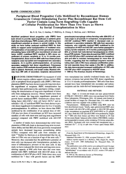
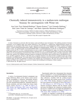
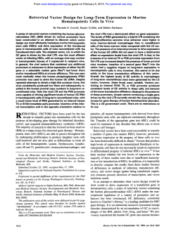
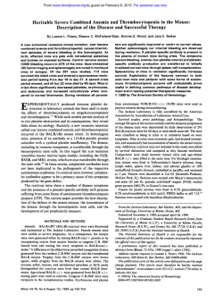
From www.bloodjournal.org by guest on February 6, 2015. For personal use only.
In Vivo Stem Cell Function of Interleukin-3-Induced Blast Cells
By Jun-lchi Tsunoda, Seiji Okada, Junko Suda, Kazunari Nagayoshi, Hiromitsu Nakauchi, Kiyohiko Hatake,
Yasusada Miura, and Toshio Suda
The treatment of mice with high doses of 5-fluorouracil
(5-FU) results in an enrichment of primitive hematopoietic
progenitors. Using this procedure, we obtained a new class
of murine hematopoietic colonies that had very high secondary plating efficiencies in vitro and could differentiate into
not only myeloid cells but also into lymphoid lineage cells.
The phenotypes of interleukin-3 (IL-3) induced blast colony
cells were Thy-l-positive and lineage-marker-negative. We
examined whether these blast colony cells contained primitive hematopoietic stem cells in vivo and could reconstitute
hematopoietic tissues in lethally irradiated mice. Blast colony cells could generate macroscopic visible spleen colonies
on days 8 and 12, and 5 x lo3 blast cells were sufficient t o
protect them from lethally irradiation. It was shown that 6 or
8 weeks after transplantation of 5 x I O a blast cells, donor
male cells were detected in the spleen and thymus of the
female recipients but not in the bone marrow by Southern
blot analysis using Y-encoded DNA probe. After 10 weeks,
bone marrow cells were partially repopulated from donor
cells. In a congenic mouse system, donor-derived cells (Ly5.2)
were detected in the thymus and spleen 6 weeks after
transplantation. Fluorescence-activated cell sorter analyses
showed that B cells and macrophages developed from donor
cells in the spleen. In the thymus, donor-derived cells were
found in CD4, CD8 double-positive, single-positive, and double-negative populations. Reconstitution of bone marrow
was delayed and myeloid and lymphoid cells were detected
10 weeks after transplantation. These results indicate that
IL-3-induced blast cells contain the primitive hematopoietic
stem cells capable of reconstituting hematopoietic organs in
lethally irradiated mice.
0 1991by The American Society of Hematology.
H
(5-FU)
DNA. In addition, we used Ly-5 congenic mice for the
analysis of hematopoietic reconstitution by blast colony
cells. We found that blast colony cells with extensive
proliferative potential could generate macroscopic spleen
colonies by days 8 and 12, provide protection from lethal
irradiation, and reconstitute hematopoietic tissues in vivo.
ODGSON AND BRADLEY found that the treatment of mice with high doses of 5-fluorouracil
resulted in an enrichment of primitive hematopoietic progenitors.’ Using this procedure, it was reported that
interleukin-3 (IL-3) induced a new class of murine hematopoietic colonies, termed blast cell colonies, which had
extremely high secondary plating efficiency’ and could
differentiate into not only myeloid-lineage but also into
lymphoid-lineage cells in vitro.’ Although the differentiation and proliferation capacity of blast colony cells was well
examined in vitro, this potential was not well examined in
vivo. In this study, we tried to clarify whether or not blast
colony cells contained primitive hematopoietic stem cells.
Hematopoietic stem cells are defined as cells that give
rise to all lineages of hematopoietic cells, have the ability to
maintain hematopoiesis in the long t e m , and have the
ability of self-renewal. In vivo hematopaietic stem cells are
determined using the spleen colony assay (colony-forming
unit-spleen [CFU-SI), the 30-day radioprotection assay,
and by determining their marrow repopulating ability.
Using Y-encoded DNA probe6 when male cells are transplanted into female mice the presence of male cells in the
myeloid and lymphoid tissue of reconstituted female recipients can be assayed by Southern blot analysis of extracted
From the Division of Hematology, Department of Medicine, Jichi
Medical School, Tochigi-ken,Japan; and the Laboratov of Molecular
Regulation of Aging, Frontier Research Prokam, The Institute of
Physical and Chemical Research (Riken), Tsukuba, Japan.
Submitted November 21,1990; accepted March 19, 1991.
Supported in part by Grants-in-Aidfrom the Ministry of Education,
Science and Culture of Japan.
Address reprint requests to Toshio Suda, MD, Division of Hematol00,Department of Medicine, Jichi Medical School, Minamikawachimachi, Tochigi-ken,329-04, Japan.
The publication costs of this article were defrayed in part by page
charge payment. This article must therefore be hereby marked
“advertisement” in accordance with 18 U.S.C.section 1734 solely to
indicate this fact.
0 1991 by The American Society of Hematology.
0006-497119117802-0020$3.0010
318
MATERIALS AND METHODS
Animals. Male and female BDFl (C57BLl6 x DBA/2) mice, 8
to 10 weeks old, were purchased from Sizuoka Laboratory Animal
Center (Sizuoka, Japan). Male BDFl mice were administered
5-FU (Adria Laboratories, Colombus, OH) through the tail vein at
a dosage of 150 mgkg body weight. Spleen cells were harvested 4
days later and single cell suspensions were prepared from pooled
spleens. C57BLlS.Ly5.1 mice were originally provided by Dr T.
Takahasi (Aichi Cancer Center, Aichi, Japan) and raised in our
colony.
Hematopoietic factors. Murine recombinant IL-3 (rIL-3) was
obtained from the serum-free culture supernatants of COS-1 cells
transfected with a clone of IL-3 gene using a PCD-X vector
provided by Dr T. Yokota (DNAX Research Institute of Molecular
and Cellular Biology Inc, Palo Alto, CA) with a specific activity of
10s U/mg protein.’
Blast colony cells. Methylcellulose culture was performed in
35-mm Don-tissue culture dishes (Falcon, Oxnard, CA). One
milliliter of culture consisted of 1.2 X lo6 5-FU-treated spleen
cells, a-minimal essential medium (a-MEM), 1.2% methylcellulose (Aldrich Chemical Company, Milwaukee, WI), 30% fetal
calf serum (FCS; Flow Laboratories, North Ryde, New South
Wales, Australia), 1% deionized bovine serum albumin (BSA
Sigma Chemical Company, St Louis, MO), 1 x
mol/L
2-mercaptoethanol (Eastman Organic Chemicals, Rochester, NY),
and 100 U rIL-3. Dishes were cultured at 37°C in a fully humidified
atmosphere of 5% CO, in air. After 6 to 7 days of culture, blast cell
colonies were lifted with a 3-pL Eppendorf micropipette under
inverted microscopy, and resuspended in a medium containing
10% FCS.
Characterization of blast colony cells. Cell surface phenotype of
blast colony cells was investigated by membrane staining. Blast
colony cells were incubated for 30 minutes on ice with fluorescein
isothiocyanate (F1TC)-, phycoerythrin (PE)-, or biotin-conjugated
monoclonal antibodies (MoAbs) (Sca-1, Thy-1, Mac-1, CD4, CD8,
Ly5.2) and wheat germ agglutinin (WGA).’ ’’ Sca-1 antibody was a
gift from Dr Y. Aihara (Yokohama City University, Kanagawa,
Blood, Vol78, No 2 (July 15), 1991: pp 318-322
From www.bloodjournal.org by guest on February 6, 2015. For personal use only.
319
FUNCTION OF INTERLEUKIN-%INDUCED BLAST CELLS
Japan). Anti-Ly5.2 antibody was kindly provided by Dr H. Yakura
(Tokyo Metropolitan Institute for Neurosciences, Tokyo, Japan).
The specificity and origins of other MoAbs used in this study are as
For stainingwith biotinylated antibodies, PE- or Texas
de~cribed.'~
Red (TR)-coupled streptoavidin was used as secondary reagent.
After the final wash with staining medium (3% FCS and 0.1%
sodium azide in phosphate-bufferedsaline [PBS]) twice, cells were
resuspended in staining medium supplemented with propidium
iodide (1 &mL), and were analyzed by a FACStarP'"s (Becton
Dickinson, Mountain View, CA). Multiparameter data were collected and analyzed using FACS-DESK (Version 1.8) run on a
Digital Micro VAX-111, configured as de~cribed.'~
The dead cells
stained with propidium iodide were gated out by FACS at the time
of analysis.
CFU-S assay. The number of spleen colonies was examined by
the modified method of Till and M~Culloch.'~~'~
All recipients were
female mice administered 9.5 Gy total body irradiation (TBI)from
a dual 13'Cs source at a dose of 1.00 Gy/min. Blast colony cells were
lifted up under the inverted microscope and resuspended in
a-MEM.Appropriate numbers of blast colony cells were injected
intravenously at 0.5 to 5 x lo3blast cells per mouse through the tail
vein. The recipient female mice were killed on days 8 and 12.
Spleens were removed and fixed in Bouin's solution, and spleen
colonies were counted under a dissection microscope. As a
negative control, medium was injected into irradiated mice.
Protection from lethal irradiation by blast colony cells. Pooled
blast colony cells were injected into each irradiated mouse at 0.5 to
5 x lo3through the tail vein. After transplantation, the percent of
surviving mice was determined 30 days after the injection? As a
negative control, pure medium was injected. All mice were kept in
laminar air flow room conditions with the acidified drinking water
at pH 2.8. All mice injected with medium died within 14 days after
transplantation.
DNA analysis after transplantation. To confirm that blast cell
colonies contain pluripotent hematopoietic stem cells, we injected
the cells obtained from male mice into irradiated female mice.
DNA was purified by proteinase K digestion and phenolchloroform extraction." For Southern blot analysis, samples of
male and female DNA from normal BDFl mice were used as
positive and negative controls. Samples of DNA were digested with
BamHI, electrophoresed through a 0.8% agarose gel, and transferred to a nitrocellulose membrane. After hybridization to pY2
probe, autoradiography was performed at -70°C with Fuji X-ray
film (Fuji Photo Film Co, Ltd, Kanagawa, Japan) for 24 to 72
hours. To determine the relative contribution of host- and donorcell-derived material in each DNA sample, DNA from a known
mixture of male and female BDFl mouse spleen cells was also
blotted.
Lymphohematopoietic repopulation of blast colony cells. Eightto 10-week-old C57BU6.Ly5.1 mice were lethally irradiated and
then injected with appropriate numbers of blast cells obtained
from C57BU6.Ly5.2 mice through the tail vein. Six to 10 weeks
later, lymphohematopoietic reconstitution in the spleen, thymus,
and bone marrow was examined by multiparameter FACS analysis
using anti-Ly5.2 and other MoAbs?
Although intensity of Thy-1 antigen of the blast cells was
widely distributed, Thy-lhigh
cells predominated.
To examine the in vivo proliferative ability of blast colony
cells, we injected 1 x lo) to 5 x lo) blast cells into 9.5 Gy
irradiated BDFl mice and scored the number of spleen
colonies on days 8 and 12 (Table 1). Simultaneously, the
incidence of CFU-S in normal bone marrow and spleen
cells and 5-FU-treated spleen cells was examined. As a
negative control, pure medium was injected. Three thousand blast colony cells formed 13 and 6 spleen colonies on
days 8 and 12, respectively. Considering that the f factor is
around 0.1, 23 blast cells contain one day 8 CFU-S. The
incidence of day 12 CFU-S was lower than that of day 8
CFU-S in blast colony cells as well as in untreated bone
marrow cells and spleen cells, while in 5-FU-treated mouse
spleen cells the incidence was reversed.
We examined the survival rate of lethally irradiated mice
that were transplanted with different numbers of blast cells
as the assay for in vivo hematopoietic stem cell activity.
When 2.5 x lo3 blast cells were transplanted, 50% of
recipient mice survived for 30 days or more. Moreover, 5 x
lo' blast cells were sufficient to protect mice from lethal
irradiation (Fig 1). As a negative control, pure medium was
injected; all such recipient mice were dead within 14 days
after transplantation. To exclude the possibility that stem
cells were generated from non-blast cells, we collected cells
from colonies containing more mature cells and injected
them into irradiated mice. The latter mice were dead within
2 weeks after transplantation and had no visible spleen
colonies.
To investigate the long-term hematopoiesis in each
hematopoietic organ by donor cells, we analyzed hematopoietic reconstitution using the Y-specific probe pY-2 and Ly5
congenic mouse system. As positive and negative controls,
DNA obtained from the thymuses of male and female mice
was also assayed. Six to 10 weeks after 5 x lo) male blast
cells were transplanted to female mice, the recipients' bone
marrow, spleen, and thymus DNA was extracted. Six and 8
weeks after transplantation, a 10-kb pY2 band probe was
observed in the spleen and thymus but not in the bone
marrow. After 10 weeks, the pY-2 band was detected in the
bone marrow, suggesting that hematopoietic reconstitution
by blast cells occured in the spleen, thymus, and bone
marrow (Fig 2).
Table 1. Incidence of Day 8 and Day 12 Spleen Colonies in Normal
Bone Marrow, Spleen, 5-FU-Treated Spleen Cells,
and Blast Cell Colonies
~
Cells
Cells
RESULTS
To examine the characteristics of blast colony cells, we
lifted about 200 blast colonies that contained 100 to 250
cells and pooled them in each experiment. At first, we
analyzed the surface phenotypes of blast colony cells. They
were WGA-positive and SCA-1-positive and had no lineage markers including B220, CD4, CD8, and Mac-1.
Bone marrowcells
Spleen cells
5-FU spleen cellst
Blast colony cells*
Injected
5
5
5
3
x
x
x
lo'
lo6
loE
x lo3
~~
No. of CFU-S
(means ? SD)'
Day 8
Day 12
11.9 f 1.8
19.7 f 0.9
1.7 f 1.9
12.7 f 1.2
9.0 2 1.7
4.8 f 1.7
8.0 f 0.8
6.2 2 1.2
'Values representthe means 2 SD from five mice.
tSpleen cells were derived from mice that had been injectedwith 150
mg of 5-FU per kilogram body weight 4 days before the harvest.
*Blast cell colonies were removed and resuspended in a medium.
From www.bloodjournal.org by guest on February 6, 2015. For personal use only.
TSUNODA ET AL
320
I
i o2
103
Lp5.2(FITC) LogFhwwceme
104
Number of Blast Cells Injected
Fig 1. Thirty day. of survival of lethally irradiated mice in which
various numbers of IL-3 blast colony cells were transplanted. In two
performances of each experiment, five mice were examined and
represent the average.
Furthermore, to clarify the lineage of the cells generated
from blast cells in vivo, transplantation experimcnts using
congenic micc were performcd. Hematopoietic cells wcrc
analysed by FACS to ascertain whcthcr thcy expressed the
donor type marker, LyS.2, in each hematopoietic organ. Six
wceks after transplantation, Ly5.2-positive cells wcrc dctcctcd in the splccn and thymus but not in thc bonc
marrow. Ten wccks after transplantation, donor-derived
cells predominated in the three organs, including the bonc
marrow (Fig 3). In the bonc marrow, Mac-1-positive
(myeloid), B220-positive (B cells), and Thy-1-positive (T
cells) were present among the LyS.2-positivc cells 10 wceks
after transplantation (data not shown). In the spleen. 6
wceks aftcr transplantation, B cells and Thy-1-positive cells
8 weeks
I
P
10 weeks
"
10 kB
Probe: pY2
Fig 2 Demonstration by Southem analysis of Vancoded DNA in
bone marrow, spleen, and thymus of female mice transplanted with
5 x 10'male blast cells. Recipient mice were killed 8 or 10 weeks later.
As controls, male and female DNA in thymus was blotted. At each
point, three mice were analyzed in three independent experiments.
-
Fig 3. Lymphohematopoletic reconstitution by blast colony cells.
Five thousand blast cells (C57SU6, Ly-5.2) were transplanted intravenously into lethally irradiated Ly-5 congenic mice (C57BU6, Ly-5.1).
Donor-derived (Ly-5.2) cells were analyzed in bone marrow, spleen,
and thymus 6 and 10 weeks later. (A) Bone marrow. (8) Spleen. IC)
Thymus. I--)
Control; (--) 6 weeks; (-) 10 weeks after transplantation. As a control, cells from each organ were stained with only PE and
analyzed.
were detected in addition to a very small number of
Mac-1-positive cells (Fig4A through C). In the thymus, we
detected CD4TD8' cclls, CD4TD8- cells, CD4+CD8cclls, and CD4TD8' cells in the Ly5.2-positive fraction
(Fig4D through F). Figure 4F shows the phenotypes of only
donor cells after gating LyS.2-positive fraction. Relativc
numbers of CD4 singlc-positive and double-negativc cells
were larger in donor ccll fraction compared with the
subpopulation of thymic cells from untreated mice.
DISCUSSION
Pluripotent hematopoietic stem cells are defined as cells
that give rise to all lineages of hematopoietic cells and also
have self-renewal capacity?"".'" It has been shown that
blast colony cclls dcrivcd from 5-FU-treated mouse splccn
cells could diffcrcntiate into not only the myeloid lincagc
but also into the lymphoid lineage in vitro.2AHowever, such
differentiation and proliferation capacities of blast cells
have not been examined in vivo. In this study, in vivo stem
cell functions of blast cells were assayed by CFU-S, radiation protection, and hematopoietic reconstitution methods.
Analysis of the surface phenotype of blast colony cells
showed that thcy were WGA'. SCA-l*, Thy-1'. B220-,
CD4-, and CD8-. This phenotype pattern is characteristic
of stem cells, as reported by Spangrude et at" and Visser et
al," except in so far as Thy-1 was not low but rather high in
IL->induced blasts. It was reported that IL-3 induccd a
Thy-1 antigen.'" A FACS clone-sorting study showed that
thc Tl1y-1~'~~
fraction formed more colonies than the Thy1'" or negative fraction (Suda, unpublished data).
Three thousand blast colony cells could generate about
13 and 6 macroscopic spleen colonies on day 8 and 12,
respectively. Blast colony cells contain more day 8 CFU-S
than day 12 CFU-S. Although the incidence of CFU-S in
blast cells is 20 times higher than in untreated bone marrow
cclls, the day 8 CFU-S/day 12 CFU-S ratio is not different
between them. On the other hand, the ratio was reversed in
5-FU-treated mouse spleen cells, suggesting that primitive
stem cells were selectively preserved in the latter. Thus, the
population of IL-3-induced blast cells seems to be similar
From www.bloodjournal.org by guest on February 6, 2015. For personal use only.
FUNCTION OF INTERLEUKIN-%INDUCED BLAST CELLS
321
X
:>
Fig 4. Representative analyses of spleen (A
through C) and thymus (0 through F) in a recipient 6
weeks after transplantationthat were shown in Fig 3.
Five thousand blast cells (C57BU6, Lyfi.2) were transplanted intravenously into lethally irradiated Ly5.1
congeneic mice. After gating only the Ly5.Z-positive
(donor origin) cells, thymic cells were analyzed for
the expression of CD4 and CD8. These cells were
stained with Ly5.2 (TR), Mac-1 (FITC), Thy-1 (PE),
E220 (allophycocyanin), CD4 (PE), or CD8 (RTC).
Thymus and spleen cells are represented by 2%
probability contour plotting. At each point, three
mice were examined in three independent experiments.
to that of normal bone marrow cells but not to 5-FUtreated mouse spleen cells from which IL-3 blast cells were
derived. To obtain a 50% survival rate in irradiated mice,
2.5 x 10' blast cells were required. Because about 3 x 10'
unfractionated bone marrow cells were required: stem
cells that could save the mice from lethal irradiation were
enriched about 10-fold in 1L-%induced blast cells. It is
shown that blast cells contain primitive hematopoietic stem
cells in vivo. More mature cells derived from blast colony
cells may support the recipients through the early critical
days after transplantation. We cannot exclude the possibility that spleen colonies were derived from co-isolated cells
but not from blast cells. However, it is considered to be
unlikely because the background cells alone could not form
any spleen colonies, and an approximately 20-fold enrichment of CFU-S was obtained in the fraction of blast colony
cells.
Long-term hematopoiesis by transplanted blast cells was
confirmed using Y-specific probe and congenic mouse
transplantation. From these studies, it was concluded that
IL-3-induced blast cells are able to differentiate into
myeloid cells, B cells, and T cells. Due to the lack of an
appropriate culture system for early T cells, the capacity of
IL-finduced blast cells to differentiate into T-cell lineage
has not yet been shown, although CD3-positive large
granular lymphocytes have been shown to develop from
blast cells in the presence of macrophages and IL-2.lThis is
the first demonstration that IL-finduced blast cells are not
$1
j
CDB(FITC)
restricted to myeloid and B-cell lineages, but that they can
differentiate into CD4+/CD8+cells in the thymus. The
repopulation of the spleen and thymus precedes that of the
bone marrow. When we transplanted WGA+Lin-cells into
irradiated mice in a separate study, bone marrow was found
to be reconstituted as well as the spleen and thymus within
6 weeks after transplantation."' Therefore, it was suggested
that in vivo differentiation of blast cells was different from
in vitro differentiation, because about 50% of 1L-finduced
blast cells formed myeloid colonies in in vitro culture
systems.' It remains to be clarified whether or not IL-3
affects primitive stem cells by committing them to differentiation pathways. IL-3 is shown to have a number of biologic
activities; in addition to a mast cell growth-promoting
activity and granulocyte/macrophagecolony-stimulating activity, lymphopoietic effects have been reported, such as
pre-B-cell-stimulating activity and activity to induce an
enzyme 20a-hydroxysteroid dehydrogenase in T lymphocytes.".z
In conclusion, IL-finduced blast cells contained primitive hematopoietic stem cells capable of colony formation in
vivo, repopulating hematopoiesis over the long term, and
differentiating into myeloid and lymphoid cells in vivo.
ACKNOWLEDGMENT
We thank S. Kurokawa for skillful technical assistance and M.
Yoshida for preparing the manuscript.
REFERENCES
1. Hodgson GS. Bradley TR:Properties of haematopoietic stem
4. Suda T,Ohara A, Suda J, Okada S,Tokuyama N, Miura Y,
cells surviving 5-fluorouracil treatment: Evidence for a pre-CFU-S
Sudo T, Nishikawa S-I,Nakauchi H: Early B cell differentiation
cell? Nature 281:381, 1979
from hematopoietic stem cells in the presence of stromal cells and
interleukin-7 (IL-7). in Gorin NC. Douay L (eds): Experimental
2. Suda T, Suda J, Ogawa M: Proliferative kinetics and differentiation of murine blast cell colonies in culture: Evidence for
Hematology Today-1989. New York, NY,Springer-Verlag. 1989
variahle Goperiods and constant doubling rates of early pluripo5. Kurihara N. Suda T, Miura Y, Nakauchi H. Kodama H, Hiura
tent hemopoietic progenitors. J Cell Physiol 117:308,1983
K, Hakeda Y, Kumegawa M: Generation of osteoclasts from
3. Minato N, Hattori M, SudoT, Kano S, Miura Y, Suda J, Suda
isolated hematopoieticprogenitor cells. Blood 74:1295, 1989
T Differentiation in vitro of T3* large granular lymphocytes with
6. Lamar EE. Palmer E: Y-encoded. species-specific DNA in
characteristic cytotoxic activity from an isolated hematopoietic
mice: Evidence that the Y chromosome exists in two polymorphic
progenitor colony. J Exp Med 167762.1988
forms in inhred strains. Cell 37171, 1984
From www.bloodjournal.org by guest on February 6, 2015. For personal use only.
322
7. Yokota T, Lee F, Rennick D, Hall C, Arai N, Mosmann T,
Nabel G, Cantor H, Arai K-I: Isolation and characterization of a
mouse cDNA clone that expresses mast-cell growth-factor activity
in monkey cells. Proc Natl Acad Sci USA 81:1070,1984
8. Spangrude GJ, Heimfeld S, Weissman I L Purification and
characterization of mouse hematopoietic stem cells. Science 241:
58,1988
9. Scheid MP, Triglia D: Further description of the Ly-5 system.
Immunogenetics 9:423, 1979
10. Dialynas DP, Wilde DB, Marrack P, Pierres A, Wall KA,
Havran W, Otten G, Loken MR, Pierres M, Kappler J, Fitch F W
Characterization of the murine antigenic determinant, designated
L3T4a, recognized by monoclonal antibody GK1.5: Expression of
L3T4a by functional T cell clones appears to correlate primarily
with class I1 MHC antigen-reactivity. Immunol Rev 74:29, 1983
11. Ledbetter JA, Herzenberg LA: Xenogeneic monoclonal
antibodies to mouse lymphoid differentiation antigens. Immunol
Rev 47:63,1979
12. Visser JWM, Bauman JGJ, Mulder AH, Eliason JF, deLeeuw
AM: Isolation of murine pluripotent hemopoietic stem cells. J Exp
Med 59:1576,1984
13. Nicola NA, Burgess AW, Staber FG, Johnson GR, Metcalf
D, Battye F L Differential expression of lectin receptors during
hemopoietic differentiation: Enrichment for granulocyte-macrophage progenitor cells. J Cell Physiol103:217,1980
14. Moore WA, Kautz RA: Data analysis in flow cytometry, in
Weir DM, Herzenberg LA, Blackwell C, Herzenberg LA (eds):
The Handbook of Experimental Immunology (ed 4). Edinburgh,
Scotland, Blackwell, 1986, p 30.1
15. Till JE, McCulloch EA: A direct measurement of the
TSUNODA ET AL
radiation sensitivity of normal mouse bone marrow cells. Radiat
Res 14:213,1961
16. Magli MC, Iscove NN, Odartchenko N Transient nature of
early hematopoietic spleen colonies. Nature 295527,1982
17. Maniatis T, Fritisch EF, Sambrook J: Molecular Cloning: A
Laboratory Manual. Cold Spring Harbor, NY,Cold Spring Harbor
Laboratory, 1982
18. Rennick DM, Lee FD, Yokota T, Arai K-I, Cantor H, Nabel
GJ: A cloned MCGF cDNA encodes a multilineage hematopoietic
growth factor: Multiple activities of interleukin 3. J Immunol
134:910,1985
19. Okada S, Suda T, Suda J, Tokuyama N, Nagayoshi K, Miura
Y, Nakauchi H: Effects of interleukin-3, interleukin-6, and granulocyte colony-stimulating factor on sorted murine splenic progenitor
cells. Exp Hematol 19:42,1991
20. Ihle JN, Keller J, Oroszlan S, Henderson LE, Copeland TD,
Fitch F, Prystowsky MB, Goldwasser E, Schrader JW, Palaszynski
E, Dy M, Lebel B: Biologic properties of homogeneous interleukin
3. I. Demonstration of WEHI-3 growth factor activity, mast cell
growth factor activity, P cell-stimulating factor activity, colonystimulating factor activity, and histamine-producing cell-stimulating factor activity. J Immunol131:282,1983
21. Palacios R, Henson G, Steinmetz M, McKearn JP: Interleukin-3 supports growth of mouse pre-B-cell clones in vitro. Nature
309:126,1984
22. Ihle JN, Pepersack L, Rebar L: Regulation of T cell
differentiation: In vitro induction of 20u-hydroxysteroid dehydrogenase in splenic lymphocytes from athymic mice by a unique
lymphokine. J Immunol 126:2184,1981
From www.bloodjournal.org by guest on February 6, 2015. For personal use only.
1991 78: 318-322
In vivo stem cell function of interleukin-3-induced blast cells
J Tsunoda, S Okada, J Suda, K Nagayoshi, H Nakauchi, K Hatake, Y Miura and T Suda
Updated information and services can be found at:
http://www.bloodjournal.org/content/78/2/318.full.html
Articles on similar topics can be found in the following Blood collections
Information about reproducing this article in parts or in its entirety may be found online at:
http://www.bloodjournal.org/site/misc/rights.xhtml#repub_requests
Information about ordering reprints may be found online at:
http://www.bloodjournal.org/site/misc/rights.xhtml#reprints
Information about subscriptions and ASH membership may be found online at:
http://www.bloodjournal.org/site/subscriptions/index.xhtml
Blood (print ISSN 0006-4971, online ISSN 1528-0020), is published weekly by the American
Society of Hematology, 2021 L St, NW, Suite 900, Washington DC 20036.
Copyright 2011 by The American Society of Hematology; all rights reserved.

© Copyright 2026