
SensoLyte AFC Caspase Sampler Kit *Fluorimetric*
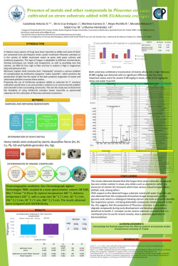
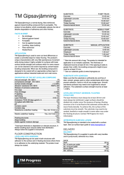
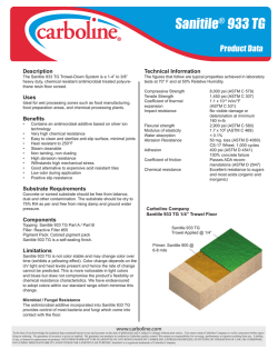
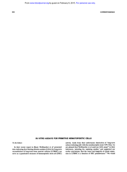
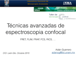
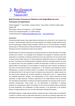
SensoLyte® AFC Caspase Sampler Kit
*Fluorimetric*
Catalog #
71117
Kit Size
8 X 100 Assays
Convenient Format: Mix-and-read one-step homogeneous assay.
Optimized Performance: Optimal conditions for assaying the activity of caspase.
Enhanced Value: Less expensive than the sum of individual components.
High Speed: Minimal hands-on time.
Assured Reliability: Detailed protocol and references are provided.
______________________________________________________________________________
Kit Components, Storage and Handling
Component
Component A
Component B
Description
Caspase substrates
Peptide sequence: Appendix I, Table 1
Ex/Em=380 nm/500 nm upon cleavage
AFC, fluorescence reference standard
Ex/Em=380 nm/500 nm
Quantity
8 substrates
10 mM, 70 μL
10 mM, 20 μL
Component C
Assay buffer
80 mL
Component D
DTT
1 M, 1 mL X 2 vials
Component E
10X Lysis Buffer
20 mL
Other Materials Required (but not provided)
• 96-well microplate: Black tissue culture microplate with or without clear bottom are
recommended.
• Fluorescence microplate reader: Capable of excitation at 380±30 nm and emission at 500±30
nm
Storage and Handling
• Store all components at -20°C
• Keep Components A and B away from light
• For convenience, Components C and E can be stored at 4°C
©AnaSpec, Inc. • 34801 Campus Dr. • Fremont, CA 94555
Tel 800-452-5530 • 510-791-9560 • [email protected] • www.anaspec.com
1
______________________________________________________________________________
Introduction
Apoptosis is a programmed, cell-autonomous death process. It plays important roles in
physiological and pathological events1, ranging from normal fetal development to diseases, such
as cancer2, organ failure and neurodegenerative diseases. During apoptosis, caspases execute the
disassembly of the cellular components by proteolytic cleavage of a variety of substrates, such as
poly-(ADP ribose) polymerase (PARP)3, DNA-dependent protein kinase (DNA-PK),
topoisomerases, and protein kinase C (PKC)δ.4 At least ten caspases have been discovered. Some
of these caspases identify and cleave a specific peptide substrate, while others recognize the
same peptide substrate4.
The SensoLyte® AFC Caspase Substrate Sampler Kit contains 8 AFC-based peptide
substrates (Appendix I, Table 1) for using as fluorogenic indicators in assaying caspase
activities. This kit provides a convenient platform for profiling substrate specificity of caspases
and optimizing assay condition for caspases. AFC-based substrates are widely used to monitor
caspase activity at the wavelength of excitation/emission = 380 nm/500 nm
______________________________________________________________________________
Protocol
Note: Warm all the kit components until thawed to room temperature before starting the experiments.
Note 2: You can formulate your own assay buffer and design your own assay experiment. For convenience, this kit
includes an assay buffer and the following suggested assay procedures.
Note 3: For instrument calibration, refer to Appendix II (recommended for first-time users).
1. Prepare working solutions.
1.1
Assay buffer: Add 20 μL of 1 M DTT (Component D) per 1 mL of assay buffer
(Component C).
Note: Use freshly prepared DTT-containing assay buffer for each experiment.
1.2
Caspase substrate solution: Dilute caspase substrate (Component A) 1:20-1:1000 in
assay buffer.
Note: Different substrate concentrations can be tested in order to calculate the Km and Kcat.
1.3
Prepare caspase samples:
•
When using purified caspase: Add bovine serum albumin (BSA) at 1 mg/mL in
assay buffer. Dilute caspase to an appropriate concentration (100 –1 nM) in assay
buffer.
Note: BSA is not included in the kit and should be provided by investigator.
•
When using cell culture: Seed 1X 10⁴-5 cells/well and add apoptosis inducing
agents. Incubate cells at 37°C for the desired time period (4-8 hr). Set up the
following controls:
Negative control: Contains cells without apoptosis inducing compound.
Substrate control: Contains culture medium only.
Test compound control: Contains culture medium and test compound.
©AnaSpec, Inc. • 34801 Campus Dr. • Fremont, CA 94555
Tel 800-452-5530 • 510-791-9560 • [email protected] • www.anaspec.com
2
The recommended total volume is 100-150 μL.
•
When using caspase-containing cell lysate: Refer to Appendix III for sample
preparation.
2. Initiate the enzymatic reaction.
2.1
When using purified caspase or cell lysate, add 50 μL/well samples to a 96-well plate.
Set up the following controls:
Negative control: Add 50 μL/well of cell lysate without inducing apoptosis.
Substrate control: Add 50 μL/well of assay buffer when using purified enzyme,
or 50 μL/well 1X lysis buffer (diluted Component E) when using cell lysate.
2.2
When using cell culture, retrieve the plate from 37°C incubator.
2.3
Add 50 μL/well caspase substrate solution into each well. Mix the reagents completely
by shaking on a plate shaker for 30-60 sec at 100-200 rpm.
2.4
Measure fluorescence signal:
•
For kinetic reading: Immediately start measuring fluorescence intensity at
Ex/Em=380 nm/500 nm continuously and record data every 5 min for 30 to 60 min.
•
For end-point reading: Incubate reaction at room temperature for 30 to 60 min on a
plate shaker at 100-200 rpm. Keep plate away from direct light. Measure
fluorescence intensity at Ex/Em=380 nm/500 nm.
Note: If the caspase activity is low in your samples, incubation time can be extended up to 18 hr before taking the
end-point reading.
•
Data analysis: Refer to Appendix IV.
©AnaSpec, Inc. • 34801 Campus Dr. • Fremont, CA 94555
Tel 800-452-5530 • 510-791-9560 • [email protected] • www.anaspec.com
3
______________________________________________________________________________
Appendix I: Peptide sequences
Table 1: Caspase substrate sequences.
Substrate
No.
Cat No.
Substrate Name
Substrate Sequence
SB1
25271
Caspase-1 substrate
Ac-YVAD-AFC
SB2
25285
Caspase-1 substrate
Ac-WEHD-AFC
SB3
25264
Caspase-2 substrate
Ac-VDVAD-AFC
SB4
25270
Caspase-8 substrate
Ac-IETD-AFC
SB5
25273
Caspase-3/7 substrate
Ac-DEVD-AFC
SB6
21685
Caspase-3/7 substrate
Z-DEVD-AFC
SB7
25276
Caspase-9 substrate
Ac-LEHD-AFC
SB8
25272
Caspase-6 substrate
Ac-VEID-AFC
©AnaSpec, Inc. • 34801 Campus Dr. • Fremont, CA 94555
Tel 800-452-5530 • 510-791-9560 • [email protected] • www.anaspec.com
4
______________________________________________________________________________
Appendix II: Instrument Calibration
•
AFC fluorescence reference standard: Dilute 10 mM AFC (Component B) to 30 μM in
assay buffer (Component C). Perform 2-fold serial dilutions to obtain 15, 7.5, 3.75, 1.88,
0.94, 0.47 μM AFC solution, include a water only sample (0 μM). Add 100 μL/well of
the serially diluted AFC solutions from 30 μM to 0 μM into a black 96-well plate.
•
Measure the fluorescence intensity of the reference standards at Ex/Em=380 nm/500 nm.
Adjust the sensitivity of the fluorometer until satisfactory signals can be read. Use the
same setting of sensitivity in the enzymatic reaction of the standard operation protocol.
•
Plot AFC fluorescent reference standard as RFU (relative fluorescent unit) versus
concentration as Figure 1.
Note: This reference standard curve is used to calibrate for the variation of different instruments and the different
batches of experiments. It can also serve as an indicator of the amount of final product in the caspase enzymatic
reaction. You may perform the standard curve for each experiment.
1800
1600
Figure 1. AFC reference standard. AFC
was serially diluted in 1X assay buffer. 100
μL of AFC at each concentration was
added and fluorescence was recorded at
Ex/Em=360±40 nm/460±40 nm (FLx800,
Bio-Tek Instruments). Samples were done
in duplicates.
1400
RFU
1200
1000
800
600
400
200
0
0
5
10
15
20
25
30
35
AFC (uM)
©AnaSpec, Inc. • 34801 Campus Dr. • Fremont, CA 94555
Tel 800-452-5530 • 510-791-9560 • [email protected] • www.anaspec.com
5
______________________________________________________________________________
Appendix III
Prepare caspase-containing sample from cell extract.
• Induce apoptosis in cell culture using a method of your choice.
• Prepare 1X lysis buffer by adding 1 mL of 10X lysis buffer (Component F) to 9 mL of
deionized water.
• Suspension cells are collected by centrifugation at 500 X g for 5 minutes. For adherent cells,
simply aspirate the growth medium.
• Add an appropriate amount of 1X lysis buffer to cells or cell pellet, e.g. 300 μL 1X lysis
buffer for one well of 6-well plate. Scrape off the adherent cells or re-suspend the cell pellet,
and then collect the cell suspension in a microcentrifuge tube.
• Rotate the cell suspension on a rotating apparatus for 30 min at 4°C.
• Centrifuge the cell suspension at 2500 X g for 10 min at 4°C.
• Collect the cell supernatant for caspase assay or store it at -80°C.
Prepare caspase-containing sample from tissue.
• Prepare 1X lysis buffer by adding 1 mL of 10X lysis buffer (Component F) to 9 mL of
deionized water.
• Tissue samples should be homogenized in 1X lysis buffer, and then centrifuged for 15 min at
10,000x g at 4°C. Supernatant, which contains caspase, can be frozen at -80 °C until use.
©AnaSpec, Inc. • 34801 Campus Dr. • Fremont, CA 94555
Tel 800-452-5530 • 510-791-9560 • [email protected] • www.anaspec.com
6
______________________________________________________________________________
Appendix IV: Data Analysis
•
The fluorescence reading from substrate control wells is the background fluorescence.
The readings from other wells need to be subtracted with this background fluorescence to
get the relative fluorescence unit (RFU).
•
For kinetic reading:
Plot data as RFU versus time for each sample.
Determine the range of initial time points during which the reaction is linear. 10-15%
conversion appears to be the optimal range.
Obtain the initial reaction velocity (Vo) in RFU/min. Determine the slope of the
linear portion of the data plot.
A variety of data analyses can be done, e.g., determining inhibition %, EC50, IC50, Km,
Ki, etc.
•
For endpoint reading:
Plot data as RFU versus concentration of test compounds.
A variety of data analyses can be done, e.g., determining inhibition %, EC50, IC50, etc.
1.2e+6
1.0e+6
RFU
8.0e+5
6.0e+5
4.0e+5
2.0e+5
0.0
0.001
0.01
Camptothecin (uM)
0.1
1
Figure 2. Dose-response curve of
Camptothecin. 1X10⁵/well Jurkat
cells were treated with
Camptothecin for 5 hrs. 50 μL/well
of AFC caspase-3 substrate (SB5)
solution was added to the apoptotic
cells and incubated at room
temperature for 30 min. Endpoint
fluorescence signal was measured
at Ex/Em=380/500 nm
(FlexStation II384, Molecular
Device, CA) EC50= 0.054±0.002
μM.
__________________________________________________________________
References
1.
2.
3.
4.
Thornberry, NA. and Y. Lazebnik, Science 281, 1312-1316 (1998).
Reed, JC. J. Clin. Oncol. 17, 2941-2953 (1999).
Lazebnik, YA. et al., Nature 371, 346-347 (1994).
Villa, P. et al., Trends Biochem. Sci. 22, 388-393 (1997).
©AnaSpec, Inc. • 34801 Campus Dr. • Fremont, CA 94555
Tel 800-452-5530 • 510-791-9560 • [email protected] • www.anaspec.com
7


© Copyright 2026