
Differential Cytokine Expression in Human Blood Monocyte
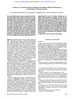
From www.bloodjournal.org by guest on February 6, 2015. For personal use only.
Differential Cytokine Expression in Human Blood Monocyte Subpopulations:
A Polymerase Chain Reaction Analysis
By Marion Frankenberger, Thomas Sternsdorf, Heinrich Pechumer, A. Pforte, and H.W. Loms Ziegler-Heitbrock
The subpopulation of strongly CDl4-positive (CD14++)
monocytes and monocytes coexpressing the CD16 antigen
and low levels of CD14 (CD14+/CD16+ cells) were isolated
by fluorescence-activated cell sorting (FACS) followed by
stimulation with lipopolysaccharide (LPS) at 1 pg/mL. Polymerase chain reaction (PCR) after reverse transcription of
isolated mRNA (RT-PCRI revealed similar levels of tumor
necrosis factor (TNF) transcripts in both subpopulations. By
contrast, transcripts for interleukin-10 (IL-10) were only detectable in CD14++ monocytes, whereas CD14+/CD16+ cells
produced no detectable IL-10 transcripts after 4 hours. Only
after 16 hours of LPS stimulation was a low level of IL-10
transcripts discernible in CD14+/CD16+ monocytes. The
same pattern was seen at the protein level in that TNF in
LPS-stimulated supernatants was comparable for both sub-
populations, whereas IL-10 was detected in CD14++ monocytes but not in CD14+/CD16+ cells. To avoid interference of
cell activation by CD14 and CD16 antibodies, cells were also
isolated based on the high and low level of CD33 antigen
expression. Again, weakly CD33-positive cells, which comprise the CD14+/CD16+ cells, showed no or only minimal IL10 mRNA. When comparing blood monocyte subpopulations
with alveolar macrophages (AM), AM showed high levels of
LPS-stimulated TNF, whereas IL-10 transcripts were undetectable. Our data show that CD14+/CD16+blood monocytes
produce high levels of proinflammatory cytokines like TNF,
whereas the anti-inflammatory IL-10 is low or absent, a pattern similar to what is seen in AM.
0 7996 by The American Society of Hematology.
H
Isolation of AM. Bronchoalveolar lavage was performed according to routine procedures in apparently healthy donors after
informed consent had been obtained. The study was approved by
the Ethics Committee of the University of Munich.
Immunojuorescence and cell sorting. Separated monocytes or
mononuclear cells were then stained for 30 minutes at 4°C in 0.5mL vol with the anti-CD14 antibody My4-fluorescein conjugate
(Coulter, Krefeld, Germany) and the anti-CD16 antibody Leu1 lcphycoerythrin conjugate (Becton Dickinson, Heidelberg, Germany)
under saturating conditions. For some experiments, cells were only
stained with the anti-CD33 antibody My9 (Coulter) and goat-antimouse FITC as detection antibody.
Cell sorting of the stained cells was performed with an EPICS V
753 flow cytometer (Coulter) with 488-nm excitation. Purities were
more than 90% for both subsets as determined by re-analysis.
After sorting, cells were either immediately lysed with 200 pL
RNAzol B (WAK-Chemie, Bad Homburg, Germany) at a concentration of 2 x IO4 cells per aliquot or stimulated with LPS (10 ng/mL
to 1 pg/mL) in RPMI1640 with 10% fetal calf serum for another 4
to 16 hours and then lysed in RNAzol B at the same concentration.
The lysates were stored at -20°C.
RT-PCR. Quantitative PCR for TNF mRNA was performed with
an internal cRNA standard according to the method of Wang et al.’4
After cell lysis, 5 to 10 x IO5 copies of the cRNA standard
(pAW109; Perkin Elmer Cetus, Uberlingen, Germany) and 14 pg
transfer RNA as carrier were added per sample. PCR for IL-IO
mRNA was performed in the same manner without internal standard,
but using the same RNA as for TNF PCR. After isolation, the RNA
was reverse-transcribed with oligo(dT) as primer. PCR amplification
ETEROGENEITY of human blood monocytes was
suggested by earlier studies that demonstrated some
functional differences for fractions with different sedimentation beha~i0r.I.~
However, research on such subsets was
hampered by the fact that they could not be isolated as pure
populations, no markers were available to clearly distinguish
them, and they could not be enumerated. We recently identified a novel subpopulation of monocytes that coexpress
CD16 (Fcy receptor 111) and low levels of CD14 (receptor
for lipopolysaccharide [LPS]). These CD14+/CD16+ cells
can be distinguished from the major population of strongly
CD14-positive (CD14++)monocytes, which are CD16-.6,7
CD14’/CD16’ cells were shown to be more mature and to
resemble alveolar macrophages (AM).’ Furthermore, they
were found to be increased in sepsis patients and in patients
with human immunodeficiency virus infection.’-’ Earlier
functional studies indicated that these cells had a generally
lower level of cytokine production,” and similar data were
obtained by others who examined populations enriched for
CD14+/CD16’ cells by ~edimentation.’~
Our earlier results
were obtained with cells subjected to lengthy gradient separation, elutriation, and fluorescence-activated cell sorting
(FACS) separation. With the advent of reverse transcriptasepolymerase chain reaction (RT-PCR) technology for analysis of mRNA levels, it became feasible to perform such
studies on low numbers of cells that could be obtained within
a few hours. Using this approach, we now show that CD14’1
CD16’ cells are in fact capable of producing proinflammatory cytokines like tumor necrosis factor (TNF). However,
expression of the antiinflammatory cytokine interleukin-10
(IL-10) is consistently minimal or absent. This suggests that
CD14+/CD16+cells may serve a function as efficient immunostimulatory and proinflammatory monocytes.
MATERIALS AND METHODS
Isolation of monocytes. Four hundred-milliliter volumes of peripheral blood were drawn from apparently healthy donors and defibrinated with the aid of glass beads. Monocytes were then isolated
under LPS-free conditions by Nycohep density gradient (1.068 specific density, W9506020; Nycomed Oslo, Norway) according to the
manufacturer’s instruction. In some experiments, we used mononuclear cells isolated by Ficoll-Hypaque density-gradient separation.
Blood, Vol 87, No 1 (January 1). 1996:pp 373-377
From the Institute f o r Immunology, and the Department of Internal Medicine, Klinikum Innenstadt, University of Munich, Munich,
Germany.
Submitted April IO, 1995; accepted August 23, 1995.
Supported by grants from Deutsche Forschungsgemeinschaji
(SFB 21 7) and from the Verum Foundation, Miinchen, Germany.
Address reprint requests to H.W. Liims Ziegler-Heitbrock, MD,
Institute f o r Immunology, University of Munich, Goethestr 3 I , 80336
Miinchen, Germany.
The publication costs of this article were defrayed in part by page
charge payment. This article must therefore be hereby marked
“advertisement” in accordance with I8 U.S.C. section 1734 solely to
indicate this fact.
0 1996 by The American Society of Hematology.
0006-4971/96/87OI-0015$3.00/0
373
From www.bloodjournal.org by guest on February 6, 2015. For personal use only.
FRANKENBERGER ET AL
374
was performed with the gene Amp RNA-PCR kit (#N8080017; Perkin Elmer Cetus). Thirty-five to 38 cycles for TNF or 45 cycles of
amplification for IL-IO were performed with a Hybaid Thermal Reactor (model HBTRI; Biometra, Gottingen, Germany) with sets of
1 minute at 94°C. I minute at 59°C. and 40 seconds at 72°C. The
PCR product was separated on a 1.4% (TNF) or 2% (IL-IO and pactin) agarose gel containing ethidium bromide. Polaroid photographs with UV exposure were taken with 4 x 5 Polaroid film (F4388; Sigma, Deisenhofen, Germany). The positive was used for
presentation, whereas the negative, after development with sodium
bisulfite (18%wt/vol, S-9OOO; Sigma), was used for laser densitometry. The following primers were used: TNF, 5'-primer, 5' CAG AGG
GAA GAG TTC CCC AG 3'. and 3'-primer, 5' CCT TGG TCT
GGT AGG AGA CG 3'; IL-Ip, 5'-primer, 5' AAA CAG ATG AAG
TGC TCC TTC CAG G 3'. and 3'-primer, 5' TGG AGA ACA CCA
CTT GTT GCT CCA 3'; IL-6, 5'-primer, 5' ATG AAC TCC TTC
TCC ACA AGC 3'. and 3'-primer, 5' CTA CAT TTG CCG AAG
AGC CCT CAG GCT GGA CTG 3'; IL-IO, 5'-primer, 5' ATG
CCC CAA GCT GAG AAC CAA GAC CCA 3'. and 3'-primer, 5'
TCT CAA GGG GCT GGG TCA GCT ATC CCA 3'; and &actin,
5'-primer, 5' GTG GGG CGC CCC AGG CAC CA 3', and 3'primer, 5' CTC CTT AAT GTC ACG CAC GAT TTC 3'.
Assaysfor cytokine protein. For determination of TNF and IL10 protein, 2 X IO5 cells were stimulated with LPS ( I pg/mL) in
400 p L of RPMI 1640 with 10% fetal calf serum, and supernatants
were harvested after 4 hours.
TNF protein was determined in serially diluted 100-pL vol using
the Wehi 164lActinomycin D bioassay with reference to a recombinant TNF standard (IO' Ulmg) as previously described."
IL- 10 protein was determined by an enzyme-linked immunosorbent assay kit (Dianova, Hamburg, Germany) according to manufacturer's instructions.
Statistical analysis was performed with Student's r-test.
RESULTS
Two-color immunofluorescence analysis of human blood
monocytes defines a population of CD14++cells and a popu-
C D14+/CD16+
u
l
4
a
U
0
LPS
+U
+
2
a
+
U
\
+
4
CI
+
?
+
e
-n
t -- i
?NF\
Standard
Fig 2. PCR analysis for TNF mRNA in monocyte subpopulations.
FACS-purified subpopulations (>94% purity) were either left untreated or cultured for 2 hours with LPS (10 ng/mL). Cells were lysed,
cRNA standard was added, and purified RNA was reverse-transcribed
and then amplified by PCR. Densitometry for the upper band specific
for endogenous TNF gave 30.6 and 33.4 arbitrary units for LPS-treated
CD14" and CD14'/CD16' cells, respectively (1representative of 9
experiments).
lation with coexpression of CD14 and CD16 (CD14'/CD16'
cells) (Fig I). In an average of 19 experiments, these CD14'/
CD16' cells accounted for 18% 2 4% (mean 2 SD) of all
monocytes. For analysis of cytokine expression, these two
subsets were isolated to more than 90% purity by FACS and
cultured with or without LPS at 1 &mL for 4 hours. Cell
samples were lysed with RNAzol, and after addition of
cRNA standard, RNA was isolated and reverse-transcribed
and PCR was performed with primers specific for TNF. A
representative example in Fig 2 demonstrates minimal or no
transcripts in unstimulated monocyte subpopulations,
whereas a strong band is visible for the internal standard.
LPS-stimulated cells showed a high level of TNF transcripts
for both CD14" and CD14'/CD16' cells, whereas the internal standard was competitively reduced. Comparable levels
of TNF transcripts were also obtained with low levels of
LPS ( I O ng/mL, data not shown). PCR analysis of IL-1
and IL-6 mRNA levels also gave similar values for both
subpopulations (Table 1). When the same samples were subTable 1. mRNA Levels for Cvtokines in Monocvte SUbDoDUlatiOnS
mRNA Level
CD14"
(96 of arbitrav units)
No. of
CDl4
Fig 1. T w o t o l o r immunofluorescence analysis of blood monocyte subpopulations. Isolated monocytes were stained with CD14FITC and CD16 phycoerythrin and analyzed by FACS. CD14" regular
monocytes were 45% and double-positive CD14'/CD16' cells were
18% in this example.
Cytokine
Experiments
TNF
IL-18
IL-6
IL-10
9
4
4
7
CD14"
CD14*/CD16'
100
100
100
100
106 2 26
94 t 6
101 2 3
0
Cell sorter-purified populations were stimulated with LPS at 1 p g l
mL for 4 hours and mRNA was reverse-transcribed and amplified by
PCR, followed by agarose gel electrophoresis and laser densitometry.
Arbitrary units obtained for CD14" monocytes were set at 100%.
Values for CD14'/CD16' are the mean z SD.
From www.bloodjournal.org by guest on February 6, 2015. For personal use only.
CYTOKINES IN MONOCYTE SUBPOPULATIONS
jected to PCR for IL-IO, only CD14" cells exhibited a
signal, whereas CD14'/CDI 6' cells were negative at 4 hours
(Fig 3). To exclude that CD14'/CD16' cells simply show
slower kinetics for IL- I O mRNA induction, cells were tested
after 16 hours. These analyses showed a further increased
level of IL-IO mRNA prevalence in CD14" cells. In some
experiments, a weak signal was discernible in CD14'/CD16'
cells at this point in time (not shown).
A similar pattern was seen when looking at cytokine protein with comparable TNF levels and low IL-IO values in
CD14+/CD16' monocytes. Here, TNF values were 244 2
101 U/mL for CD14" cells and 525 2 124 U/mL for
CD14'/CDI 6' cells (not significant). Levels of IL- IO protein
were 27.2 5 1 1.2 pg/mL for CD14", whereas no IL- IO was
detectable in CD14'/CD16' cells (n = 3).
To exclude the possibility that the CD14 antibody, which
is used for identification and isolation of monocyte subsets,
does block LPS action and thereby prevents production of
IL-IO, we isolated the subsets by a different strategy. For
this, we exploited the fact that regular CD14" cells show
high levels of CD33 (CD33"), whereas CD14'/CD16' cells
CD33'
CD33"
CD33
LPS 4h
0
+us
+us
T
F
n
++
w
r
n
u
u
+
\
w
r
n
u
n
LPS 16h
+\o
r
Fig 4. Isolation of monocyte subpopulation based on CD33 antigen density. Cells were FACS-separatedbased on high or low antigen
density. Test samples were controlled by additional staining with
anti-CD16. Isolated CD33' cells were 50% CD16' and CD33" cells
were 8% CD16' in this example.
n
u
+
\
w
.-
n
u
Fig 3. PCR analysis for IL-10 mRNA in monocyte subpopulations.
FACS-purified subpopulations (>97% purity) were either left untreated or cultured for 4 or 16 hours with LPS (1 pg/mL). RNA was
purified, reverse-transcribad, and amplified by PCR (1 representative
of 7 experiments].Densitometryfor stimulated 14" cells gave values
of 37.3 for 4 hours and 62.5 for 16 hours, whereas no signal was
evident for 14'/16' cells.
exhibit only low CD33 levels (CD33"). Sorting was performed with gates as in Fig 4. When test samples of the
resultant populations were stained with CD16, only 8% of
CD33" cells were CD16'. whereas the CD33" fraction
contained 50% CD16' cells. These isolated cells that had
been exposed only to CD33 antibody were then stimulated
with LPS, and transcript levels were determined by PCR.
Figure 5 demonstrates that CD33" cells, which represent
CD 14" regular monocytes, show high transcript levels for
IL-10 at 4 and 16 hours. By contrast, CD33' cells, which
comprise the CD14+/CD16' monocytes, had no or only minimal levels of IL-IO mRNA. Furthermore, when 14'/16'
and 14" cells were isolated by FACS directly from FicollHypaque-purified mononuclear cells instead of NycoPrepenriched monocytes, CD14'/CDI 6' cells also showed more
than ninefold lower IL-IO mRNA levels (n = 2, data not
shown). These data show that CD14'/CD16' monocytes are
in fact incapable of producing significant amounts of IL-10
mRNA.
In previous studies, we have demonstrated that CD14'/
CD 16' monocytes share several phenotypic properties with
AM. We have therefore asked if this similarity extends to
functional properties like cytokine production. For this purpose, bronchoalveolar lavage cells from healthy donors were
isolated and compared with CD14" and CD14'/CD16'
blood monocytes from the same donor. Figure 6 demonstrates that LPS-stimulated AM produce TNF transcripts at
levels comparable to blood monocytes. However, IL- IO production was absent in AM, with or without CD14 staining
From www.bloodjournal.org by guest on February 6, 2015. For personal use only.
376
FRANKENBERGER ET AL
and cell sorting. Hence, AM and CD14'/CD16' cells are
similar also with respect to the pattern of cytokine production.
LPS
fa
w
DISCUSSION
I-
Early studies on monocyte subpopulations have revealed
that fractions containing somewhat smaller cells exhibit
lower reactive oxygen production and higher antigen presentation ~apacity.'.~.~
Furthermore, more dense monocytes were
found to exhibit higher cytokine production.' However, isolation of subpopulations by such sedimentation procedures
did not result in clearly separated pure subsets. Furthermore,
subpopulations could not be enumerated as can be done with
cell-surface markers. We have recently identified the novel
subset of blood monocytes that coexpress CD14 and CD16
antigens. These CD14+/CD16' cells account for about 10%
of all blood monocytes." When compared with CD14"
monocytes, CD14'/CD16+ cells exhibit lower phagocytosis
capacity and H202production,' and show lower levels of
CDI Ib and CD33 and higher levels of MHC class 11.'
In previous studies on cytokine production, we found that
CD I4'/CDI 6' monocytes generated lower levels of the cy-
f
Y
0
v,
r'
Q:
++
3
d
CI
0
TNF
11-10
L P S 4h L P S 16h
Oh
++
+
++
+
++
+
" " "
" " "
n
a
n
n
n
n
U
U
V
U
U
U
IL-IO
Fig 6. Comparison of cytokine production in blood monocyte subpopulations and AM. FACS-purified subpopulations (>90% purity)
and AM from the same donor (either without or with CD14 staining
and cell sorting) were stimulated for 4 hours with LPS (1 pglmL).
Cells were lysed, and mRNA was isolated and amplified by PCR (1
of 3 experiments).
tokines TNF, IL-I, and IL-6. Similar results were obtained
by Wang et al," who reported lower levels of TNF and ILI in slow-sedimentation fractions of cells that were characterized by lower CD14 and lower C D l l b expression. We
now report that CD 14'/CD 16' monocytes and CD 14' cells
show comparable levels of TNF, IL-1, and IL-6. The reasons
for the discrepancy between these previous studies and the
results presented herein are unclear, but it may be that
CD14'/CD16' monocytes are more sensitive to physical
alterations inflicted on them during isolation. This may not
lead to cell death, but could lead to impaired responsiveness.
Our previous approach included density-gradient isolation,
elutriation, and cell sorting, which altogether required more
than 24 hours to obtain lo6 cells. Our current procedure
involves only one gradient separation followed by flow cytometry sorting for a few hours to obtain only the IO5 cells
that are sufficient for RT-PCR analysis.
Although analysis in such cells revealed efficient production of the cytokines TNF, IL-I, and IL-6 at the transcript
level, IL-IO mRNA is consistently low to absent in CD14'/
CD16' monocytes. One could still argue that the isolation
procedure involving NycoPrep density-gradient separation
+
Actin
Fig 5. PCR analysis for IL-10 mRNA in monocyte subpopulations.
FACS-purified 0 3 3 ' and CD33*+ cells 1>90% purity) were either left
untreated or treated with LPS (1FglmLl for 4 or 16 hours. Cells were
lysed, and RNA was isolated, reverse-transcribed, and amplified by
PCR. Densitometry for LPS-stimulated CD33" cells gave 40.3 arbitrary units for 4 hours and 91.3 for 16 hours. CD33' cells had no signal
at 4 hours and 7.4 arbitrary units at 16 hours (1 of 3 experiments).
From www.bloodjournal.org by guest on February 6, 2015. For personal use only.
CYTOKINES IN MONOCYTE SUBPOPULATIONS
to enrich for monocytes will result in impairment of IL-10
production in CD14+/CD16+ cells, but we have obtained
similar results when using FACS purification of Ficoll-Hypaque-isolated mononuclear cells. Nonaltering procedures
like stimulation of whole blood followed by intracellular
staining for cytokines and cell-surface staining for CD14 and
CD16 are required to resolve this question. Still, our data,
which show none to minimal IL-10 expression together with
undiminished production of TNF, argue against the possibility that physical damage is the reason for the inability to
produce IL-10. Of note, AM isolated without any antibody
staining or cell sorting also exhibited the same pattern of
high TNF and no IL-10.
The standard isolation procedure involves staining with
monoclonal antibodies directed against CD14 and CD16 antigens. Blockade of the CD14 LPS receptor or a negative
signal induced by engagement of the CD16 antigen could
be invoked as a mechanism to explain the absence of IL10 mRNA in CD14+/CD16+ monocytes. We have therefore
exploited the fact that CD14+/CD16' monocytes show a
low level at the myelomonocytic stem-cell antigen, CD33.'
CD33" cells are identical to CD14++cells, whereas CD33+
cells contain CD14+/CD16+ cells and monocytes that are
negative for CD16. These latter cells may form an additional
distinct human blood monocyte subset. Still, half of the
CD33+ cells were the CD14+/CD16+ monocytes, and these
cells have no or a minimal IL-10 signal after LPS stimulation. Hence, it appears that CD14+/CD16+ monocytes are
in fact incapable of producing significant amounts of the
antiinflammatory cytokine, IL- 10.
Comparison of blood monocytes and tissue macrophages
did demonstrate that AM, with low CD1 lb, CD14, and CD33
and high CD16 and MHC class I1 expression, are similar in
phenotype to CD14+/CD16+ cells.' Hence, we have asked
whether AM would also be functionally similar to CD14+/
CD16+ blood monocytes. In fact, our data show efficient
production of TNF but no IL-10 &A
in AM. Since both
CD14+/CD16+blood monocytes and AM exhibit low levels
of the LPS receptor CD14, one could assume that signaling
through these few receptor molecules is not sufficient to
induce IL-10. On the other hand, TNF production induced
by LPS is not impaired. Furthermore, human B cells, which
express low levels of CD14, can be induced to express IL10 efficiently.I6It appears that IL-10 production is silenced
in CD14+/CD16+ monocytes and in AM by other mechanisms. In any event, efficient production of the proinflammatory cytokine TNF in the absence of expression of the
antiinflammatory cytokine IL- 10 suggests that these cells
will serve a function as proinflammatory cells that trigger
an efficient immune response. This may have implications
for patients with solid tumors and for patients after M-CSF
therapy, both of which recently were reported to have increased numbers of CD14+/CD16+ monocytes.17."
REFERENCES
1. Arenson EBJ, Epstein MB, Seeger RC: Volumetric and functional heterogeneity of human monocytes. J Clin Invest 65613, 1980
311
2. Yasaka T, Mantich NM, Boxer LA, Baehner RL:Functions of
human monocyte and lymphocyte subsets obtained by countercurrent
centrifugal elutriation: Differing functional capacities of human
monocyte subsets. J Immunol 127:1515, 1981
3. Khansari N, Chou YK, Fudenberg HH: Human monocyte heterogeneity: Interleukin 1 and prostaglandin Ez production by separate
subsets. Eur J Immunol 15:48, 1985
4. Esa AH, Noga SJ, Donnenberg AD, Hess AD: Immunological
heterogeneity of human monocyte subsets prepared by counterflow
centrifugation elutriation. Immunology 59:95, 1986
5. Turpin J, Hersh EM, Lopez-Berestein G: Characterization of
small and large human peripheral blood monocytes: Effects of in
vitro maturation on hydrogen peroxide release and on the response
to macrophage activators. J Immunol 136:4194, 1986
6. Ziegler-Heitbrock HWL, Passlick B, Flieger D: The monoclonal antimonocyte antibody My4 stains B lymphocytes and two
distinct monocyte subsets in human peripheral blood. Hybridoma
7521, 1988
7. Passlick B, Flieger D, Ziegler-Heitbrock HWL: Identification
and characterization of a novel monocyte subpopulation in human
peripheral blood. Blood 74:2527, 1989
8. Ziegler-Heitbrock HWL, Fingerle G, Strobel M, Schraut W,
Stelter F, Schutt C, Passlick B, Pforte A: The novel subset of CD14+/
CD16+ blood monocytes exhibits features of tissue macrophages.
Eur J Immunol 23:2053, 1993
9. Fingerle G, Pforte A, Passlick B, Blumenstein M, Strobel M,
Ziegler-Heitbrock HWL The novel subset of CD14+/CD16+blood
monocytes is expanded n sepsis patients. Blood 82:3170, 1993
10. Locher C, Vanham G, Kestens L, Kruger M, Ceuppens JL,
Vingerhoets J, Gigase P Expression patterns of Fcy receptors, HLADR and selected adhesion molecules on monocytes from normal and
HIV-infected individuals. Clin Exp Immunol 98:115, 1994
11. Nockher WA, Bergmann L, Scherberich JE: Increased soluble
CD14 serum levels and altered CD14 expression of peripheral blood
monocytes in HIV-infected patients. Clin Exp Immunol98:369,1994
12. Ziegler-Heitbrock HWL, Strobel M, Kieper D, Fingerle G,
Schlunck T, Petersmann I, Ellwart J, Blumenstein M, Haas JG:
Differential expression of cytokines in human blood monocyte subpopulations. Blood 72503, 1992
13. Wang S-Y, Mak KL, Chen LY, Chou MP, Ho C K Heterogeneity of human blood monocyte: Two subpopulations with different
sizes, phenotypes and functions. Immunology 77:298, 1992
14. Wang AM, Doyle MV, Mark DF: Quantitation of mRNA by
the polymerase chain reaction. Proc Natl Acad Sci USA 86:9717,
1989
15. Ziegler-Heitbrock HWL, Kafferlein E, Haas JG, Meyer N,
Strobel M, Weber C, Flieger D: Gangliosides suppress tumor necrosis factor production in human monocytes. J Immunol 148:1753,
1992
16. Ziegler-Heitbrock HWL, Pechumer H, Petersmann I, Durieux
JJ, Vita N, Labeta MO, Strobel M: CD14 is expressed and functional
in human B cells. Eur J Immunol 24:1937, 1994
17. Weiner LM, Li W, Holmes M, Catalan0 RB, Dovnarsky M,
Padavic K, Alpaugh RK: Phase I trial of recombinant macrophage
colony-stimulating factor and recombinant y-interferon: Toxicity,
monocytosis and clinical effects. Cancer Res 54:4084, 1994
18. Saleh MN, Goldmann SJ, LoBuglio AF, Beall AC, Sabio H,
McCord MC, Minasian L, Alpaugh K, Weiner LM, Munn DH:
CD16+ monocytes in patients with cancer: Spontaneous elevation
and pharmacological induction by recombinant human macrophagecolony-stimulating factor. Blood 85:2910, 1995
From www.bloodjournal.org by guest on February 6, 2015. For personal use only.
1996 87: 373-377
Differential cytokine expression in human blood monocyte
subpopulations: a polymerase chain reaction analysis
M Frankenberger, T Sternsdorf, H Pechumer, A Pforte and HW Ziegler-Heitbrock
Updated information and services can be found at:
http://www.bloodjournal.org/content/87/1/373.full.html
Articles on similar topics can be found in the following Blood collections
Information about reproducing this article in parts or in its entirety may be found online at:
http://www.bloodjournal.org/site/misc/rights.xhtml#repub_requests
Information about ordering reprints may be found online at:
http://www.bloodjournal.org/site/misc/rights.xhtml#reprints
Information about subscriptions and ASH membership may be found online at:
http://www.bloodjournal.org/site/subscriptions/index.xhtml
Blood (print ISSN 0006-4971, online ISSN 1528-0020), is published weekly by the American
Society of Hematology, 2021 L St, NW, Suite 900, Washington DC 20036.
Copyright 2011 by The American Society of Hematology; all rights reserved.

© Copyright 2026