
Crystallographic Studies of the Escherichia coli Quinol
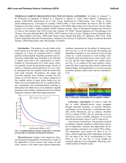
THE JOURNAL OF BIOLOGICAL CHEMISTRY Vol. 277, No. 18, Issue of May 3, pp. 16124 –16130, 2002 Printed in U.S.A. Crystallographic Studies of the Escherichia coli Quinol-Fumarate Reductase with Inhibitors Bound to the Quinol-binding Site* Received for publication, January 25, 2002, and in revised form, February 15, 2002 Published, JBC Papers in Press, February 15, 2002, DOI 10.1074/jbc.M200815200 Tina M. Iverson‡§, Ce´sar Luna-Chavez¶储**, Laura R. Croal‡‡, Gary Cecchini¶储, and Douglas C. Rees‡§§ From the ‡Division of Chemistry and Chemical Engineering, Howard Hughes Medical Institute, California Institute of Technology, Pasadena, California 91125, the ¶Molecular Biology Division 151-S, Department of Veterans Affairs Medical Center, San Francisco, California 94121, and the 储Department of Biochemistry and Biophysics, University of California, San Francisco, California 94143, and the ‡‡Division of Biology, California Institute of Technology, Pasadena, California 91125 The quinol-fumarate reductase (QFR) respiratory complex of Escherichia coli is a four-subunit integralmembrane complex that catalyzes the final step of anaerobic respiration when fumarate is the terminal electron acceptor. The membrane-soluble redox-active molecule menaquinol (MQH2) transfers electrons to QFR by binding directly to the membrane-spanning region. The crystal structure of QFR contains two quinone species, presumably MQH2, bound to the transmembrane-spanning region. The binding sites for the two quinone molecules are termed QP and QD, indicating their positions proximal (QP) or distal (QD) to the site of fumarate reduction in the hydrophilic flavoprotein and iron-sulfur protein subunits. It has not been established whether both of these sites are mechanistically significant. Co-crystallization studies of the E. coli QFR with the known quinol-binding site inhibitors 2-heptyl-4-hydroxyquinoline-N-oxide and 2-[1-(p-chlorophenyl)ethyl] 4,6-dinitrophenol establish that both inhibitors block the binding of MQH2 at the QP site. In the structures with the inhibitor bound at QP, no density is observed at QD, which suggests that the occupancy of this site can vary and argues against a structurally obligatory role for quinol binding to QD. A comparison of the QP site of the E. coli enzyme with quinone-binding sites in other respiratory enzymes shows that an acidic residue is structurally conserved. This acidic residue, Glu-C29, in the E. coli enzyme may act as a proton shuttle from the quinol during enzyme turnover. * This work was supported in part by the Department of Veterans Affairs (to G. C.), National Institutes of Health Grants GM61606 (to G. C.) and GM45162 (to D. C. R.), and National Science Foundation Grant MCB-9729778 (to G. C.). The costs of publication of this article were defrayed in part by the payment of page charges. This article must therefore be hereby marked “advertisement” in accordance with 18 U.S.C. Section 1734 solely to indicate this fact. The atomic coordinates and structure factors (code 1KF6, 1KFY, and 1L0V) have been deposited in the Protein Data Bank, Research Collaboratory for Structural Bioinformatics, Rutgers University, New Brunswick, NJ (http://www.rcsb.org/). § Present address: Dept. of Biochemistry and Howard Hughes Medical Institute, MS 013, Brandeis University, Waltham, MA 02454. ** Present address: Center for Biophysics and Computational Biology, MC-147, University of Illinois at Urbana-Champaign, Urbana, IL 61801. §§ To whom correspondence should be addressed: Division of Chemistry and Chemical Engineering, Howard Hughes Medical Institute, California Institute of Technology, MC 147-75CH, Pasadena, CA 91125. Tel.: 626-395-8393; Fax: 626-744-9524; E-mail: [email protected]. The Escherichia coli quinol-fumarate reductase (QFR)1 respiratory complex catalyzes the terminal step of anaerobic respiration when fumarate acts as the terminal electron acceptor (1). During this type of anaerobic respiration, electrons are donated to QFR by menaquinol (MQH2) molecules in the membrane. The electrons are transferred to a covalently-bound flavin adenine nucleotide at the active site through three distinct iron-sulfur clusters and ultimately are used to reduce fumarate to succinate (2, 3). The QFR respiratory complex is composed of four polypeptide chains. Two of these chains, the flavoprotein (FrdA) and the iron protein (FrdB), comprise the soluble domain, which is involved in fumarate reduction, whereas the remaining two subunits (FrdC and FrdD) are membrane-spanning polypeptides involved in the electron transfer with quinones. High sequence identity between the soluble domain of the E. coli QFR and the soluble domain of succinate-quinone oxidoreductase (SQR, complex II) of the aerobic respiratory chain places these two complexes in the same family (4). Consistent with the sequence relationship, both enzymes from E. coli can bidirectionally catalyze the interconversion of succinate and fumarate (5), and each can functionally replace the other to support growth (6, 7). In contrast to the soluble domain, the transmembrane anchor subunits of the complex II family have little sequence identity and exhibit variable polypeptide and cofactor composition. Nevertheless, similar structural models for the membrane-spanning subunits have been proposed for the QFR and SQR respiratory complexes (8). The structures of QFR respiratory complexes from E. coli (9) and Wolinella succinogenes (10) have been determined. Although these QFRs exhibit similar polypeptide-folds and cofactor arrangements in the soluble domain, there are distinctive features in the subunit organization and cofactor composition of the membrane-spanning domains. The structure of the QFR respiratory complex from E. coli (Fig. 1A) (9) contains two menaquinone molecules of unknown oxidation state designated QP and QD that are associated with the membrane-spanning region (Fig. 1B). Both of the binding sites for these quinol molecules had been predicted through a variety of techniques (11–15). QP is located near the [3Fe:4S] cluster of the soluble domain, whereas QD is positioned at the opposite side of the membrane. The minimum separation distance between the quinol rings of QP and QD is ⬃25 Å. (9). Although electron transfer has been observed to proceed at this distance (16), the 1 The abbreviations used are: QFR, quinol-fumarate reductase; MQH2, menaquinol; SQR, succinate-quinone oxidoreductase; PEG, polyethylene glycol; HQNO, 2-heptyl-4-hydroxyquinoline-N-oxide; DNP19, 2-[1-(p-chlorophenyl)ethyl]4,6-dinitrophenol. 16124 This paper is available on line at http://www.jbc.org Quinol Inhibitors of E. coli Quinol-Fumarate Reductase 16125 FIG. 1. Polypeptide -fold and electron transfer distances in QFR. A, ribbon diagram views of the E. coli QFR separated by a 90° rotation about a vertical axis. The flavoprotein is shown in blue, the iron protein is in red, and the transmembrane anchors are in dark green (FrdC) and purple (FrdD). The approximate boundary of the membrane is indicated with a black line. B, inter-cofactor distances of the E. coli enzyme. The known cofactors are superimposed onto an outline of the enzyme. C, inter-cofactor distances in the W. succinogenes enzyme. The b-type hemes associated with the membrane anchor reduce the electron transfer distance between a predicted distal quinol-binding site (data not shown). Figs. 1, 3, and 4 were made using Molscript (56), Bobscript (57), and Raster3D (58). separation is much greater than the distance typically observed among redox cofactors that participate in physiological electron transfer (17). This raises the question of whether both quinolbinding sites are functionally relevant or whether quinol binding at the QD site fulfills a structural role. If intervening heme(s) were present between these two quinol-binding sites, as is the case in other members of the QFR/SQR family (3, 4), electron transfer would be expected to readily proceed from QD to the active site (Fig. 1C). As quinone molecules are carriers of both protons and electrons, the transmembrane positioning of quinone (Fig. 2A)/ quinol (Fig. 2B) molecules during oxidation and reduction can 16126 Quinol Inhibitors of E. coli Quinol-Fumarate Reductase FIG. 2. Chemical structures for oxidized menaquinone-8 (MQ-8) (A), reduced menaquinol-8 (B), HQNO (C), and DNP-19 (D). MQ-8 is the primary menaquinone found in E. coli membranes, but smaller proportions of MQ-6, MQ-7, and MQ-9 are additionally present in the organism (59). lead to proton translocation across the membrane exemplified by the Q-cycle mechanism employed by cytochrome bc1 (18). It has been proposed that some fumarate reductases could participate in energy transduction if the oxidation of the quinol occurs at the QD site and is coupled to the reduction of a quinone positioned on the opposite side of the membrane in a second respiratory protein such as a hydrogenase or formate dehydrogenase (19). In the crystal structure of the two-heme QFR from W. succinogenes (10), a cavity search predicts that quinol molecules may bind at a site positioned similarly to QD (20, 21). The mutation of Glu-C66 near this cavity results in a loss of enzyme activity, whereas the protein retains a fully native structure (21), providing evidence that this location acts as a quinol-binding site. There is no evidence for a site positioned similarly to QP in the W. succinogenes complex; however, the presence of a QP site is not necessary for energy transduction when coupled to another respiratory complex. To assess whether both the QD and the QP quinol-binding sites of the E. coli QFR respiratory complex are functionally relevant for electron transfer, the E. coli QFR respiratory complex has been co-crystallized with the known quinol-binding inhibitors 2-heptyl-4-hydroxyquinoline-N-oxide (HQNO) (Fig. 2C) and 2-[1-(p-chlorophenyl)ethyl] 4,6-dinitrophenol (DNP-19) (Fig. 2D), and the structures were determined at resolutions of 2.7 and 3.6 Å, respectively. Additionally, the native E. coli QFR structure has been further refined at 3.3 Å resolution for more accurate comparison to the inhibitor-bound forms. These studies demonstrate unequivocally that both inhibitors bind to the E. coli QFR at the QP site near the [3Fe:4S] cluster. EXPERIMENTAL PROCEDURES Protein Preparation and Crystallization—QFR was expressed and purified as previously described (22). All crystallizations were performed using a protein concentration of 30 mg/ml in 0.7% (w/v) of the polyoxyethylene detergent C12E9 (Thesit). Previously, optimal crystal formation occurred while using polyethylene glycol (PEG) 10,000 as the precipitant (9, 22); however, the variability between protein batches necessitated the use of different molecular weights of PEGs as the precipitant for each preparation. In this study, crystals grown using PEG 5000 monomethyl ether displayed the highest diffraction limit. Crystallization reactions were performed by the vapor diffusion method with hanging drops equilibrated over 12.5% PEG 5000 monomethyl ether, 85 mM magnesium acetate, 100 mM sodium citrate, pH 5.8, 100 M EDTA, 0.001% dithiothreitol, and the appropriate inhibitor. Inhib- itor concentrations were 100 M HQNO (Ki ⫽ 0.2 M) or 10 mM DNP-19 (Ki ⫽ 5.2 M) (23, 24). Crystals grew in the orthorhombic space group P212121 with unit cell dimensions listed in Table I. Prior to cryocooling, crystals were soaked in a solution containing 30% ethylene glycol and the crystallization components. Structure Determination and Refinement—Data from co-crystals of QFR and HQNO were collected at 93 K at a wavelength of 1.00 Å on Stanford Synchrotron Radiation Laboratories (SSRL) beamline 9-2 using a Quantum 4 CCD detector, whereas data from co-crystals of QFR and DNP-19 were collected at a wavelength of 0.98 Å on SSRL beamline 9-1 using a MAR345 image plate detector. Data were processed using Denzo and Scalepack (25). As the structures are isomorphous, the structure of QFR bound to HQNO was refined following rigid body refinement of the original 3.3-Å resolution model (9) with the CNS program (26). The DNP-19 model was refined starting with the final model of HQNO bound to QFR after rigid body refinement. All models were built using the program O (27). Refinement was carried out using CNS (26) and REFMAC (28, 29). Tight non-crystallographic symmetry restraints were employed in regions of the molecule that exhibited similar electron densities but were released in crystal contacts and surface regions in which the structures of the two complexes clearly differed. Individual temperature factors were refined for the model of HQNO bound to QFR. For DNP-19 bound to QFR and the additional refinement performed on the previously reported native structure, group temperature factors were employed. The Rfree was calculated for a test set of reflections identical to those used for the original structure determination with additional reflections randomly selected for the QFR ⫹ HQNO data set (Table I). RESULTS Binding of Quinone Inhibitors to the Membrane Anchor Subunits—Two of the most potent inhibitors of the reactions of E. coli QFR with quinones are HQNO (Ki ⫽ 0.2 M) and DNP-19 (Ki ⫽ 5.2 M) (23, 24). These inhibitors were chosen for cocrystallization studies with purified QFR to investigate their binding sites in the membrane domain of the enzyme. Crystallographic analysis of the binding of HQNO and DNP-19 to the E. coli QFR complex reveals density for both of these inhibitors at the QP site (Fig. 3 and Table I). HQNO is nearly isosteric with MQH2 and binds at the QP site in a manner similar to that previously identified for MQH2. Two hydrogen bond donors, Lys-B228 N and Trp-D14 N⑀ are positioned within hydrogenbonding distance of the negatively charged N-oxide (Fig. 4A). These two hydrogen bonds would satisfy the hydrogen bonding capabilities of menaquinone or semimenaquinone. The hydroxyl group on the other side of the HQNO ring is positioned Quinol Inhibitors of E. coli Quinol-Fumarate Reductase 16127 TABLE I Data collection and refinement statistics Numbers in parentheses indicate values for the highest resolution bin. Unit cell (Å) a b c Resolution (Å) No. of observations Unique observations Rfree observations Completeness (%) Rsyma I/ Rcrystb Rfree root mean square deviation bond lengths root mean square deviation bond angles QP M QD PDB entry Wilson B (Å2) a b Native ⫹HQNO ⫹DNP-19 96.6 138.1 275.3 50–3.3 219,456 49,332 1005 87.2 (90.0) 0.093 (0.277) 15.1 (6.3) 0.245 0.290 0.014 1.8 ° MQ 4 MQ 1FUM, 1L0V 75 96.5 137.8 273.5 50–2.7 292,370 93,174 1953 91.6 (74.0) 0.063 (0.287) 25.0 (6.2) 0.231 0.280 0.019 1.9 ° HQNO 5 – 1KF6 71 96.2 137.8 271.0 50–3.6 124,660 38,919 819 91.1 (90.8) 0.104 (0.306) 12.0 (4.1) 0.264 0.301 0.017 1.9 ° DNP 3 – 1KFY 37 Rsym ⫽ ¥兩Ii ⫺ 具I典兩/¥Ii, where i is the “ith” measurement and 具I典 is the weighted mean of I. Rcryst ⫽ ¥储Fobs兩 ⫺ 兩Fcalc储/¥兩Fobs兩. Rfree is the same as Rcryst for data omitted from refinement. FIG. 3. Binding of quinol and inhibitor molecules in the transmembrane-spanning region of the E. coli QFR. The C␣ trace of the membrane anchors is shown with the same coloring as in Fig. 1. The soluble domain of the complex has been omitted for clarity. Difference Fourier 兩Fo兩 ⫺ 兩Fc兩 electron density (green) is contoured at 3 and displayed at QP, QD, and M after molecules binding to those sites were omitted. Cavities calculated using the program VOIDOO (60) are displayed at the M site and are shown in blue. A, QFR at 3.3 Å resolution binds quinol molecules at both the QP and QD sites and additionally has density associated with the cavity M at the center of the membrane. B, QFR at 2.7 Å resolution shows clear density for an HQNO molecule bound at QP but lacks appreciable density at the QD site. The intensity of the density at position M has increased to 5. C, QFR at 3.6 Å resolution bound to DNP-19. Density at the QD site does not exceed background levels. Although the calculated cavity at the position M is larger, the density associated with it barely exceeds 3. within hydrogen-bonding distance of the side chains of Glu-C29 O⑀2 and Arg-D81 N1. Although DNP-19 also binds at the QP position, the binding site is shifted by over 2 Å with respect to HQNO or MQH2 (Fig. 4A), and the inhibitor molecule only makes hydrogen-bonding contacts with one side of the quinolbinding pocket. The hydrogen bonds form between the DNP-19 O2 and Lys-B228 N and between the DNP-19 O13 and TrpD14 N⑀, whereas Glu-C29 and Arg-D81 do not make contacts. Significant positional differences between the QFR proteins in these structures are limited to the movement of the side chains of Gln-B225, Arg-C28, and Leu-C89 in the DNP-19-bound enzyme (Fig. 4A). These side chains would sterically clash with DNP-19 if they adopted the same conformation as exhibited in the other structures. Kinetic analyses have established that HQNO is a non-competitive inhibitor of QFR, whereas DNP-19 acts as a pure competitive inhibitor (23, 24), and have suggested that both inhibitors bind at or near the same site. As the DNP-19 compound is not deeply inserted into QP but rather blocks the entrance to this site, this inhibitor probably acts by sterically preventing quinol from binding as predicted previously (23). Neither inhibitor was observed to bind to the QD site; yet intriguingly, electron density for QD, which was strong in the 3.3 Å resolution structure determination, is absent when either HQNO or DNP-19 is bound to the QP-binding site (Fig. 3). Unassigned Density in the Membrane-spanning Domain—In the 3.3-Å resolution structure containing MQH2 bound at both the QP and QD positions, an unassigned feature in the electron density with a height of 4 above background was visible between these two sites (Fig. 3A). This extra density, designated “M,” coincides with the only major cavity found in the E. coli QFR structure. The density in this location is additionally present in the inhibitor structures (Fig. 3, B and C) and becomes stronger with improved resolution, reaching 5 in the HQNO co-structure. For comparison, the omission of the HQNO inhibitor from the HQNO co-structure results in a difference Fourier density of 5.8, whereas the omission of the quinone molecules bound at QD and QP from the 3.3-Å resolution structures result in difference Fourier densities of 4.5 and 3.9, respectively. It is possible that this additional density at M represents a partially ordered cofactor that has not been previously detected. The distance between the M site and each of the other established quinol-binding sites is ⬃13 Å. The side chains near the density (Fig. 4B) associated with this cavity 16128 Quinol Inhibitors of E. coli Quinol-Fumarate Reductase FIG. 4. Comparison of inhibitors binding at QP and side chains surrounding the M site. A, comparison of inhibitor-binding position and side chain movements. Residues from FrdB have the carbon atoms colored in red, residues from FrdC have the carbon atoms colored in dark green, whereas residues from FrdD have the carbon atoms colored in purple. Oxygen atoms are colored in red, nitrogen atoms are blue, and sulfur atoms are in yellow. The inhibitor HQNO has the carbon atoms colored in black, whereas the inhibitor DNP-19 has the carbon atoms colored in white. The orientation of the view is looking onto the QP-binding site from the surface of the membrane. The side chain positions are taken from the QFR structure with HQNO bound (Protein Data Bank code 1KF6) and are within the error of the side chain positions from the structure of QFR with MQ bound (Protein Data Bank code 1FUM). The differences in side chain position between the DNP-19 bound enzyme and the other two structures that were greater than the error of the determined structures were limited to Gln-B225, Arg-C28, and Leu-C89. The altered positions of these three side chains that allow for DNP-19 binding are shown with the carbon atoms in white. B, the position M from the structure bound to HQNO with surrounding side chains. The orientation of the molecule in this view is the same as in Fig. 3B. The approximate location of a binding site can be inferred from a cavity calculated using the program VOIDOO (60) (cyan) and difference Fourier (兩Fo兩 ⫺ 兩Fc兩) electron density contoured at 3 (green). The coloring of atoms is the same as seen in Fig. 4A. The position of the quinone at QP is taken from the QFR structure with quinones bound (Protein Data Bank code 1L0V), and the quinone molecule is shown with the carbon atoms colored in gray. tend to be polar, whereas those in the region not associated with any density tend to be apolar, giving this cavity the hydrophobicity characteristics of a quinone-binding site. Sitedirected mutagenesis suggests that the residues lining this location are critical for enzyme function (14). However, the location of this density in the center of the membrane would be unusual for a quinol-binding site when compared with those structurally characterized to date. The binding of a metal ion at this site is also unlikely, because neighboring residues appear too distant to act as metal ligands and no significant peaks are observed near this site in anomalous difference Fourier maps. Thus, the identity of the species at this M site cannot be determined at present. DISCUSSION Mechanistic Role for the QP-binding Site—QP is positioned ⬃8 Å from the [3Fe:4S] cluster, implicating this quinol-binding site as the immediate electron donor to the redox cofactors in the soluble domain. The fully reduced quinol bound at QP must release two electrons and two protons during oxidation to the quinone. As the [3Fe:4S] cluster is a one electron acceptor, QFR must be able to stabilize both the fully reduced quinol and at least transiently the semiquinone oxidation states at the QPbinding site during turnover. The binding of HQNO to the QP site strengthens the hypothesis that the semiquinone form can be stabilized at this site (15), because HQNO is proposed to be a mimic of the semiquinone intermediate. In both the QFR structure with HQNO bound at QP and the structure with MQH2 bound, the side chain of Glu-C29 lies within hydrogen-bonding distance of the hydroxyl of the HQNO or MQH2 ring. The proximity of the Glu-C29 residue and the fact that the HQNO elicits a significant change in the EPR line shape of the [3Fe:4S] cluster (30) are consistent with HQNO binding near the Qp site (Fig. 3). The obligatory presence of an acidic residue near the exchangeable quinone-binding site is emerging as a theme in respiratory proteins. The Qi ubiquinone-binding site of cytochrome bc1 (31–33) contains the conserved Asp-229 (chicken numbering) residue within hydrogenbonding distance of the ubiquinone. Similarly, the proposed ubiquinol-binding site in quinol oxidase (34) contains a conserved Asp-75, and the co-structures of cytochrome bc1 with the Qo-binding inhibitor stigmatellin shows that Glu-272 of the cytochrome b subunit lies within hydrogen-bonding distance of stigmatellin (35). In the W. succinogenes QFR, Glu-C66 has been shown to be an essential component of the menaquinol oxidation site (21), whereas in the Bacillus subtilis SQR, the conserved Asp-C52 is part of the predicted HQNO-binding site (36). Additionally, in the Paracoccus denitrificans SQR, the mutation of Asp-D88 to a glycine confers resistance to the known quinone-binding inhibitor carboxin (37). In eukaryotes, the small membrane anchor (FrdD/SdhD/cyt bS) gene of QFR/ SQR (38 – 41) contains three conserved aspartate residues, and the alteration of one, Asp-92 (human numbering of uncleaved sequence), is associated with hereditary paraganglioma (42). Site-directed mutagenesis (43) and azido-3[H]-linked quinone labeling (40) predicts that Asp-92 is located within a region important for ubiquinone binding. Because variants of cytochrome bc1 altering Glu-272 of the cytochrome b subunit impair enzyme turnover while maintaining ubiquinol binding to Qo (35), Glu-272 is proposed to participate in proton shuttling rather than be involved in stabilizing the binding of a protonated quinone species. Similarly, in the soluble quinohemoprotein amine dehydrogenase from Pseudomonas putida, Asp-33␥ is presumed to be the proton shuttle (44) for the covalently attached cysteine tryptophylquinone. Although the photosynthetic reaction center does not contain an acidic residue that directly hydrogen-bonds to the ubiquinone, two acidic residues in Rhodobacter sphaeroides reaction center, Glu-L212 and Asp-L213, are located within 5 Å of the semiquinone bound at QB (45) and have been implicated as proton shuttles (46). A role with the carboxyl group primarily involved in proton transfer is consistent with the EPR analysis of the FrdC E29L variant of the E. coli QFR (15). Quinol molecules have two hydroxy groups located on opposite sides of the molecule (Fig. 2); each is deprotonated during oxidation. As discussed above, the acidic residue Glu-C29 may act as the proton shuttle during quinol oxidation. In cyto- Quinol Inhibitors of E. coli Quinol-Fumarate Reductase chrome bc1 (both Qi and Qo), ubiquinol oxidase and the W. succinogenes QFR, a conserved histidine residue is located on the opposite side of the quinone-binding site from the acidic residue. Because the photosynthetic reaction center quinonebinding site also contains a conserved histidine residue (HisL190), searches for consensus quinone-binding sequences have focused on conserved histidine residues (47). The E. coli QFR departs from the use of a histidine in the quinol-binding site. Although several histidine side chains are near the QP site and may therefore act as secondary proton shuttles in a chain from Glu-C29 (Fig. 4A), none of them is optimally positioned to form hydrogen bonds with the quinol hydroxyl groups. Instead, on the opposite side of the quinol, DNP-19, and HQNO molecules from Glu-C29, all of these molecules are positioned within hydrogen-bonding distance of Trp-D14 N⑀ and Lys-B228 N that could participate in the proton transfer pathway. By analogy to the photosynthetic reaction center in which the quinone bound at the QB site undergoes a propeller twist during reduction (45), multiple positions of the quinone may also be utilized in QFR to generate additional contacts with residues involved in the proton shuttle. In addition to HQNO and DNP, the other known inhibitors of the E. coli QFR have been either shown or predicted to bind to the QP site (15, 24), and the available kinetic data are consistent with a single dissociable binding site in the QFR complex (24, 48). Furthermore, although the E. coli QFR is insensitive to many of the other known complex II quinone-binding inhibitors, the locations of mutations that confer resistance to carboxin and 2-thenoyltrifluoroacetone in the single heme containing P. denitrificans SQR (37, 49) suggest that both of these inhibitors bind at a site positioned similarly to QP. Thus, the insensitivity of the E. coli QFR to these inhibitors does not reflect the binding of these inhibitors to a non-functional site. The M-binding Site—Three molecules of ubiquinone have recently been proposed to bind specifically to the two quinonebinding sites, Qi and Qo, of cytochrome bc1 respiratory complex (50). One molecule is proposed to bind at the ubiquinone reduction center, Qi, whereas two are proposed to bind at the ubiquinol oxidation site, Qo. In the E. coli QFR structure, the entity at the M site is separated from QP by the side chain of Arg-D81 (Fig. 4B). Thus, the double occupancy model proposed for the cytochrome bc1 Qo site (50, 51) would require an alternative position for the side chain of Arg-D81. The position of Arg-D81 on an ␣-helix and facing in toward the center of the protein would make it difficult for this side chain to move out of the pocket into solvent, constraining any repositioning relative to the M cavity. Although it seems more likely that M is a separate entity from QP, the rearrangement of Arg-D81 to form a larger binding pocket cannot be entirely ruled out. The QD-binding Site—Quinol oxidation at the QD site of the E. coli QFR has not been demonstrated. However, one intriguing aspect of the QFR structures with inhibitor bound at the QP site is that no density corresponding to quinol bound at QD is observed. In the cytochrome bc1 complex, the binding of either of the inhibitors stigmatellin or methoxyacrylate stilbene at Qo reduces the affinity for the inhibitor or quinol at the second Qo site in the dimer (52). As a result of the anti-cooperative binding behavior, these two compounds can inhibit the cytochrome bc1 complex with a stoichiometry of 0.5 molecules of inhibitor/ Qo-binding site. In the E. coli QFR, the absence of quinol density at QD when an inhibitor is tightly associated at QP could suggest a similar anti-cooperative behavior in these binding sites. Although the native structure of QFR containing only quinones bound shows density at both QP and QD (9), the density at QP is much weaker. This finding may reflect a disorder in quinol binding or it may reflect partial occupancy of 16129 the site or both. Although the experiments presented here cannot establish the functional relevance of QD, the observation that the occupancy at the QD site decreases in the structures with inhibitors bound to the QP site indicates that the binding of a quinone species at the QD site is not required for protein stability. Electron Transfer during Catalysis—QFRs and SQRs exhibit variability in membrane subunit composition, heme content, and quinol/quinone association, suggesting that electron transfer through the membrane-spanning region differs between the various enzymes of this family. The experiments on many heme-containing members of the QFR/SQR family have yielded evidence for the participation of a site positioned similarly to QD during electron transfer (21, 40, 43, 53); however the molecular characteristics of these functional sites resemble those observed in QP in the E. coli QFR-respiratory complex. Quinol oxidation at a site positioned similarly to QD indicates that transmembrane electron transfer occurs and would be obligatory for the generation of a transmembrane electrochemical potential gradient during turnover (54, 55). The existence of the M site near the middle of the membrane possibly identifies an additional center that could participate in an electron transfer pathway between QP and QD in the E. coli QFR. A more definitive proposal for the mechanistic role of QD during catalysis by the E. coli QFR will require determination of whether the non-functional mutants generated at the QD and M positions retain the native structure and identification of the molecular species at the M site. Acknowledgments—We thank A. P. Yeh, R. B. Bass, and A. Dunn for experimental assistance, H. Miyoshi for generously providing the DNP-19 inhibitor compound, Y. F. C. Lau for providing sequences of the mouse SQR before publication, C. L. Drennan and D. Ringe for the use of computer facilities, and T. Ohnishi, I. Schro¨ der, P. L. Dutton, H. B. Gray, and R. A. Marcus for enlightening discussions. REFERENCES 1. Gottschalk, G. (1986) Bacterial Metabolism, 2nd Ed, Springer-Verlag New York Inc., New York 2. Kro¨ ger, A., Geisler, V., Lemma, E., Theis, F., and Lenger, R. (1992) Arch. Microbiol. 158, 311–314 3. Ackrell, B. A. C., Johnson, M. K., Gunsalus, R. P., and Cecchini, G. (1992) in Chemistry and Biochemistry of Flavoenzymes (Mu¨ ller, F., ed) Vol. 3, pp. 229 –297, CRC Press, Inc., Boca Raton, FL 4. Ha¨ gerha¨ ll, C. (1997) Biochim. Biophys. Acta 1320, 107–141 5. Pershad, H. R., Hirst, J., Cochran, B., Ackrell, B. A. C., and Armstrong, F. A. (1999) Biochim. Biophys. Acta 1412, 262–272 6. Guest, J. R. (1981) J. Gen. Microbiol. 122, 171–179 7. Maklashina, E., Berthold, D. K., and Cecchini, G. (1998) J. Bacteriol. 180, 5989 –5996 8. Ha¨ gerha¨ ll, C., and Hederstedt, L. (1996) FEBS Lett. 389, 25–31 9. Iverson, T. M., Luna-Chavez, C., Cecchini, G., and Rees, D. C. (1999) Science 284, 1961–1966 10. Lancaster, C. R. D., Kro¨ ger, A., Auer, M., and Michel, H. (1999) Nature 402, 377–385 11. Ruzicka, F. J., Beinert, H., Schepler, K. L., Dunham, W. R., and Sands, R. H. (1975) Proc. Natl. Acad. Sci. U. S. A. 72, 2886 –2890 12. Salerno, J. C., Harmon, H. J., Blum, H., Leigh, J. S., and Ohnishi, T. (1977) FEBS Lett. 82, 179 –182 13. Westenberg, D. J., Gunsalus, R. P., Ackrell, B. A. C., and Cecchini, G. (1990) J. Biol. Chem. 265, 19560 –19567 14. Westenberg, D. J., Gunsalus, R. P., Ackrell, B. A. C., Sices, H., and Cecchini, G. (1993) J. Biol. Chem. 268, 815– 822 15. Ha¨ gerha¨ ll, C., Magnitsky, S., Sled, V. D., Schro¨ der, I., Gunsalus, R. P., Cecchini, G., and Ohnishi, T. (1999) J. Biol. Chem. 274, 26157–26164 16. Winkler, J. R., DiBilio, A., Farrow, N. A., Richards, J. H., and Gray, H. B. (1999) Pure Appl. Chem. 71, 1753–1764 17. Page, C. C., Moser, C. C., Chen, X., and Dutton, P. L. (1999) Nature 402, 47–52 18. Mitchell, P. (1961) Nature 191, 144 –148 19. Ohnishi, T., Moser, C. C., Page, C. C., Dutton, P. L., and Yano, T. (2000) Structure 8, R23–R32 20. Iverson, T. M., Luna-Chavez, C., Schro¨ der, I., Cecchini, G., and Rees, D. C. (2000) Curr. Opin. Struct. Biol. 10, 448 – 455 21. Lancaster, C., Gro, R., Haas, A., Ritter, M., Mantele, W., Simon, J., and Kroger, A. (2000) Proc. Natl. Acad. Sci. U. S. A. 97, 13051–13056 22. Luna-Chavez, C., Iverson, T. M., Rees, D. C., and Cecchini, G. (2000) Protein Expression Purif. 19, 188 –196 23. Yankovskaya, V., Sablin, S. O., Ramsay, R. R., Singer, T. P., Ackrell, B. A. C., Cecchini, G., and Miyoshi, H. (1996) J. Biol. Chem. 271, 21020 –21024 24. Maklashina, E., and Cecchini, G. (1999) Arch. Biochem. Biophys. 369, 223–232 25. Otwinowski, Z. (1993) in CCP4 Study Weekend Data Collection and Processing 16130 Quinol Inhibitors of E. coli Quinol-Fumarate Reductase (Sawyer, L., Isaacs, N., and Bailey, S., eds), pp. 56 – 62, SERC Daresbury Laboratory, Daresbury, UK 26. Bru¨ nger, A., Adams, P., Clore, G., DeLano, W., Gros, P., Grosse-Kunstleve, R., Jiang, J., Kuszewski, J., Nilges, M., Pannu, N., Read, R., Rice, L., Simonson, T., and Warren, G. (1998) Acta Crystallogr. Sec. D 54, 905–921 27. Jones, T. A., Zou, J. Y., Cowan, S. W., and Kjeldgaard, M. (1991) Acta Crystallogr. Sec. A 47, 110 –119 28. Bailey, S. (1994) Acta Crystallogr. Sec. D 50, 760 –763 29. Murshudov, G. N., Vagin, A. A., and Dodson, E. J. (1997) Acta Crystallogr. Sec. D 53, 240 –255 30. Rothery, R. A., and Weiner, J. H. (1998) Eur. J. Biochem. 254, 588 –595 31. Xia, D., Yu, C., Kim, H., Xia, J., Kachurin, A. M., Zhang, L., Yu, L., and Deisenhofer, J. (1997) Science 277, 60 – 66 32. Iwata, S., Lee, J. W., Okada, K., Lee, J. K., Iwata, M., Rasmussen, B., Link, T. A., Ramaswamy, S., and Jap, B. K. (1998) Science 281, 64 –71 33. Zhang, Z. (1998) Nature 392, 677– 684 34. Abramson, J., Riistama, S., Larsson, G., Jasaitis, A., Svensson-Ek, M., Laakkonen, L., Puustinen, A., Iwata, S., and Wikstro¨ m, M. (2000) Nat. Struct. Biol. 7, 910 –917 35. Crofts, A. R., Barquera, B., Gennis, R. B., Kuras, R., Guergova-Kuras, M., and Berry, E. A. (1999) Biochemistry 38, 15807–15926 36. Smirnova, I. A., Ha¨ gerhall, C., Konstantinov, A. A., and Hederstedt, L. H. (1995) FEBS Lett. 359, 23–26 37. Matsson, M., and Hederstedt, L. (2001) J. Bioenerg. Biomembr. 33, 99 –105 38. Bullis, B. L., and Lemire, B. D. (1994) J. Biol. Chem. 269, 6543– 6549 39. Saruta, F., Hirawake, H., Takamiya, S., Ma, Y., Aoki, T., Sekimizu, K., Kojima, S., and Kita, K. (1996) Biochim. Biophys. Acta 1270, 1–5 40. Shenoy, S. K., Yu, L., and Yu, C. A. (1997) J. Biol. Chem. 272, 17867–17872 41. Hirawake, H., Taniwaki, M., Tamura, A., Amino, H., Tomitsuka, E., and Kita, K. (1999) Biochim. Biophys. Acta 1412, 295–300 42. Baysal, B. E., Ferrell, R. E., Willett-Brozick, J. E., Lawrence, E. C., Myssiorek, D., Bosch, A., van der Mey, A., Taschner, P. E. M., Rubinstein, W. S., Myers, E. N., Richard, C. W. I., Cornelisse, C. J., Devilee, P., and Devlin, B. (2000) Science 287, 848 – 851 43. Shenoy, S. K., Yu, L., and Yu, C. A. (1999) J. Biol. Chem. 274, 8717– 8722 44. Satoh, A., Kim, J.-K., Miyahara, I., Devreese, B., Vandenberghe, I., Hacisalihoglu, A., Okajima, T., Kuroda, S., Adachi, O., Duine, J. A., Van Beeumen, J., Tanizawa, K., and Hirotsu, K. (2002) J. Biol. Chem. 277, 2830 –2834 45. Stowell, M., McPhillips, T., Rees, D., Soltis, S., Abresch, E., and Feher, G. (1997) Science 276, 812– 816 46. Okamura, M. Y., and Feher, G. (1992) Annu. Rev. Biochem. 61, 861– 896 47. Fisher, N., and Rich, P. R. (2000) J. Mol. Biol. 296, 1153–1162 48. Rothery, R. A., Chatterjee, I., Kiema, G., McDermott, M. T., and Weiner, J. H. (1998) Biochem. J. 332, 35– 41 49. Matsson, M., Ackrell, B. A. C., Cochran, B., and Hederstedt, L. (1998) Arch. Microbiol. 170, 27–37 50. Bartoschek, S., Johansson, M., Geierstanger, B. H., Okun, J. G., Lancaster, C. R. D., Humpfer, E., Yu, L., Yu, C. A., Griesinger, C., and Brandt, U. (2001) J. Biol. Chem. 276, 35231–35234 51. Ding, H., Moser, C. C., Robertson, D. E., Tokito, M. K., Daldal, F., and Dutton, P. L. (1995) Biochemistry 34, 15979 –15996 52. Gutierrez-Cirlos, E. B., and Trumpower, B. L. (2002) J. Biol. Chem. 277, 1195–1202 53. Oyedotun, K. S., and Lemire, B. D. (1999) J. Biol. Chem. 274, 23956 –23962 54. Geisler, V., Ullmann, R., and Kro¨ ger, A. (1994) Biochim. Biophys. Acta 1184, 219 –226 55. Kim, Y. J. (2000) J. Microbiol. Biotechnol. 10, 48 –50 56. Kraulis, P. J. (1991) J. Appl. Crystallogr. 24, 946 –950 57. Esnouf, R. (1997) J. Mol. Graph. 15, 133–138 58. Merritt, E. A., and Murphy, M. E. P. (1994) Acta Crystallogr. Sec. D 50, 869 – 873 59. Collins, M. D., and Jones, D. (1981) Microbiol. Rev. 45, 316 –354 60. Kleywegt, G. J., and Jones, T. A. (1994) Acta Crystallogr. Sec. D 50, 175–185


© Copyright 2026