
COESITE AT THE LONAR CRATER: THE IMPORTANCE OF PRE
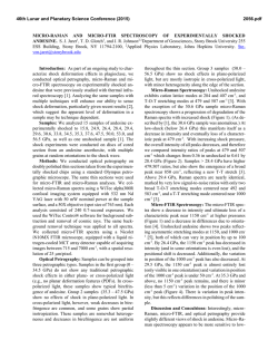
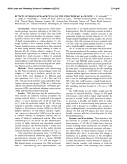
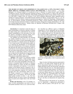
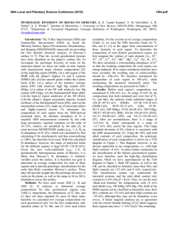
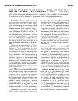
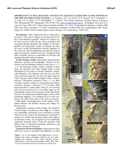
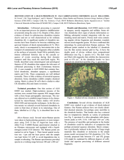
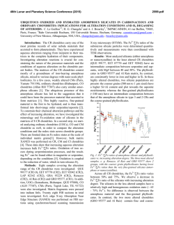
46th Lunar and Planetary Science Conference (2015) 2086.pdf COESITE AT THE LONAR CRATER: THE IMPORTANCE OF PRE-IMPACT ALTERATION AND SHOCK HETEROGENEITY. S. J. Jaret1, T. D. Glotch1, B. L. Phillips1, S. P. Wright2,3, and D. T. King, Jr.3, 1 Department of Geosciences, Stony Brook University 255 ESS Building, Stony Brook, NY 11794-2100, 2Planetary Science Institute, Tucson, AZ 3Department of Geosciences, Auburn University, AL 36849-3505, [email protected] Introduction: Coesite, a high-pressure SiO2 polymorph [1-2], is a well-known mineral associated with meteorite impact structures [3]. Even though it can be produced in extreme tectonic environments (e.g., eclogites [4]), natural coesite was first discovered in impactites, and its formation is often linked to shock metamorphism. Interestingly, coesite has only been reported from 28 of the ~190 known impact structures. Of these reports, overwhelmingly, coesite identification has not been made in-situ. Common methods for coesite identification include dissolving host silicates to concentrate coesite and whole-rock or powder spectroscopic (IR and XRD) analysis. Published photomicrographs of coesite are exceedingly uncommon (e.g., Ries [5]; Bosumtwi [6-7]). Petrographic context, however, may provide insight into coesite formation, but unfortunately such context is not commonly described in the literature. Here we present the first identification of coesite from the Lonar Crater (India) from both optical petrography and detailed micro-Raman imaging. Specific spatial location of coesite in these samples likely reflects the complex nature of a secondary silica precipitate within amygdaloidal basalts, which was then shocked. Methods: We conducted optical petrography and micro-Raman spectroscopy on doubly polished thin sections. Thin sections were slightly thicker than standard sections (~45 m rather than standard 30 m). We collected micro-Raman spectra using a WiTec alpha300R confocal imaging system equipped with 532 nm Nd YAG laser with 50 mW nominal power at the sample surface, and a spot size of 763 nm. Images were acquired over a 175 x 175 m area with acquisition times of 0.1 sec. Samples: We identified coesite in three samples, LC09-253, LC09-294, and LC09-256 (see [8] for detailed sample descriptions). Each is a shocked basalt of shock class 2 [9]. Labradorite has been transformed to maskelynite and is fully optically isotropic. Pyroxene grains, however, show little deformation and remain birefringent and lack fractures. Shock barometry suggests an average shock pressure in these rocks of ~2528 GPa [9]. Coesite occurs within bright white amygdules inside 1-2 mm vesicles. Optical Petrography: In thin section (Figure 1-2), 2 phases are identifiable optically within the vesiclefills. Phase 1 is clear in plane-polarized light, and optically isotropic in cross-polarized light. Small patches of brown glass are common, particularly associated with fractures. No flow textures are present, which could indicate it is a form of diaplectic glass. However, we cannot rule out the possibility of a high-pressure fused glass, and it is likely that this glass is a mix of multiple types of amorphous silica. Preliminarily Nuclear Magnetic Resonance spectroscopic results suggest the presence of a second amorphous phase. Neither micro-Raman nor optical petrography are able to easily resolve distinct silica-rich amorphous phases. Phase 2 occurs as 30-40 m high-relief, greenish spherical aggregates of smaller crystallites, consistent with coesite (“granular coesite” of [12]). Granular coesite occur primarily along the rim of the amygdule, but also concentrated in patches of the interior. Under higher power, individual aggregates can be recognized within the close-packed network of coesite occurring in the rim (Figure 2). Figure 1: Plane-Polarized light image of an amygdule containing amorphous SiO2 and coesite. Coesite is concentrated along the rim and in patches of the interior. Red box shown in Figure 2. Figure 2: ppl image of coesite rim shown in Figure 1. The rim itself is a closely packed network of coesite aggregates. Area in red shown in Figure 3. Micro-Raman Spectroscopy: We used microRaman spectroscopy to distinguished between the 2 optically-identifiable phases present: 1) SiO2 glass, characterized by a broad peak near 449 cm-1 and a dramatic drop-off in intensity at 494 cm-1, and 2) coesite, indicated by a spectrum with characteristic 46th Lunar and Planetary Science Conference (2015) peaks at 113, 173, 267, 429, and a strong Si-O mode at 521 cm-1 (Figures 3-4). Granular coesite, however, are not entirely pure coesite, and many are a mix of amorphous SiO2 and coesite. Figure 3: MicroRaman image of the amygdule rim. Red (Cs) indicates the primary coesite peak (521 cm-1), Green (Si) indicates amorphous SiO2 (449 cm-1 peak). Granular coesite contains internal amorphous silica. Figure 4: Selected micro-Raman spectra of coesite (“Cs” on Figure 3) and amorphous SiO2 (“Si” in Figure 3). Discussion: Raman imaging and optical petrography reveal coesite of 2 petrographic types: 1) coesite that occurs as isolated balls of granular coesite within amorphous SiO2 and 2) densely packed network of granular coesite concentrated along the rim and in the interior of amygdules. The individual or small groups of coesite crystals matches descriptions from other impact structures (e.g., Bosumtwi, [6-7], Xiuyan, [12]). The overlapping network of coesite aggregates, has not been reported from other impact structures. In these regions, coesite is so closely packed that they obscure optical recognition of individual aggregates. In slightly thicker sections, changing the focal depth shows that the close-packing is in 3 dimensions. This texture likely reflects complex interactions between the shock wave, host rock, vesicle wall, and amygdule. Pore space is a known source of shock heterogeneity, causing spikes in temperature (and to some extent pressure) due to perturbations of the shock wave [9-10]. Here, it is even more complex since the amygdule fills the vesicle, restricting the pore from collapsing during shock. Thus, we suggest the coesite crystal- 2086.pdf lized from a melt after spikes in temperature (at slightly elevated pressures) concentrated along the rim and interior as the shock wave passes through the host basalt, hit a free-surface of the vesicle wall, and then traveled into the amygdule itself. Intense refraction of the shock wave likely caused reverberations between the amygdule and the vesicle wall. Shock reverberation has been proposed for formation of natural impactproduced lechatelierite [11] but may also be an important process here. The coarse grained granular coesite is consistent with crystallization from a high-T melt [12]. Comparison with unshocked basalt from nearby Deccan flows shows quartz and opalline silica as common secondary minerals [8-9]. While the coesite in the shocked samples likely is transformed from crystalline quartz, we cannot rule out the possibility that it formed after a less-crystalline material. Conclusion: We present the first observation of coesite from the Lonar Crater. This is also the first report of a high-pressure polymorph as the product of a shocked secondary precipitate. The unusual texture of densely packed granular coesite concentrated along amygdule rims likely reflects extremely heterogeneous shock conditions due to complex interactions between the shock wave and SiO2 precipitates. Secondary silica is a common component of the target rocks at many impact structures, and so shocked secondary SiO2 may be more common than previously recognized on Earth [13] and Mars [14] References: [1] Coes, 1953. Science 118, 131132/. [2] Chao et al., 1960. Science 132, 220-222. [3] French and Koeberl, 2010. Earth Sci. Rev. 98, 123170. [4] Smith, 1984. Nature 310,461-464 [5] von Engelhardt and Stöffler, 1968. In Shock Metamorphism of Natural Materials, 159-168. [6] Morrow, 2007. MAPS 42, 591-609. [7] Ferrière et al., 2009. Eur. J. Mineral. 21, 203-217. [8] Wright et al., 2011. JGR 116, E09006, doi:10.1029/2010JE003785. [9] Kieffer et al., 1976. 7th Lunar Sci. Conf. 1391-1412. [10] Osinksi, 2007. MAPS 42, 1945-1960. [11] Stöffler and Langenhorst, 1994. Meteoritics 29, 155-181. [12] Chen et al., 2010. EPSL 297, 306-314. [13] Schmieder et al., 2011. MAPS 46, 574-586. [14] Michalski et al., (2003), Geophys. Res. Lett., 30, 2008, doi:10.1029/2003GL018354, 19. Acknowledgements: This work is supported by NASA Earth & Space Science Fellowship, RIS4E/SERVI and NASA PG&G award NNX14AP52G.
© Copyright 2026