
The Mood Stabilizer Lithium Potentiates the Antidepressant-Like
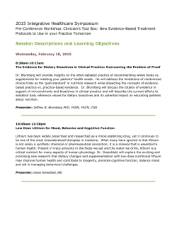
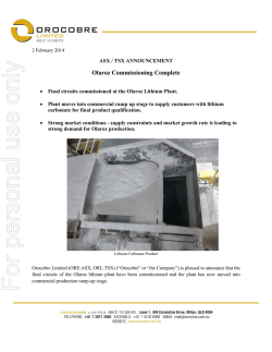
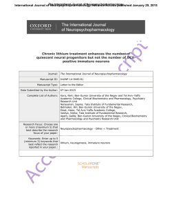
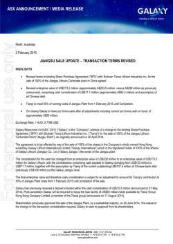
International Journal of Neuropsychopharmacology Advance Access published January 31, 2015
International Journal of Neuropsychopharmacology, 2015, 1–13
doi:10.1093/ijnp/pyu102
Research Article
research article
The Mood Stabilizer Lithium Potentiates the
Antidepressant-Like Effects and Ameliorates
Oxidative Stress Induced by Acute Ketamine in a
Mouse Model of Stress
Chi-Tso Chiu, PhD; Lisa Scheuing, BS; Guangping Liu, BS; Hsiao-Mei Liao, PhD;
Gabriel R. Linares, PhD; Dora Lin, BA; De-Maw Chuang, PhD
Section on Molecular Neurobiology, National Institute of Mental Health, National Institutes of Health,
Bethesda, MD.
Correspondence: De-Maw Chuang, PhD, Section on Molecular Neurobiology, National Institute of Mental Health, National Institutes of Health, Building
10, Room 3D41, 10 Center Drive MSC 1363, Bethesda, MD 20892–1363 ([email protected]); and Chi-Tso Chiu, PhD, Section on Molecular Neurobiology,
National Institute of Mental Health, National Institutes of Health, Building 10, Room 3D39, 10 Center Drive MSC 1363, Bethesda, MD 20892–1363 (chiuc@
mail.nih.gov).
Abstract
Background: Evidence suggests that mammalian target of rapamycin activation mediates ketamine’s rapid but transient
antidepressant effects and that glycogen synthase kinase-3β inhibits this pathway. However, ketamine has associated
psychotomimetic effects and a high risk of abuse. The mood stabilizer lithium is a glycogen synthase kinase-3 inhibitor with
strong antisuicidal properties. Here, we used a mouse stress model to investigate whether adjunct lithium treatment would
potentiate ketamine’s antidepressant-like effects.
Methods: Mice received chronic restraint stress and long-term pre- or postketamine lithium treatment in drinking water.
The effects of lithium on ketamine-induced antidepressant-like effects, activation of the mammalian target of rapamycin/
brain-derived neurotrophic factor signaling pathways, oxidative stress, and dendritic spine density in the brain of mice were
investigated.
Results: Subtherapeutic (600 mg/L) lithium-pretreated mice exhibited an antidepressant-like response to an ineffective
ketamine (2.5 mg/kg, intraperitoneally) challenge in the forced swim test. Both the antidepressant-like effects and restoration
of dendritic spine density in the medial prefrontal cortex of stressed mice induced by a single ketamine (50 mg/kg) injection
were sustained by postketamine treatment with 1200 mg/L of lithium for at least 2 weeks. These benefits of lithium treatments
were associated with activation of the mammalian target of rapamycin/brain-derived neurotrophic factor signaling pathways
in the prefrontal cortex. Acute ketamine (50 mg/kg) injection also significantly increased lipid peroxidation, catalase activity,
and oxidized glutathione levels in stressed mice. Notably, these oxidative stress markers were completely abolished by
pretreatment with 1200 mg/L of lithium.
Conclusions: Our results suggest a novel therapeutic strategy and justify the use of lithium in patients who benefit from
ketamine.
Keywords: ketamine, lithium, mTOR, GSK-3, BDNF
Received: September 18, 2014; Revised: October 31, 2014; Accepted: November 21, 2014
Published by Oxford University Press on behalf of CINP 2015. This work is written by (a) US Government employee(s)
and is in the public domain in the US.
1
2 | International Journal of Neuropsychopharmacology, 2015
Introduction
Methods
Depression in individuals with either major depressive disorder (MDD) or bipolar disorder (BD) is one of the leading causes
of the global disease burden. Almost all current antidepressant
drugs in clinical use require weeks to months to take full effect
(Adell et al., 2005), and a significant proportion of patients do not
respond to available agents (Insel and Wang, 2009). Ketamine,
a noncompetitive N-methyl-D-aspartate receptor antagonist,
has been safely used as an anesthetic and analgesic agent
for many years. Recent clinical and preclinical research indicates that, in treatment-resistant MDD and BD subjects, a single subanesthetic dose of ketamine not only produces a rapid
antidepressant effect within hours of administration, but also
improves suicidal ideation (Berman et al., 2000; Zarate et al.,
2006; DiazGranados et al., 2010).
Activation of mammalian target of rapamycin (mTOR) and
subsequent synaptogenesis in the prefrontal cortex (PFC) have
been suggested to mediate ketamine’s rapid antidepressant
effects (Li et al., 2010; Dwyer and Duman, 2013). The loss of
synaptic function in the PFC, as well as the downregulation
of synaptic proteins such as the postsynaptic density protein
95 (PSD95), were associated with depressive-like behaviors
in a rodent model of chronic stress (Li et al., 2011). Through
activation of the mTOR signaling pathway, acute injection of
ketamine rapidly increased levels of these synaptic proteins
and dendritic spine density; in contrast, inhibition of mTOR
signaling prevented these synaptic actions and the antidepressant-like effects of ketamine in experimental animals (Li
et al., 2010).
In clinical populations, repeated ketamine treatment is
usually necessary to avoid subsequent relapse (Zarate et al.,
2006; Ibrahim et al., 2012). However, ketamine has been used
as a recreational drug and has a high risk of abuse. Repeated
administration of ketamine can cause a variety of side effects,
including hallucinations and cognitive impairments, and psychotomimetic symptoms (Krystal et al., 2005). In fact, ketamine
was found to induce schizophrenia-like behaviors in humans
(Krystal et al., 2003), and treating animals with subanesthetic
doses of ketamine is a pharmacological model of schizophrenia (Gunduz-Bruce, 2009). In addition, administration of subanesthetic doses of ketamine increases oxidative stress in the
rodent brain (Zuo et al., 2007; de Oliveira et al., 2009; da Silva
et al., 2010). These adverse effects limit ketamine’s potential for
widespread clinical use.
Accumulating evidence indicates that the mood stabilizer lithium has strong antisuicidal properties (Cipriani
et al., 2005) and holds promise for treating other neurological
and neurodegenerative diseases via its diverse mechanisms
of action (Chiu and Chuang, 2010; Chiu et al., 2013). Among
them, lithium’s ability to inhibit glycogen synthase kinase-3
(GSK-3), a ubiquitous serine-threonine kinase, has been considered critical to mediating its numerous mood-stabilizing
and neuroprotective effects. Ketamine’s rapid antidepressant effects require GSK-3 inhibition (Beurel et al., 2011). In
addition, GSK-3 negatively regulated mTOR in mouse brain
(Sarkar et al., 2008). These findings indicate that lithium and
ketamine may have a signaling convergence on the mTOR
pathway and a possible mechanistic synergy on their antidepressant-like effects. The present study was undertaken
to investigate whether combining ketamine with the mood
stabilizer lithium could benefit ketamine’s antidepressant
effects in mice as well as protect against the oxidative stress
associated with ketamine use.
Animals and Chronic Restraint Stress
Male CD-1 mice were purchased from Charles River Laboratory
(Wilmington, MA), and chronic restraint stress was performed as
previously described (Omata et al., 2011). Briefly, mice were placed
into a Plexiglas tube (2.5 cm in diameter) individually for 2 hours
once a day for 2 weeks (supplementary Figure S1a). All procedures
for animal experiments were approved by the Animal Care and
Use Committee of the National Institutes of Health (NIH).
Drug Treatment and Measurement of Serum Lithium
Concentration
Ketamine hydrochloride (10 mg/mL; Vedco, St. Joseph, MO) was
freshly diluted in saline (0.9%) before use and injected intraperitoneally at a volume of 10 mL/kg of body weight. To ensure
a steady-state serum concentration, mice were pretreated with
lithium chloride through drinking water (600 or 1200 mg/L) for 3
weeks prior to acute saline or ketamine challenge (supplementary
Figure S1b). For postketamine lithium treatment, another cohort of
stressed mice received lithium chloride treatment (1200 mg/L) in
drinking water following a single ketamine challenge (supplementary Figure S1c). Serum lithium concentrations of mice were measured by MEDTOX Laboratories (Saint Paul, MN; test code 60063).
Behavioral Tests
Mice underwent the open-field test (OFT) 60 minutes after acute
ketamine injection, followed by the forced swim test (FST) 30
minutes later. To investigate the behavioral effects of postketamine lithium treatment, both tests were repeated in the same
order after 1 and 2 weeks of lithium treatment using the same
group of mice (supplementary Figure S1).
OFT
Under bright illumination from regular ceiling lights, mice were
placed individually in the center of a clear open-field chamber
(27.9 cm × 27.9 cm × 20 cm), and their horizontal locomotor activities were measured by using an automated photo-beam openfield system (Med-Associates, St. Albans, VT) for 30 minutes.
FST
Briefly, each mouse was placed for 6 minutes in a 25-cm–high,
16-cm–diameter transparent cylindrical tank filled with room
temperature water (~22°C). Immobility was scored during the
last 4 minutes of the session, and a decrease in swimming time
was considered a measure of depressive-like behaviors. All test
sessions were analyzed off-line by ANY-maze video-tracking
system (Stoelting, Wood Dale, IL).
Tail-Suspension Test
Briefly, mice were suspended individually by the tail using nonirritating adhesive tape placed at about one-half the total tail
length to a hook connected to a horizontal bar. Each test session
lasted for 6 minutes and total immobility time was analyzed offline by ANY-maze system (Stoelting).
Brain Sample Preparation and Western-Blot Analysis
Brain tissues were homogenized in 20 mM 4-(2-hydroxyethyl)1-piperazineethanesulfonic acid containing 0.32 M sucrose and
centrifuged at 700 g for 10 minutes at 4°C. The supernatants
Chiu et al. | 3
were centrifuged again at 14,000 g for 10 minutes at 4°C, and the
pellets were resuspended in T-PER reagent (Thermo Scientific,
Rockford, IL). Proteins were separated and transferred onto a
nitrocellulose membrane. Blots were immunostained overnight
at 4°C with primary antibody against total GSK-3β (BD, Franklin
Lakes, NJ), phospho-GSK-3β at Ser9, total Akt (the serine/threonine kinase, also known as protein kinase B or PKB), phosphoAkt at Ser473, total extracellular signal-regulated kinases (ERKs),
phospho-ERK at Thr202/Tyr204, total mTOR, phospho-mTOR at
Ser2448, total P70S6 kinase (P70S6K), phospho-P70S6K at Thr389,
total eukaryotic elongation factor-2 (eEF2), phospho-eEF2 at
Thr56, PSD95 (all from Cell Signaling, Beverly, MA), total tropomyosin-related kinase B (TrkB; Millipore, Billerica, MA), phospho-TrkB at Tyr817, or the house-keeping gene β-actin (Abcam,
Cambridge, MA). Membranes were then incubated with secondary antibodies (LI-COR, Lincoln, NE) for 1 hour at room temperature. Finally, blotted proteins were detected and quantified using
the Odyssey infrared imaging system (LI-COR).
Analysis of Oxidative Stress
Mice were sacrificed by decapitation 20 minutes after acute
ketamine challenge, and the brains were dissected and homogenized according to the buffer requirements of each assay.
Thriobarbituric Acid Reactive Substances Assay
Assay of thriobarbituric acid reactive substances, byproducts
of lipid peroxidation, was performed according to the manufacturer’s instructions (Cayman Chemical, Ann Arbor, MI). The
production of malondialdehyde was normalized by protein
concentration.
Catalase Activity Assay
This assay was performed according to the manufacturer’s
instructions (Cayman Chemical). The production rate of formaldehyde was normalized by protein concentration.
Glutathione Assay
Analyses of reduced and oxidized glutathione levels were conducted per the manufacturer’s instructions (Cayman Chemical).
The oxidized glutathione content was expressed as the ratio to
total (reduced and oxidized) glutathione.
Analysis of Dendritic Spine Density
Mice were sacrificed and brains were subjected to Golgi-staining
(FD NeuroTechnologies, Columbia, MD) at the time indicated.
Briefly, coronal sections of 100 µm in thickness were prepared,
and both basal and apical dendrites (~50 and ~100 µm from soma,
respectively) of pyramidal neurons in layer V of medial PFC (anterior cingulate and prelimbic) were chosen for quantitative analysis. Images were captured by an Olympus BX61 microscope, and
the length of dendritic segments was determined by using ImageJ
software from NIH. Spine numbers in ~30-µm segments were
measured manually by investigators blind to the experimental
conditions. Two segments from each neuron were analyzed, and
the results were expressed as number of spines per µm.
Statistical Analyses
All statistical analyses were performed using GraphPad Prism
(GraphPad, San Diego, CA). Data are expressed as mean ± SEM
and analyzed using t test or 1-way analysis of variance. When
necessary, multiple comparisons between groups were assessed
with posthoc Student–Newman–Keuls multiple comparison
test. Statistical significance was considered at P < .05.
Results
Antidepressant-Like Effects of Ketamine in CD-1
Mouse Model of Stress
To mimic a clinical situation, the chronic restraint stress paradigm was used for the detection of antidepressant-like effects
of ketamine and lithium. The depressive-like behaviors of this
stress model can be suppressed by 20 mg/kg of the antidepressant desipramine (Omata et al., 2011). In the present study, chronic
restraint paradigm markedly increased immobility time in the
FST and tail suspension test (TST) by 140.8 ± 12.1% (t(14) = 2.93,
P = .011) and 124.2 ± 7.6% (t(14) = 2.59, P=.021), respectively, compared
with unstressed mice (Figure 1a). A wide range of ketamine doses
(1–100 mg/kg) has been reported to effectively produce antidepressant-like effects in various mouse strains (Mantovani et al.,
2003; Hayase et al., 2006; Kos et al., 2006; Popik et al., 2008; Browne
and Lucki, 2013). We thus performed a dose response test in our
CD-1 mouse model of stress and found that the minimum dose
for ketamine to suppress immobility time in the FST was 50 mg/kg
(72.2 ± 6.5% of control, F[3, 26] = 3.02, P < .05) (Figure 1b). This effective
dose of ketamine was confirmed by a decreased immobility time in
the TST performed 24 hours after FST (72.6 ± 8.7% of control, F[3, 26]
= 0.046, P < .05) (Figure 1b). However, mice that received this dose of
ketamine also exhibited hyperlocomotion as measured by the OFT
(169.8 ± 13.2% of control, F[3, 26] = 5.03, P < .01) (Figure 1c).
Preketamine Treatment with a Subtherapeutic Dose
of Lithium Potentiates the Antidepressant-Like
Effects Induced by a Low Dose of Ketamine
Long-term treatment is usually necessary for lithium to reach a
steady-state serum level and exert its effects. Our pilot study indicated that the serum concentration of lithium in CD-1 mice after
3 weeks of treatment with 600 mg/L of the drug in drinking water
(0.202 ± 0.029 mEq/L; n = 8) was below the therapeutic levels for
human patients with BD (~0.5–1.2 mEq/L) (American Psychiatric
Association, 2002). No differences were observed for measures of
body weight (Figure 2a) or locomotor activity (Figure 2b) between
controls and mice that received this dose of lithium for 3 weeks.
In stressed mice, pretreatment with 600 mg/L of lithium
alone had no significant effects on immobility time in the FST
(Figure 2c). Interestingly, stressed mice that had been pretreated
with this subtherapeutic dose of lithium had a robust antidepressant-like response to acute challenge with a very low dose
(2.5 mg/kg) of ketamine (78.76 ± 4.38% of control, F[5, 52] = 4.34,
P < .05) (Figure 2c) that, administered alone, had no significant
antidepressant effect (Figure 1b). Treatment with this dose of lithium alone or together with a 2.5-mg/kg ketamine challenge did
not affect locomotor activity of stressed mice (Figure 2d). These
data show that the combination of “ineffective” doses of lithium
and ketamine can produce synergy on antidepressant-like effects
and suggest a signaling convergence between these two drugs.
Preketamine Treatment with a Subtherapeutic Dose
of Lithium Potentiates the Activation of mTOR/
BDNF-TrkB Signaling Pathways Induced by a Low
Dose of Ketamine
As mentioned above, activation of the mTOR pathway in the PFC
is critical to ketamine’s rapid antidepressant-like effect. In the
4 | International Journal of Neuropsychopharmacology, 2015
at threonine 56 induced by eEF2 kinase, a downstream effector
of the mTOR pathway that is inhibited by P70S6K (Wang et al.,
2001). Again, pretreatment with 600 mg/L of lithium together
with acute injection of 2.5 mg/kg of ketamine robustly decreased
eEF2 phosphorylation (60.25 ± 4.46%, P < .05) and upregulated the
expression of synaptic protein PSD95 (150.44 ± 14.91%, P < .05)
in the PFC compared with control stressed mice. Neither drug
alone had significant effects (Figure 3a).
In addition to the mTOR pathway, GSK-3β inhibition (Beurel
et al., 2011) and acute increases in brain-derived neurotrophic
factor (BDNF) protein levels (Garcia et al., 2008; Autry et al.,
2011), including activation of the downstream effectors Akt
and ERK (Li et al., 2010), are also necessary for ketamine’s rapid
antidepressant actions. Similar to the observations described
above, we found that only pretreatment with 600 mg/L of lithium plus subsequent challenge with 2.5 mg/kg of ketamine
significantly increased phosphorylation levels of TrkB (a BDNF
receptor; 123.02 ± 6.49%, P < .05), Akt (184.18 ± 8.57%, P < .01), ERK
(156.59 ± 14.37%, P < .05), and GSK-3β (206.21 ± 10.40%, P < .01)
(Figure 3b) in the PFC compared with control stressed mice.
Treatment with either drug alone had no significant effects. It
is interesting to note that chronic restraint stress alone markedly decreased GSK-3β phosphorylation in this brain region
(76.10 ± 5.23%, t(12) = 3.69, P < .01) compared with unstressed
mice (Figure 3b).
Preketamine Treatment with a Low Therapeutic
Dose of Lithium Suppresses Acute Ketamineinduced Oxidative Stress
Figure 1. Dose response of acute ketamine challenge in stressed mice. Chronic
restraint stress produced depressive-like behaviors in CD-1 mice, as assessed
by increased immobility time in the forced swim test (FST) and tail-suspension
test (TST) (a). In stressed mice, acute injection of ketamine at a dose of 50 mg/kg
not only significantly suppressed immobility in both tests (b), but also increased
locomotor activity measured by the open-field test (OFT) (c). Data are mean ± SEM
(n = 6–8). +P < .05, t test; *P < .05, **P < .01, compared with control groups, according
to Student–Newman–Keuls multiple comparison test after a 1-way analysis of
variance (ANOVA).
present study, mice that had undergone chronic restraint stress
showed a trend towards decreased phosphorylation of mTOR
and its downstream effector P70S6K in the PFC, though the
effect was not statistically significant compared with unstressed
mice (Figure 3a). In stressed mice, acute injection with a behaviorally ineffective dose (2.5 mg/kg) of ketamine or pretreatment
with 600 mg/L of lithium alone had no significant effects, while
a combination with these two treatments strongly enhanced
the phosphorylation of mTOR (136.30 ± 6.47%, P < .01) and P70S6K
(163.25 ± 11.58%, P < .01) in the PFC (Figure 3a).
We also found that stressed mice showed increased phosphorylation of eEF2 (145.37 ± 9.96% of unstressed mice, t(12) = 4.47,
P < .01) (Figure 3a), an essential component required for polypeptide elongation in protein synthesis (Wang et al., 2001). The
activity of eEF2 is negatively regulated by its phosphorylation
In our mouse model of stress, we found that acute injection with
ketamine at a dose that produces an antidepressant-like effect
(50 mg/kg) markedly increased lipid peroxidation (141.49 ± 7.29%
of control, P < .01) (Figure 4a), catalase activity (120.64 ± 5.79% of
control, P < .01) (Figure 4b), and levels of oxidized glutathione
(130.90 ± 7.75% of control, P < .01) (Figure 4c) in the PFC 20 minutes
after injection. Similar results were also observed in the hippocampus (lipid peroxidation: 173.40 ± 14.26%, P < .01; catalase activity:
145.69 ± 9.92%, P < .01; oxidized glutathione: 142.60 ± 6.62%, P < .05)
and striatum (lipid peroxidation: 140.86 ± 12.37%, P < .01; catalase
activity: 121.37 ± 6.14%, P < .05; oxidized glutathione: 138.70 ± 7.27%,
P < .05; compared with control stressed mice) (Figure 4).
To investigate whether lithium can protect against the oxidative stress induced by ketamine, mice were pretreated with a
higher dose (1200 mg/L) of lithium in drinking water for 3 weeks
before ketamine challenge. The serum concentration of lithium after 3 weeks of treatment with this dose was at the lower
end (0.483 ± 0.052 mEq/L; n = 8) of the therapeutic spectrum for
human patients with BD. Long-term treatment with this dose of
lithium did not affect either body weight (Figure 2a) or locomotor activity of mice (Figure 2 ). Compared with control stressed
mice, lithium pretreatment robustly suppressed ketamineinduced lipid peroxidation (89.35 ± 13.09%, P < .01) (Figure 4a),
catalase activity (85.16 ± 3.40%, P < .01) (Figure 4b), and levels of
oxidized glutathione (103.49 ± 7.73%, P < .01) (Figure 4c) in the
PFC. These ketamine-induced oxidative metabolism markers in the hippocampus (lipid peroxidation: 121.23 ± 10.34%,
P < .01; catalase activity: 109.80 ± 7.42%, P < .01; oxidized glutathione: 106.85 ± 10.40%, P < .05) and striatum (lipid peroxidation: 94.04 ± 7.48%, P < .01; catalase activity: 100.37 ± 5.64%, P < .01;
oxidized glutathione: 106.12 ± 8.19%, P < .05, compared with
control stressed mice) were also reduced by lithium pretreatment (Figure 4). Although stress is an important contributor to
intracellular reactive oxygen species (ROS) generation, we did
Chiu et al. | 5
Figure 2. Preketamine treatment with a subtherapeutic dose of lithium potentiates a low dose of ketamine-induced antidepressant-like effects. In CD-1 mice, longterm treatment with a sub- (600 mg/L) or low therapeutic dose (1200 mg/L) of lithium chloride in drinking water for 3 weeks did not affect body weight (a) or locomotor
activity (b). Pretreatment with 600 mg/L of lithium alone had no effect on immobility time in saline-challenged stressed mice (lithium alone group), but significantly
potentiated the response induced by a very low dose of ketamine (2.5 mg/kg) in the forced swim test (FST) (lithium + ketamine group) (c). The locomotor activity of
stressed mice measured by the open-field test (OFT) was not affected by any given treatment (d). Data are mean ± SEM (n = 6–12). *P < .05, **P < .01, according to Student–
Newman–Keuls multiple comparison test after a 1-way analysis of variance (ANOVA).
not observe a significant increase in ROS formation induced by
chronic restraint stress (data not shown).
Postketamine Treatment with a Low Therapeutic
Dose of Lithium Maintains the Antidepressant-Like
Effect and Prevents the Hyperlocomotion Induced by
a Single Injection of Ketamine
Although a single infusion of ketamine exerts rapid antidepressant effects, these typically last for only 1 week in human
patients and experimental animals (Berman et al., 2000; Zarate
et al., 2006; Autry et al., 2011; Ibrahim et al., 2012). We thus
sought to investigate whether lithium treatment after ketamine
injection might prolong its antidepressant-like effect in our
mouse stress model, thereby assessing its potential as a relapse
prevention therapeutic strategy.
To ensure the potential effects of lithium, the higher dose
(1200 mg/L) was preferred in this part of tests. Treatment with
this dose of lithium that produced a low therapeutic serum
concentration for 1 (Figure 5a) or 2 weeks (Figure 5b) produced
no antidepressant-like effect in the FST in saline-challenged
stressed mice. In contrast, the stressed mice that had received
a single injection with 50 mg/kg of ketamine still showed
decreased immobility time in the FST after 1 week compared
with control (F[3, 28] = 3.69, P = 0.024). Postketamine treatment
with 1200 mg/L of lithium for 1 week did not further suppress
this effect (Figure 5a). Consistent with findings in the literature, the ketamine-induced antidepressant-like effects were
not sustained for 2 weeks (Figure 5b). However, the immobility
time of mice that received both ketamine injection and lithium
treatment remained decreased after 2 weeks compared with
control (F[3, 28] = 5.18, P = .005) (Figure 5b). On the other hand, the
hyperlocomotion associated with ketamine injection lasted for at
least 2 weeks (Figure 5c-d). Interestingly, this ketamine-induced
long-lasting hyperlocomotion was completely prevented by postketamine lithium treatment measured at either 1 (F[3, 28] = 3.88,
P = .019) (Figure 5c) or 2 weeks (F[3, 28] = 3.73, P = .023) (Figure 5d)
following ketamine injection, while the same lithium treatment
did not affect the OFT in saline-challenged mice.
Postketamine Treatment with a Low Therapeutic
Dose of Lithium Prolongs the Activation of mTOR/
BDNF-TrkB Signaling Pathways Induced by a Single
Injection of Ketamine
One week after a single injection of 50 mg/kg ketamine, we
observed a trend towards elevated phosphorylation of mTOR
(123.17 ± 15.73%, not significant), P70S6K (154.76 ± 15.36%,
P < .05) (Figure 6a), TrkB (122.19 ± 5.18%, not significant), Akt
(137.52 ± 8.42%, P < .05), ERK (120.39 ± 14.93%, not significant), and
GSK-3β (156.77 ± 13.79%, P < .05) (Figure 7a), while eEF2 phosphorylation was decreased (76.56 ± 4.47%, P < .05) (Figure 6a).
Although not significant, PSD95 expression also appeared to
be upregulated in the PFC (124.79 ± 11.27%) compared with control stressed mice (Figure 6a). Treatment with 1200 mg/L of lithium for 1 week had no significant effects in saline-challenged
stressed mice and did not affect the regulatory effects of ketamine described above (Figures 6a and 7a).
Compared with unstressed mice, GSK-3β phosphorylation
remained significantly decreased in the PFC of stressed mice
6 | International Journal of Neuropsychopharmacology, 2015
Figure 3. Preketamine treatment with a subtherapeutic dose of lithium potentiates the activation of mammalian target of rapamycin (mTOR)/brain-derived neurotrophic factor (BDNF)-tropomyosin-related kinase B (TrkB) signaling pathways induced by acute challenge with a low dose of ketamine. Mice were pretreated with
600 mg/L of lithium for 3 weeks, and brain tissues were collected 60 minutes after a single injection of saline (lithium alone group) or a very low dose (2.5 mg/kg) of
ketamine (lithium + ketamine group). Typical Western blots and quantified results are shown. The phosphorylation levels of mTOR, P70S6K, eukaryotic elongation factor-2 (eEF2), and the expression of postsynaptic density protein 95 (PSD95) (a), as well as the phosphorylation levels of TrkB, Akt, extracellular signal-regulated kinase
(ERK), and glycogen synthase kinase-3β (GSK-3β) (b) in the prefrontal cortex (PFC) were normalized to the levels of total protein or β-actin and expressed as percentage
of control group. Data are mean ± SEM (n = 4–8). ++P < .01, t test; *P < .05, **P < .01, according to Student–Newman–Keuls multiple comparison test after a 1-way analysis of
variance (ANOVA). P, phosphorylated protein; T, total protein; US, unstressed.
1 week (68.23 ± 6.46%, t(10) = 3.33, P < .01) (Figure 7a), but not 2
weeks, after chronic restraint stress (Figure 7b). Similar to the
observation in the behavioral study, the cellular changes induced
by ketamine alone lasted for only approximately 1 week. No significant effects were obtained 2 weeks after a single injection
with 50 mg/kg of ketamine (Figures 6 and 7 ). However, postketamine treatment with lithium for 2 weeks prolonged and
potentiated the regulatory effects of ketamine on the expression of PSD95 (141.91 ± 11.25%, P < .01) as well as phosphorylation
of mTOR (145.15 ± 12.02%, P < .05), P70S6K (153.70 ± 8.97%, P < .05),
eEF2 (71.30 ± 5.25%, P < .05) (Figure 6b), TrkB (150.38 ± 19.44%,
P < .05), Akt (142.66 ± 7.60%, P < .05), ERK (146.24 ± 7.86%, P < .05),
and GSK-3β (139.08 ± 5.20%, P < .05) (Figure 7b) compared with
control stressed mice. These results suggest that the behavioral
effects of lithium treatment on ketamine’s antidepressant-like
effects appear to be closely associated with GSK-3β inhibition
and stimulation of the mTOR/BDNF-TrkB signaling pathways.
Postketamine Treatment with a Low Therapeutic
Dose of Lithium Maintains the Restoration of
Dendritic Spine Density Induced by a Single
Injection of Ketamine
An increase in the density of dendritic spines in the medial PFC
has been reported to be one of the mTOR-dependent mechanisms
underlying ketamine’s antidepressant-like effects (Li et al., 2010;
Liu et al., 2013). We thus analyzed the effects of postketamine
lithium treatment on alterations in spine density induced by a
single injection of ketamine. Compared with unstressed mice,
the spine densities were found decreased in the medial PFC at 1
(69.55 ± 3.20%, t(70) = 7.78, P < .01) and 2 weeks (78.85 ± 3.96%, t(70)
= 3.81, P < .01) after chronic restraint stress (Figure 8). Compared
with control stressed mice, the spine densities in this brain region
still remained increased 1 week after a single injection with 50 mg/
kg of ketamine (134.79 ± 6.62% of control, F[3, 140] = 15.80, P < .01),
while this effect was not further potentiated by 1-week postketamine treatment with 1200 mg/L of lithium (153.07 ± 5.50% of
control) (Figure 8a-b). Ketamine-induced normalization in spine
density was not sustained for 2 weeks (103.71 ± 5.01% of control,
not significant). However, the density of spines remained elevated
in the medial PFC of mice that received both ketamine injection and lithium treatment for 2 weeks compared with control
stressed mice (128.31 ± 4.36%, F[3, 140] = 6.26, P < .01) (Figure 8a, c).
Discussion
The present study found that pretreatment of stressed mice with
600 mg/L of lithium markedly potentiated the antidepressantlike effect induced by 2.5 mg/kg of ketamine, which by itself was
ineffective. This is particularly important, because this low-dose
Chiu et al. | 7
Figure 4. Preketamine treatment with a low therapeutic dose of lithium suppresses acute ketamine-induced oxidative stress. Acute ketamine injection at a dose that
produces antidepressant-like effect (50 mg/kg) markedly increased lipid peroxidation (a), catalase activity (b), and the levels of oxidized glutathione (c) in the prefrontal
cortex (PFC), hippocampus (HPC), and striatum (STR) of stressed mice 20 minutes after injection. Compared with control stressed mice, preketamine treatment with
1200 mg/L of lithium for 3 weeks robustly suppressed the oxidative metabolism markers induced by acute ketamine (lithium + ketamine group) in these brain regions.
Data are mean ± SEM (n = 6–10). *P < .05, **P < .01, according to Student–Newman–Keuls multiple comparison test after a one-way analysis of variance (ANOVA).
combination might avoid the possible adverse effects associated
with individual drugs. Indeed, we observed an elevated level of
oxidative stress in the mouse brain in conjunction with ketamine’s rapid antidepressant-like action, but this side effect was
mitigated by pretreatment with 1200 mg/L of lithium. Also notable
was that the antidepressant-like effect induced by a single injection with 50 mg/kg of ketamine was prolonged by postketamine
treatment with 1200 mg/L of lithium for at least 2 weeks. We demonstrated that the behavioral benefits of lithium treatment on
ketamine’s antidepressant effects were associated with GSK-3β
inhibition, stimulation of the mTOR/BDNF-TrkB signaling pathways, and restoration in dendritic spine density in the medial PFC.
These data support a recent rat study showing that GSK-3 inhibition potentiates the antidepressant-like effects of subthreshold
doses of ketamine (Liu et al., 2013), and are consistent with a clinical observation that lithium-treated BD patients expressed greater
antianhedonic responses to ketamine (Lally et al., 2014).
Presumably due to significant strain differences in sensitivity
to ketamine, the effective dose (50 mg/kg) for ketamine to produce antidepressant-like effects in our mouse model of stress
was high and caused hyperlocomotion. However, this dose was
the result of our dose-response experiment and is also comparable with other studies. For example, acute ketamine was
reported to induce antidepressant-like effects at doses of 30 to
8 | International Journal of Neuropsychopharmacology, 2015
Figure 5. Postketamine treatment with a low therapeutic dose of lithium prolongs the antidepressant-like effects and prevents the hyperlocomotion induced by a
single injection of ketamine. Stressed mice received long-term treatment with 1200 mg/L of lithium in drinking water immediately after a single injection of saline
(lithium alone group) or 50 mg/kg of ketamine (ketamine + lithium group). The effects of lithium on ketamine-induced antidepressant-like effects (assessed by the
forced swim test [FST]) (a-b) and locomotor activity (assessed by the open-field test [OFT]) (c-d) were measured after 1 (a, c) and 2 weeks (b, d) of lithium treatment. Data
are mean ± SEM (n = 8).*P < .05, **P < .01, according to Student–Newman–Keuls multiple comparison test after a 1-way analysis of variance (ANOVA).
100 mg/kg in male ICR mice using FST (Hayase et al., 2006), 50
and 66 mg/kg in male C57BL/6J/Han mice using TST (Kos et al.,
2006), and 50 mg/kg in male Swiss mice using FST (Popik et al.,
2008). Mice that received 50 mg/kg of ketamine challenge indeed
appeared slightly impaired during the first 30 minutes after
injection; however, no obvious lingering sedative or anesthetic
conditions were observed while performing the OFT and FST. In
addition, since hyperlocomotion is known to affect immobility
in the FST, we performed TST as an additional indicator in the
ketamine dose-response study. Although also dependent on a
motor readout, the TST avoids the need of swimming and has
been suggested to be more relevant for the study of animals
with compromised motor coordination (Cryan et al., 2005). Most
importantly, stressed mice that received both ketamine and lithium treatment showed decreased immobility time in the FST
but did not exhibit hyperlocomotion (Figures 2 and 5).
Compared with other published reports (Dehpour et al., 1995,
2002; Ghasemi et al., 2009), the serum lithium concentrations of
stressed mice in our study were relatively low. We speculated
that this discrepancy might be caused by restraint stress, which
may reduce water consumption, or because of the difference in
the experimental conditions (eg, sucrose in water) or animals.
As expected, long-term treatment with either 600 or 1200 mg/L
of lithium alone produced no effects on behavioral tests and
most of the biochemical measurements (including GSK-3β
phosphorylation). This may explain why the potentiation effects
of postketamine lithium were obscure when ketamine’s effects
were still evident (1 week) but became obvious after ketamine’s
effect subsided (2 weeks). In fact, our preliminary data showed
that treatment with 2400 mg/L of lithium not only produced a
therapeutic serum concentration (0.897 ± 0.250 mEq/L, n = 8) in
stressed mice but also decreased immobility time in the FST
(data not shown).
Ketamine-induced increase in dendritic spine density in the
medial PFC was identified dependent on activation of mTOR
pathway (Li et al., 2010) and can be mimicked by GSK-3 inhibition (Liu et al., 2013). Studies have noted that the expression of
both mTOR and P70S6K is decreased in the postmortem brain of
depressed subjects (Jernigan et al., 2011), suggesting a compromised mTOR pathway. We observed a trend towards decreased
phosphorylation of these 2 proteins (Figure 3a) and a sustained
reduction in dendritic spine density in the PFC of stressed mice
(Figure 8). In addition, rats that suffered from chronic mild stress
showed increased GSK-3β expression in the hippocampus (Silva
et al., 2008), and elevated GSK-3 activity was found in postmortem samples from individuals with MDD (Karege et al., 2007).
Consistent with previous hippocampal data (Omata et al., 2011),
chronic restraint stress also significantly decreased GSK-3β
phosphorylation in the PFC (Figure 3b), and this effect lasted for
at least 1 week after chronic restraint stress ended (Figure 7).
These findings provide additional justification for the use of this
restraint paradigm in the present study. It is interesting to note
that the increased GSK-3β phosphorylation in rat PFC observed
during ketamine’s rapid antidepressant actions was not affected
by mTOR or P70S6K antagonists (Zhou et al., 2014), further supporting the notion that GSK-3β is an upstream regulator of mTOR.
BDNF plays a central role in synaptic plasticity and mediates the clinical efficacy of antidepressants and anxiolytic drugs.
Chiu et al. | 9
Figure 6. Postketamine treatment with a low therapeutic dose of lithium maintains the activation of the mammalian target of rapamycin (mTOR) signaling pathway
induced by a single injection of ketamine. Stressed mice received long-term treatment with 1200 mg/L of lithium in drinking water immediately after a single injection
of saline (lithium alone group) or 50 mg/kg of ketamine (ketamine + lithium group), and brain tissues were collected after 1 (a) or 2 weeks (b) of lithium treatment. Typical Western blots and quantified results are shown. Phosphorylation levels of mTOR, P70S6K, eukaryotic elongation factor-2 (eEF2), and the expression of postsynaptic
density protein 95 (PSD95) in the prefrontal cortex (PFC) were normalized to the levels of total protein or β-actin and expressed as percentage of control group. Data
are mean ± SEM (n = 6–8). *P < .05, **P < .01, according to Student–Newman–Keuls multiple comparison test after a 1-way analysis of variance (ANOVA). P, phosphorylated
protein; T, total protein; US, unstressed.
Ketamine-induced antidepressant-like effects were absent in
BDNF knockout mice (Autry et al., 2011) and abolished by inhibitors of Akt or ERK; both are upstream regulators of mTOR (Li
et al., 2010). We found that lithium treatment also potentiated
and prolonged acute ketamine’s effect on phosphorylation of
TrkB, Akt, and ERK in stressed mice, suggesting the activation of
this pathway. By inhibiting GSK-3β, lithium was demonstrated to
activate BDNF promoter IV in primary cortical neurons (Yasuda
et al., 2009) and upregulate BDNF expression in the rat brain
(Fukumoto et al., 2001). Presumably through BDNF-TrkB receptor signaling, lithium treatment may subsequently enhance
activation of its downstream effectors Akt and ERK. Moreover,
Akt mediates the phosphorylation of GSK-3β while ketamine
exerts its rapid antidepressant effects (Zhou et al., 2014). By disrupting the formation of β-arrestin 2/protein phosphatase 2A
(PP2A)/Akt complex that inactivates Akt, lithium was reported
to activate Akt and in turn inhibit GSK-3 (Beaulieu et al., 2008).
These findings suggest a positive feedback loop and indicate
that mTOR may act as a node receiving multiple upstream regulatory effects induced by lithium.
Under nutrient-deprived stress conditions, activated eEF2
kinase was found to block protein translation by phosphorylating
eEF2 (Carlberg et al., 1990). We similarly observed an increased
eEF2 phosphorylation in stressed mice (Figure 3a). Echoing this
finding, other studies reported reductions in eEF2 phosphorylation in the PFC (Carrier and Kabbaj, 2013) and hippocampus
(Autry et al., 2011) of rodents following ketamine administration.
Moreover, ketamine-induced augmentation of BDNF synthesis
was found to be eEF2 dependent (Monteggia et al., 2013), and
inhibitors of eEF2 kinase produced a fast-acting antidepressantlike effect in mice (Autry et al., 2011). These results suggest that
eEF2 is sensitive to stress and involved in mediating ketamine’s
antidepressant-like effects. In contrast to eEF2 kinase, eEF2 can
be positively regulated by dephosphorylation with PP2A (Nairn
and Palfrey, 1987). Lithium was found to upregulate PP2A activity
in the rat brain (Tsuji et al., 2003). Therefore, lithium may have
multiple regulatory effects on eEF2 dephosphorylation in addition to activation of the mTOR/P70S6K signaling pathway that
inhibits eEF2 kinase.
Oxidative stress plays a significant role in the pathogenesis of many neurological and psychiatric diseases such as
schizophrenia and BD (Kuloglu et al., 2002). Elevated ROS production was observed from 10 minutes in mouse brains (da
Silva et al., 2010) to 30 minutes in rat brains (de Oliveira et al.,
2009) after acute injection of subanesthetic doses of ketamine.
Consistently, we found that ROS production was significantly
elevated 20 minutes after ketamine injection at a dose that produces antidepressant-like effects (Figure 4). Many studies have
shown that restraint stress increases ROS formation (Fontella
et al., 2005). Perhaps due to the variations in severity of the
restraint stress or the oxidative metabolism markers measured, we did find a trend towards increased ROS production in
10 | International Journal of Neuropsychopharmacology, 2015
Figure 7. Postketamine treatment with a low therapeutic dose of lithium maintains the inhibition of glycogen synthase kinase-3β (GSK-3β) and activation of brainderived neurotrophic factor (BDNF)-tropomyosin-related kinase B (TrkB) signaling pathway induced by a single injection of ketamine. Stressed mice received long-term
treatment with 1200 mg/L of lithium in drinking water immediately after a single injection of saline (lithium alone group) or 50 mg/kg of ketamine (ketamine + lithium
group), and brain tissues were collected after 1 (a) or 2 weeks (b) of lithium treatment. Typical Western blots and quantified results are shown. Phosphorylation levels
of TrkB, Akt, extracellular signal-regulated kinase (ERK), and GSK-3β in the prefrontal cortex (PFC) were normalized to the levels of total protein and expressed as percentage of control group. Data are mean ± SEM (n = 6–8). ++P < .01, t test; *P < .05, **P < .01, according to Student–Newman–Keuls multiple comparison test after a 1-way
analysis of variance (ANOVA). P, phosphorylated protein; T, total protein; US, unstressed.
Figure 8. Postketamine treatment with a low therapeutic dose of lithium maintains the dendritic spine density restored by a single injection of ketamine. Representative Golgi-stained sections of spines on dendrites of pyramidal neurons in layer V of medial prefrontal cortex (PFC) are shown (scale bar indicates 5 µm) (a). Stressed
mice received long-term treatment with 1200 mg/L of lithium in drinking water immediately after a single injection of saline (lithium alone group) or 50 mg/kg of
ketamine (ketamine + lithium group). Quantified data were obtained from brain samples of unstressed (US) and stressed mice after 1 (b) or 2 weeks (c) of lithium treatment. Data are mean ± SEM (n = 36). ++P < .01, t test; **P < .01, according to Student–Newman–Keuls multiple comparison test after a 1-way analysis of variance (ANOVA).
Chiu et al. | 11
the brain of stressed mice, but the effect was not statistically
significant.
Lithium has been reported to ameliorate ROS levels in an
animal model of mania (Frey et al., 2006) and suppress elevated
oxidative metabolism markers, including thriobarbituric acid
reactive substances and catalase in unmedicated manic patients
(Machado-Vieira et al., 2007). The present study is the first to
demonstrate that the oxidative stress induced by ketamine can
be completely blocked by pretreatment with a low therapeutic
dose of lithium (Figure 4), underscoring lithium’s neuroprotective aspects. Similar to our observations, ketamine was found to
produce hyperlocomotion in rodents at doses related to its antidepressant effects (da Silva et al., 2010). Interestingly, antioxidant treatment inhibited ketamine-induced hyperlocomotion in
mice (de Araujo et al., 2011). In our study, treatment with a low
therapeutic dose of lithium prevented ketamine-induced hyperlocomotor activity (Figure 5c-d). Although further experiments
are needed, these data suggest that lithium’s antioxidant effects
may be part of its underlying mechanisms. Taken together, the
antioxidant effects of lithium provide an additional benefit and
justification for its adjunctive use with ketamine in the treatment of depression.
In conclusion, the present findings highlight the ability of
lithium—when given in conjunction with ketamine—to augment and prolong both clinical efficacy and remission in the
treatment of depression. Numerous clinical and preclinical studies have underscored ketamine’s remarkable antidepressant
effects. Nevertheless, given the inevitable relapse of depressive
symptoms and the potential for abuse, ketamine is not a viable
long-term clinical option. Thus, in providing a novel therapeutic
strategy to solve this important clinical issue, our results could
be of significant clinical relevance.
Supplementary Material
For supplementary material accompanying this paper, visit
http://www.ijnp.oxfordjournals.org/
Acknowledgments
This work was supported by the Intramural Research Program
of the National Institute of Mental Health, National Institutes of
Health, Department of Health and Human Services (IRP-NIMHNIH-DHHS). The authors thank Ioline Henter of the NIMH, NIH,
for critical review and editorial assistance with this manuscript.
Statement of Interest
None.
References
Adell A, Castro E, Celada P, Bortolozzi A, Pazos A, Artigas F (2005)
Strategies for producing faster acting antidepressants. Drug
Discov Today 10:578–585.
American Psychiatric A (2002) Practice guideline for the treatment of patients with bipolar disorder (revision). Am J Psychiatry 159:1–50.
Autry AE, Adachi M, Nosyreva E, Na ES, Los MF, Cheng PF, Kavalali ET, Monteggia LM (2011) NMDA receptor blockade at rest
triggers rapid behavioural antidepressant responses. Nature
475:91–95.
Beaulieu JM, Marion S, Rodriguiz RM, Medvedev IO, Sotnikova TD,
Ghisi V, Wetsel WC, Lefkowitz RJ, Gainetdinov RR, Caron MG
(2008) A beta-arrestin 2 signaling complex mediates lithium
action on behavior. Cell 132:125–136.
Berman RM, Cappiello A, Anand A, Oren DA, Heninger GR, Charney DS, Krystal JH (2000) Antidepressant effects of ketamine
in depressed patients. Biol Psychiatry 47:351–354.
Beurel E, Song L, Jope RS (2011) Inhibition of glycogen synthase
kinase-3 is necessary for the rapid antidepressant effect of
ketamine in mice. Mol Psychiatry 16:1068–1070.
Browne CA, Lucki I (2013) Antidepressant effects of ketamine:
mechanisms underlying fast-acting novel antidepressants.
Front Pharmacol 4:161.
Carlberg U, Nilsson A, Nygard O (1990) Functional properties of
phosphorylated elongation factor 2. Eur J Biochem 191:639–
645.
Carrier N, Kabbaj M (2013) Sex differences in the antidepressantlike effects of ketamine. Neuropharmacology 70:27–34.
Chiu CT, Chuang DM (2010) Molecular actions and therapeutic
potential of lithium in preclinical and clinical studies of CNS
disorders. Pharmacol Ther 128:281–304.
Chiu CT, Wang Z, Hunsberger JG, Chuang DM (2013) Therapeutic potential of mood stabilizers lithium and valproic acid:
beyond bipolar disorder. Pharmacol Rev 65:105–142.
Cipriani A, Pretty H, Hawton K, Geddes JR (2005) Lithium in the
prevention of suicidal behavior and all-cause mortality in
patients with mood disorders: a systematic review of randomized trials. Am J Psychiatry 162:1805–1819.
Cryan JF, Mombereau C, Vassout A (2005) The tail suspension
test as a model for assessing antidepressant activity: review
of pharmacological and genetic studies in mice. Neurosci
Biobehav Rev 29:571–625.
da Silva FCC, do Carmo de Oliveira Cito M, da Silva MIG, Moura
BA, de Aquino Neto MR, Feitosa ML, de Castro Chaves R,
Macedo DS, de Vasconcelos SMM, de França Fonteles MM,
de Sousa FCF (2010) Behavioral alterations and pro-oxidant
effect of a single ketamine administration to mice. Brain Res
Bull 83:9–15.
de Araujo FY, de Oliveira GV, Gomes PX, Soares MA, Silva MI,
Carvalho AF, de Moraes MO, de Moraes ME, Vasconcelos SM,
Viana GS, de Sousa FC, Macedo DS (2011) Inhibition of ketamine-induced hyperlocomotion in mice by the essential oil
of Alpinia zerumbet: possible involvement of an antioxidant
effect. J Pharm Pharmacol 63:1103–1110.
de Oliveira L, Spiazzi CM, Bortolin T, Canever L, Petronilho F,
Mina FG, Dal-Pizzol F, Quevedo J, Zugno AI (2009) Different
sub-anesthetic doses of ketamine increase oxidative stress in
the brain of rats. Prog Neuropsychopharmacol Biol Psychiatry
33:1003–1008.
Dehpour AR, Farsam H, Azizabadi-Farahani M (1995) Inhibition
of the morphine withdrawal syndrome and the development
of physical dependence by lithium in mice. Neuropharmacology 34:115–121.
Dehpour AR, Sadr SS, Azizi MR, Namiranian K, Farahani M,
Javidan AN (2002) Lithium inhibits the development of physical dependence to clonidine in mice. Pharmacol Toxicol
90:89–93.
DiazGranados N, Ibrahim LA, Brutsche NE, Ameli R, Henter ID,
Luckenbaugh DA, Machado-Vieira R, Zarate CA Jr (2010) Rapid
resolution of suicidal ideation after a single infusion of an
N-methyl-D-aspartate antagonist in patients with treatment-resistant major depressive disorder. J Clin Psychiatry
71:1605–1611.
Dwyer JM, Duman RS (2013) Activation of mammalian target of
rapamycin and synaptogenesis: role in the actions of rapidacting antidepressants. Biol Psychiatry 73:1189–1198.
12 | International Journal of Neuropsychopharmacology, 2015
Fontella FU, Siqueira IR, Vasconcellos AP, Tabajara AS, Netto CA,
Dalmaz C (2005) Repeated restraint stress induces oxidative
damage in rat hippocampus. Neurochem Res 30:105–111.
Frey BN, Valvassori SS, Reus GZ, Martins MR, Petronilho FC, Bardini K, Dal-Pizzol F, Kapczinski F, Quevedo J (2006) Effects of
lithium and valproate on amphetamine-induced oxidative
stress generation in an animal model of mania. J Psychiatry
Neurosci 31:326–332.
Fukumoto T, Morinobu S, Okamoto Y, Kagaya A, Yamawaki S
(2001) Chronic lithium treatment increases the expression of
brain-derived neurotrophic factor in the rat brain. Psychopharmacology (Berl) 158:100–106.
Garcia LS, Comim CM, Valvassori SS, Reus GZ, Barbosa LM,
Andreazza AC, Stertz L, Fries GR, Gavioli EC, Kapczinski F,
Quevedo J (2008) Acute administration of ketamine induces
antidepressant-like effects in the forced swimming test and
increases BDNF levels in the rat hippocampus. Prog Neuropsychopharmacol Biol Psychiatry 32:140–144.
Ghasemi M, Sadeghipour H, Poorheidari G, Dehpour AR (2009)
A role for nitrergic system in the antidepressant-like effects
of chronic lithium treatment in the mouse forced swimming
test. Behav Brain Res 200:76–82.
Gunduz-Bruce H (2009) The acute effects of NMDA antagonism:
from the rodent to the human brain. Brain Res Rev 60:279–286.
Hayase T, Yamamoto Y, Yamamoto K (2006) Behavioral effects of
ketamine and toxic interactions with psychostimulants. BMC
Neurosci 7:25.
Ibrahim L, Diazgranados N, Franco-Chaves J, Brutsche N, Henter
ID, Kronstein P, Moaddel R, Wainer I, Luckenbaugh DA, Manji
HK, Zarate CA Jr (2012) Course of improvement in depressive
symptoms to a single intravenous infusion of ketamine vs
add-on riluzole: results from a 4-week, double-blind, placebocontrolled study. Neuropsychopharmacology 37:1526–1533.
Insel TR, Wang PS (2009) The STAR*D trial: revealing the need for
better treatments. Psychiatr Serv 60:1466–1467.
Jernigan CS, Goswami DB, Austin MC, Iyo AH, Chandran A, Stockmeier CA, Karolewicz B (2011) The mTOR signaling pathway in
the prefrontal cortex is compromised in major depressive disorder. Prog Neuropsychopharmacol Biol Psychiatry 35:1774–1779.
Karege F, Perroud N, Burkhardt S, Schwald M, Ballmann E, La
Harpe R, Malafosse A (2007) Alteration in kinase activity but
not in protein levels of protein kinase B and glycogen synthase kinase-3beta in ventral prefrontal cortex of depressed
suicide victims. Biol Psychiatry 61:240–245.
Kos T, Popik P, Pietraszek M, Schafer D, Danysz W, Dravolina O,
Blokhina E, Galankin T, Bespalov AY (2006) Effect of 5-HT3
receptor antagonist MDL 72222 on behaviors induced by ketamine in rats and mice. Eur Neuropsychopharmacol 16:297–310.
Krystal JH, D’Souza DC, Mathalon D, Perry E, Belger A, Hoffman R
(2003) NMDA receptor antagonist effects, cortical glutamatergic
function, and schizophrenia: toward a paradigm shift in medication development. Psychopharmacology (Berl) 169:215–233.
Krystal JH, Perry EB Jr, Gueorguieva R, Belger A, Madonick SH, AbiDargham A, Cooper TB, Macdougall L, Abi-Saab W, D’Souza
DC (2005) Comparative and interactive human psychopharmacologic effects of ketamine and amphetamine: implications for glutamatergic and dopaminergic model psychoses
and cognitive function. Arch Gen Psychiatry 62:985–994.
Kuloglu M, Ustundag B, Atmaca M, Canatan H, Tezcan AE, Cinkilinc N (2002) Lipid peroxidation and antioxidant enzyme levels in patients with schizophrenia and bipolar disorder. Cell
Biochem Funct 20:171–175.
Lally N, Nugent AC, Luckenbaugh DA, Ameli R, Roiser JP, Zarate
CA (2014) Anti-anhedonic effect of ketamine and its neural
correlates in treatment-resistant bipolar depression. Translational psychiatry 4:e469.
Li N, Lee B, Liu RJ, Banasr M, Dwyer JM, Iwata M, Li XY, Aghajanian
G, Duman RS (2010) mTOR-dependent synapse formation
underlies the rapid antidepressant effects of NMDA antagonists. Science 329:959–964.
Li N, Liu RJ, Dwyer JM, Banasr M, Lee B, Son H, Li XY, Aghajanian
G, Duman RS (2011) Glutamate N-methyl-D-aspartate receptor antagonists rapidly reverse behavioral and synaptic
deficits caused by chronic stress exposure. Biol Psychiatry
69:754–761.
Liu RJ, Fuchikami M, Dwyer JM, Lepack AE, Duman RS, Aghajanian
GK (2013) GSK-3 inhibition potentiates the synaptogenic and
antidepressant-like effects of subthreshold doses of ketamine. Neuropsychopharmacology 38:2268–2277.
Machado-Vieira R, Andreazza AC, Viale CI, Zanatto V, Cereser V, Jr.,
da Silva Vargas R, Kapczinski F, Portela LV, Souza DO, Salvador M,
Gentil V (2007) Oxidative stress parameters in unmedicated and
treated bipolar subjects during initial manic episode: a possible
role for lithium antioxidant effects. Neurosci Lett 421:33–36.
Mantovani M, Pértile R, Calixto JB, Santos ARS, Rodrigues ALS
(2003) Melatonin exerts an antidepressant-like effect in the
tail suspension test in mice: evidence for involvement of
N-methyl-d-aspartate receptors and the l-arginine-nitric
oxide pathway. Neurosci Lett 343:1–4.
Monteggia LM, Gideons E, Kavalali ET (2013) The role of eukaryotic elongation factor 2 kinase in rapid antidepressant action
of ketamine. Biol Psychiatry 73:1199–1203.
Nairn AC, Palfrey HC (1987) Identification of the major Mr
100,000 substrate for calmodulin-dependent protein kinase
III in mammalian cells as elongation factor-2. J Biol Chem
262:17299–17303.
Omata N, Chiu CT, Moya PR, Leng Y, Wang Z, Hunsberger JG,
Leeds P, Chuang DM (2011) Lentivirally mediated GSK-3beta
silencing in the hippocampal dentate gyrus induces antidepressant-like effects in stressed mice. Int J Neuropsychopharmacol 14:711–717.
Popik P, Kos T, Sowa-Kucma M, Nowak G (2008) Lack of persistent
effects of ketamine in rodent models of depression. Psychopharmacology (Berl) 198:421–430.
Sarkar S, Krishna G, Imarisio S, Saiki S, O’Kane CJ, Rubinsztein
DC (2008) A rational mechanism for combination treatment
of Huntington’s disease using lithium and rapamycin. Hum
Mol Genet 17:170–178.
Silva R, Mesquita AR, Bessa J, Sousa JC, Sotiropoulos I, Leao P,
Almeida OF, Sousa N (2008) Lithium blocks stress-induced
changes in depressive-like behavior and hippocampal cell
fate: the role of glycogen-synthase-kinase-3beta. Neuroscience 152:656–669.
Tsuji S, Morinobu S, Tanaka K, Kawano K, Yamawaki S (2003) Lithium, but not valproate, induces the serine/threonine phosphatase activity of protein phosphatase 2A in the rat brain,
without affecting its expression. J Neural Transm 110:413–425.
Wang X, Li W, Williams M, Terada N, Alessi DR, Proud CG (2001)
Regulation of elongation factor 2 kinase by p90(RSK1) and p70
S6 kinase. EMBO J 20:4370–4379.
Yasuda S, Liang MH, Marinova Z, Yahyavi A, Chuang D-M (2009)
The mood stabilizers lithium and valproate selectively activate the promoter IV of brain-derived neurotrophic factor in
neurons. Mol Psychiatry 14:51–59.
Zarate CA Jr, Singh JB, Carlson PJ, Brutsche NE, Ameli R, Luckenbaugh DA, Charney DS, Manji HK (2006) A randomized trial of
an N-methyl-D-aspartate antagonist in treatment-resistant
major depression. Arch Gen Psychiatry 63:856–864.
Chiu et al. | 13
Zhou W, Dong L, Wang N, Shi JY, Yang JJ, Zuo ZY, Zhou ZQ
(2014) Akt Mediates GSK-3beta phosphorylation in the rat
prefrontal cortex during the process of ketamine exerting
rapid antidepressant actions. Neuroimmunomodulation
21:183–188.
Zuo DY, Wu YL, Yao WX, Cao Y, Wu CF, Tanaka M (2007) Effect of
MK-801 and ketamine on hydroxyl radical generation in the
posterior cingulate and retrosplenial cortex of free-moving
mice, as determined by in vivo microdialysis. Pharmacol Biochem Behav 86:1–7.

© Copyright 2026