
Chronic lithium treatment enhances the number of quiescent neural
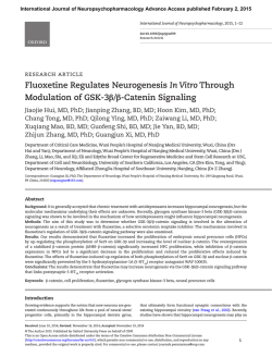
International Journal of Neuropsychopharmacology
International Journal ofThe
Neuropsychopharmacology
Advance Access published January 29, 2015
Chronic lithium treatment enhances the number of
quiescent neural progenitors but not the number of DCXpositive immature neurons
Journal:
The International Journal of Neuropsychopharmacology
Manuscript ID:
IntJNP-14-0449.R1
Manuscript Type:
Letter to the Editor
Date Submitted by the Author:
Complete List of Authors:
Research Focus: Choose one
or more (maximum 5) that
best describe the research
focus of your paper:
Keywords: Enter up to 5
(minimum 3) keywords that
best reflect the research
reported in your paper.:
07-Jan-2015
Kara, Nirit; Ben-Gurion University of the Negev and Tel Aviv-Yaffo
Academic College, Clinical Biochemistry and Pharmacology, Psychiatry
Research Unit
Narayanan, Sapna; Tata Institute of Fundamental Research,
Belmaker, RH; Ben-Gurion University of the Negev,
Einat, Haim; Tel Aviv-Yaffo Academic College,
Vaidya, Vidita; Tata Institute of Fundamental Research,
Agam, Galila; Ben-Gurion University of the Negev, Clinical Biochemistry
and Pharmacology and Psychiatry Research Unit
Neuropsychopharmacology - Other < Treatment
lithium, neurogenesis, immature neurons
Page 1 of 11
The International Journal of Neuropsychopharmacology
Chronic lithium treatment enhances the number of quiescent neural
progenitors but not the number of DCX-positive immature neurons
Kara N1,2,3,4, Narayanan S5, Belmaker RH2, Einat H4,6, Vaidya VA5, Agam G1,2,3
1
Department of Clinical Biochemistry and Pharmacology and 2Psychiatry Research
Unit, 1,2Faculty of Health Sciences, 1,2Ben-Gurion University of the Negev and
3
Mental Health Center, Beer-Sheva, Israel; 4Tel Aviv-Yaffo Academic College, Tel
Aviv-Yaffo, Israel; 5Tata Institute of Fundamental Research, Mumbai, India;
6
University of Minnesota, Minnesota, USA
Correspondence:
Galila Agam, PhD
J. Dreyfus Professor of Biochemistry in Psychiatry
Psychiatry Research Unit and
Department of Clinical Biochemistry and Pharmacology
Faculty of Health Sciences
Ben-Gurion University of the Negev
Beersheva, Israel
Tel: 972-8-6401737
Fax: 972-8-6401740
E-mail: [email protected]
Vidita A. Vaidya, PhD
Associate Professor
Department of Biological Sciences
Tata Institute of Fundamental Research
Colaba, Mumbai 400005, India
Tel: +91 22 22782608
Fax: +91 22 22804610
E-mail: [email protected]
Statistical Summary
Word count - body of the manuscript: 1252
- abstract: 232
Number of references - 11
Number of figures - 1; no tables
Category: letter to the editor
© The Author 2014. Published by Oxford University Press on behalf of CINP.
This is an Open Access article distributed under the terms of the Creative Commons Attribution License (http://creativecommons.org/licenses/by
/4.0/), which permits unrestricted reuse, distribution, and reproduction in any medium, provided the original work is properly cited.
1
The International Journal of Neuropsychopharmacology
Page 2 of 11
Abstract
Background The term adult neurogenesis constitutes a series of
developmental steps including the birth, survival, differentiation, maturation and even
death of newborn progenitor cells within neurogenic niches. Within the hippocampus
progenitors reside in the neurogenic niche of the subgranular zone (SGZ) in the
dentate gyrus (DG) subfield. At the different stages, designated type-I, type-IIa, typeIIb, type-III and granule cell neurons, the cells express a series of markers enabling
their identification and visualization.
Lithium has been shown to increase hippocampal cell proliferation in the SGZ
of the hippocampal DG subfield of adult rodents and to stimulate the proliferation of
hippocampal progenitor cells in vitro but data regarding lithium's ability to increase
neuronal differentiation and survival is equivocal.
Methods To clarify the effect of lithium on adult hippocampal neurogenesis
we identified the effect of chronic lithium treatment on distinct stages of hippocampal
progenitor
development
using
adult
Nestin-GFP
transgenic
mice
and
immunofluorescent techniques.
Results The present observations confirm that lithium targets the initial stages
of progenitor development enhancing the turnover of quiescent neural progenitors
(QNP)/putative stem-cells corroborating previous reports. However, the enhanced
QNP-turnover does not translate into an increased number of immature neurons. We
also observed a steep decline in the number of type-III immature neurons with
complex tertiary-dendrites suggesting that lithium alters the morphological maturation
of newborn neurons.
Conclusions Our results do not corroborate previous reports of lithiuminduced enhanced numbers of newly generated neurons.
2
Page 3 of 11
The International Journal of Neuropsychopharmacology
The term adult neurogenesis constitutes a series of developmental steps
including the birth, survival, differentiation, maturation and even death of newborn
progenitor cells within neurogenic niches. During the proliferation, fate choice and
eventual maturation of these progenitors they transiently express a series of markers
including nestin, glial fibrillary acidic protein (GFAP), doublecortin (DCX) and
neuronal nuclei (NeuN), enabling the identification and visualization of adult
progenitors at discrete stages of their development. Within the hippocampus
progenitors reside in the neurogenic niche of the subgranular zone (SGZ) in the
dentate gyrus (DG) subfield. Slowly dividing radial glia-like progenitors (type-I
cells/quiescent neural progenitors-QNPs), expressing both nestin and GFAP, divide
into transiently amplifying, rapidly dividing progenitors (type-IIa cells/Amplifying
neural progenitors-ANPs) that express nestin, but are no longer immunopositive for
GFAP. Type-IIa cells differentiate into neuroblasts (type-IIb cells) expressing both
nestin and DCX. Finally, migrating neuroblasts and immature neurons (type-III cells)
express DCX and elaborate their dendritic and axonal arbors to mature into granule
cell neurons expressing NeuN that integrate into functional hippocampal circuitry
[Figure 1A (Ming and Song, 2011)].
Lithium has been shown to increase hippocampal cell proliferation in the SGZ
of the hippocampal DG subfield of adult rodents and to stimulate the proliferation of
hippocampal progenitor cells in vitro (Chen et al., 2000). However, existing data
regarding lithium's ability to increase neuronal differentiation and survival is
equivocal. While some studies show that lithium induces differentiation towards a
neuronal fate, as demonstrated by an increase in the number of NeuN-positive cells
both in vitro and in vivo (Kim et al., 2004), in contrast reports also indicate that
despite the increase in the number of proliferating cells in the DG the percentage of
NeuN-positive neurons generated remains unchanged (Chen et al., 2000; Li et al.,
2011). In addition, a recent study suggests that the effects of lithium on progenitor
turnover and survival are restricted to the ventral hippocampus, and emerge only in
animals concomitantly subjected to stress (O'Leary et al., 2012). The focus of the
current study was to identify the effects of lithium on distinct stages of hippocampal
progenitor development to clarify the effect of lithium on adult hippocampal
neurogenesis (AHN).
Male Nestin-GFP transgenic mice (2-months) expressing transgenic greenfluorescent-protein (GFP) under the Nestin promoter (Yu et al., 2005), a kind gift of
3
The International Journal of Neuropsychopharmacology
S.G. Kernie (Columbia University, USA), were bred and group-housed at the TataInstitute of Fundamental Research. Mice were supplied with food and water adlibitum and maintained on a 12/12 hrs light/dark cycle at 24±1oC. The protocol was
approved by the Tata-Institute of Fundamental Research animal-experimentation
ethics-committee and was in-line with the NIH-guide for the care and use of
laboratory animals. Mice were treated with regular powdered food (RF) or lithiumsupplemented RF (Li) for 14 days. The Li group received chow mixed with 0.2% LiCl
for five days followed by 0.4% LiCl for ten additional days (O'Brien et al., 2004).
Mean plasma Li levels were 1.15 mM ± 0.38 (S.D.). Mice were sacrificed by
transcardial perfusion with 4% paraformaldehyde; brains removed, allowed to sink in
30% sucrose, frozen and 40 µm sections generated on a sliding microtome. To detect
GFP/GFAP/DCX-positive cells a series of four hippocampal sections were
selected/brain and GFP/GFAP/DCX triple immunofluorescent staining performed
(Kapoor et al., 2012). To address the changes in AHN, immunohistochemistry with
DCX, a microtubule-associated protein was performed. The number of GFP- and
DCX-positive cells within the DG was quantified in four sections/animal using a
Zeiss Axioskop at 40X in a blind manner. The morphological status of DCX-positive
immature neurons was assessed by categorizing them as DCX+ cells with/without
tertiary-dendrites. For quantification of triple immunofluorescence, 100 GFP-positive
cells were assessed/animal to determine the number of nestin-GFP/GFAP and
nestin/DCX double-positive cells using z-plane sectioning with 1 µm steps on a Zeiss
LSM5 Exciter laser-scanning microscope to confirm co-localization.
Chronic lithium-treatment did not alter the total number of nestin-GFPpositive progenitors within the SGZ of the DG (Figure 1B), however it significantly
increased the percentage of Nestin-GFP/GFAP double-positive cells, indicating
increased QNPs/type-I cell proliferation (Figure 1C&D). Lithium-treatment did not
affect the percentage of Nestin-GFP/DCX double-positive cells (Figure 1E) indicating
a lack of effect on type-IIb (neuroblasts). Lithium-treatment also had no effect on the
number of DCX-positive cells/section indicating that immature neuron number was
unaltered (Figure 1F). However, the significant (~75%) decline in the percentage of
DCX-positive newborn neurons bearing complex tertiary-dendrites suggests
decreased morphological maturation (Figure 1G).
4
Page 4 of 11
Page 5 of 11
The International Journal of Neuropsychopharmacology
The results confirm that lithium targets the initial stages of progenitor
development enhancing the turnover of QNP/putative stem-cells corroborating
previous reports (Chen et al., 2000) indicating lithium-induced enhanced progenitor
proliferation assessed with bromodeoxyuridine (BrdU) within the SGZ. Our findings
extend this observation demonstrating that lithium targets the QNP stage similarly to
electroconvulsive-seizure treatment which enhances QNP-turnover, but distinctly
from pharmacological antidepressants reported to influence ANPs-turnover (Encinas
et al., 2006; Segi-Nishida et al., 2008). However, it should be noted that Green and
Nolan (2008) did not find lithium-induced change in BrdU-labelled cells under
proliferation conditions in vitro (Green and Nolan, 2012). This discrepancy might
stem from the difference between the in vivo vs. the in vitro conditions. Similarly to
Green and Nolan, we found no effect of lithium on total number of DCX-positive
immature neurons suggesting that the observed enhanced QNP-turnover does not
translate into an increased number of immature neurons. Strikingly, we also discerned
a steep decline in the number of DCX-positive immature neurons with complex
tertiary-dendrites suggesting that lithium alters newborn neurons morphological
maturation.
Our results do not corroborate previous reports of lithium-induced
enhanced neurogenesis, which reported enhanced numbers of generated newborn
neurons (Kim et al., 2004). However, both Chen et al. (2000) and Li et al. (2011)
observed lithium-induced increased hippocampal cell-proliferation but did not find a
specific increase of neuronal differentiation. While lithium treatment increased the
number of BrdU-positive cells in the adult DG, numbers of BrdU- and NeuN-double
positive cells remained unchanged. Methodological issues might provide explanation
for the different results: 1) In-vitro vs. in-vivo lithium-treatment; 2) Different markers
used to identify the neurogenesis stages; 3) Different regimes of in-vivo lithium
administration.
We further report that lithium significantly decreased the number of type-III
cells exhibiting tertiary-dendrites by 75%. Mature dendrites are more complex and
have tertiary branches which extend to the outer molecular layer. This result is
seemingly contradictory with the well-established lithium-induced elevated cell
survival attributed to lithium's inhibition of glycogen-synthase kinase-3. However,
O’Leary et al. (2012) recently reported that chronic lithium treatment significantly
decreased the survival of newly-born cells in the DG. Newborn neurons have primary
dendrites that further split-off into secondary and tertiary dendritic branches. Hence,
5
The International Journal of Neuropsychopharmacology
Page 6 of 11
our result corroborates with lithium-induced decreased survival of newly-born
neurons. It is noteworthy that in the nematode Caenorhabditis-elegans (C. elegans)
lithium-treatment as well as mutations in the gene encoding for the enzyme inositolmonophosphatase
(IMPase),
inhibited
by
therapeutically-relevant
lithium
concentrations, cause incorrect localization of neuronal synaptic proteins and
behavioral defects, both rescued by expression of IMPase or by supplementation of
inositol (the product of IMPase) (Tanizawa et al., 2006). These results suggest that
IMPase is involved in the localization of synaptic components and in normal
behavior. It is tempting to speculate that our observation of reduced number of typeIII neurons with tertiary-dendrites and the decreased survival of newborn neurons
(O'Leary et al., 2012) following chronic lithium treatment are mediated by the drug's
inhibitory effect on IMPase.
Our results suggest that while lithium targets the QNP stage similarly to other
fast-acting antidepressants, this does not translate into enhanced numbers of newborn
neurons and rather appears to eventually evoke a decline in the morphological
maturation of newborn neurons.
6
Page 7 of 11
The International Journal of Neuropsychopharmacology
References
Chen G, Rajkowska G, Du F, Seraji-Bozorgzad N, Manji HK (2000)
Enhancement of hippocampal neurogenesis by lithium. J Neurochem 75:17291734.
Encinas JM, Vaahtokari A, Enikolopov G (2006) Fluoxetine targets early
progenitor cells in the adult brain. Proc Natl Acad Sci 103:8233-8238.
Kapoor R, Desouza LA, Nanavaty IN, Kernie SG, Vaidya VA (2012) Thyroid
hormone accelerates the differentiation of adult hippocampal progenitors. J
Neuroendocrinol 24:1259-1271.
Kim JS, Chang MY, Yu IT, Kim JH, Lee SH, Lee YS, Son H (2004) Lithium
selectively increases neuronal differentiation of hippocampal neural progenitor
cells both in vitro and in vivo. J Neurochem 89:324-336.
Li H, Li Q, Du X, Sun Y, Wang X, Kroemer G, Blomgren K, Zhu C (2011)
Lithium-mediated long-term neuroprotection in neonatal rat hypoxia-ischemia is
associated with antiinflammatory effects and enhanced proliferation and
survival of neural stem/progenitor cells. Journal of cerebral blood flow and
metabolism. J Cereb Blood Flow Metab 31:2106-2115.
Ming GL, Song H (2011) Adult neurogenesis in the mammalian brain:
significant answers and significant questions. Neuron 70:687-702.
O'Brien WT, Harper AD, Jove F, Woodgett JR, Maretto S, Piccolo S, Klein PS
(2004). Glycogen synthase kinase-3beta haploinsufficiency mimics the behavioral
and molecular effects of lithium. J Neuroscience 24:6791-6798.
O'Leary OF, O'Connor RM, Cryan JF (2012). Lithium-induced effects on adult
hippocampal neurogenesis are topographically segregated along the dorsoventral axis of stressed mice. Neuropharmacol 62:247-255.
Segi-Nishida E, Warner-Schmidt JL, Duman RS (2008). Electroconvulsive
seizure and VEGF increase the proliferation of neural stem-like cells in rat
hippocampus. Proc Natl Acad Sci 105:11352-11357.
Tanizawa Y, Kuhara A, Inada H, Kodama E, Mizuno T, Mori I (2006). Inositol
monophosphatase regulates localization of synaptic components and behavior in
the mature nervous system of C. elegans. Genes Dev 20:3296-3310.
7
The International Journal of Neuropsychopharmacology
Figure legend
Figure 1: Effect of chronic lithium treatment on the number and percentage of different
cell types in the hippocampus
(a) Lithium treatment significantly increased the percentage of Nestin-GFP/GFAP double
positive cells by about two fold; (b) Lithium treatment had no effect on the percentage of
nestin-GFP positive cells; (c) Lithium treatment had no effect on the percentage of NestinGFP/DCX double-positive cells; (d) Lithium treatment had no effect on the number of DCX
positive cells; (e) Lithium treatment significantly decreased the number of type III cells
exhibiting tertiary dendrites by about 75%.
n=7/group in all experiments.
Results are means +/- S.E.M.
* indicates p<0.05 (student t-test).
8
Page 8 of 11
Page 9 of 11
The International Journal of Neuropsychopharmacology
Acknowledgement
None
9
The International Journal of Neuropsychopharmacology
Statement of Interest
The authors declare they have no conflict of interest related to this manuscript.
10
Page 10 of 11
Page 11 of 11
The International Journal of Neuropsychopharmacology
297x210mm (150 x 150 DPI)


© Copyright 2026