
PDF (1532K) - World Journal of Gastroenterology
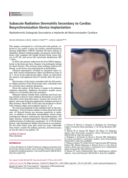
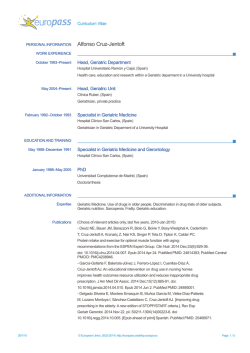
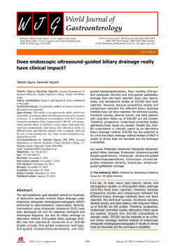
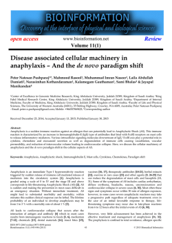
World J Gastroenterol 2015 January 28; 21(4): 1091-1098 ISSN 1007-9327 (print) ISSN 2219-2840 (online) Submit a Manuscript: http://www.wjgnet.com/esps/ Help Desk: http://www.wjgnet.com/esps/helpdesk.aspx DOI: 10.3748/wjg.v21.i4.1091 © 2015 Baishideng Publishing Group Inc. All rights reserved. MINIREVIEWS Benign esophageal lesions: Endoscopic and pathologic features Shu-Jung Tsai, Ching-Chung Lin, Chen-Wang Chang, Chien-Yuan Hung, Tze-Yu Shieh, Horng-Yuan Wang, Shou-Chuan Shih, Ming-Jen Chen Abstract Shu-Jung Tsai, Ching-Chung Lin, Chen-Wang Chang, ChienYuan Hung, Tze-Yu Shieh, Horng-Yuan Wang, Shou-Chuan Shih, Ming-Jen Chen, Division of Gastroenterology, Department of Internal Medicine, Mackay Memorial Hospital, Taipei 10001, Taiwan Shu-Jung Tsai, Ching-Chung Lin, Chen-Wang Chang, Chien-Yuan Hung, Tze-Yu Shieh, Horng-Yuan Wang, MingJen Chen, Department of Nursing, Nursing and Management, Mackay Junior College of Medicine, Taipei 10001, Taiwan Shu-Jung Tsai, Ching-Chung Lin, Chen-Wang Chang, ChienYuan Hung, Tze-Yu Shieh, Horng-Yuan Wang, Shou-Chuan Shih, Ming-Jen Chen, Mackay Medical College, New Taipei 25243, Taiwan Author contributions: Chen MJ conducted the review design; Tsai SJ and Lin CC wrote the article; Lin CC and Chen MJ prepared the endoscopic photographs; Hung CY and Shieh TY conducted the literature review; Shih SC supported this work and critically read the manuscript; Wang HY and Chen MJ supported this work and supervised the final editing; all authors read and approved the final manuscript. Open-Access: This article is an open-access article which was selected by an in-house editor and fully peer-reviewed by external reviewers. It is distributed in accordance with the Creative Commons Attribution Non Commercial (CC BY-NC 4.0) license, which permits others to distribute, remix, adapt, build upon this work non-commercially, and license their derivative works on different terms, provided the original work is properly cited and the use is non-commercial. See: http://creativecommons.org/ licenses/by-nc/4.0/ Correspondence to: Ming-Jen Chen, MD, Division of Gastroenterology, Department of Internal Medicine, Mackay Memorial Hospital, No. 92, Sec. 2, Chungshan North Road, Taipei 10001, Taiwan. [email protected] Telephone: +886-2-25433535-2260 Fax: +886-2-25433642 Received: July 23, 2014 Peer-review started: July 24, 2014 First decision: September 15, 2014 Revised: September 23, 2014 Accepted: October 21, 2014 Article in press: October 21, 2014 Published online: January 28, 2015 WJG|www.wjgnet.com Benign esophageal lesions have a wide spectrum of clinical and pathologic features. Understanding the endoscopic and pathologic features of esophageal lesions is essential for their detection, differential diagnosis, and management. The purpose of this review is to provide updated features that may help physicians to appropriately manage these esophageal lesions. The endoscopic features of 2997 patients are reviewed. In epithelial lesions, the frequency of occurrence was in the following order: glycogenic acanthosis, heterotopic gastric mucosa, squamous papilloma, hyperplastic polyp, ectopic sebaceous gland and xanthoma. In subepithelial lesions, the order was as follows: hemangioma, leiomyoma, dysphagia aortica and granular cell tumor. Most benign esophageal lesions can be diagnosed according to their endoscopic appearance and fin dings on routine biopsy, and submucosal lesions, by endoscopic resection. Management is generally based upon the confidence of diagnosis and whether the lesion causes symptoms. We suggest endoscopic resection of all granular cell tumors and squamous papillomas because, while rare, these lesions have malignant potential. Dysphagia aortica should be considered in the differential diagnosis of dysphagia in the elderly. Key words: Benign tumor; Esophagus; Epithelial lesions; Subepithelial lesions; Endoscopy © The Author(s) 2015. Published by Baishideng Publishing Group Inc. All rights reserved. Core tip: Benign esophageal tumors have a lower detection rate due to the fact that most patients are asymptomatic. The majority of these benign lesions are asymptomatic, and diagnoses are often made incidentally during investigations for other symptoms. Although biopsy or excision is required for a definitive 1091 January 28, 2015|Volume 21|Issue 4| Tsai SJ et al . Benign esophageal lesions diagnosis, understanding the endoscopic appearances provides essential help for differential diagnosis. In epithelial lesions, the frequency of occurrence was in the following order: glycogenic acanthosis, heterotopic gastric mucosa, squamous papilloma, hyperplastic polyp, ectopic sebaceous gland and xanthoma. In subepithelial lesions, the order was as follows: hemangioma, leiomyoma, dysphagia aortica and granular cell tumor. A Tsai SJ, Lin CC, Chang CW, Hung CY, Shieh TY, Wang HY, Shih SC, Chen MJ. Benign esophageal lesions: Endoscopic and pathologic features. World J Gastroenterol 2015; 21(4): 1091-1098 Available from: URL: http://www.wjgnet. com/1007-9327/full/v21/i4/1091.htm DOI: http://dx.doi. org/10.3748/wjg.v21.i4.1091 B INTRODUCTION Esophageal benign lesions have a diverse spectrum of etiologies in terms of clinical course and under lying pathologic features. Benign esophageal tu mors, while uncommon compared with esophageal carcinoma, can sometimes cause dysphagia but often have insignificant clinical outcomes. Endoscopic findings are essential for detection, diagnosis, staging, and treatment planning. Benign esophageal [1] tumors are rare, with a prevalence ≤ 0.5% , while benign tumors represent 20% of esophageal [2] neoplasms on autopsy . Since many of these tu mors are small and asymptomatic, few benign esophageal lesions attract clinical attention. Benign esophageal lesions could be detected more often with the widespread use of endoscopes, radiologic [3] imaging , and increased awareness of the disease. Esophageal lesions can be classified in two different ways; histologically depending on the involved layer into epithelial or subepithelial lesions and endoscopically depending on endoscopic features such as flat, raised, or cystic lesions. In this article, we review the endoscopic and pathological features of esophageal benign lesions. In all, 149 benign esophageal lesions in 2997 endoscopic examinations are retrospectively reviewed. We re moved the esophageal epithelial lesions by biopsy or resected the subepithelial lesions by endoscopic mucosal resection or endoscopic submucosal dis section for histological analysis. In this article, we divide these lesions into epithelial or subepithelial lesions based on the final histological findings. The Institutional Review Board at Mackay Memorial Hos pital approved this project. Figure 1 Heterotopic gastric mucosa. A: Heterotopic gastric mucosa appears salmon-colored under conventional endoscopy and is recognized as flat or slightly elevated; B: Narrow band imaging facilitates mucosal surface evaluation of heterotopic gastric mucosa by adjusting reflected light to enhance the contrast between the esophageal mucosa and the gastric mucosa and may improve the diagnosis of heterotopic gastric mucosa. age: 48 years, range: 25-69 years), which appears salmon-colored under conventional esophagogas troduodenoscopy (EGD) and is recognized as flat or slightly elevated (Figure 1A), while the iodine reaction is negative. Narrow band imaging facilitates mucosal surface evaluation by adjusting the refle cted light to enhance the contrast between the esophageal mucosa and the HGM and may improve the diagnostic rate of HGM (Figure 1B). It is easily overlooked because it is typically located just below the upper esophageal sphincter. [4-6] The prevalence of HGM is 0.1%-10% , and [5,6] most patients have no symptoms . Some pati ents present with pharyngeal globus sensation, heartburn, acid regurgitation, or dysphagia because HGM produces mucin and acid. Two types of HGM have been recognized: one with foveolar epithelium and fundic glands and the other with only foveolar [7] epithelium . The foveolar epithelium produces neutral mucins. The number of reported cases with neoplastic changes or malignant transformation is very low and these cases are considered extremely [8] rare . Management is generally required only if symptoms or complications develop. EPITHELIAL LESIONS Heterotopic gastric mucosa Squamous cell papilloma In all, 21 patients had histologically proven heteroto pic gastric mucosa (HGM) (12 men, 9 women; mean WJG|www.wjgnet.com In all, 20 patients had histologically proven squamous cell papilloma (SCP) (3 men, 17 women; mean age: 1092 January 28, 2015|Volume 21|Issue 4| Tsai SJ et al . Benign esophageal lesions A B Figure 3 Hyperplasia polyp. Hyperplasia polyp occurs as a polypoid lesion and is located on edematous inflamed gastric folds at the gastroesophageal junction. Figure 2 Squamous cell papilloma. A: Squamous cell papilloma is recognized as whitish-pink, wart-like exophytic projections on conventional endoscopy; B: Narrow band imaging facilitates mucosal surface evaluation of squamous cell papilloma and shows that microvessels in the lesion are not dilated. 49 years, range: 26-68 years), which is recognized as whitish-pink, wart-like exophytic projections on conventional endoscopy (Figure 2A) and is a rare benign esophageal lesion, with a prevalence of [9,10] 0.01%-0.45% . Narrow band imaging facilitates mucosal surface evaluation of squamous cell pa pilloma and shows that microvessels in the lesion are not dilated (Figure 2A). Most cases of SCP are [10,11] solitary and asymptomatic . The etiology of SCP is not fully understood, although there are two possible hypotheses: chronic mucosal irrita [12] tion and infection with human papillomaviruses . The hypotheses of inflammatory reactions such as gastroesophageal reflux disease were based on the high frequency of SCP occurring in the lower third of the esophagus. To our best knowledge, whether SCP is associated with human papillomavirus is [13] controversial . Papillomatosis in the proximal esophagus seems to favor involvement of the human [14] papillomavirus . In the majority of the cases, SCP of the eso phagus can be removed using endoscopic biopsy forceps because most are only a few millimeters in size. Larger papillomas can be removed using endoscopic mucosectomy. We suggest removing these lesions endoscopically because, while rare, these lesions have malignant potential. Figure 4 Xanthoma. Xanthomas are endoscopically recognized as elevated, granular, yellowish, fern-like lesions and scattered on a normal mucosal surface. perplasia polyp (HP) (12 men, 6 women; mean age: 36 years, range: 18-69 years). In our patients, HPs were most common in the region of the esophago cardiac junction (67%), followed by the distal esophagus (27%) and mid-esophagus (6%). HP appears as a polypoid lesion on edematous inflamed gastric folds at the gastroesophageal junction (Figure 3). HPs of the esophagus and esophagogastric junc tion region are characterized by the presence of mixed inflammatory infiltrates with plasma cells, eosinophils, fibroblasts, and inflamed stroma. HPs are associated with concurrent erosive esophagitis in the majority of cases, but other potential etiologies including medication-induced pill esophagitis, infection, and previous anastomosis or polypectomy [15] have been reported . These results suggest that the pathogenesis of HP is a mucosal regenerative [16] response to surrounding mucosal injury . Treatment for HP is similar to that for gastro esophageal reflux. In our experience, HP regresses after anti-acid therapy such as proton pump inhibitors. Careful clinical history and biopsy of the nonpolypoid mucosa are essential for determining the clinicopathological context in which the HP developed. When HP is found with Barrett’s esophagus, en [17] doscopic mucosal resection is recommended . Hyperplastic polyp In all, 18 patients had histologically proved hy WJG|www.wjgnet.com 1093 January 28, 2015|Volume 21|Issue 4| Tsai SJ et al . Benign esophageal lesions A B Figure 5 Ectopic sebaceous gland. A: The number of ectopic sebaceous gland is variable from single to B: more than one hundred yellowish plaques measuring 1 to 2 mm in the esophagus. A B Figure 6 Glycogenic acanthosis. A: Esophagogastroduodenoscopy reveals multiple, uniformly sized, oval or round glycogenic acanthosis usually < 1 cm, involving otherwise normal esophageal mucosa; B: In chromoscopy with iodine spray, glycogenic acanthosis is recognized as slightly elevated iodine-positive, brownish areas. Xanthoma (ESGs). We reported on esophageal xanthomas localized in the lower esophagus in a 62-year-old man. EGD revealed some well-defined, fern-like, and yellowish lesions scattered over the middle and lower eso phagus (Figure 4). In terms of endoscopic findings, xanthoma is most commonly solitary, 2-10 mm across, and located in the lower esophagus, but cases of multifocal lesions [18,19] have been reported . Microscopically, they consist of fat accumulation in foamy histiocytes beneath [20] the squamous epithelium . One study showed the most common location of ectopic xanthoma in the gastrointestinal tract was the stomach (76%), followed by the esophagus (12%) and duodenum [21] (12%) . The etiology of esophageal xanthoma remains unknown, but one study theorized that these xanthomas were derived from focal mucosal damage, and that lipids derived from broken down cell membranes are captured by interstitial [13] histiocytes . This may explain why they occur less frequently in the esophagus than stomach because the esophageal mucosa can better tolerate mucosal [21] injury . Xanthoma must be distinguished on endoscopy from other yellowish lesions such as carcinoid tumor, granular cell tumors, and ectopic sebaceous glands WJG|www.wjgnet.com ESGs In all, three patients (one man and two women; mean age: 52 years, range: 45-69 years) had histologically proven ESGs within the esopha gus. EGD revealed variable numbers from single (Figure 5A) to > 100 (Figure 5B) yellowish plaques measuring 1-2 mm in diameter in the middle and lower esophagus. ESGs have been found in various tissues, such as the lips and mouth, external genitalia, parotid [22] glands, palms, soles, and various organs . Ty pically, the mean age of affected patients is ap proximately 50 years, and the condition has equal [23] gender distribution . The numbers of ESGs in the esophagus varied from single to > 100, while their [23,24] size was 1-20 mm (the majority were < 0.5 cm) . The ESGs are most likely the result of a metaplastic process rather than a congenital anomaly because the ESGs are derived from endodermal tissue unlike the sebaceous glands, which are derived from [22,25] ectodermal tissue . The apparent low incidence of this condition may be because of the lack of obvious clinical signs and symptoms. Most cases have been discovered incidentally by endoscopy during a referral for a 1094 January 28, 2015|Volume 21|Issue 4| Tsai SJ et al . Benign esophageal lesions A B Figure 7 Leiomyoma. A: Leiomyoma commonly arises from the muscularis propria layer of the esophagus and presents as submucosal tumor; B: Leiomyoma arising from the muscularis mucosae can present as a polypoid intraluminal tumor. A B Figure 8 Granular cell tumor. A: Granular cell tumor is endoscopically recognized as a firm, yellowish subepithelial tumor covered with the normal mucosa in the esophagus; B: Endoscopic ultrasonography of granular cell tumor shows a homogenous hypo-echogenic tumor extending from the muscularis mucosa layer, and the musculais propria is not involved. gastrointestinal tract examination. Most cases have no significant overall changes in ectopic sebaceous [26] gland number, size, or shape during follow-up . We suggest that ESGs do not need further treatment. SUBEPITHELIAL LESIONS Leiomyoma Leiomyoma was observed in three patients (3 men; mean age: 37 year, range: 25-43 years). Leiomyomas were located at the middle esophagus in 1 case and at the distal esophagus in 2 cases. The mean size of leiomyoma is 1.26 ± 0.4 cm. It commonly arises from the muscularis propria layer of the esophagus and presents as a submucosal tumor (Figure 7A). In rare circumstances, those cases arising from the muscularis mucosae can present as polypoid intraluminal tumors (Figure 7B). Leiomyomas are the most common benign eso phageal neoplasm, accounting for roughly two[29] thirds of all benign tumors of this organ . Because they arise from smooth muscle cells, they are located mainly in the middle and distal thirds of the esophagus but are uncommon in the upper third of the esophagus, where the muscular layer is predominately skeletal in origin. Most patients are asymptomatic, but dysphagia and pain may develop depending on lesion size and encroachment degree into the esophageal lumen. [30] Treatment options include endoscopic enucleation , Glycogenic acanthosis Glycogenic acanthosis was demonstrated as nodules involving otherwise normal esophageal mucosa in 66 patients (42 men and 24 women; mean age: 52 years, range: 45-79 years). Glycogenic acan thosis is defined as nodules involving otherwise normal esophageal mucosa. This is endoscopically recognized as slightly elevated areas on conven tional endoscopy (Figure 6A) and slightly elevated iodine-positive areas on chromoscopy (Figure 6B). These lesions most commonly appear as multiple, uniformly sized, and oval or round elevations < 1 [27] cm in diameter . Biopsies after periodic acid-Schiff staining demonstrate that the nodules that represent glycogenic acanthosis are combinations of cellular [27] hyperplasia and increased cellular glycogen . Although glycogenic acanthosis was thought to be related to gastroesophageal reflux, antireflux therapy improved symptoms but failed to eradicate [27,28] glycogenic acanthosis lesions . WJG|www.wjgnet.com 1095 January 28, 2015|Volume 21|Issue 4| Tsai SJ et al . Benign esophageal lesions A B Figure 9 Hemangioma. On endoscopy, esophageal hemangioma appears cystic and bluish-red and can be pressed with biopsy forceps. [31] submucosal tunneling endoscopic resection , and [32] surgical enucleation or observation. Esophageal leiomyomas have a benign clinical course and typically do not recur after surgery. Granular cell tumor Granular cell tumor was observed in one 39-year-old health woman who presented with acid regurgitation and discomfort sensation when swallowing. EGD revealed one 12 mm × 15 mm firm, slightly elevated, whitish-to-yellow, and smooth nodular tumor covered by an intact epithelium (Figure 8A). Endoscopic ultrasonography showed one homogenous hypoechogenic tumor extending from the muscularis mucosa layer (Figure 8B). The muscularis propria was not involved. Endoscopic submucosal dissection was performed. Granular cell tumors are the secondary common cause of non-epithelial tumors in the esophagus. Most of the patients with esophageal granular cell [33] tumor are asymptomatic , but some complain of mild dysphagia or retrosternal discomfort. Although most granular cell tumors have a clinically indolent course, it is estimated that 1%-3% are malignant [34] with a 5-year survival rate < 35% . Microscopically, granular cell tumors are composed of nests of ovoid or polygonal cells separated by collagen bundles. The tumor has malignant potential in the presence of necrosis, increased mitotic count, vesicular nuclei [35] with large nucleoi, and high Ki67 index . Resection is the main treatment for granular cell tumors. Endoscopic mucosal resection and endoscopic submucosal dissection were introduced and were considered the therapy of choice for tumors within the subepithelial layer or submucosa separated from the muscularis propria. Figure 10 Dysphagia aortica. A: Esophagogastroduodenoscopy reveals a pulsatile extrinsic compression at about 25 cm from the incisor; B: The chest computed tomography showed aortic arch and descending aorta tortuosity with compression into adjacent esophagus. The arrow indicates the esophagus. cystic and bluish-red and can be pressed with biopsy forceps (Figure 9). The prevalence of eso phageal hemangiomas in the general population was 0.04% based upon the findings of an autopsy [36] series . The majority of these hemangiomas are cavernous; however, capillary lesions have been described. Although usually solitary, multiple lesions can be seen in congenital blue rubber bleb nevus [37] syndrome . Esophageal hemangiomas are usually found incidentally. When symptomatic, they are most often associated with bleeding and dysphagia. In such cases, surgical resection has been performed, but endoscopic resection has also been accomplished [38] safely . Although esophageal hemangiomas are uncommon, careful consideration during endoscopy [39] is required to avoid the misdiagnosis of varices . Dysphagia aortica In dysphagia aortica was observed in three patients (1 man, 2 women; mean age: 68 years, range: 63-78 years). They visited our clinics with postprandial abdominal fullness and progressive dysphagia to solid meals. The esophagus begins on the right side of the thoracic aorta and then, descends and crosses the aorta anteriorly across its lower third. EGD reveals external compression of the middle-to-lower esophagus by the tortuous aorta with pulsations from the great vessels (Figure 10A). Hemangioma (venous bleb) Esophageal hemangiomas were observed in 13 patients (4 men, 9 women; mean age: 67 years, range: 45-82 years). On endoscopy, they appear WJG|www.wjgnet.com 1096 January 28, 2015|Volume 21|Issue 4| Tsai SJ et al . Benign esophageal lesions Dysphagia aortica is a rare etiology of dysphagia resulting from the extrinsic compression of the esophagus by a thoracic aortic aneurysm or the tortuosity and elongation of the thoracic aorta (Figure 10B). The clinical findings resemble those of esophageal malignancy or esophageal motility disorders. Esophageal compression by a vascular [40] structure is a common cause of dysphagia . Nonaneurysmatic aortic dysphagia is usually observed in the elderly, especially in hypertensive women [41,42] with enlarged heart and kyphosis . The treatment of dysphagia aortica depends on symptom severity. Symptoms of mild dysphagia often improve with diet modifications (e.g., by avoiding lying down immediately after taking drugs or food, eating small but frequent meals, and chewing well) and treatment of the coexistent disease, such as heart failure or hypertension. Some patients with [43,44] more severe symptoms may respond to surgery . Dysphagia aortica should be considered in the differential diagnosis of dysphagia, especially in the growing elderly population with underlying car diovascular disease or hypertension. 4 5 6 7 8 9 CONCLUSION 10 Benign esophageal lesions have a lower detection rate due to the fact that most patients are asy mptomatic. But it could be easily found with the widespread use of endoscopes and the increasing awareness of this disease. The majority of these benign tumors are asymptomatic, and diagnoses are often made incidentally during investigations for other complaints or symptoms. Although biopsy or excision is required for a definitive diagnosis, understanding the endoscopic appearances provides essential help for differential diagnosis. We suggest endoscopic resection of all granular cell tumors and squamous papillomas because, while rare, these lesions have malignant potential. Hyperplastic polyps could regress after anti-acid therapy such as proton pump inhibitors. We suggest that ectopic sebaceous glands and xanthoma do not need further treatment. Esophageal hemangiomas are uncommon, and careful consideration during endoscopy is required to avoid the misdiagnosis of varices. Dysphagia aortica should be considered in the differential diagnosis of dysphagia in the elderly. 11 12 13 14 15 16 17 REFERENCES 1 2 3 18 Choong CK, Meyers BF. Benign esophageal tumors: introduction, incidence, classification, and clinical features. Semin Thorac Cardiovasc Surg 2003; 15: 3-8 [PMID: 12813683 DOI: 10.1016/ S1043-0679(03)70035-5] Attah EB, Hajdu SI. Benign and malignant tumors of the esophagus at autopsy. J Thorac Cardiovasc Surg 1968; 55: 396-404 [PMID: 5644217] Lewis RB, Mehrotra AK, Rodriguez P, Levine MS. From the radiologic pathology archives: esophageal neoplasms: radiologic- WJG|www.wjgnet.com 19 20 1097 pathologic correlation. Radiographics 2013; 33: 1083-1108 [PMID: 23842973 DOI: 10.1148/rg.334135027] Yüksel I, Usküdar O, Köklü S, Başar O, Gültuna S, Unverdi S, Oztürk ZA, Sengül D, Arikök AT, Yüksel O, Coban S. Inlet patch: associations with endoscopic findings in the upper gastrointestinal system. Scand J Gastroenterol 2008; 43: 910-914 [PMID: 19086275 DOI: 10.1080/00365520801986619] Maconi G, Pace F, Vago L, Carsana L, Bargiggia S, Bianchi Porro G. Prevalence and clinical features of heterotopic gastric mucosa in the upper oesophagus (inlet patch). Eur J Gastroenterol Hepatol 2000; 12: 745-749 [PMID: 10929900 DOI: 10.1097/00042737-200 012070-00005] Poyrazoglu OK, Bahcecioglu IH, Dagli AF, Ataseven H, Celebi S, Yalniz M. Heterotopic gastric mucosa (inlet patch): endoscopic prevalence, histopathological, demographical and clinical characteristics. Int J Clin Pract 2009; 63: 287-291 [PMID: 17535303 DOI: 10.1111/j.1742-1241.2006.01215.x] Tang P, McKinley MJ, Sporrer M, Kahn E. Inlet patch: prevalence, histologic type, and association with esophagitis, Barrett esophagus, and antritis. Arch Pathol Lab Med 2004; 128: 444-447 [PMID: 15043461] Akanuma N, Hoshino I, Akutsu Y, Shuto K, Shiratori T, Kono T, Uesato M, Sato A, Isozaki Y, Maruyama T, Takeshita N, Matsubara H. Primary esophageal adenocarcinoma arising from heterotopic gastric mucosa: report of a case. Surg Today 2013; 43: 446-451 [PMID: 22706784 DOI: 10.1007/s00595-012-0206-9] Szántó I, Szentirmay Z, Banai J, Nagy P, Gonda G, Vörös A, Kiss J, Bajtai A. [Squamous papilloma of the esophagus. Clinical and pathological observations based on 172 papillomas in 155 patients]. Orv Hetil 2005; 146: 547-552 [PMID: 15853063] Mosca S, Manes G, Monaco R, Bellomo PF, Bottino V, Balzano A. Squamous papilloma of the esophagus: long-term follow up. J Gastroenterol Hepatol 2001; 16: 857-861 [PMID: 11555097 DOI: 10.1046/j.1440-1746.2001.02531.x] Carr NJ, Monihan JM, Sobin LH. Squamous cell papilloma of the esophagus: a clinicopathologic and follow-up study of 25 cases. Am J Gastroenterol 1994; 89: 245-248 [PMID: 8304311] Poljak M, Orlowska J, Cerar A. Human papillomavirus infection in esophageal squamous cell papillomas: a study of 29 lesions. Anticancer Res 1995; 15: 965-969 [PMID: 7645987] Odze R, Antonioli D, Shocket D, Noble-Topham S, Goldman H, Upton M. Esophageal squamous papillomas. A clinicopathologic study of 38 lesions and analysis for human papillomavirus by the polymerase chain reaction. Am J Surg Pathol 1993; 17: 803-812 [PMID: 8393303 DOI: 10.1097/00000478-199308000-00005] Politoske EJ. Squamous papilloma of the esophagus associated with the human papillomavirus. Gastroenterology 1992; 102: 668-673 [PMID: 1310082] Abraham SC, Singh VK, Yardley JH, Wu TT. Hyperplastic polyps of the esophagus and esophagogastric junction: histologic and clinicopathologic findings. Am J Surg Pathol 2001; 25: 1180-1187 [PMID: 11688578 DOI: 10.1097/00000478-200109000-00009] Chang WH, Shih SC, Wang HY, Chang CW, Chen CJ, Chen MJ. Acquired hyperplastic gastric polyps after treatment of ulcer. J Formos Med Assoc 2010; 109: 567-573 [PMID: 20708507 DOI: 10.1016/S0929-6646(10)60093-9] De Ceglie A, Lapertosa G, Blanchi S, Di Muzio M, Picasso M, Filiberti R, Scotto F, Conio M. Endoscopic mucosal resection of large hyperplastic polyps in 3 patients with Barrett’s esophagus. World J Gastroenterol 2006; 12: 5699-5704 [PMID: 17007025] Becheanu G, Dumbrava M, Arbanas T, Diculescu M, HoyeauIdrissi N, Fléjou JF. Esophageal xanthoma--report of two new cases and review of the literature. J Gastrointestin Liver Dis 2011; 20: 431-433 [PMID: 22187711] Licci S, Campo SM, Ventura P. Verruciform xanthoma of the esophagus: an uncommon entity in an unusual site. Endoscopy 2010; 42 Suppl 2: E330 [PMID: 21170833 DOI: 10.1055/s-0030-1255944] Herrera-Goepfert R, Lizano-Soberón M, García-Perales M. Verruciform xanthoma of the esophagus. Hum Pathol 2003; 34: January 28, 2015|Volume 21|Issue 4| Tsai SJ et al . Benign esophageal lesions 21 22 23 24 25 26 27 28 29 30 31 814-815 [PMID: 14506645 DOI: 10.1016/S0046-8177(03)00236-3] Gencosmanoglu R, Sen-Oran E, Kurtkaya-Yapicier O, Tozun N. Xanthelasmas of the upper gastrointestinal tract. J Gastroenterol 2004; 39: 215-219 [PMID: 15064997 DOI: 10.1007/s00535-0031288-3] Guiducci AA, Hyman AB. Ectopic sebaceous glands. A review of the literature regarding their occurrence, histology and embryonic relationships. Dermatologica 1962; 125: 44-63 [PMID: 13902791 DOI: 10.1159/000254952] Kim YS, Jin SY, Shim CS. Esophageal ectopic sebaceous glands. Clin Gastroenterol Hepatol 2007; 5: A23 [PMID: 16904949 DOI: 10.1016/j.cgh.2006.06.013] Wei IF, Chang CC, Fang CL, Hsieh CR, Wang JJ, Lou HY, Cheng T. Education and imaging. Gastrointestinal: ectopic sebaceous glands in the esophagus. J Gastroenterol Hepatol 2008; 23: 338 [PMID: 18289363 DOI: 10.1111/j.1440-1746.2007.05303.x] Nakanishi Y, Ochiai A, Shimoda T, Yamaguchi H, Tachimori Y, Kato H, Watanabe H, Hirohashi S. Heterotopic sebaceous glands in the esophagus: histopathological and immunohistochemical study of a resected esophagus. Pathol Int 1999; 49: 364-368 [PMID: 10365859 DOI: 10.1046/j.1440-1827.1999.00874.x] Wang WP, Wang WS, Tsai YC. Multiple tiny ectopic sebaceous glands discovered throughout entire esophageal tract. Dig Dis Sci 2009; 54: 2754-2757 [PMID: 19117122 DOI: 10.1007/s10620008-0676-1] Vadva MD, Triadafilopoulos G. Glycogenic acanthosis of the esophagus and gastroesophageal reflux. J Clin Gastroenterol 1993; 17: 79-83 [PMID: 8409304] Nazligül Y, Aslan M, Esen R, Yeniova AÖ, Kefeli A, Küçükazman M, Dülger AC, Celik Y. Benign glycogenic acanthosis lesions of the esophagus. Turk J Gastroenterol 2012; 23: 199-202 [PMID: 22798107] Lee LS, Singhal S, Brinster CJ, Marshall B, Kochman ML, Kaiser LR, Kucharczuk JC. Current management of esophageal leiomyoma. J Am Coll Surg 2004; 198: 136-146 [PMID: 14698321 DOI: 10.1016/j.jamcollsurg.2003.08.015] Guo J, Liu Z, Sun S, Liu X, Wang S, Ge N. Ligation-assisted endoscopic enucleation for treatment of esophageal subepithelial lesions originating from the muscularis propria: a preliminary study. Dis Esophagus 2014; Epub ahead of print [PMID: 24592944 DOI: 10.1111/dote.12192] Ye LP, Zhang Y, Mao XL, Zhu LH, Zhou X, Chen JY. Submucosal tunneling endoscopic resection for small upper gastrointestinal subepithelial tumors originating from the muscularis propria layer. 32 33 34 35 36 37 38 39 40 41 42 43 44 Surg Endosc 2014; 28: 524-530 [PMID: 24013472 DOI: 10.1007/ s00464-013-3197-8] Hu X, Lee H. Complete thoracoscopic enucleation of giant leiomyoma of the esophagus: a case report and review of the literature. J Cardiothorac Surg 2014; 9: 34 [PMID: 24528601 DOI: 10.1186/1749-8090-9-34] Xu GQ, Chen HT, Xu CF, Teng XD. Esophageal granular cell tumors: report of 9 cases and a literature review. World J Gastroenterol 2012; 18: 7118-7121 [PMID: 23323018 DOI: 10.3748/wjg.v18.i47.7118] David O, Jakate S. Multifocal granular cell tumor of the esophagus and proximal stomach with infiltrative pattern: a case report and review of the literature. Arch Pathol Lab Med 1999; 123: 967-973 [PMID: 10506457] Fanburg-Smith JC, Meis-Kindblom JM, Fante R, Kindblom LG. Malignant granular cell tumor of soft tissue: diagnostic criteria and clinicopathologic correlation. Am J Surg Pathol 1998; 22: 779-794 [PMID: 9669341 DOI: 10.1097/00000478-199807000-00001] Moersch HJ, Harrington SW. Benign tumor of the esophagus. Ann Otl Rhnol Laryngol 1944; 53: 800 Rasalkar DD, Chiu PW, Teoh AY, Chu WC. Oesophageal haemangioma: imaging characteristics of this rare condition. Hong Kong Med J 2010; 16: 230-231 [PMID: 20519762] Cantero D, Yoshida T, Ito T, Suzumi M, Tada M, Okita K. Esophageal hemangioma: endoscopic diagnosis and treatment. Endoscopy 1994; 26: 250-253 [PMID: 8026376 DOI: 10.1055/s-2007-1008954] Won JW, Lee HW, Yoon KH, Yang SY, Moon IS, Lee TJ. Extended hemangioma from pharynx to esophagus that could be misdiagnosed as an esophageal varix on endoscopy. Dig Endosc 2013; 25: 626-629 [PMID: 24164602 DOI: 10.1111/j.1443-1661.2012.01405.x] Hilliard AA, Murali NS, Keller AS. Dysphagia aortica. Ann Intern Med 2005; 142: 230-231 [PMID: 15684224 DOI: 10.7326/0003-48 19-142-3-200502010-00031] Keates PG, Magidson O. Dysphagia associated with sclerosis of the aorta. Br J Radiol 1955; 28: 184-190 [PMID: 14363659 DOI: 10.1259/0007-1285-28-328-184] Song SW, Chung JH, Kim SH. A Case of Dysphagia Aortica in an Elderly Patient. Int J Gerontol 2012; 6: 46-48 Cao DB, Gao Y, Sun XY, Yang SR. Dysphagia aortica secondary to descending thoracic aortic pseudoaneurysm. Ann Thorac Surg 2012; 94: 656 [PMID: 22818315 DOI: 10.1016/j.athoracsur.2012.02.020] Kische S, Werner D, Ince H. A neglected symptom of contained aortic laceration--dysphagia aortica successfully treated by endovascular stentgrafting. Catheter Cardiovasc Interv 2012; 80: 1052-1055 [PMID: 21805591 DOI: 10.1002/ccd.23265] P- Reviewer: Koch TR S- Editor: Yu J WJG|www.wjgnet.com 1098 L- Editor: Wang TQ E- Editor: Wang CH January 28, 2015|Volume 21|Issue 4| Published by Baishideng Publishing Group Inc 8226 Regency Drive, Pleasanton, CA 94588, USA Telephone: +1-925-223-8242 Fax: +1-925-223-8243 E-mail: [email protected] Help Desk: http://www.wjgnet.com/esps/helpdesk.aspx http://www.wjgnet.com I S S N 1 0 0 7 - 9 3 2 7 0 4 9 7 7 1 0 0 7 9 3 2 0 45 © 2015 Baishideng Publishing Group Inc. All rights reserved.

© Copyright 2026