
Temporary DPOAE level shifts, ABR threshold shifts
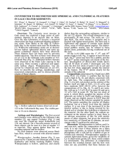
Hearing Research 196 (2004) 94–108
www.elsevier.com/locate/heares
Temporary DPOAE level shifts, ABR threshold shifts and
histopathological damage following below-critical-level
noise exposures
Gary W. Harding *, Barbara A. Bohne
Department of Otolaryngology, Washington University School of Medicine, P.O. Box 8115, 660 South Euclid, St. Louis, MO 63110, USA
Received 22 January 2004; accepted 8 March 2004
Available online 18 August 2004
Abstract
DPOAE temporary level shift (TLS) at 2f1 f2 and f2 f1, ABR temporary threshold shift (TTS), and detailed histopathological
findings were compared in three groups of chinchillas that were exposed for 24 h to an octave band of noise (OBN) centered at 4 kHz
with a sound pressure level (SPL) of 80, 86 or 92 dB (n = 3, 4, 6). DPOAE levels at 39 frequencies from f1 = 0.3 to 16 kHz (f2/f1 = 1.23;
L2 and L1 = 55, 65 and 75 dB, equal and differing by 10 dB) and ABR thresholds at 13 frequencies from 0.5 to 20 kHz were collected
pre- and immediately post-exposure. The functional data were converted to pre- minus post-exposure shift and overlaid upon the
cytocochleogram of cochlear damage using the frequency-place map for the chinchilla. The magnitude and frequency place of components in the 2f1 f2 TLS patterns were determined and group averages for each OBN SPL and L1, L2 combination were calculated.
The f2 f1 TLS was also examined in ears with focal lesions equal to or greater than 0.4 mm. The 2f1 f2 TLS (plotted at f1) and TTS
aligned with the extent and location of damaged supporting cells. The TLS patterns over frequency had two features which were
unexpected: (1) a peak at about a half octave above the center of the OBN with a valley just above and below it and (2) a peak
(often showing enhancement) at the apical boundary of the supporting-cell damage. The magnitudes of the TLS and TTS generally
increased with increasing SPL of the exposure. The peaks of the TLS and TTS, as well as the peaks and valleys of the TLS pattern
moved apically as the SPL of the OBN was increased. However, there was little consistency in the pattern relations with differing L1,
L2 combinations. In addition, neither the 2f1 f2 nor f2 f1 TLS for any L1, L2 combination reliably detected focal lesions (100%
OHC loss) from 0.4 to 1.2 mm in size. Often, the TLS went in the opposite direction from what would be expected at focal lesions.
Recovery from TLS and TTS was also examined in seven animals. Both TLS and TTS recovered partially or completely, the magnitude depending upon exposure SPL.
2004 Elsevier B.V. All rights reserved.
Keywords: DPOAE; ABR; Noise; Organ of Corti; Histopathology; Chinchilla
1. Introduction
*
Corresponding author. Tel./fax: +1-314-362-7497.
E-mail address: [email protected] (G.W. Harding).
Abbreviations: ABR, auditory brainstem response; DPOAE, distortion product otoacoustic emission; IHC, inner hair cell; OBN, octave band of noise; OC, organ of Corti; OHC, outer hair cell; SPL,
sound pressure level; TLS, temporary level shift; TTS, temporary threshold shift
0378-5955/$ - see front matter 2004 Elsevier B.V. All rights reserved.
doi:10.1016/j.heares.2004.03.011
Distortion product otoacoustic emissions (DPOAE)
are presumed to be produced by the summed activity
of outer hair cells (OHC) (e.g., Chang and Norton,
1996). Auditory brainstem responses (ABR) appear to
be dominated by the activity of inner hair cells
(IHC). The magnitude and extent of permanent ABR
threshold shifts correlate well with IHC and nerve fiber
G.W. Harding, B.A. Bohne / Hearing Research 196 (2004) 94–108
loss (e.g., Nordmann et al., 2000). It has been tacitly assumed that anything which produces a functional deficit in or complete loss of a substantial number of OHCs
would have a profound effect upon DPOAEs. In some
instances, when the cochleae were examined histopathologically, this assumption was mostly confirmed (e.g.,
Canlon and Fransson, 1995; Martin et al., 2002). However, in other studies in which correlations were made
between OHC loss and DPOAEs, this assumption
was not supported (e.g., Harding et al., 2002; Avan
et al., 2003).
In a previous report (Harding et al., 2002), we examined the frequency-place alignment of noise-induced
DPOAE level shifts and ABR threshold shifts (preminus post-exposure) with a detailed assessment of the
histopathological damage from a high-level, short-duration noise exposure (4 kHz OBN, 108 dB SPL, 1.75 h). It
was found that: (1) the best correlation of DPOAE level
shifts with the noise-induced hair-cell loss required plotting these data at f1; (2) level shifts reflected different
correlates for temporary and permanent hearing loss
(i.e., supporting-cell damage leading to stereocilia
uncoupling versus hair-cell loss); (3) level shifts did not
detect relatively large focal lesions (i.e., >0.6 mm,
100% OHC loss) and (4) partial recovery of level shifts
occurred in regions of complete organ of Corti (OC) loss
(i.e., OC wipeout).
Subsequently, it was found that in these cochleae,
there were discontinuities in the reticular lamina (Ahmad et al., 2002) and that these lesions appear to be a
consequence of above-critical-level exposures (i.e., SPL
above which there is no relation between total exposure
energy and hearing or hair-cell loss; Harding and
Bohne, 2004). In the present report, we examined the
changes in DPOAE levels and ABR thresholds with below-critical-level, longer duration noise exposures (i.e.,
SPLs at which there is a clear relation between total
exposure energy and hair-cell loss), particularly as they
relate to the effects immediately post-exposure. We
hypothesized that by removing the secondary effects of
high-level noise exposures (i.e., those occurring postexposure), DPOAE level shifts would more clearly
reflect underlying primary effects (i.e., those occurring
during the exposure; Bohne and Harding, 2000).
2. Materials and methods
2.1. Animals
Thirteen 1 to 2-year-old chinchillas were used in the
study. The animals were anesthetized during DPOAE
levels and ABR thresholds testing with a mixture of
ketamine (40 mg/ml), acepromazine (1 mg/ml) and atropine (0.04 mg/ml), given at a dosage of 1 ml/kg body
weight. Supplemental anesthetic doses (0.33 ml/kg) were
95
administered as needed during ABR testing. First, ABR
thresholds were determined in both ears, followed by
collection of DPOAE levels in both ears while the anesthesia level was a little lighter. Baseline DPOAE levels
and ABR thresholds were determined one day prenoise-exposure using the same methods described below
for functional testing post-exposure. The protocol for
animal use was approved by Washington UniversityÕs
Animal Studies Committee (#20000131 and 20030093;
Adverse Effects of Noise on Hearing: Basic Mechanisms; B.A. Bohne, PI).
2.2. Noise exposure
The animals were exposed awake while individually
housed in a cage suspended in the middle of a reverberant, soundproof booth. The exposure was an octave
band of noise (OBN) with a center frequency of 4 kHz
and a sound pressure level (SPL) of 80 (n = 3), 86
(n = 4) or 92 (n = 6) dB for 24 h. A Bru¨el and Kjaer
2203 sound level meter with a 12.7-mm microphone
was used to calibrate sound pressure ±1 dB. Some of
the data from the group of animals exposed at 80 dB
SPL were reported previously (Harding et al., 2002).
Further analysis of the results from these animals is
presented here.
2.3. Functional testing
DPOAEs were recorded at 39 frequencies from
f1 = 0.3 to 16 kHz and ABR thresholds at 13 frequencies
from 0.5 to 20 kHz. DPOAEs were elicited with tones
produced by a pair of Etymotic ER-2 insert
earphones and recorded with an Etymotic ER-10B+
microphone. ABRs were elicited with clicks and tone
pips using the same ER-2 earphones. DPOAE levels
were collected at equal and unequal input levels
(L1 and L2) ranging from 75 down to 55 dB in 10 dB
steps. The combinations used in each animal are shown
in Table 1. For the noise exposures used, 2f1 f2
DPOAE magnitudes dropped into the noise floor in
the region of maximal shift when L1 or L2 was below
55 dB (data not shown). The frequency ratio (f2/f1)
was 1.23 (see Harding et al., 2002 for recording details).
DPOAE levels and ABR thresholds were determined in
both ears immediately post-exposure in all animals. Six
animals were sacrificed shortly after testing to correlate
DPOAE temporary level shift (TLS) and ABR temporary threshold shift (TTS) with the pattern of cochlear
injury and degeneration. Seven animals were allowed to
recover a variable length of time (i.e., 6–24 days) before
their cochleae were fixed for histopathological examination. Functional testing in these latter animals was
performed several times during the recovery period,
the number depending upon recovery duration.
96
G.W. Harding, B.A. Bohne / Hearing Research 196 (2004) 94–108
Table 1
Animals, exposure SPLs, and L1, L2 combinations used in the present study
Animal
SPL(dB)
L1, L2 (dB)
75, 75
75, 65
963
964
965
80
80
80
x
x
x
xa
xa
Total ears
6
4
86
86
86
86
x
Total ears
2
960b
967
974
975
985
986
987
988
989
991
b
c
65, 65
65, 55
55, 55
x
x
x
xa
xa
x
x
x
6
4
6
x
x
x
x
x
x
x
x
x
0
6
6
6
x
x
x
x
xc
x
x
x
xc
x
x
x
x
x
xc
x
x
x
x
x
xc
x
x
x
x
x
xc
x
11
7
11
11
11
0
x
x
x
92
92
92
92
92
92
Total ears
a
75, 55
0
6
L1 = L2 10 dB.
Recorded at slightly different frequencies from the others.
989-Right not included due to poor pattern correlations with the others in this group.
2.4. Tissue processing
Under deep anesthesia, the cochleae were fixed in
vivo by perfusing a solution of 1% osmium tetroxide
in DaltonÕs buffer through the perilymphatic scalae for
5 min. After both cochleae were fixed, they were separated from the skull and immersed in a large volume
of cold fixative for 2 h. After fixation, the cochleae were
washed in HankÕs balanced salt solution (three 15-min
changes) then placed in 70% ethanol and refrigerated
overnight. The following day, the cochleae were dehydrated in a graded series of ethanol followed by propylene oxide, infiltrated with Durcupan then embedded in
the same medium (see Bohne and Harding, 1993 for details). After polymerization for 48 h, the cochleae were
dissected into flat preparations that were analyzed for
hair-cell loss and other histopathological changes by
phase-contrast microscopy at magnifications of 625
and 1250·.
2.5. Quantitative
analysis
and
qualitative
histopathological
Organ-of-Corti length was measured and missing
IHC, OHC, and inner and outer pillar cells were
counted from apex to base. The percentage of missing
nerve fibers was estimated (see Bohne et al., 1990 for
details). The apex-to-base extent of buckled pillar cells
and partly collapsed DeitersÕ cells were determined qualitatively. A cytocochleogram was prepared for each
cochlea to present these data relative to the percentage
distance from the OC apex.
2.6. Data processing
DPOAE level shifts and ABR threshold shifts were
calculated by subtracting post-exposure from pre-exposure magnitudes. The differential noise floor (i.e., the
maximum measurable shift) was determined by subtracting post-exposure noise floor magnitudes from
pre-exposure DPOAE magnitudes. The distortion product, 2f1 f2, was the focus of this analysis and comparison with cochlear damage. However, the results from
f2 f1 distortion products were also examined in part.
The 2f1 f2 DPOAE level shift and ABR threshold shift
results were overlaid upon the cytocochleograms using
the frequency-place map for the chinchilla (Eldredge
et al., 1981) to align the functional changes on the cytocochleogram. The logarithmic mean was used to calculate
the mean of DPOAE TLS and ABR TTS across animals. The harmonic mean was used to calculate the
mean frequencies at which components in the responses
occurred.
G.W. Harding, B.A. Bohne / Hearing Research 196 (2004) 94–108
3. Results
3.1. Temporary level shift
An example of the correlation among TLS, TTS
and histopathology is shown in Fig. 1. OHC loss (solid
line on the cytocochleogram, Fig. 1(a)) was minimal
across frequency except for a 1-mm region from 4 to
Fig. 1. (a) Cytocochleogram and functional data from cochlea 988L
(exposed at 92 dB SPL). Percent (left y-axis) OHC loss (solid line)
and IHC loss (short dashed line) as a function of percent distance
(frequency place in kHz) from the OC apex (x-axis). The magnitude
of TLS (open circles; plotted at f1), differential noise floor (dotted
line), and TTS (filled circles) are overlaid (right y-axis). OHC focal
lesion (100% loss) from 4 to 6 kHz. Qualitative histopathology
results (top) and nerve fiber loss (bottom). Pillar cells were buckled
from 1 to 8 kHz (OC box; lower horizontal bar) and third row
OHCs were misshapen and out-of-position from 1.5 to 8 kHz (OC
box; upper horizontal bars). Endolymphatic space was decreased
from normal (ES box; lower horizontal bar) and the stria vascularis
was vacuolated (ES box; upper horizontal bar). There was no nerve
fiber loss (MNF LOSS box). (b) TLS for L1, L2 combinations not
shown in (a) (L1, L2 = 75, 65, squares; 75, 55, stars; 65, 65, diamonds;
55, 55, triangles). Region of focal lesion in A is indicated by vertical
gray bar. Error bars omitted for clarity. TLS y scales in (a) and (b)
differ.
97
6 kHz with 100% loss. IHC loss (short-dashed lines
on cytocochleogram, Fig. 1(a)) was minimal throughout the cochlea. OHCs in the third row were misshapen from 1.5 to 4 kHz and 6 to 10 kHz. The
supporting cells, especially the pillars, were buckled
from 1 to 8 kHz (horizontal bars in OC box, Fig.
1(a)). ReisnerÕs membrane had been displaced toward
the reticular lamina over 1 to 20 kHz from its normal
position and the endolymphatic space was reduced
(lower horizontal bar in ES box). This pathology was
visible in the dissection microscope when the OC segments were viewed at a radial angle. Viewed by light
microscopy at 1250·, the stria vascularis contained
vacuoles from its apical tip to 0.3 kHz (upper horizontal bar in ES box). Among the ears in the present
study, these latter two pathologies were seen only in
this ear and were thought to have been present before
the noise exposure.
The immediate post-exposure TLS at L1, L2 = 65, 55
dB (open circles) and the TTS (solid circles) are overlaid
upon the cytocochleogram. These data are plotted at f1
because this alignment produced the best agreement between noise-induced DPOAE level shifts and hair-cell
loss (see also Harding et al., 2002). The level shift curves
would move exactly two points to the right if the data
were aligned at f2.
The TLS was maximal (40 dB) at about 8 kHz and
had a local valley (30 dB) at about 3 kHz with an
intervening local peak (20 dB) at 4 kHz. These two
local valleys were at the noise floor. There was a negative peak ( 10 dB) centered at about 1 kHz with
a leading local valley (20 dB) in the noise floor.
The maximal TTS (58 dB) occurred at 4 kHz (see also
another example of this frequency-specific pattern in
Fig. 3). The TTS at 0.5–1 kHz was unusually large
which was probably due to the vacuolated stria vascularis which may have resulted in a reduced EP at the
apex of the cochlea.
The region of maximal TLS and TTS aligned with the
location and extent of the buckled pillar cells (Fig. 1(a)).
The TLS peak at 1 kHz aligned with the border between
normal and buckled pillar cells. Curiously, the local
peak at 4 kHz aligned with the apical edge of the focal
loss of OHCs, but is in the opposite direction from that
which would be expected. Moving the TLS and noise
floor curves two points to the right (i.e., plotting the
data at f2) would place the local peak in the center of
the lesion. However, the degree of alignment with the
buckled pillar cells would be reduced if the data were
plotted at f2. The TLSs from the other four L1, L2 combinations are shown in Fig. 1(b). Their patterns were
similar in some ways, particularly at the focal lesion
(vertical gray bar), and different in others, such as in
the region of the boundary between normal and buckled
pillar cells. The biggest difference overall was with the
75, 55 dB L1, L2 combination (stars).
98
G.W. Harding, B.A. Bohne / Hearing Research 196 (2004) 94–108
3.2. Mean temporary level shift
The mean 2f1 f2 DPOAE TLSs for the three exposures (80 dB SPL, circles; 86 dB SPL, squares; 92 dB
SPL, diamonds) and five L1, L2 combinations (75, 65;
75, 55; 65, 65; 65, 55; and 55, 55) are shown in Figs.
2(a)–(e). The data from the right ear of 989 were not
included in the means because of poor pattern correlations (r = 0.4 to 0.6) compared to all the others in the 92
dB SPL group (r = 0.7 to 0.96). The data have been
plotted at f1 for the reasons stated above. Error bars
have been omitted for clarity, but the logarithmic
Fig. 2. (a–e) Mean DPOAE TLS for L1, L2 combinations shown and exposure SPLs (80 dB, circles; 86 dB squares; 92 dB, diamonds) and differential
noise floor (NF; solid dots) plotted at frequency f1. (f) Mean ABR TTS (same symbols as in a–e). Error bars omitted for clarity. (At 80 dB SPL: * L1,
L2 = 65, 75; ** L1, L2 = 55, 65).
G.W. Harding, B.A. Bohne / Hearing Research 196 (2004) 94–108
variances were similar to those shown in Table 2. In
Fig. 2(a), the L1, L2 combination 75, 65 was not run
in the 80 dB SPL animals, so the results at 75, 75 dB
are shown. Similarly, the 65, 55 combination was not
run in the 80 dB animals, so the results from 65, 65
dB from Fig. 2(c) are repeated in Fig. 2(d) for
comparison.
In general, the TLS in the region from 2 to 12 kHz
increased with increasing SPL of the exposure. The
TLS was minimal from 0.3 to 1.5 kHz, gradually increased to a maximum from 2 to 12 kHz, and returned
to baseline at 13–16 kHz. However, the patterns of shift
were variable. Within the 2–12 kHz region, some TLSs
were ÔUÕ shaped and others were high-frequency (basally) skewed and ÔVÕ shaped. For the most part, the
maximum shift moved to a lower frequency (apically)
as the SPL of the exposure was increased and the range
of affected frequencies increased. In many instances,
there appeared to be a ÔWÕ shaped bimodal maximum
(e.g., Fig. 2(c), 92 dB SPL, 4 and 8 kHz). The frequency place of the valleys and intervening local peak
was variable. In addition, there was almost always a
leading, sometimes negative, local peak (e.g., Figs.
2(c) and (d), 1.5–2 kHz) with a local valley apical
to it.
The mean ABR TTS for the three exposures is shown
in Fig. 2(f). In general, the magnitude from 2 to 16 kHz
increased with increasing SPL of the exposure. The maximum shift was about 10 dB greater than for the corresponding TLS. Like the TLS, the maximum moved to a
lower frequency and the affected frequency range widened as the SPL of the exposure increased. The TTS
was wider than the TLS and each TLS fit neatly inside
its corresponding TTS.
Because the subtle features of the TLS patterns
showed variable frequency place, averaging the data
across animals and ears would tend to reduce the magnitudes of these components. Therefore, a different approach was taken to analyze the components of the
responses (see below).
3.3. Pattern measurements
Data were assembled from the individual DPOAE
recordings at each level combination to characterize
the magnitudes of the dominant features and the f1 frequency place where they occurred. Fig. 3 illustrates
(974R at L1, L2 = 55, 55 dB) how these data were collected. The magnitudes and frequency places of the
departure from baseline (a), the bottom of the local
valley (b), the maximum of the leading local peak (c),
and the end of the leading local peak or the inflection
point (d) were entered into the data set. The magnitudes of the three adjacent samples containing the local
valleys (e and g) were averaged and the results and fre-
99
quency place of the middle sample were determined.
The magnitudes and frequency places of the intervening local peak (f) and the return to baseline (h) were
also determined. The logarithmic mean magnitude
and harmonic mean frequency-place were then computed for each exposure SPL and L1, L2 combination.
In three out of the 108 cases (964L at 65, 65; 965L at
55, 55; and 991R at 75, 55), the magnitude at point c
minus that at point b (amplitude of leading local peak)
was less than 3 dB. This was considered to be within
the margin of error and these points were excluded
from the calculation for the magnitude at point c.
The magnitude of the differential noise floor at the
sampling points (a–h) was not entered.
The results from these calculations are shown in Fig.
4 for the individual L1, L2 combinations. The L1, L2
combinations are arranged somewhat differently than
in Fig. 2 in order to accommodate the results at 75,
75 from animal 960 which was run at slightly different
frequencies than the others. In each panel, distanceweighted least-squares was used to calculate smooth
curves through the mean magnitude by mean frequency
points for the available data at each of the three exposure SPLs. The magnitudes at points a and h were arbitrarily set to zero to start and end the curves at
baseline. The patterns can be seen more clearly here
than in Fig. 2. Error bars have been omitted for clarity,
but the logarithmic and harmonic variances are presented in Table 2.
In Fig. 4, the bimodal local valleys (i.e., e and g in
Fig. 3) moved to a lower frequency with increasing
SPL of the exposure. The intervening local peak (i.e., f
in Fig. 3) was much more apparent than in Fig. 2 and
moved apically with the local valley. The leading local
peak (i.e., c in Fig. 3) was much more apparent and
moved to a lower frequency with increasing SPL of
the exposure as well. However, there were some subtle
differences with changes in exposure SPL. With the 80dB exposure (circles), the magnitude of the first local
valley (e) was less than the second (g), except at 65, 55
and 55, 55 dB. The magnitude of the leading local peak
(c) was more pronounced with the 86-dB exposure
(squares) except for 75, 75 dB.
Fig. 5 shows the data plotted by exposure SPL so
that the patterns from the L1, L2 combinations (75,
75 – circles; 75, 65 – squares; 75, 55 – triangles; 65,
65 – diamonds; 65, 55 – pentagons; 55, 55 – stars)
can be compared. In general, the magnitude of the
TLS increased with increasing SPL of the exposure.
The frequency places of the first and second local valleys (e and g) and the intervening local peak (f) moved
around a bit, but generally not in a consistent way relative to the overall magnitude of L1 and L2 (Table 2).
The magnitude and frequency place of the leading local
peak was variable and did not appear to be related to
L1 and L2.
100
G.W. Harding, B.A. Bohne / Hearing Research 196 (2004) 94–108
Table 2
Means and standard deviations for magnitude (P/P0) and frequency place (1/f1) of component measures shown in Fig. 3
L1, L2 (dB)
80 dB SPL
75, 75
65, 75
65, 65
55, 65
55, 55
86 dB SPL
75, 75
75, 65
65, 65
65, 55
55, 55
92 dB SPL
75, 65
75, 55
65, 65
65, 55
55, 55
bA
cA
dA
eA
fA
gA
4.5B
3.7C
0.49B
0.04C
2.8
1.9
0.43
0.04
3.1
2.0
0.57
0.11
2.3
1.1
0.58
0.12
2.3
0.1
0.56
0.14
1.2
0.5
0.39
0.03
0.8
0.3
0.36
0.01
0.8
0.2
0.45
0.05
0.7
0.0
0.44
0.09
1.0
0.2
0.41
0.05
3.6
0.7
0.33
0.01
1.7
0.3
0.32
0.00
1.5
0.7
0.36
0.02
2.9
0.0
0.35
0.08
1.8
0.6
0.37
0.03
16.5
10.7
0.20
0.03
16.9
12.1
0.17
0.00
13.2
7.2
0.22
0.04
15.9
6.4
0.20
0.01
41.1
16.7
0.22
0.03
15.6
10.3
0.18
0.03
11.7
4.6
0.14
0.02
13.0
8.5
0.18
0.02
10.7
2.0
0.17
0.04
17.8
8.9
0.18
0.04
30.6
16.4
0.12
0.01
26.1
3.4
0.11
0.00
43.9
16.6
0.12
0.01
18.3
3.7
0.12
0.01
38.4
25.1
0.14
0.03
4.2
0.4
0.56
0.00
4.6
3.3
0.59
0.05
3.5
1.7
0.72
0.06
1.1
0.8
0.83
0.15
2.3
1.1
0.74
0.08
2.4
0.4
0.50
0.00
0.4
0.3
0.43
0.04
0.3
0.1
0.54
0.07
0.2
0.1
0.59
0.06
0.8
0.2
0.55
0.11
4.1
0.4
0.40
0.00
2.2
1.3
0.35
0.03
2.2
0.7
0.35
0.03
1.2
0.5
0.41
0.06
1.8
0.7
0.43
0.06
48.6
11.7
0.26
0.01
19.5
5.1
0.21
0.05
24.9
8.6
0.20
0.05
24.6
16.6
0.20
0.08
52.1
17.1
0.19
0.00
56.9
37.2
0.22
0.05
15.0
8.4
0.18
0.06
19.5
2.9
0.18
0.05
19.2
12.5
0.17
0.07
18.3
6.2
0.15
0.01
69.5
32.2
0.14
0.00
27.9
4.9
0.13
0.05
56.5
25.0
0.13
0.03
47.2
13.4
0.13
0.03
42.4
20.7
0.12
0.01
6.7
4.7
0.68
0.19
4.7
1.6
0.78
0.27
5.1
3.1
0.82
0.12
1.8
2.2
1.00
0.25
3.9
1.7
1.0
0.53
0.13
1.5
1.0
0.60
0.16
0.6
0.4
0.59
0.07
0.3
0.1
0.71
0.14
0.7
5.6
3.8
0.45
0.11
4.5
1.9
0.50
0.12
1.9
0.6
0.47
0.06
1.6
0.8
0.50
0.07
3.1
73.6
53.1
0.30
0.03
61.4
35.9
0.28
0.02
110.9
84.7
0.24
0.05
47.2
27.1
0.25
0.06
43.0
52.7
41.7
0.24
0.04
42.2
22.5
0.23
0.04
38.4
19.3
0.20
0.07
15.8
9.2
0.19
0.05
13.3
136.5
88.1
0.16
0.02
82.0
44.1
0.16
0.02
109.2
58.1
0.15
0.04
59.8
16.9
0.14
0.03
41.3
G.W. Harding, B.A. Bohne / Hearing Research 196 (2004) 94–108
101
Table 2 (continued)
L1, L2 (dB)
A
B
bA
cA
dA
eA
fA
gA
4.9
0.68
0.16
0.5
0.57
0.15
2.6
0.45
0.10
19.4
0.23
0.08
8.7
0.19
0.08
13.8
0.13
0.02
Sampling points from Fig. 3.
Means and Cstandard deviations of P/P0 (above) and 1/f1 (below).
Fig. 3. TLS pattern measurement points (a–h; filled circles) for
magnitudes and frequency place f1 in cochlea 974R at L1 = L2 = 55 dB.
Three adjacent points at e and g averaged.
3.4. Recovery from temporary level shift
Fig. 6 shows the TLS (open circles) and TTS (solid
circles) for 989L at 0 days of recovery and the cytocochleogram, DPOAE level shift (open squares) and ABR
threshold shift (solid squares) at 7 days of recovery.
OHC and IHC losses were minimal except in the 6–12
kHz region, a distance of 2 mm (Fig. 6(a)). There was
a 0.24-mm lesion of 100% OHC loss at 12 kHz. Apical
to this lesion was a region (6–12 kHz) with 20% OHC
loss where most of the remaining OHCs were distorted
and swollen. From 1 to 6 kHz, the OHCs were out-ofposition and slightly shrunken. The bodies of the pillar
cells had partially recovered from 2 to 6 kHz, but were
still not parallel to one another. From 6 to 12 kHz,
the pillar cells were buckled.
The TLSs (Fig. 6(a), open circles; Fig. 6(b)) and TTS
(Fig. 6(a); solid circles) were part of the analysis in Fig. 4
and thus, similar to the data presented. The level shift at
7 days did not change much between 0.4 and 1.5 kHz
(Figs. 6(a) and (c)). The level shift in the 2–12 kHz region greatly improved, but the residual level shift still
coincided with the extent of injured pillar cells. There
was a region of partial DPOAE recovery (Fig. 6(a)),
which coincided with the focal lesion and adjacent, severe pathological changes in the OHCs. With other L1,
L2 combinations (Fig. 6(c)), the recovery in the same region (vertical gray bar) was similar but varied somewhat
above and below the focal lesion. All combinations
showed moderate recovery in the region of OHC damage and loss; many with a region where the TLS was
in the opposite direction from what would be expected.
In all of the ears which were allowed to recover, the
magnitude of the DPOAE level shift in the 2–12 kHz region diminished with time. The TLS and TTS in all six
of the ears exposed at 80 dB recovered completely by
2 days, except for the TTS in 963L at the focal lesion.
Only two of the four ears exposed at 86 dB and allowed
to recover showed complete DPOAE recovery by 10–14
days; the other two had only partial recovery by 21–24
days. The TTS had not completely recovered in any of
these ears. None of the four ears exposed at 92 dB
and allowed to recover showed complete TLS and
TTS recovery by 7 or 14 days. Over recovery time, the
magnitude of the leading local peak (c in Fig. 3)
decreased and its frequency place moved basally to a
higher frequency. In those ears which showed complete
DPOAE recovery, the leading local peak disappeared
altogether.
As shown in Harding et al. (2002) (Fig. 4(b)), the
DPOAEs in ear 963L (80-dB exposure) with an equal
L1 and L2 of 75, 65 and 55 dB had completely recovered
by 13 days, including at a focal lesion. These TLS patterns were similar to those shown here and the TTS
was the same. The ABR threshold shift at 13 days of
recovery showed a notch which aligned with the 0.4mm near wipeout that involved loss of all IHCs, OHCs,
inner and outer pillar cells and many DeitersÕ cells. In
addition, 85% of the nerve fibers to the lesioned area
were missing. Unequal levels with L2 = L1 + 10 dB were
done in animal 963, but the data were not processed until recently. Surprisingly, the DPOAE level shift in the
left ear from the 55, 65 combination for L1 and L2 at
13 days of recovery showed a narrow local valley or
notch at 6 kHz which was aligned with the OHC loss
in the lesion (Fig. 7; open squares at 6 kHz). A notch
in the ABR threshold shift at the same location and
recovery time (Fig. 7; solid squares at 6 kHz) also occurred as a consequence of the IHC and nerve fiber loss
in the lesion. Interestingly, the level-shift notch was already evident at 6 and 9 days of recovery with the
L2 = L1 + 10 dB combinations.
3.5. Focal lesions
Focal lesions involving OHCs occurred in one of
the six cochleae exposed at 80 dB SPL (963L, 13-day
102
G.W. Harding, B.A. Bohne / Hearing Research 196 (2004) 94–108
Fig. 4. (a–f) Mean TLS magnitude and frequency place f1 patterns for L1, L2 combinations shown and exposure SPL (80 dB, circles; 86 dB, squares,
92 dB diamonds). (a,c) All exposure SPLs were not run at each L1, L2 combination. (At 80 dB SPL: * L1, L2 = 65, 75; ** L1, L2 = 55, 65).
recovery), three of the eight cochleae exposed at 86 dB
SPL (960L at 24 days; 967R at 10 days; and 974R at 0
days), and 11 of the 12 cochleae exposed at 92 dB SPL
(985L and R, 986L and R, 987L and R, 988L and R,
989L and R, 991L, 0–14 day recovery). All but the lesion
in 986R were smaller (0.1–0.7 mm) than the one shown
in Fig. 1 and their frequency place was variable (Table
3). At 0 days of recovery, only 15 of the 42 (36%) tested
combinations in cochleae with focal lesions had a narrow, aligned local valley (notch), 14 (33%) combinations
resulted in a narrow local peak (enhancement), while 13
(31%) combinations had neither a notch nor an
enhancement. When plotting the data at f2 rather than
f1, 10 (24%) combinations revealed a notch, 21 (50%)
combinations showed an enhancement, while 11 (26%)
combinations showed neither (Table 3). The frequencyplace of an enhancement and focal lesion was often at
the intervening local peak.
With 7–24 days of recovery, 12 of the 23 (52%) tested
combinations had an aligned notch at a focal lesion, 7
(30%) produced an enhancement, and 4 (17%) showed
neither. Plotting the data at f2, 15 (65%) showed a notch,
4 (17.5%) an enhancement, and 4 (17.5%) neither. After
recovery, there was a slightly higher likelihood that the
correspondence of a notch with a focal lesion was better
when the data were plotted at f2 rather than at f1. However, there was little consistency among the L1, L2 combinations which produced a notch, an enhancement or
neither in the two ears of the same animal or across animals. In cochleae without a focal lesion, 1 of the 12 combinations at 0 days and 10 of the 31 after 6–21 days of
recovery showed a notch in the region where focal lesions would be expected to occur, a 32% false positive
rate with the levels used here.
3.6. f2
f1 DPOAE level shift
It was shown in Harding et al. (2002) (Fig. 7(a)) that
f2 f1 had a local valley that coincided with the 0.4-mm
focal lesion at 6 kHz in 963L (Fig. 7). The f2 f1
G.W. Harding, B.A. Bohne / Hearing Research 196 (2004) 94–108
Fig. 5. (a–c) Mean TLS magnitude and frequency place (f1) patterns
for exposure SPLs shown and L1, L2 combinations (L1, L2 = 75, 75,
circles; 75, 65, squares; 75, 55, stars; 65, 65, diamonds; 65, 55,
pentagons; 55, 55, triangles). All L 1, L2 combinations were not run at
each exposure SPL. (At 80 dB SPL: * L1, L2 = 65, 75; ** L1, L2 = 55, 65).
DPOAE was examined in the ears other than 963L with
focal lesions greater than or equal to 0.7 mm in length
(the approximate distance on the chinchilla basilar
membrane between the frequency places of f1 and f2).
It was found that f2 f1 DPOAEs at some L1, L2 combinations showed a substantial notch at a focal lesion
of similar size. However, other L1, L2 combinations,
even in the same ear, did not. An example is shown in
Fig. 8 for ear 989L, the same ear in Fig. 6 for 2f1 f2
DPOAE shift. Although the f2 f1 DPOAEs had a smaller magnitude than the 2f1 f2 DPOAEs, the TLS
patterns from 2 to 12 kHz (Fig. 8(a)) were indistinguishable. At 7 days of recovery (Fig. 8(b)), the 75, 55 and 65,
103
Fig. 6. (a) Cytocochleogram and functional data for cochlea 989L
(exposed at 92 dB SPL). TLS data (symbols as in Fig. 1), and level
(open squares) and threshold (filled squares) shift at 7 days of recovery
are shown. There is a focal lesion at 12 kHz and 20% OHC loss from 6
to 12 kHz. At 7 days of recovery, pillar cell bodies were not parallel to
one another from 1.5 to 6 kHz and remained buckled from 6 to 10 kHz
(OC box; lower horizontal bars). OHCs were out-of-place, shrunken,
or swollen from 1 to 9 kHz (OC box; upper horizontal bars). The stria
vascularis was intact (ES box). (b) TLS data for L1, L2 combinations
not shown in (a) (symbols as in Fig. 1). (c) Level shifts at 7 days of
recovery for L1, L2 combinations not shown in (a) (symbols as in Fig.
1). Region of OHC loss or severely impaired function indicated by
vertical gray bar. Level shift y scale in (a) differs from (b) and (c). Error
bars omitted for clarity.
55 combinations showed a distinct local valley which
aligned with the lesion. However, the other combinations had a local peak at this location, much like those
from 2f1 f2. The f2 f1 patterns of TLS in animals
986 and 988 at 0 days of recovery were also indistinguishable from those at 2f1 f2 in the 2–12 kHz region.
104
G.W. Harding, B.A. Bohne / Hearing Research 196 (2004) 94–108
Fig. 7. Cytocochleogram and functional data immediately postexposure and at 13 days of recovery for cochlea 963L (exposed at 80
dB SPL; L1 = 55, L2 = 65 dB; symbols same as in Fig. 6a). Both the level
shift (open squares) and threshold shift (filled squares) at 13 days of
recovery show a notch at 6 kHz which coincided with the focal OC
lesion, nerve fiber loss (MNF LOSS box at bottom) and buckled pillar
cells (OC box; horizontal bars). The stria vascularis was intact (ES
box). (After Harding et al., 2002; Fig. 4b).
In animal 986 at 7 days of recovery, f2 f1 DPOAE level
shifts with all L1, L2 combinations were much like the
patterns from 2f1 f2 in this region.
4. Discussion
The data from the 92-dB exposures provide an answer to the question raised in Harding and Bohne
(2004) regarding Ôcritical levelÕ in chinchillas. In that report, critical level for OHC loss was thought to be about
90 dB. However, there were no animals included which
were exposed to a 4-kHz OBN between 86 and 108 dB.
The 86-dB exposure was clearly below critical level and
the 108-dB exposure was well above it. Here, the 92-dB
exposures produced OHC loss which was at the margin
between below versus above critical level as defined in
Harding and Bohne (2004). Thus, the cochleae in the
present study did not sustain OHC loss by both primary
and secondary mechanisms (i.e., loss occurring during
versus after the exposure) whereas the 108-dB exposed
cochleae reported in Harding et al. (2002) probably did.
The frequency range of both the TLS and TTS
aligned with the extent of injured supporting cells, especially buckled pillar cells. As the SPL of the exposure
was increased, pillar buckling extended further apically
and the magnitudes of the shifts increased. It has been
shown that this buckling is associated with a reduction
in the height of the OC (Harding et al., 1992) and an
uncoupling of the OHC stereocilia from the tectorial
membrane (Nordmann et al., 2000). We hypothesize
that OC height reduction would also disconnect the
IHC stereocilia from their contact with HensenÕs stripe.
The magnitude of the functional shifts would depend on
the degree of supporting-cell damage. As the SPL and/or
duration of the exposure is increased, the supporting
cells would collapse further and further, resulting in less
and less energy being transmitted from the basilar membrane to the reticular lamina.
The patterns of TLS in chinchillas were similar to level shifts seen in rabbits 4–5 weeks after a 6-h exposure
to a 105-dB OBN centered at 2 kHz (Howard et al.,
2003). In rabbits, the leading local peak, first local valley, and a hint of the intervening local peak were also
similar when a 5.962 kHz, 75 dB interference tone was
introduced (Martin et al., 1987). A similar level-shift
pattern was also seen in the guinea pig soon after
Table 3
Ear, recovery time and focal lesion size and frequency-place
Ear
Recovery time (days)
Focal lesion (mm)
f1 @ center (kHz)
Notch @ f1a (L1, L2; dB)
Enhancement @ f1a (L1, L2; dB)
974R
985L
985R
987L
987R
988L
988R
991L
991R
989L
960L
963L
986L
986R
967R
0
0
0
0
0
0
0
0
0
7
10
13
14
14
24
0.36
0.24
0.27
0.09
0.21
1.06
0.69
0.22
0.23
0.24 (+1.82)b
0.12 (+1.18)b
0.38
0.70
1.16 (+1.68)b
0.24
6.7
7.9
8.2
7.3
6.2
5.0
4.3
10.0
13.0
11.0 (8.0)b
7.7 (8.6)b
6.0
4.0
6.0 (4.0)b
11.0
65,
65,
55,
65,
75,
65,
55,
75,
75,
75,
–
55,
65,
65,
65,
65,
–
65,
75,
65,
75,
75,
–
–
–
–
65,
75,
75,
75,
a
b
55
55;
55
65
65
65;
55
65;
65;
65;
55, 55;
55, 55
75, 55; 65, 55
65, 55; 55, 55
75, 55; 65, 65; 65, 55; 55, 55
65; 55, 55
65
65; 55, 55
55; 55, 55
L1, L2 combinations not listed showed neither a notch nor an enhancement.
Region with 20% OHC loss and the remainder damaged.
65
65
65; 65,
65; 65,
65; 75,
65; 75,
55; 55, 55
55; 55, 55
55
55; 65, 65; 65, 55
75
65; 65, 55; 55, 55
65; 65, 55
65
G.W. Harding, B.A. Bohne / Hearing Research 196 (2004) 94–108
Fig. 8. (a) TLS for f2 f1 in cochlea 989L immediately post exposure
(same cochlea as in Fig. 6). (b) Level shift for f2 f1 at 13 days of
recovery. Symbols same as in Fig. 6(b) and (c). Region of OHC loss or
severe impairment indicated by vertical gray bar.
exposure to a 110 dB, 2.176 kHz tone for 45 min (Cianfrone et al., 1998). Chang and Norton (1996) exposed
guinea pigs to a half OBN centered at 4, 6 or 8 kHz at
either 80 or 90 dB SPL for 4 h. Chang and NortonÕs
(1996) data (Fig. 1, single animals) were digitized in order to determine the pre- and post-exposure 2f1 f2
DPOAEs (L1 = 65, L2 = 55 dB) for the three half OBNs
Fig. 9. TLS calculated from individual guinea pig data in Chang and
Norton (1996; Fig. 1) for 2f1 f2 DPOAEs, (f2/f1 = 1.26, L1 = 65, L2 = 55
dB) shortly after exposure to an 80 dB, half OBN centered at either 4
(diamonds), 6 (squares) or 8 kHz (circles).
105
at 80 dB. When the TLS was calculated (Fig. 9), the
resulting patterns at 4 and 8 kHz were remarkably similar to those presented here. Thus, the patterns shown
here are not unique to the chinchilla.
Both the maximal TLS and TTS moved to a lower
frequency (i.e., apically) as the SPL of the exposure
was increased. For the TLS, the frequency place of the
leading and intervening local peaks moved apically with
the maximal TLS. This movement contrasts with the
observations that the maximally stimulated region by
mid- or high-frequency tones or narrow bands moves
basally as intensity is increased (Cody and Johnstone,
1981; Liberman and Mulroy, 1982; as cited by Wang
et al., 2002). In addition, the group means for an 80
or 90 dB, half OBN centered at 6 kHz in Chang and
Norton (1996) did not show an apical shift of the pattern components with increasing SPL of the exposure.
This discrepancy may be the result of variability in the
frequency place of pattern details that disappeared when
data from individual animals were averaged. However,
these movements are consistent with the observations
that for tones of the same frequency, the location of
maximal basilar membrane displacement (e.g., Rhode
and Recio, 2000) and velocity (e.g., Ren and Nuttall,
2001) moves apically as SPL increases.
Although the frequency-place moved apically as SPL
of the exposure was increased, the location of the intervening local peak was about a half octave above the
center of the OBN. A similar half-octave shift was seen
with the half OBN exposure in guinea pigs (Chang and
Norton, 1996). These findings are consistent with the
half-octave shift of maximum hearing loss from a 4kHz OBN exposure (e.g., Carder and Miller, 1972; Liberman and Mulroy, 1982), as well as pure tones (e.g.,
Cody and Johnstone, 1981). In chinchillas, focal lesions
commonly form in the first turn following exposure to
either a 0.5- or a 4-kHz OBN (e.g., Bohne, 1976; Clark
and Bohne, 1978; Bohne et al., 1987). It is puzzling,
therefore, that after the noise exposures used here,
DPOAEs appeared to be less affected a half octave
above the center of the OBN than those apical and
basal to this location.
The leading local peak often showed enhancement of
TLS magnitude. The frequency-place of the local peak
generally corresponded to the boundary between buckled pillar cells (located basally) and normal pillar cells
(located apically). If the OHCs at the boundary were hyper- and/or tonically active, this phenomenon could be a
partial explanation for the tinnitus commonly associated
with TTS-producing exposures (i.e., exposures where
thresholds return to pre-exposure levels within 48 h).
At each of the exposure SPLs, it was surprising that
the frequency-place of the components in the TLS patterns moved around depending upon L1, L2 combination. There was little consistency in these movements
with decreasing L1, L2 levels, regardless of whether they
106
G.W. Harding, B.A. Bohne / Hearing Research 196 (2004) 94–108
were equal or unequal. The 1.23 f2/f1 ratio and range for
L1 and L2 used here were close to optimal over the same
ranges in maps of both the non-noise-exposed rabbit
(Whitehead et al., 1992) and guinea pig (Lukashkin
and Russell, 2001). It is not known whether the optimal
ratio and the optimal levels change after noise exposure.
The observed variation in the magnitude and frequency-place of the leading and intervening local peaks
suggests that their mechanism(s) are independent from
uncoupling of the stereocilia from the tectorial membrane. Uncoupling produces a broad, U-shaped pattern
and the leading and intervening local peaks appear to be
subtracted from this. Whether or not the leading local
peak shows an enhancement depends upon its magnitude relative to that due to uncoupling. The location
of the intervening peak then makes the pattern thereafter look like a balanced or unbalanced W-shape depending upon magnitude and where the peak occurred.
The leading local peak could reflect a local change in
the operating point of the nonlinear transfer function
(e.g., Frank and Ko¨ssl, 1997; Lukashkin and Russell,
2001) or a change in the shape of the transfer function
or a combination of both. What would cause the operating point and/or the transfer function to change is unknown. It is likely that the underlying mechanism of
these changes is intrinsic to the OC–basilar–membrane
complex. However, this phenomenon could also be explained by a local increase in OHC sensitivity. The intervening local peak appears to reflect such an increase.
The mechanism of this increase might be intrinsic to
the OC as well. The sensitivity of OHCs at one location
could also be modulated by the efferent system (Rajan,
2001) in response to the effects of the noise exposure
on the local and adjacent regions of the OC. Raveh
et al. (1998) found increased otoacoustic emissions adjacent to thermally induced lesions in the apical and middle turns of the chinchilla cochlea, regions where the OC
was normal. It is interesting that in the present study,
the frequency place of the leading and intervening local
peaks was in the neighborhood of the apical and basal
edges of the OBN, respectively. There is considerable
evidence that experimental manipulation of the efferent
system to the OHCs (e.g., electrical stimulation and
noise in the contralateral ear) reduces CAP and DPOAE
magnitudes (e.g., Puria et al., 1996). However, contrary
to what would be expected after exposure of chinchillas
to a broadband noise, cutting the efferent pathway to the
OHCs reduced cochlear microphonic and DPOAE
magnitudes as well (e.g., Zheng et al., 1997). On the
other hand, Zheng et al. (2000) found in non-noiseexposed chinchillas that DPOAEs elicited with higher level primaries (such as those used in the present study)
and lower frequencies (i.e., 1 and 2 kHz) were enhanced
by 5–20 dB.
It has been shown that notches in the ABR threshold shift occur at focal lesions only if that lesion in-
volves focal loss of IHCs (e.g., Nordmann et al.,
2000). It was our expectation that with relatively large
focal lesions (0.4–2.0 mm) involving 100% loss of
OHCs, level shifts would show a distinct notch, but
not necessarily to the noise floor, which would align
with the lesion. However, the present results contradicted this expectation. Over all recovery times, only
42% of the tested combinations run on ears with focal
lesions showed an aligned notch, 32% went in the
opposite (enhanced) direction, and 26% showed neither
a notch nor an enhancement. Although the magnitude
of hair-cell loss in the two ears of the same animal has
been shown to be highly correlated (Bohne et al.,
1986), the present study showed little consistency in
L1, L2 combinations which did or did not produce a
notch or an enhancement in the two ears of the same
animal or between animals receiving the same exposure. In a large study of chinchillas exposed to a variety of noises, Davis et al. (2004) found that the
variability in post-exposure DPOAEs made it difficult
to predict PTS or OHC loss. Whether or not other
f2/f1 ratios and/or lower input level combinations
would detect these lesions is unknown. On the other
hand, for OHC lesions greater than 0.4 mm and over
a range of input levels that are reasonably close to
optimal, the f2/f1 ratio and/or the L1, L2 combination
should not matter.
In a personal communication (2004), Glen K. Martin
pointed out that the damage to supporting cells could
explain why DPOAEs do not reliably detect focal lesions. The cochlea is not operating normally and DPOAEs are generated over a much broader region. For
example, in 989L, DPOAEs that are expected to ÔdetectÕ
the focal lesion must be generated in the presence of a
very large region of abnormality consisting of buckled
pillars and shrunken or swollen OHCs. Frequency selectivity is probably very poor and the overlap of the f1 and
f2 primaries may be affected by this damage, allowing
DPOAEs to be produced over a much broader region
than when the OC is normal.
As reported in Harding et al. (2002), the best alignment of TLS with large regions of severe cell loss required plotting the data at f1. It was also reported
that partial or complete recovery of level shifts occurred at focal lesions involving 100% loss of hair cells
and supporting cells. Most of these focal lesions were
located at the frequency place of the intervening local
peak. It was argued in Harding et al. (2002) that this
paradox could be explained by difference tones coming
from someplace other than 2f1, f2 intermodulation
(e.g., Fahey et al., 2000). This interpretation is probably valid for the results from the high-level (108 dB)
exposures. However, it is unlikely that the results presented here can be explained on this basis; not just
for focal lesions, but the leading and intervening local
peaks as well. The apical half and basal 10% of the OC
G.W. Harding, B.A. Bohne / Hearing Research 196 (2004) 94–108
in these animals was intact at 0 days. If distortion
products were coming from someplace else, the components of the response should not move their frequency
place apically if only the SPL of the exposure is
increased.
As in the individual guinea pigs and group means of
those exposed at 80 dB shown in Chang and Norton
(1996), post-exposure DPOAE levels in the 80 dB exposed chinchillas had returned to pre-exposure levels
after 2 days of recovery. Chang and NortonÕs guinea
pigs exposed at 90 dB had only partially recovered
by 8 days. Similarly, our chinchillas exposed at 86
and 92 dB had not completely recovered from their
TLS by 10–24 days. If all OHCs are missing over a
large region of the OC, the reticular lamina would be
uncoupled from the tectorial membrane in that region.
Even if nearly all IHCs are present and functionally
normal, the loss of adjacent OHCs should disconnect
the IHC stereocilia from HensenÕs stripe and result in
a permanent hearing loss and permanent reduction in
DPOAEs.
It is surprising that 2f1 f2 DPOAEs appear to be
very sensitive to the condition of the supporting cells
and the relation of these cells to stereocilia function
and relatively insensitive to focal OHC loss. Although
histopathological damage was not assessed by Chang
and Norton (1996), they found that after noise exposure
in guinea pigs, f2 f1 and 3f1 2f2 DPOAEs appeared to
be more sensitive than 2f1 f2 DPOAEs. In chinchillas,
the results from f2 f1 DPOAEs were similar to those
from 2f1 f2.
The clinical and screening use of DPOAEs to measure OHC status is an important issue. In cases of congenital hearing loss where DPOAEs are greatly
reduced or absent, the status of OHCs in the end organ
is clear. However, the implications from the present
study suggest that commonly used DPOAE paradigms
are not very reliable for detection of beginning (focal)
loss of OHCs from noise damage. Thus, the use of DPOAEs to screen industrial workers for noise induced hearing loss may often miss the opportunity for early
intervention and treatment.
5. Conclusions
1. The best correlation of DPOAE level shift with noiseinduced histopathology from lower level, longer
duration exposures occurred when these data were
plotted at f1.
2. Damaged supporting cells, especially buckled pillar
cells, correlated well with the location and extent of
the TLS and TTS.
3. The peak TLS and TTS and other components of the
TLS response moved apically as the SPL of the exposure was increased.
107
4. 2f1 f2 DPOAEs did not reliably detect relatively
large focal lesions involving 100% OHC loss.
5. There was little consistency in L1, L2 combinations
which did or did not produce a notch or an enhancement at focal lesions within the two ears of the same
animal or between animals.
6. f2 f1 TLS patterns were similar to those at 2f1 f2.
Acknowledgements
This work was supported by a Grant from NIOSH
(OH 03973), an NIDCD training Grant (DC00071),
and the Department of Otolaryngology, Washington
University School of Medicine. The contents of this article are solely the responsibility of the authors and do not
necessarily represent the official views of NIOSH. The
authors are grateful for the contributions to the histopathological analyses by Steven Lee, Nicole C. Schmitt
and Dr. Kenneth Hsu.
References
Ahmad, M., Bohne, B.A., Harding, G.W., 2002. An in vivo tracer
study of noise-induced damage to the reticular lamina. Hear. Res.
175, 82–100.
Avan, P., Bonfils, P., Gilain, L., Mom, T., 2003. Physiological
significance of distortion-product otoacoustic emissions at 2f1–f2
produced by high- versus low-level stimuli. J. Acoust. Soc. Am.
113, 430–441.
Bohne, B.A., 1976. Safe level for noise exposure?. Ann. Otol. Rhinol.
Laryngol. 85, 711–724.
Bohne, B.A., Harding, G.W., 1993. Combined organ of Corti/
modiolus technique for preparing mammalian cochleas for quantitative microscopy. Hear. Res. 71, 114–124.
Bohne, B.A., Harding, G.W., 2000. Degeneration in the cochlea after
noise damage: primary versus secondary events. Am. J. Otol. 21,
502–509.
Bohne, B.A., Bozzay, D.G., Harding, G.W., 1986. Interaural correlation in normal and traumatized cochleas: length and sensory cell
loss. J. Acoust. Soc. Am. 80, 1729–1736.
Bohne, B.A., Gruner, M.M., Harding, G.W., 1990. Morphological
correlates of aging in the chinchilla cochlea. Hear. Res. 48, 79–
92.
Bohne, B.A., Yohman, L., Gruner, M.M., 1987. Cochlear damage
following interrupted exposure to high-frequency noise. Hear. Res.
29, 251–264.
Cody, A.R., Johnstone, B.M., 1981. Acoustic trauma: single neuron
basis for the ‘‘half-octave shift’’. J. Acoust. Soc. Am. 70, 707–711.
Canlon, B., Fransson, A., 1995. Morphological and functional
preservation of the outer hair cells from noise trauma by sound
conditioning. Hear. Res. 84, 112–124.
Carder, H.M., Miller, J.D., 1972. Temporary threshold shifts from
prolonged exposure to noise. J. Speech Hear. Res. 15, 603–623.
Chang, K.W., Norton, S.J., 1996. The effects of continuous versus
interrupted noise exposures on distortion product otoacoustic
emissions in guinea pigs. Hear. Res. 96, 1–12.
Cianfrone, G., Ingrosso, A., Altissimi, G., Ralli, G., Turchette, R.,
1998. DPOAE modifications induced by pure tone overstimulation
in guinea pigs. Scand. Audiol. Suppl. 48, 37–43.
108
G.W. Harding, B.A. Bohne / Hearing Research 196 (2004) 94–108
Clark, W.W., Bohne, B.A., 1978. Animal model for the 4-kHz tonal
dip. Ann. Otol. Rhinol. Laryngol. Suppl. 51, 1–16.
Davis, B., Qiu, W., Hamernik, R.P., 2004. The use of distortion
product otoacoustic emissions in the estimation of hearing and
sensory cell loss in noise-damaged cochleas. Hear. Res. 187, 12–24.
Eldredge, D.E., Miller, J.D., Bohne, B.A., 1981. A frequency-position
map for the chinchilla cochlea. J. Acoust. Soc. Am. 69, 1091–1095.
Fahey, P.F., Stagner, B.B., Lonsbury-Martin, B.L., Martin, G.K.,
2000. Nonlinear interactions that could explain distortion product
interference response areas. J. Acoust. Soc. Am. 108, 1786–1802.
Frank, G., Ko¨ssl, M., 1997. Acoustical and electrical biasing of the
cochlea partition. Effects on the acoustic two tone distortions f2 f1
and 2f1 f2. Hear. Res. 113, 57–68.
Harding, G.W., Bohne, B.A., 2004. Noise-induced hair-cell loss and
exposure energy: analysis of a large data set. J. Acoust. Soc. Am.
115, 2207–2220.
Harding, G.W., Baggot, P.J., Bohne, B.A., 1992. Height changes in the
organ of Corti after noise exposure. Hear. Res. 63, 26–36.
Harding, G.W., Bohne, B.A., Ahmad, M., 2002. DPOAE level shifts
and ABR threshold shifts compared to detailed analysis of
histopathological damage from noise. Hear. Res. 174, 158–171.
Howard, M.A., Stagner, B.B., Foster, P.K., Lonsbury-Martin, B.L.,
Martin, G.K., 2003. Suppression tuning in noise-exposed rabbits. J.
Acoust. Soc. Am. 114, 279–293.
Liberman, M.C., Mulroy, M.J., 1982. Acute and chronic effects of
acoustic trauma: cochlear pathology and auditory nerve pathophysiology. In: Hamernik, R.P., Henderson, D., Salvi, R. (Eds.),
New Perspectives on Noise-Induced Hearing Loss. Raven Press,
New York, pp. 105–135.
Lukashkin, A.N., Russell, I.J., 2001. Origin of the bell-like dependence
of the DPOAE amplitude on primary frequency ratio. J. Acoust.
Soc. Am. 110, 3097–3106.
Martin, G.K., Candreia, C., Bohne, B.A., Harding, G.W., Stagner,
B.B., Lonsbury-Martin, B.L., 2002. An augmented acoustic environment delays age-related hearing loss in C57BL/6J mice as
revealed by DPOAEs and cochlear histopathology. Abst. Assoc.
Res. Otolaryngol. 25, 69–70.
Martin, G.K., Lonsbury-Martin, B.L., Probst, R., Scheinin, S.A.,
Coats, A.C., 1987. Acoustic distortion products in rabbit ear canal:
II. Sites of origin revealed by suppression contours and pure-tone
exposures. Hear. Res. 28, 191–208.
Nordmann, A.S., Bohne, B.A., Harding, G.W., 2000. Histopathological differences between temporary and permanent threshold shift.
Hear. Res. 139, 13–30.
Puria, S., Guinan, J.J., Liberman, M.C., 1996. Olivocochlear reflex
assays: effects of contralateral sound on compound action potentials versus ear-canal distortion products. J. Acoust. Soc. Am. 99,
500–507.
Rajan, R., 2001. Cochlear outer-hair-cell efferents and complex-soundinduced hearing loss: protective and opposing effects. J. Neurophysiol. 86, 3073–3076.
Raveh, E., Mount, R.J., Harrison, R.V., 1998. Increased otoacousticemission amplitude secondary to cochlear lesions. J. Otolaryngol.
27, 354–360.
Ren, T., Nuttall, A.L., 2001. Basilar membrane vibration in the basal
turn of the sensitive gerbil cochlea. Hear. Res. 151, 48–60.
Rhode, W.S., Recio, A., 2000. Study of mechanical motions in the
basal region of the chinchilla cochlea. J. Acoust. Soc. Am. 107,
3317–3332.
Wang, Y., Hirose, K., Liberman, M.C., 2002. Dynamics of noiseinduced cellular injury and repair in the mouse cochlea. J. Assoc.
Res. Otolaryngol. 3, 248–268.
Whitehead, M.L., Lonsbury-Martin, B.L., Martin, G.K., 1992.
Evidence for two discrete sources of 2f1 f2 distortionproduct otoacoustic emission in rabbit: I. Differential dependence on stimulus parameters. J. Acoust. Soc. Am. 91, 1587–
1607.
Zheng, X.Y., Henderson, D., Hu, B.H., Ding, D.L., McFadden, S.L.,
1997. The influence of the cochlear efferent system on chronic
acoustic trauma. Hear. Res. 107, 147–159.
Zheng, X.Y., McFadden, S.L., Henderson, D., Ding, D.L.,
Burkard, R., 2000. Cochlear microphonic and otoacoustic
emissions in chronically de-efferented chinchilla. Hear. Res.
143, 14–22.


© Copyright 2026