
Characterization of Hematopoietic Progenitors From Human
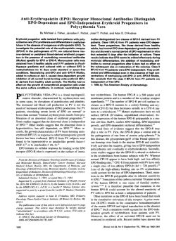
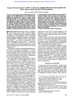
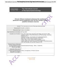
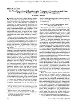
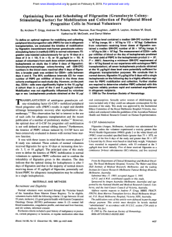
Characterization of Hematopoietic Progenitors
From Human Yolk Sacs and Embryos
By Anne Huyhn, Marc Dommergues, Brigitte Izac, Laure Croisille, Andre Katz,
William Vainchenker, and Laure Coulombel
Hematopoiesisfirst arises in the extraembryonic yolksac, and
throid colonies on replating. Analysis of the distribution of
it is generally believedthat yolk sac-derived stem cells migrate
progenitors revealed that in contrast t o erythroid progeniand seed the ietal liver at approximately week 6 of developtors, whose numberswere equally distributed between the
ment in humans. Recently, the identificationat day 8.5 to 9 of
yolk sac and the embryo, 80% of the nonerythroid progenifrom
multipotential stem cellsin intraembryonic sites different
tors were found in the embryo at stages II and 111. Interestthe liver suggests that the establishment of hematopoiesis
ingly, ahigh proportionof nonerythroid progenitors (includmight be more complexthan initially believed. In an attempt
ing high proliferative potential cells) was present in colony
to understand initial steps of hematopoiesis during human
assays initiated with cells remaining after the liver has been
ontogeny. we characterizedclonogenicmyeloidprogenitor
removed. These findings were validated in colony assays
cells in human yolksacs and corresponding embryosa t 25 to
established with CD34' cells purified from extraembryonic
50 days of development.Most erythroid colonies derivedfrom
yolk sacs and intraembryonic tissues. Increased knowledge
the yolk sacs differad from adult marrow-derived progenitors about the biology of hematopoietic stem cells early in life
in that they also contained cells of the granulomacrophagic
may helpto further understanding of the mechanisms assolineage, suggesting that they were pluripotent and exhibited
ciated with therestriction in proliferative and differentiative
potential observed in the adult hematopoieticstem cell coma different response t o cytokines. Furthermore, a subclass of
nonerythroid progenitors generated very large granulomacro- partment.
phagic colonies, some of which generated secondary ery0 7995 by The American Society of Hematology.
T
HE SEQUENCE of events during mouse development
has clearly established that the extraembryonic yolk
sac is the initial site of hematopoiesis, which begins at approximately day 7.1-3Progenitor cells undergoing differentiation toward the B lymphoid pathway were also detected in
the embryonic body at approximately day 8 to 9, although
some controversy exists on the exact time that such cells are
first detected4"and whether these intraembryonic progenitor
cells derive from intraembryonic tissues or from stem cells
migrating from the yolk sac and seeding the fetal liver."'
There is no direct experimental evidence for the latter hypothesis. In contrast, emergence of stem cells in the embryonic body has received support in birds, where yolk sac and
fetal liver hematopoiesis are independent events," and more
Thus, two groups have shown
that
recently, in
some primitive stem cells, identified in vivo by their repopulating ability,"',12 or in vitro by their ability to generate cells
from all myeloid and lymphoid lineages," originate in the
paraaortic splanchnopleura'"." or in the aorta-gonad-mesonephros (AGM) region.12 Preservation of primitive yolk sacderived hematopoiesis contrasting with altered fetal liver
erythropoiesis observed in knockout mice for c-myb" and
some transcriptional factorsI4also give credit to the hypothe-
From INSERM Unit 362, Institut Gustave Rnussy, Villejuif;and
Maternite' Port-Royal, Paris, France.
Submitted July 29, 1994; accepted July 31, 1995.
Supported by INSERM and by grants from Association
Recherche
contre le Cancer (6532 LC), Electricite' de France, and Institut
Gustave Rou.\sy.
Address reprint requests to L u r e Coulombel, MD, PhD,INSERM
U 362, Pavillon de recherche I , Institut Gustave Roussy. 39 avenue
Camille Desmoulins, 94800 Villejug France.
The publication costsof this article were defrayedin part by page
chargepayment. This article must therefore be hereby marked
"advertisement" in accordance with 18 U.S.C. section 1734 solely IO
indicate this fact.
0 1998 by The American Sociery of Hematology.
0006-4971/98/8612-0022$3.00/0
4474
sis of extrahepatic sites for hematopoietic production. In
contrast with the accumulation of evidence demonstrating
extensive potentialities for intraembryonic hematopoietic
stem cells, little is known on the properties of yolk sac
progenitor cells. Committed progenitor cells of multiple myeloid and lymphoid lineages have been found in the mouse
and in the human yolk sac,1.7,15-18
but very few studies have
identified day-12 spleen colony-forming units (CFU-S),'.]'
and the existence of long-term reconstituting cells in the yolk
sac is still debated.".I9 Characterization of the hematopoietic
stem cell properties early in life will be important in view
of recent observations that suggest age-related restrictions in
the proliferative capacity'" and potentialities"." of primitive
progenitor cells. Thus, individual progenitor cells with a
primitive phenotype (CD34++/Thyl+)isolated frombone
marrow of 16- to 18-week-old human fetuses generate myeloid and B lymphoid cells in vitro with a high frequency."
CD34+ cells from fetal liver or marrow also produce, in
some conditions, T-lymphocyte precursors" and easily reconstitute severe combined immunodeficiency (SCID)
mice,24 whereas the potential of phenotypically identical
adult marrow cells maintained in similar culture conditions
is restricted to the myeloid lineages. Also important in this
context is the observation that amplification of CD34' cells
might be more efficiently obtained in vitro from hematopoietic tissues of fetal origin.25
In this study, we characterized in detail the number and
potential of clonogenic hematopoietic progenitors in human
yolk sacs and corresponding embryonic bodies between 30
and 50 days of development. Both erythroid and nonerythroid progenitor cells were detected in colony assays initiated with yolk saccells but also with cells from the embryos.
Unique morphologic features and cytokine requirements of
colonies observed in colony assays of yolk sac and intraembryonic cells suggest that the progenitors were endowed with
properties that distinguished them from adultmarrow-derived progenitors. This was particularly true of pluripotent,
high proliferative potential progenitors thatwere detected
almost exclusively in the embryo outside the liver. sugBlood, Vol 86,No 12 (December 15). 1995: pp 4474-4485
4475
HEMATOPOIESIS IN HUMAN YOLK SACS AND EMBRYOS
gesting that extrahepatichematopoietic sites may exist in
humans as well.
MATERIALS AND METHODS
Sample Collection and Staging
Abortion products were collected after termination of pregnancy
by administration of oral mifepristone and parental misoprostol
(prostaglandin E l [PGEl]). The project was approved by the National Ethics Committee, and written consent was obtained from
every patient. Yolk sacs and corresponding embryos were dissected
free of the surrounding tissues and the stage of development was
estimated by measuring crown-rump length (CRL) and by examining
general developmental features of the embryo as reported by Hamilton and Mossman." Although the menstrual history of every patient
was available, we chose to classify the embryos according to their
morphologic stage of development. We analyzed 34 yolk sacs and
35 embryos. Using CRL and development features described by
Hamilton and Mossman,26we distinguished four stages of development. Prominent features of stage I (approximately 25 to 30 days)
included a large connection between yolk sac and midgut, an elongated embryo, poor vascularization, absence of clear liver rudiment
under the microscope. Embryos at stages I1 (approximately 30 to 35
days) and 111 (35 to 40 days) were markedly curved, with marked
pontine and cervical flexures and apparent limb buds. The yolk sac
was connected to the embryo through a long vitelline duct, which
facilitated its separation from the embryo. Stages I1 and I11 were
mscriminated by the subdivision of limb buds in stage 111, an obvious
liver rudiment, marked forebrain vesicles, and cervical and pontine
flexures. Four embryos at stage IV (greater than 40 days) were
also obtained. The liver rudiment was clearly identified under the
microscope at stage 111, and whenever possible, it was dissected and
analyzed separately.
Unique technical difficulties due to the collection of human embryos were encountered in this study and deserve two comments.
(1) The earliest human embryos used in this study were at approximately 25 days of development, which is probably later than the
time at which intraembryonic progenitors were first identified in the
mouse species. (2) Individual experiments were performed from one
sample generating low numbers of cells, a limitation that precluded
detailed comparisons within each experiment.
Cell Preparation
Extraembryonic yolk sacs and embryos were separately digested
enzymatically in 0.1% collagenase in Hanks' balanced salt solution
(HBSS) supplemented with 30% fetal calf serum (FCS; JBIO Laboratory, Les Ulis, France) and DNAse (type I, 100 mg/mL; Sigma,
St Louis, MO). Ten fetal livers were also prepared using a similar
procedure. Cells were incubated for 30 to 90 minutes at 37°C in
the enzyme solution, vigorously pipetted to dissociate the tissues,
pelleted, washed once, and incubated overnight in tissue culture
dishes in a-minimal essential medium (aMEM) plus 30% FCS at
37"C, 5% COz. This step facilitated the dissociation of aggregates
present in the cell suspension immediately after collagenase digestion. These strongly adhered and spread during the overnight incubation. Cytokines, recombinant human (rhu) stem cell factor (SCF;
supplied by AMGEN; Thousand Oaks, CA), interleukin (1L)-3, IL-6
(supplied by Genetics Institute, Cambridge, MA), and erythropoietin
(Epo; AMGEN), were systematically added during the incubation
period to minimize cell death. The next day, nonadherent cells were
collected, and adherent cells were detached with trypsin in a singlecell suspension, washed, and pooled with the nonadherent cells.
Pooled cells were counted and plated in methylcellulose assays supplemented with various combinations of recombinant growth factors
(see below).
Isolation and lmmunophenotyping of CD34+ Cells From
Yolk Sac, Embryonic Liver, and Extrahepatic Embryo
Immunologic phenotyping was performed in three samples on cell
suspensions obtained as described above from liver, embryo, and
extraembryonic yolk sac, except that adherent cells were recovered
after overnight incubation with a nonenzymatic solution (Sigma
Chemicals). Cells were labeled in uMEM supplemented with 5%
FCS and 100 pg/mL DNAse (type I; Sigma Chemicals) with phycoerythrin (PE)-conjugated HPCA-2 monoclonal antibody (MoAb;
CD34) and one of the following MoAbs directly coupled to fluorescein isothyocyanate (FITC) and recognizing the following antigens:
CD38, CD19 (Becton Dickinson, San JosB, CA), CDIlb, CD33,
HLA-DR, GPIIIa (CD61; Dako, Trappes, France), Thy1 (Pharmingen, San Diego, CA), and UlexEuropeaus (Vector Laboratories).
Acquisition of at least 10,OOO events was performed on a FACSsort
(Becton Dickinson). Analysis of the data was performed with the
Cellquest software (Becton Dickinson). Systematically, 7-amino-a~tinomycine D (7-AAD; Sigma) was added tothe samples, and a
gate was defined that included only 7-AAD-negative (viable) cells.
Compensation was set up with control cells stained with CD34-PW
IgGl -FITC and CD38-FITCAgGl-PE.
In four experiments, CD34' cells were sorted from yolk sac, liver,
and embryonic cell suspensions labeled with the PE-HPCA-2 MoAb
on a FACS Vantage (Becton Dickinson) equipped with an INNOVA
70-4 Argon ion laser (Coherent Radiation, Palo Alto, CA) tuned at
488 nm and operating at 500 mW. Positivity for the CD34 antigen
was determined in the 7-AAD-negative gate using a PE-irrelevant
IgG1. To eliminate the CD34', 7-AAD-negative debris with a very
low forward side scatter, whichusually contaminated the sorted
fraction and may indicate cell death in the CD34+ population, we
selected 7-AAD- CD34' cells with a forward side scatter higher
than 300.
Assessment of Hematopoietic Progenitor Cells in
Semisolid Colony Assays
Nonadherent cells were plated in 0.8% methylcellulose in Iscove's
modified Dulbecco's medium (purchased from Stem Cell Technologies, Vancouver, Canada) supplemented with 30% FCS, 1% deionm o m 0-2 mercaptoethized bovine serum albumin (BSA), and
anol. Optimal conditions for detecting erythroid progenitors included
rhuEpo (2 U/mL), rhuSCF (50 ng/mL), and rhuIL-3 (100 U/mL).
For assessment of granulocytic progenitors, rhu granulocyte colonystimulating factor (rhuG-CSF; AMGEN) and rhu granulocyte-macrophage colony-stimulating factor (rhuGM-CSF; Immunex, Seattle,
WA) were added to the previous cytokine combinations or used
alone in independent dishes. The optimum concentrations of cytokines usedin this study were definedin colony assays initiated
with normal adult marrow cells. However, dose-response curves for
rhuSCF and rhuEpo were performed with embryonic cells in three
experiments. When colony assays were initiated with unfractionated
cells, the concentration of cells per dish varied from between 1 X
IO4 and 2 X lo4 to 2 X lo", depending on the total number of cells
obtained after dissociation of the tissue. Thus, colony assays were
established with 1.5 X IO4 to 2 X lo4 yolk sac cells per dish, 5 X
lo4 to 10 X lo4 cells from the embryo, and 2 X IO" cells from the
liver. CD34+ cells purified from the different compartments were
plated at a concentration of 1,OOO to 2,000 cells per dish. Dishes
were incubated at 37°C in an air atmosphere supplemented with 5%
CO2 and saturated with humidity. Progenitors were scored several
times: after 4 to 5 days in culture to not miss small erythroid colonyforming unit (Cm-E)-derived colonies/clusters and at days 8 to I O
and 13 to 14 when large erythroid and nonerythroid colonies were
present. Dishes were again scored at days 15 to 18. Typical small,
hemoglobinized colonies (less than 100 cells) were scored as C m -
4476
E. Other hemoglobinized colonies were large and exhibited a very
consistent and peculiar phenotype that distinguished them from standard burst-forming unit-erythroid (BFU-E)-derived colonies”
or colony-forming unit-granulocyte, erythroid, monocyte, megakaryocyte
(CFU-GEMM; see Results).Therefore, they werereferred to as
erythroid progenitors. A subclass of nonerythroid colonies also had
a distinct morphology and a very large size (greater than100,000
cells), indicating that theywerederivedfrom
a progenitor with a
veryhigh proliferativepotential(see Results). Thesewerescored
separately also.
HUYHN ET AL
as development progressed was more pronounced and was
observed independently of results expression (per organ or
per nucleated cells; Fig 1). Thus, atstages I and 11, the
absolute numbers of nonerythroid progenitors were comparable with those of erythroid progenitors (Fig 2: ie, 143 +27 [mean i SEM, n = IO] and95 5 38 [n = 71, respectively), but decreased at stages 111 (71 2 20, n = 8 ) and IV
(61 +- 26, n = 4). When expressed per IO5 nucleated cells,
these numbers were 224 ? 57 at stage I, S3 2 19 at stage
11, 27 i 7at stage 111, and 15 I
5.5at stage IV, and the
differences were statistically significant.
Analysis of Growth Factor RNA in the Yolk Sac and
We made twoadditional observations. ( I ) Almost all large
Embryos by Reverse Transcription-Polymerase Chain
erythroid colonies hadapeculiar phenotype, illustrated in
Reaction (RT-PCR)
Fig 3, with a tight center of hemoglobinized cells surrounded
RNA was extracted either from yolk sac and embryos frozen in
by dispersed cells. Cytologic examination of cytospun cololiquid nitrogen without previous processing or from cells lysed in
nies revealed a majority of differentiated erythroblasts mixed
isothiocyanate guanidinium (isothiocyanate guanidium,
4 mol& sowith some macrophages and granulocytesin most of the
dium citrate, 25 mmol/L pH7; sarcosyl, 0.5%; and 2 mercaptoethacolonies. The mixed phenotype of erythroid colonies was
nol, 0. I mol/L) after collagenase digestion. Total cellularRNA was
isolated by the modified Chomczynski and Sacchi method adapted
not reproduced foradult marrow-derived BFU-E but has
to small cell numbers.2x Presence of RNA encoding various cytobeenreported with cord bloodBFU-E-derived
colonies”’
kines was determinedusing the PCRmethod.First-strandcDNA
(and our unpublished observations, January 1995) and sugwas synthesizedfromtotalcellular RNA with hexanucleotides as
gests that these colonies arise from a class of BFU-E with
primers with avianmyeloblastosisvirus(AMV)reverse
tranpotentialities different from standard BFU-E. The rnorpholscriptase, as previously described.” Presenceof RNA in the sample
ogy of‘ erythroblasts in the colonies was unremarkable and
wasdetermined by the amplification of human 02-microglobulin
identical to that of erythroblasts found in adult marrow BFUgene, which is expressed at this stage of development (our unpubE-derived
erythroid colonies. Notably,analysis of slides
lishedobservations, March 1994). Onlysamples in which we had
made with cells from embryos and yolk sacs before culture
observed the presence of RNAof human 82-microglobulin were
revealed that erythroid cells in all but one samplewere nucletested by RT-PCR for cytokine expression. Primers specific for human GM-CSF, Epo, SCF, IL-3,and 82-microglobulin were chosen,
(2) A secondobservation
ated with nonuclearexpulsion.
as previously described in details,’” and amplification products were
concernsthe very low numbers of smallhemoglobinized
revealed by Southern blots using an oligonucleotiderecognizing
coloniesklusters derived from conventional CFU-E, even
internal sequence. RNA extracted from human phytohemagglutininwhen dishes were first scored within 4to S days after plating.
stimulated peripheral blood lymphocytes servedas a positive control
The mean (?SEM) number of CFU-E per yolk sac was 9 5
for GM-CSF and IL-3 primers, cells from the Hep G2 line for Epo,
6 at stage I. S ? 2.7 at stage 11, 36 2 20 at stage 111, and
and human marrow fibroblasts for SCF.
30 2 13 at stage IV. In contrast, at stage W . 12,000 2 4,760
CFU-E were counted per liver (n = 10).
RESULTS
We next studied the growthfactor requirements of progenHematopoietic progenitor cells were assessed in 35 emitor cells generating large erythroidcolonies. Growthof large
bryos and 34 yolk sacs. In 27 of these samples, the extraemerythroid colonies in vitro from yolk sac cellswas absolutely
bryonic yolk sac andthe embryo were from the same sample. dependent on the presence of rhuEpo. Some erythroid colonies grew in the presence of rhuEpo alone. However, their
Characterization of Hematopoietic Clanogenic Progenitor
number was considerably increased by the addition of anCells in the Yolk Sac
other cytokine. Themean (2SEM) fold-increase in the numbers of erythroid colonies induced by the addition of rhuILBoth erythroid and nonerythroidprogenitors generating
3. rhuSCF,or both torhuEpo was 10 +- 4 (n = 9)for
colonies of more than 100 cells were found in every yolk
rhuIL-3,6
+- 3 (n = 9) for rhuSCF, and2 1 t 4 when rhuEpo.
sac studied (Fig 1). The absolute number (mean ? SEM) of
rhuIL-3, and rhuSCF werepresent(Fig 4). The effects of
largeerythroidcolonies
observedperyolk
sacwas very
cytokines wereindependent of the stage of development.
similar from stage I through IV: 106 ? 18 (n = 10) of these
Interestingly, rhuSCF was usually less efficient than IL-3 in
were detected at stage 1, 1 12 L 38 (n = 7) at stage 11, and
stimulating
the development of erythroid colonies from the
189 ? 51 (n = 8) at stage 111 (Fig 2). Notably, high numbers
yolk sacs, a result that contrasted with what is usually
ob(483 ? 247, n = 4) of large erythroid colonies were observed
served with BFU-E from adult marrow. By comparison, in
at stage IV in the yolk sac. However, as the mean number
colony assays initiated with cells from embryonic livers, a
of nucleated cells per yolk sac increased from 1.1 X 10’ ?
substantialnumber of erythroid colonies developed in the
0.24 X 10’ to 3.4 X 10’ t- 0.9 X 10’ from stage I to IV, the
presence of rhuEpo alone (Fig 4B), and rhuIL-3, rhuSCF,
concentration in erythroid colonies was, in fact, decreasing.
and a combination of both increased the number of erythroid
Nevertheless, these numbers were far below the numbers of
colonies (fold-increase) by 1.6 5 0.5 (n = 41, 2.1 t 0.4 (n
progenitors generating similar colonies present in the liver
= 4),and2.4
t 0.4 (n = 7), respectively, over numbers
at stage IV of development (65,000 ? 25,700; n = 10).
observed with rhuEpoalone.
Dose-response curvesfor
By comparison, the decrease in nonerythroid progenitors
HEMATOPOIESIS INHUMAN YOLKSACS
1250
C
m
g
4411
AND EMBRYOS
5
A
0
1000
Q
n
p
n
C
al
S
W
1000
0
0
en
500
q
2
0
0
:
750
0)
0
500
0
c
0
e
c
g
c
.-
750
Q
0)
B
e0
L
0
.-c
C
1250
P
0
250
:
4
250
0
0
Stage
I
II
111
I
IV
I1
111
IV
Fig 1. Numbers of erythroid (A) and nonerythroid (B) progenitor cells in human extraembryonic yolk sacs and human embryosat different
stages of development. Thehematopoietic progenitor cell content of 29 yolk sacs and 27 embryos was determined byplating the cells after
enzymatic digestion in methylcellulose colony assays in the presence of rhuEpo, rhuSCF, and rhulL-3. Colonies were scoredat days 10 to 14
as erythroid or nonerythroid. Each symbol represents the number of the indicated progenitor cell type found in an individual yolk sac IO) or
embryo (0).Ten yolk sacs were studied at stage I, seven at stage II, eight at stage 111, and four at stage IV. Eleven embryos werestudied at
stage 1, nine at stage II, and seven at stage 111. Results from stage IV are not indicated, as only liver cells were assessed.
rhuEpo and rhuSCF in colony assays from three embryonic
samples and control adult marrow cells simultaneously did
not reveal major differences (data not shown): 50% maximal
erythroid progenitor stimulation was seen in control marrow
for rhuEpo concentrations of 10 to 50 mU/mL, and 0.5 U/
mL rhuEpo was required for optimum stimulation of colony
8oo
600
size and number. The effect of rhuSCF was first seen at 1
ng/mL, with maximal effect (judged by colony size) at 10
ng/mL.
Development of yolk sac-derived, nonerythroid progenitor
cells also absolutely required the addition of cytokines, and
although low numbers of cells in the starting sample precluded any detailed comparison of the effect of each granulocyte-specific cytokine on these progenitors, similar numbers
were obtained with the combinations rhuSCF plus rhuIL-3
and rhuGM-CSF plus rhuG-CSF with or without the addition
of agar leukocyte-conditioned medium (A-LCM; data not
shown). Cytologic analysis of cytospun colonies showed mature granulocytes of the neutrophilic and eosinophilic lineages as well as macrophages.
U
l
T
T
Stage
Fig 2. Comparison of the number of erythroid and nonerythroid
progenitor cells in yolk sacs and embryosat different stages of development. The hematopoietic progenitor cell content of 25 yolk sacs
and 27 embryos was determinedby plating the cells after enzymatic
digestion in methylcellulosecolony assaysin the presence of rhuEpo,
rhuSCF, and rhulL-3. Colonies were scoredat days 10 to 14 as erythroid or nonerythroid. Numbers of samplesat each stage are as in
Fig 1. Each histogram represents the mean 2 SEM of the absolute
number of erythroid and nonerythroid progenitor cells for yolk sacs
(U) and embryos (W).
Characterization of Hematopoietic Clonogenic Progenitor
Cells Detected in the Embryo
Comparison of the numbers of progenitor cells found in
yolk sacs and embryos. At each of the four developmental
stages studied, the progenitor cell content was determined
simultaneously in the extraembryonic yolk sac andin the
corresponding embryo using standard colony assays (Figs 1
and 2). Comparison of the absolute numbers of erythroid
and nonerythroid progenitors found in the yolk sac and inthe
embryo revealed differences in the distribution of progenitor
cells.
At stage I in every sample, the majority of erythroid progenitors (generating large erythroid colonies) was located in
the yolk sac (Fig 1A). However, at stage 11, higher numbers
of erythroid progenitors were found in the embryo than in
the yolk sac in three of seven samples. To better illustrate
the different behavior of erythroid and nonerythroid progenitors, we calculated for each individual paired sample the
ratio of absolute numbers of progenitor cells found in the
embryo versus in the yolk sac. For erythroid progenitors,
this ratio was 0.2 ? 0.1 at stage I, 3 2 1.3 at stage 11, and
HUYHN ET AL
Fig 3. Photomicrographof a
typical large erythroid colony
obtained after 12 days of culture
in methylcellulose colonyassays
initiated with humanyolk sac
cells
and
stimulated with
rhuEpo,
rhuSCF,
and
rhulL-3.
Note the crown of characteristic
individual cells irradiating from
the tight center oferythroid cells
(original magnification, ~1201.
1.5 2 1.3 at stage 111. Only liver-derived erythroid progenitors were studied at stage IV. Interestingly, although only
small numbers of colonies were scored (which precludes any
firm conclusion), erythroid progenitor cells from embryos
exhibited the same response to cytokines as that described
above for yolk sac erythroid progenitor cells and were dependent on the presence of Epo for the production of hemoglobinized colonies (data not shown). Also characteristic was
the absence of CFU-E: 26 2 24, 21 ? 9, and 27 2 18 of
these colonies were counted per embryo at stages I, 11, and
111, respectively (mean 2 SEM).
In contrast, the distribution of nonerythroid progenitors
was unequal at stage 11 and later, and numbers of progenitors
in the embryos were higher than those found in the corresponding yolk sacs. This was illustrated by a ratio between
embryo- and yolk sac-derived nonerythroid progenitors of 7
? 2.5 and 12 2 3.8 at stages 11 and Ill, respectively (Fig
IB and Fig 2).
In five experiments to determine whether these differences
were explained by the hematopoietic activity of the liver,
we analyzed separately the region of the liver rudiment and
the remaining extrahepatic embryonic tissues. As shown in
Table I, in experiments 1 through 3. the numbers of nonerythroid progenitors found in the liver rudiment were equal to
or lower than numbers in the extrahepatic tissues, indicating
that hematopoietic activity was not predominant in the liver.
In contrast, in experiments 4 and 5 (Table l ) , one of which
was from a stage IV embryo, very high numbers of erythroid
colonies and, to a lesser extent, nonerythroid colonies were
observed in assays initiated with cell suspensions from the
v)
FETALLIVER
a,
0
0
0
75
600
50
400
25
200
Ao
Bo
9
0
0
2
l
g*
- +'K
v)
p:$
5 E
._
C
ctl
cn
2
a
Fig 4. Response of erythroid progenitors grown fromyolk sacs and embryost o recombinant human cytokines. Cellsfrom 10 yolk sacs (A)
and nine livers (B; stage IV) were plated in duplicate methylcellulosecolony assays in the presence of rhuEpo alone (2 U/mL, 0).rhuEpo +
rhuSCF (50 ng/mL, 01, rhuEpo + rhulL-3 (100 U/mL, 1, or rhuEpo + rhuSCF + rhulL-3 (W. Erythroid colonies were scored after10 t o 14 days
in culture. As the response to cytokines was independent of the stage of development, results obtained from samples studied at different
stages were pooled. Each histogram represents the mean (+SEMI of colony numbersfound in the 10 yolk sac and nine liver samples.
HEMATOPOIESIS IN HUMAN YOLKSACS
4479
AND EMBRYOS
Table 1. Hematopoietic Progenitors in Embryonic Livers,
Extrahepatic Embryonic Tissues,and Extraembryonic Yolk Sacs
Hematopoietic Colonies
Nonerythroid
Large erythroid
ExpIOrgan
Stage
No./Organ
No./lO' Cells
NoJOrgan
No.110' Cells
1
Liver
II
Embryo
Heart
28
49
5
6
1
2
8
31 1
3
2
6
1
L
Liver
Embryo
Yolk sac
II
418
37
150
60
1
34
357
430
150
51
3
34
Liver
Embryo
111
232
147
12
1
300
448
15
3
Liver
Embryo
Heart
Yolk sac
111
3,400
142
153
136
1
17
64
480
15
5
21
19
<l
3
9
Liver
Embryo
Yolk sac
IV
170,000
2,290
1,200
1,400
13
192
25,370
1,074
120
215
3
4
28
5
1
19
Embryos and yolk sacs were isolated, and the region of the hepatic
rudiment was dissected out from the embryos and digested separately in 0.1% collagenase. In experiments l and 4,the cardiac cavities
were separately assessed also. The hematopoietic progenitor content
of all tissues was assessed in methylcellulose colony assays stimulated by Epo, SCF, and IL-3. Results are expressed either as absolute
numbers of the indicated progenitors per organ or per lo5 plated
cells.
Abbreviation: Exp, experiment.
liver, indicating that this organ was a major source of hematopoietic progenitors. In two experiments, progenitors present in the heart cavities were evaluated as an indicator of
circulating progenitors (Table 1).
IdentiJcation in the embryo of hematopoietic progenitors
with a very high proliferative potential. A subset of nonerythroid progenitors exhibited a unique phenotype essentially
characterized by the generation of very large colonies organized in clusters (see Fig 5). These colonies contained a
mean number of 7.3 X lo4cells (n = 50),which were almost
exclusively mature granulocytes and macrophages when examined after May-Griinwald-Giemsa staining of colonies individually plucked at days 12 to 14. These high proliferative
potential progenitors represented 20% to 25% of nonerythroid progenitors, and optimal numbers of colonies were
obtained in assays stimulated by rhuSCF and rhuIL-3. Cytokines classically used to stimulate granulocytic differentiation, ie, rhuG-CSF and rhuGM-CSF, increased neither the
size nor the numbers of these macroscopic colonies over that
observed with rhuSCF plus rhuIL-3. To determine if the
potentialities of these progenitor cells were restricted to the
granulomacrophagic lineage, we individually replated 84 of
these colonies in a secondary methylcellulose assay in conditions used for primary assays. Of these 84 primary colonies,
25 (30%) gave rise to secondary colonies, and 9 of 25 generated both erythroid and granulocytic secondary colonies (Table 2), suggesting that the primary high proliferative potential progenitor was, in fact, pluripotent. No secondary
macroscopic colonies were seen. By comparison, primary
mixed erythroid colonies (n = 15) and granulocytic colonies
were picked at days 14 to15 from the same dishes and
replated in similar conditions. As expected, 6 of 15 mixed
colonies generated secondary colonies, mostly CFU-E (data
not shown). Granulocytic colonies did not generate secondary colonies.
Interestingly, these very large granulomacrophagic colonies were detected preferentially in colony assays established
with intraembryonic cells, and only very few were observed
in colony assays initiated with yolk sac or liver cells. We
have not found such progenitors in colony assays established
with adult bone marrow stimulated only withrhuSCF, rhuIL3, and rhuEpo, a combination that allowed the development
of only small granulocytic colonies (data not shown). As
detailed in the next section, these progenitor cells were present in assays established with low numbers of CD34' cells
purified from human embryos.
Assessment by Colony Assays of the Hematopoietic
Potential of CD34+ Cells Sorted From Yolk Sac, Liver,
and Embryos
In four samples, cells were labeled withPE-HPCA2,
sorted, and plated in methylcellulose colony assays (Table
3). In two experiments (Table 3, experiments 3 and 4), we
assessed simultaneously the clonogenic potential of CD34'
cells sorted from yolk sacs, livers, and extrahepatic parts of
the embryos. The cloning efficiency ofCD34' cells was
4.6% in the yolk sac (n = 2), 1.85% 2 0.77% in the embryo
(n = 4), and 3.2% in the liver rudiment (n = 2). The total
number of CD34+ clonogenic progenitors counted per organ
approximated the total number calculated from assays performed with unfractionated cells (see Figs 1 and 2 and Table
l), thus confirming that early in human ontogeny, clonogenic
hematopoietic progenitor cells express the CD34 antigen.
Importantly, unique phenotypic features reported above for
erythroid and nonerythroid high proliferative potential progenitor-derived colonies grown in assays of unfractionated
cells were also seen in colony assays initiated withlow
numbers of CD34+ cells, thus making it unlikely that accessory cells contributed to these results through the release of
endogenous activities. CD34+ high proliferative progenitors
generated colonies of on average 200,000 cells in assays
supplemented with rhuSCF, rhuIL-3, and rhuEpo, and 4 of
10 primary colonies generated secondary granulocytic colonies after replating (see Table 2, experiment 8). The distribution of progenitors in CD34+ assays paralleled that observed
when assays were initiated with unfractionated cells. Although low numbers of progenitors precluded any firm conclusion, in both experiments where liver cells and nonliver
embryonic cells were separately assessed, these macroscopic
colonies were seen in assays from nonliver embryonic cells
(respectively, 32 and 6.5), but none were detected among
CD34+ cells of the liver rudiment from the same samples
(Table 3, experiments 3 and 4). The slight predominance of
4480
ET AL
HUYHN
Fig 5. Photomicrograph of a
colony derived from an HPP-CFC
detected in methylcellulose colony assays from cells of a stage
II human embryo. Colony assays
were stimulated by
rhuEpo,
rhuSCF,
and
rhulL-3
(original
magnification, x 48).
nonerythroid progenitors in the embryo as compared with
the yolk sac was also confirmed (258 v 205 in experiment
3 and 82 v 71 in experiment 4).
Flow cytometric analyses of CD34 subsets in yolk sac
(Fig 6a), liver (Fig 6b), and embryo (Fig 6c) were also
performed: CD34' cells in the yolk sac and extrahepatic
tissues of the embryo expressed neither CD38 nor CD33
antigen. This phenotype characterizes primitive hematopoietic progenitor cells in the adult marrow butmay also be
shared by endothelial cells and other nonhematopoietic cell
subsets that represent a high proportion of embryonic CD34'
c e k 3 ' Interestingly, a fraction of CD34' cells in the liver
rudiment expressed CD38 (Fig 6b). The Thy1 antigen was
detected on a high proportion of CD34' and CD34- cells
from all three tissues, as was the lectin Ulex Europeaus (data
not shown). HLA-DR was undetectable, andCD1 1 b was
expressed on a subset of CD34' cells and also on CD34cells from another embryo (data not shown). These data have
been confirmed on three different samples.
Expression qf Cyrokine Genes in Yolk Sacs and Embryos
Table 2. Characterizationof the Replating Potential of
Embryo-Derived Hematopoietic Progenitors With a
Very High Proliferative Capacity
Exp
1
2
3
4
5
6
7
8
Total
No. of
No. of Primary
Primary
Colonies
Colonies Giving
Secondary Colonies
20
12
17
6
6
12
8
9
10
84
2
6
2
2
2
1
4
25
Colony Phenotype in
Secondary Plates
E
+G
G
1
0
4
0
2
1
1
E
2
0
1
0
0
0
0
4
0
13
3
After collagenase digestion and overnight incubation, embryonic
cells, either unfractionated (exp 1 through 7) or CD34' (exp 8) were
plated in methylcellulose colony assays inthe presence of SCF, IL-3,
and Epo. After 12 to 15 days in culture,primary colonies identified as
HPP-CFC (see Fig3) were individuallylifted and replated in secondary
colony assays in conditions identical to the primary assays. Secondary colonies of different phenotypeswere scored at day 12.
Abbreviations: E, erythroid; G, granulocytic; E + G, presence ofboth
erythroid and
granulocytic
colonies
in
the same
dish.
As a first approach to characterize cytokines regulating
human hematopoietic differentiation at early developmental
stages, we examined by RT-PCR the expression ofRNA
coding for Epo, SCF, GM-CSF, and IL-3 in cells from six
yolk sacs and five embryos. RNA was extracted either from
tissues frozen immediately after collection (five embryos and
six yolk sacs) or after collagenase digestion and overnight
incubation (Fig 7 and Table 4). No attempt wasmade to
separate hematopoietic cells from nonhematopoietic components. The SCF gene was highly expressed in every sample,
as expected from previous studies.." The Epo gene was transcribed in every sample examined from liver rudiment and
yolk sac. In contrast, expression of GM-CSF and IL-3 genes
was negative in ex vivo samples, but transcription of the
GM-CSF gene, in contrast with that of IL-3, was induced in
adherent cells from all three tissues after their incubation in
culture.
DISCUSSION
In a search for models suitable for identifying expression
of human hematopoietic stem cell self-renewal and multipotentialities, we started to investigate the properties of hematopoietic progenitors found very early in human ontogeny.
Use of this material was motivated by recent observations
age
HEMATOPOIESIS IN HUMAN YOLK SACS AND EMBRYOS
4481
Table 3. Clonogenic Progenitor Cells Detected Among CD34' Cells Sorted From Human Yolk Sacs, Liver Rudiment. and Embwos
in Four Diffarent Experiments
Progenitors/10,000 CD34+ Cells (total progenitordorgan)
EXP
1
2
3
II
II
111
4
111
Embryo
Embryo
Embryo
Liver
Yolk sac
Embryo
Liver
Yolk sac
Total CD34+
Erythroid
10,400
13,000
16,000
13,000
3,600
9,200
2,100
5,500
17 (17)
305 (396)
45 (72)
155 (201)
266 (96)
32 (29)
250 (52)
262 (145)
Nonerythroid
67 (67)
110 (143)
75
(120)
82' (106)
236 (185)
45 (41)
165* (35)
128 (71)
Proliferative
High
10 (IO)
0 (0)
20 (32)
0 (0)
45 (16)
7 (6.5)
0 (0)
0 (0)
Cell suspensions from yolk sacs and embryonic tissues at different stages of development were digested as described in Materials and
Methods and labeled with anti-CD34 MoAb conjugated to PE. CD34+ cells were sorted on a FACS vantage as described (see Materials and
Methods) and plated in methylcellulose colony assays with SCF, IL-3, Epo, and G-CSF. The total number of CD34' cells sorted is indicated.
* Colonies were mainly composed of macrophages with very few pure granulocytic colonies.
showing that primitive progenitors with the most extensive
potentialities and proliferation capacity might be found preferentially at early stages of development."s20321
As afirst
approach, we precisely evaluated the properties of hematopoietic clonogenic progenitor cells in different compartments
of the human embryo and in the extraembryonic yolk sac.
Our study extends previous results on the assessment of
erythroid and nonerythroid progenitor cells in human yolk
sacs at 5 to 7 weeks' and provides new information on the
phenotype of these progenitor cells, their cytokine requirements, and their distribution between embryonic and extraembryonic compartments.
Most erythroid and some nonerythroid progenitors identified in human yolk sacs and embryos, when stimulated by
rhuSCF and rhuIL-3, expressed properties that distinguished
them as earlier cells than the BFU-E and CFU-GM identified
in adult bone marrow using similar colony assays. Thus,
most erythroid colonies included some macrophages and mature granulocytes, suggesting that the progenitor was pluripotent. Within the population of nonerythroid progenitors, a
subset was identified that produced very large granulomacrophagic colonies in vitro in response to rhuSCF and rhuIL3. Such properties characterized primitive high proliferative
potential colony-forming cells (HPP-CFC) identified in agar
colony assays of 5-fluorouracil (SW)-treated mouse bone
marrow and that have been shown to be hierarchically close
to in vivo repopulating cells33 and may also be shared by
cord blood CD34+ progenitors." In addition to their high
proliferative capacity, some of these progenitors identified
in human embryos were pluripotent, as suggested by the
replating capacity of primary colonies that yielded both erythroid and granulocytic secondary colonies. This result
underlines the fact that a significant number of progenitors
believed to be restricted to the granulocyte-macrophage lineage based on the phenotype of the primary colonies that
they have generated may also be capable of erythroid differentiation or of lymphoid differentiation, as recently demonstrated in the mouse.35However, the exact potential of these
HPP-CFC-like progenitors and their position in the hematopoietic hierarchy remain to be elucidated in appropriate
assays. Second, intraembryonic erythroid and nonerythroid
progenitor cells expressed the CD34+ antigen, as demonstrated by the results of colony assays established with
CD34+ cells purified from yolk sacs and embryos. These
progenitors were negative for the CD38 and CD33 antigens,
as suggested by flow cytometric analysis, although this is
difficult to ascertain because the plating efficiency was very
low and we did not directly analyze the clonogenic potential
of sorted CD34+CD38- cells. Thus, all CD34+ cells from
yolk sacs and embryos were negative for the expression of
CD33 and CD38 antigens, a phenotype usually characteristic
of immature progenitors, at least in adult bone m a r r ~ w . ~ ~ . ~ '
It is possible, however, that during ontogeny the expression
of antigens classically used to subdivide CD34' cells will
prove to be different from what is expected at a given maturation stage based on results reported with adult cells. Phenotypic analysis might also be skewed by uncontrolled technical problems inherent to the use of human embryos, such
as selective loss of more mature CD34+CD38+progenitors
during the time elapsed between chemical induction of abortion and collection of the samples.
Nevertheless, results from colony assays performed with
CD34+ cells purified from embryonic sources confirmed that
unique features observed in colony assays established with
unfractionated cells did not result from the endogenous release of stimulatory molecules by accessory nonhematopoietic cells contaminating the assay, but rather resulted from
unique, intrinsic properties of the progenitors. The contribution of cytokines produced by cells within the colony itself
is still possible and will have to be further analyzed.
The distribution of progenitor cells between yolk sac and
intraembryonic tissues that we observed also raised some
questions: high numbers of both erythroid and nonerythroid
progenitor cells were consistently detected in the embryo
as early as stage I. Second, nonerythroid progenitors were
predominantly found in the embryo, whereas erythroid progenitors were more equally distributed. Thus, at stages I1
and III, there were 7 to 10-fold more nonerythroid progenitors but only twofold more erythroid progenitors in the embryo as compared with the extraembryonic yolk sac. Because
the whole embryo was digested, we could not determine
whether the cells were circulating or arose from a precise
HUYHN ET AL
4482
Yolk sac
a
t
0
C
t
4
THY 1
Extra-hepatic embryo
4
CD19
GPlllA
Embryonic liver
b
P
P
2
2
P
2
n
0-
2
2
0
CD38
THY1
CD33
Fig 6. f l o w c y t o m d c analysea of unfractlonated cells from the
yolk sac (a),liver rudiment (bl, and exhehepatic tissues (c)of a stage
111 embryo. Cells were incubated simultaneously with PE-HPCA2
(CD341and FITC-labeled MoAbs recognizing CD38,Thyl, CD33, CD19,
and GPIIIA. Positivity or negativity for the PE- or MC-labeled MoAbs
was determined using control cells labeled with MC-CD38 and PEIgG1 (a) (A panels) or PE-HPCA2 and an FITC-labeled lgGl (B panels).
Cells were simultaneously stained with 7-AAD, and only 7-AAD-negative cells were analyzed in a morphologic gate (R11 defined by the
light scatter profile (see upper left panels).Representative cytograms
of the two-color staining were performed in the R1 gate.
bryonic progenitors that we have identified in this study,
three arguments, although indirect, suggest that they might
not arise from the liver. The most convincing isone
provided
by the comparison of numbers of colonies observed in colony assays initiated with cells from the liver rudiment with
those observed in colony assays from embryos from which
the liver rudimenthas been removed. High numbers of both
50% of all
intraembryonic source. The liver is the primary hematopoi- erythroid and nonerythroid progenitors (up to
intraembryonic progenitors) were found in assays from both
etic organ from 6 weeks of development in humans, but in
sources, and high proliferative potential progenitor cells genother species, hematopoietic activity has clearly been demerating large colonies (see Fig 5 ) were observed in colony
onstrated earlier in other intraembryonic regions. In birds,
assays from nonliver cells and were not observedin assays
the analysis of histologic sections and the use of chimeras
established with liver cells. This was true whether the assays
have proven that definitive hematopoiesis arises from the
were initiated with unfractionated or CD34+ cells. The preparaaortic region: and more recently, hematopoietic stem
dominant locationof CFU-S in the AGM region outside the
cells capable of generating myeloid andB- and T-lymphoid
liver has also been demonstrated in the mouse.” It is imcells have been localized in the paraaortic splanchnopleura
portant to mention, however, that the distribution of progeniand AGM region of mouse
embryos at days 8.5 to
The existenceof intraaortic sitesof hematopoietic production tors within embryonic compartments is likely to vary very
quickly, and that our observations probably hold true only
has also beensuggestedonhistologicsectionsofhuman
ata veryprecisetimeofdevelopmentcorrespondingto
embryos at stage 11, and this region has been shown to produce clonogenic progenitors in short term assays. However, stages II to III. This does not question the fact that the liver
direct proof that the CD34+ cells identified on tissue sections
is later the primary source of primitive stem cells. Finally,
it should be mentioned that our finding of hematopoietic
can express hematopoietic potential in functional assays is
activity in the yolk sac does not necessarily prove that prostill lacking?’”
Although we can only speculate on the origin of intraem- genitors were produced in situ, and the observation that a
HEMATOPOIESIS IN HUMAN YOLKSACS
AND EMBRYOS
4483
P
io
v)
>
OD
W
0
m
F
v)
A
>
L
(Y
F
*
v)
SCF
177bp
GM-CSF
159bp
Fig 7. PCR analysis of cytokineexpression in samples from human yolksacs,fetal livers, and embryos.
cDNA was prepared from total cellular RNA extracted from positive control (first
line), negative
control (water; second line), and different samples
of fetal livers (FL), yolk sacs (YS), and embryos (E).
YS8,E8,YS12,FL13,FL15,FL18,
El9a, E19b. and
YS20 refer t o samples analyzed either ex vivo or directly aftercollagenase treatment. In some samples,
cells dissociated with collagenase were incubated
overnight, and nonadherent (NA) oradherent (A H241
cells were used for PCR analysis. Some adherent
cells were used after two (P2), three (P3). or four(P41
passages in culture. Hybridization was performed
with "P-labeled appropriateinternaloligonucleotides for human SCF (A), GM-CSF (B), IL-3 (C), and
Epo (D).
IL-3
211bp
ca
0 EPO
156bp
Table 4. Detection of Cytokine mRNA Expression b y RT-PCR in Cell
Samples Isolated From ExtraembryonicYolk Sacs,
Livers, and Embryos
rnRNA*
Organ
Embryo
Ex vivo
Adh 24h
Adh P2
Yolk sac
Ex vivo
Adh 24h
Adh P4
Liver
Ex vivo
Adh P2
Adh P3
N Adh 24h
No. of Samples
Assessed
Epo
SCF
GM-CSF
IL-3
0
0
0
5
1
1
It
5
0
0
1
1
0
1
1
6
1
1
5
1
0
5
1
1
0
1
1
0
0
0
4
2
4
0
1
. o
2
0
2
2
1
2
0
2
1
2
0
0
0
0
RNA was extracted from the different organs either immediately
after collection (ex vivo) of thesamples or after enzymatic treatment
and overnight incubation.
Abbreviations: Adh, adherent cells after 24 hours of incubation (24h)
or after two (P2). three (P3), or four(P41 passages;N Adh, nonadherent
cells.
The presence of RNA encoding Epo, GM-CSF, 11-3, or SCF was
determined using the PCR method. Numbers refer t o the number of
positive samples.
t Only four samples were tested for the presence of Epo mRNA.
few high proliferative progenitors were, indeed, observed in
yolk sac colony assays could indicate their migration from
an external site. With respect to that question, analysis of
globin gene expression in individual colonies grown from
the different embryonic sources will be helpful.
Comparison of the biologic properties of progenitors
grown from intra- and extrahepatic embryonic cells may also
provide meaningful indications. Thus, the Epo response of
intraembryonic progenitors differed from that observed for
progenitors detected in assays from liver suspensions, in
that Epo alone did not stimulate the growth of extrahepatic
embryonic erythroid progenitor cells, but triggered the development of a high number of large erythroid colonies from
liver cells, as previously described at later developmental
~ t a g e s . ~Also,
' . ~ ~ typical CFU-E were usually not detected in
colony assays initiated with cell suspensions from extrahepatic embryonic cells or yolk sacs, but were present in high
numbers in colony assays from fetal liver cells. Such inappropriately low numbers of CFU-E have already been reported in mouse yolk sacs13*16 butnot in human yolk sac.'
A parallel could be drawn with the adult situation, where no
CFU-E circulate and circulating BFU-E, although originating in the marrow, express properties different from those
of marrow-derived BFU-E."" CFU-E may also have been
lost during the time elapsed between abortion induction and
collection of the products or may be hypersensitive to collagenase, although mechanical dispersion of yolk sac cells in
two experiments did not result in an increase in CFU-E
numbers. None of these conditions, however, was detrimen-
4404
HUYHN ET AL
tal to fetal liver-derived CFU-E processed simultaneously,
and neither collagenase nor trypsin treatment were toxic to
adult marrow4’ or liver CFU-E (data from this studyand
Rich4*).
Despite the heterogeneity in the response of erythroid progenitors from different embryonic sources to Epo alone, production of hemoglobinized erythroblasts from both yolk sacs
and embryonic progenitors was absolutely dependent on the
addition of rhuEpo, and we did not detect any hypersensitivity to either this hormone or rhuSCF. This pointisstill
controversial in the literature, and the growth of pure erythroid embryonic liver progenitors in the presence of SCF
only has been reported, which may be partly explained by
the local production of E ~ O . ~With
’ . ~respect to the in vivo
situation, the biologic significance of the in vitro cytokine
response of progenitors is unclear. In vivo, mRNA encoding
SCF and Epo were bothdetected in embryos or yolk sacs, but
neither IL-3 nor GM-CSF genes were transcribed. Similar
cytokine transcription patterns have been reported in day8.5 mouse embryo and yolk sacs and also within embryoid
bodies where hematopoiesis develops efficiently in the absence of added growth factors.& It is possible that the proliferation and differentiation of intraembryonic hematopoietic
progenitor cells are under the control of as yet unidentified
molecules produced by unique local yolk sac or embryonic
environments during embryonic development.
The results described in this study suggest that progenitor
cells identified in the humanyolk sac, embryo, and liver
differ based on their potentialities and response to cytokines.
The next challenge will be to investigate the ability of individual CD34’ cells purified from yolk sacs and different
parts of the embryos to generate differentiated cells of myeloid lineages and to undergo commitment into the B and T
lineages using long-term culture assays on competent stromal cell^^'^"^^^ or in vivo mouse chimeric reconstitution
assay^?^,^^ This may help in gaining an understanding of the
potential age-related mechanisms associated with restriction
of potential in the hematopoietic stem cell compartment.
ACKNOWLEDGMENT
We are indebted to Dr E. Aubenyandthestaffofthe
Centre
d’orthogbnie de I’H6pital Broussais in Paris, France, for collecting
human samples. We also thank AMGEN and Genetics Institute for
their generous gifts of human recombinant cytokines and F. Wendling for help in PCR procedure.
REFERENCES
1. Migliaccio G, Migliaccio A, Petti S, Mavilio F, Russo G, Lazzaro D, Lazzaro D, Testa U, Marinucci M, Peschle C: Human embryonic hemopoiesis. Kinetics of progenitors and precursors underlying
the yolk sac liver transition. J Clin Invest 78:51, 1986
2. Moore M, Metcalf D: Ontogeny ofthe hemopoietic system:
Yolk sac origin of in vivo and in vitro colony-forming cells in the
developing mouse embryo. Br J Haematol 18:279, 1970
3. Tavassoli M: Embryonic and fetal hemopoiesis: An overview.
Blood Cells 1:269, 1991
4. Ogawa M, Nishikawa S, Ikuta F, Yamamura F, Naito M, Takahashi K, Nishikawa S: B cell ontogeny in murine embryo studied
by a culture system with the monolayer of a stromal cell clone ST2.
B cell progenitor develops first in the embryonal body rather than
in the yolk sac. EMBO J 7:1337, 1988
5. Palacios R, Imhof BI: At day 8.5 of mouse development the
yolk sac, not the embryo proper, has lymphoid precursor potential
in vivo and in vitro. Proc Natl Acad Sci USA 90:6581, 1093
6. Cumano A, Furlonger C, Paige C: Differentiation and characterization of B-cell precursors detected in the yolk sac and embryo
body of embryos beginning at the I O to 12 somite stage. Proc Natl
Acad Sci USA 90:6429, 1993
7. Huang H, Zettergren LD, Auerbach R: In vitro differentiation
of B cells and myeloid cells from the early mouse embryo and its
extraembryonic yolk sac. Exp Hematol 22:19, 1994
8. Weissman I, Papaioannou V, Gardner R: Fetal hematopoietic
origins of the adulthematolymphoid system, in Clarkson B, Marks P,
Till J (eds): Differentiation of Noma1 and Neoplastic Hematopoietic
Cells. Cold Spring Harbor Conferences on Cell Proliferation. Cold
Spring Harbor, NY, Cold Spring Harbor Laboratory, 1978, p 33
9. Dieterlen-Litvre F: Onthe origin of haemopoietic stem cells
in the avian embryo: An experimental approach. J Embryo1 Exp
Morph 33:607, 1975
IO. Godin I, Garcia-Perero J, Coutinho A, Dieterlen-Like F,
Marcos M: Para-aortic splanchnopleura from early mouse embryos
contains Bla cell progenitors. Nature 364:67, 1993
1 I . Godin I, Dieterlen-Likvre F, Cumano A: Emergence of
multipotent hemopoietic cells in the yolk sac and paraaortic splanchnopleura in mouse embryos, beginning at 8.5 days post-coitus. Proc
Natl Acad Sci USA 92:773, 1995
12. Medvinski A, Samoylina L, Muller A, Dzlerzak E: An early
pre-liver intraembryonic source of CFU-S in the developing mouse.
Nature 364:64, 1993
13. Mucenski M, McLain K, Kier A, Swerdlow S, Schreiner C,
Miller T, Pietryga D, Scott W, Potter S: A functional c-myb gene is
required for normal murine fetal hepatic hematopoiesis. Cell 65:677.
1991
14. Tsai F, Keller G, Kuo F, Weiss M, Chen J, Rosenblatt M.
Alt F, Orkin S: An early haematopoietic defect in mice lacking the
transcription factor GATA-2. Nature 371:221, 1994
15. Wong P, Chung S, Chui D, Eaves C: Properties of the earliest
clonogenic precursors to appear in the developing murine yolk sac.
Proc Natl Acad Sci USA 83:3851, 1986
16. Labastie M, Thiery J, Le Douarin N: Mouse yolk sac and
intraembryonic tissues produce factors able to elicit differentiation of
erythroid burst-forming units and colony-forming units respectively.
Dev Biol 81:1453,1984
17. Huang HH, Auerbach R: Identification and characterization
of hematopoietic stem cells from the yolk sac of the early mouse
embryo. Proc Natl Acad Sci USA 90:lOl IO, 1993
18. Johnson G, Barker D: Erythroid progenitor cells and stimulating factors during murine embryonic and fetal development. Exp
Hematol 13:200,1985
19. Toles J, Chui D, Beldeck L, Stan E, Barker J: Hemopoietic
stem cells in murine embryonic yolk sac and peripheral blood. Proc
Natl Acad Sci USA 86:7456, 1989
20.VaziriH, Dragowska W, Allsopp R, Thomas T, Harley C,
Lansdorp PM: Evidence for a mitotic clock in human hematopoietic
stem cells: Loss of telomeric DNA with age. ProcNatlAcadSci
USA 91:9857, 1994
21. Baum C, Weissman I, Tsukamoto A, Buckle A, Peault B:
Isolation of a candidate human hematopoietic stem cell population.
Proc Natl Acad Sci USA 89:2804, 1992
22. Huang S, Terstappen LW: Lymphoid and myeloid differentiation of single human CD34’, HLA-DRf, CD38- hematopoietic stem
cells. Blood 83:1515, 1994
23. Plum J, DeSmedt M, Defresne MP, Leclercq G, Vanderkerckhove B: Human CD34’ fetal liver stem cells differentiate to T cells
in a mouse thymic microenvironment. Blood 84:1587, 1994
24. Kollmann T. Kim A, Zhuang X, Hachamovitch M, Goldstein
HEMATOPOIESIS IN HUMAN YOLK SACS AND EMBRYOS
H: Reconstitution of SCID mice with human lymphoid and myeloid
cells after transplantation with human fetal bone marrow without
the requirement for exogenous human cytokines. Proc Natl Acad
Sci USA 91:8032, 1994
25. Lansdorp P, Dragowska W, Mayani H: Ontogeny-related
changes in proliferative potential of human hematopoietic cells. J
Exp Med 178:787, 1993
26. Hamilton W, Mossman H: Chapter VIII, in Hamilton WJ,
Boyd JD, Mossman H W (eds): Human Embryology. New York,
NY, Macmillan, 1972, p 174
27. Eaves C, Eaves A: Erythropoiesis in culture. Clin Haematol
13:371, 1984
28. Chomczynski P, Sacchi N: Single-step method of RNA isolation by acid guanidinium thiocyanate-phenol-chloroform extraction.
Anal Biochem 162:156, 1987
29. Auffray I, Dubart A, Izac B, Vainchenker W, Coulombel L:
A murine stromal cell line promotes the proliferation of a human
factor-dependent leukemic cell line. Exp Hematol 22:417, 1994
30. Broxmeyer HE, Hangoc G, Cooper S, Ribeiro RC, Graves
V, Yoder M: Growth characteristics and expansion of human umbilical cord blood and estimation of its potential for transplantation in
adults. Proc Natl Acad Sci USA 89:4109, 1993
31. Young P, Baumhueter S, Lasky L: The sialomucin CD34 is
expressed on hematopoietic cells and blood vessels during murine
development. Blood 85:96, 1995
32. Matsui Y, Zsebo K, Hogan B: Embryonic expression of a
haematopoietic growth factor encoded by the SI locus.and the ligand
for c-kit. Nature 347:667, 1990
33. Mac Niece I, Bradley T, Kriegler A, Hodgson G: Subpopulations of mouse bone marrow high-proliferative-potential cells. Exp
Hematol 14:856, 1986
34. Lu L, Xiao M, Shen RM, Grigsby S, Broxmeyer H: Enrichment, characterization and responsiveness of single primitive
CD34'"
human umbilical cord blood hematopoietic progenitors
with high proliferative and replating potential. Blood 81:41, 1993
35. Hirayama F, Shih JP, Awgulewitsch A, Warr GW, Clark
SC, Ogawa M: Clonal proliferation of murine lymphohemopoietic
progenitors in culture. Proc Natl Acad Sci USA 895907, 1992
36. Andrews R, Singer J, Bernstein I: Human hematopoietic precursors in long-term culture: Single CD34+ cells that lack detectable
4485
T cell, B cell, and myeloid antigens produce multiple colony-forming
cells when cultured with marrow stromal cells. J Exp Med 172:355,
1990
37. Issaad C, Croisille L, Katz A, Vainchenker W, Coulombel L:
A murine stromal cell line allows the proliferation of very primitive
human CD34++/CD38- progenitor cells in long-term cultures and
semi-solid assays. Blood 81:2916, 1993
37a. Tavian M, Coulombel L, Luton D, San Clemente H, Dieterlen-Likvre F, Pkault B: Aorta-associated CD34' hematopoietic
cells in early human embryo. Blood (in press)
38. Emerson S, Thoma S, Ferrar J, Greenstein J: Developmental
regulation of erythropoiesis by hematopoietic growth factors: Analysis on populations of BFU-E from bone marrow, peripheral blood
and fetal liver. Blood 74:49, 1989
39. Valtieri M, Gabbianelli M, Pelosi E, Bassano E, Petti S, Russo
G, Testa U, Peschle C: Erythropoietin alone induces erythroid burst
formation by human embryonic but not adult BFU-E in unicellular
serum-free cultures. Blood 74:460, 1989
40. Clarke BJ, Housman D: Characterization of an erythroid precursor cell of high proliferative capacity in normal human peripheral
blood. Proc Natl Acad Sci USA 74:1105, 1977
41. Coulombel L, Eaves AC, Eaves CJ: Enzymatic treatment of
long-term human marrow cultures reveals the preferential location
of primitive hemopoietic progenitors in the adherent layer. Blood
62:291, 1983
42. Rich I: The developmental biology of hematopoiesis: Effect
of growth factors on the colony formation by embryonic cells. Exp
Hematol 20:368, 1992
43. Rich I, Noe G, Vogt C, Zsebo K The initiation of hernatopoiesis in the mouse embryo. Blood 78:257, 1991 (suppl 1)
44. Keller G, Kennedy M, Papayannopoulou T, Wiles M: Hematopoietic commitment during embryonic stem cell differentiation in
culture. Mol Cell Biol 13:473, 1993
45. Sutherland H, Eaves C, Dragowska W, Lansdorp P: Characterization and partial purification of human marrow cells capable of
initiating long-term hematopoiesis in vitro. Blood 74:1563, 1989
46. Vormoor J, Lapidot T, Pflumio F, Risdon G, Patterson B,
Broxmeyer H, Dick J: Immature human cord blood progenitors engraft and proliferate to high levels in severe combined immunodeficiency mice. Blood 83:2489, 1994
© Copyright 2026