
Aging and DNA methylation
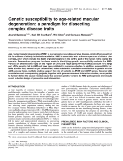
Jung and Pfeifer BMC Biology (2015) 13:7
DOI 10.1186/s12915-015-0118-4
OPINION
Open Access
Aging and DNA methylation
Marc Jung1* and Gerd P Pfeifer2*
Abstract
In this Opinion article, we summarize how changes in
DNA methylation occur during aging in mammals and
discuss examples of how such events may contribute
to the aging process. We explore mechanisms that
could facilitate DNA methylation changes in a
site-specific manner and highlight a model in which
region-specific DNA hypermethylation during aging is
facilitated in a competitive manner by destabilization
of the Polycomb repressive complex.
Introduction
Aging can be described as a slow, time-dependent
decline of a set of multiple biological functions. In some
biological pathways, functional decline can be defined
in a mono-causal way, such as the decline of resting
metabolism, whereas in other pathways the scope of the
decline is rather broad and elusive, such as that for reduced stability of epigenetic patterns. Although epigenetic patterns change dramatically during development,
these early events are biologically programmed and necessary, whereas alterations of the epigenome in adult
somatic tissue may reflect aging-associated deleterious
events. Efforts have been made to encapsulate processes
of aging into well-defined categories. These range from
accumulation of genomic damage that leads to chromosomal instability and telomere shortening, reactive oxygen species-induced damage to mitochondrial functions
and reduced energy production, stem cell depletion, accumulation of damaged proteins via loss of proteostasis,
processes leading to senescence and changes in intercellular communications, age-related effects of the insulin
and IGF-1 signaling pathways and, lastly, alterations of
the epigenome. Many of these altered pathways are
thought to be the prime components of age-related diseases including cancer, neurodegenerative diseases, atherosclerosis and inflammation. A detailed description of
these categories can be found in the review by López-Otín
* Correspondence: [email protected]; [email protected]
1
Beckman Research Institute, Duarte, CA 91010, USA
2
Van Andel Research Institute, Grand Rapids, MI 49525, USA
et al. [1], which defined these categories as the ‘hallmarks’
of aging. It is expected that there is extensive crosstalk between categories and because epigenetic changes are often
regulatory events and can lead to altered gene expression,
they can impact other hallmarks of aging, such as by
silencing of DNA repair genes or anti-inflammatory genes.
Here, we would like to focus on the epigenetic contributions to aging. More specifically, we will discuss the relationship between aging and changes in DNA methylation.
DNA methylation patterns are shaped by two opposing
processes of adding and removing a methyl group at position five of cytosine in DNA. The first functions of DNA
methylation to be discovered were related to gene regulation and cell differentiation [2]. Selective maintenance of
DNA methylation at specific loci is essential for controlling differential expression of the paternal and maternal
alleles in mammals [3], known as genomic imprinting. In
particular, X-chromosome inactivation is an often-cited
example where DNA methylation is required for longterm silencing of a locus [4]. After the developmental
phase, the genome of somatic cells will consist of roughly
1% methylated DNA cytosines, mostly affecting CpG
dinucleotides [5]. While there exists great variability between the established patterns of DNA methylation, there
is a consistent landmark in the form of CpG islands, which
are unmethylated GC-rich regions with high densities of
CpGs and often correlated with promoter regions [6].
After DNA methylation patterns have been established
during embryogenesis, key questions in the field are how
regulatory mechanisms define and maintain them in
specific tissues, how the environment can facilitate
changes in methylation patterns during a lifespan and
what the impacts of these changes are.
Predictability of DNA methylation during aging
The earliest studies, which related DNA methylation
changes to the aging process, were investigating different
organs and life stages of humpback salmon [7] and found
that 5-methylcytosine (5mC) levels during ontogenesis
were significantly decreasing. Attesting to the evolutionary
importance of DNA methylation, these results could be
later extended to mammals [8]. The highest amount of
© 2015 Jung and Pfeifer; licensee BioMed Central. This is an Open Access article distributed under the terms of the Creative
Commons Attribution License (http://creativecommons.org/licenses/by/4.0), which permits unrestricted use, distribution, and
reproduction in any medium, provided the original work is properly credited. The Creative Commons Public Domain
Dedication waiver (http://creativecommons.org/publicdomain/zero/1.0/) applies to the data made available in this article,
unless otherwise stated.
Jung and Pfeifer BMC Biology (2015) 13:7
5mC was observed in embryos and then followed a seemingly gradual decrease. In rodents it was shown that the
decrease of 5mC was inversely related to lifespan [9]. A
link between global DNA hypomethylation and senescence could be further strengthened by experiments with
fibroblast cultures [10] and by the observation that the
DNA methylation inhibitor 5-aza-2′-deoxycytidine substantially shortened the lifespan of cells [11]. A comparison between the DNA methylomes of CD4+ T cells of
newborns and centenarians, using whole genome bisulfite
sequencing, verified the overall hypomethylation as a function of age. Newborn CpG methylation was more homogeneous compared to the centenarian methylomes,
indicating a scattered demethylation over a lifetime [12].
However, these data were based on cross-sectional studies
(groups of different individuals of different age). Longitudinal studies (same individuals at two or more time
points) are rare. One such longitudinal study examining
global levels of methylation in white blood cells found
roughly the same number of participants with a decrease
in methylation as with an increase [13]. Such substantial
intra-individual change in opposite directions would likely
be missed by age-specific cross-sectional analysis.
Global DNA hypomethylation also does not mean that
individual sites become hypomethylated during aging.
The gene encoding the estrogen receptor (ER) was the
first gene shown to become hypermethylated in colon
with increasing age [14]. These findings led to genomic
studies addressing the question of whether at a genomewide level the methylation patterns of a specific set of
genes could be used to predict the age of individuals.
The continuous accumulation of genome methylation
data in the form of Infinium HumanMethylation BeadChip assays (Illumina) enabled a systematic evaluation of
this question. In one case, Florath et al. [15] could identify from 962 whole blood DNA samples a set of 162
CpG sites that were predictive for the age of the donors.
While the general concept of an epigenetic clock was
already established [16], a breakthrough in the data analysis was the work of Horvarth [17], who used not only
one specific tissue but a collection of different tissues
from 8,000 samples. Indeed, he was able to identify a
tissue-independent set of 393 CpG sites that could predict the age of the donors with a correlation of 0.96 and
an error of 3.6 years remarkably well. More recently, at
least for blood DNA, the number of CpG sites needed
for age prediction could be reduced to just three [18],
establishing that only a very few selected CpG sites are
sufficient for reliable age prediction. Thus, there is
substantial evidence for an ‘epigenetic clock’ model.
On a functional level, we have to note that the common canon that increasing DNA methylation in a promoter leads to decreased gene expression and vice versa
cannot be generalized. For example, in human peripheral
Page 2 of 8
blood mononuclear cells (PBMCs), the overall correlation of gene expression and DNA methylation changes
was weak, arguing against a model where age-related
DNA methylation changes would generally exert a biological role via gene expression changes [19].
Another challenge when studying aging via DNA
methylation changes is confounding effects, which affect
DNA methylation over a long time period as well. As
such it is known that type 2 diabetes and associated
obesity have an effect on the methylome [20,21]. The
quality of air we breathe could have an impact on the
overall methylation pattern [22] as well as more specific
toxic exposures over a longer time period [23]. Also life
style decisions, as diverse as exercise and aspirin usage
[24] or smoking [25], can have significant effects. Even
physical environmental parameters have been shown to
have an effect on DNA methylation [26]. Although it is
not unreasonable to expect associations between environmental factors or exposures and DNA methylation
patterns, reproducing such effects in independent studies will be required before any firm conclusions that they
are confounders can be reached. Often, information
about the lifestyle, eating habits, air quality, body mass
index, recorded diseases, and living conditions is lacking
from age-related DNA methylation studies. Apart from
the ‘clock-specific’ age signatures, without this information it is impossible to attribute changes in DNA methylation over time to intrinsic age-specific effects.
Mechanisms of DNA methylation changes during
aging
When looking for functional overlaps with other genomic
features, the DNA hypermethylation of Polycomb group
protein (PcG) target genes is emerging from both agingand cancer-related studies as a common theme. Polycomb
group proteins form complexes that associate with DNA
and chromatin and normally function as repressors of
genes involved in embryonic development and cell-fate
decisions [27-30]. The function of the Polycomb repressive
complex 2 (PRC2) is to tri-methylate lysine 27 on histone
H3 (to create H3K27me3), which is a histone modification
maintained during cell division [31]. PRC2 mediates transcriptional silencing via its H3K27me3 mark, which is
either subsequently or independently bound by a subset of
Polycomb repressor complex 1 (PRC1) protein complexes,
leading to further chromatin compaction and acting as a
stable silencing mechanism [32,33]. Polycomb group
protein target genes have been shown to be prone to
hypermethylation in diverse types of cancer [34-39] and a
recent study in breast cancer showed the close relationship to age-related DNA methylation changes [40]. It is
still not exactly clear how Polycomb complexes are targeted to specific genomic regions but growing evidence
supports the theory that DNA methylation patterns are
Jung and Pfeifer BMC Biology (2015) 13:7
involved in regulating PcG accessibility and vice versa.
Further evidence for Polycomb targeting to CpG islands
comes from a Dnmt3a/b−/− mouse embryonic stem cell
model, suggesting that a high density of unmethylated
CpG dinucleotides is sufficient for PcG recruitment [41].
One study, investigating genome-wide DNA methylation
changes by comparing C57BL/6 mice at 3 months of age
to mice at 35 months of age, found that Polycomb target
genes and various tumor suppressor genes were enriched
among the age-dependently hypermethylated genes [42].
There is substantial crosstalk between DNA methylation
and the Polycomb mark. Recent studies showed that
increased H3K27me3 occupancy occurred at regions of
the genome that are normally highly DNA-methylated and
then become demethylated after inhibition of DNA methylation by 5-aza-2′-deoxycytidine (5-aza-dC) [43]. One specific mechanism by which the Polycomb complexes
recognize unmethylated DNA regions is via the protein
KDM2B, which recruits PRC1 and PRC2 [44].
How can unmethylated regions bound by the Polycomb complex become methylated in an age-dependent
manner? One theory of abnormal methylation during
aging involves the recruitment of de novo DNA methyltransferases by the Polycomb complex [45]. We propose
an alternative theory where the targeting of the PcG
machinery to unmethylated CpG-rich target sequences
erodes with age, allowing DNA methylation at PcG
target genes to increase slowly over time. This would
suggest a competitive model where unmethylated DNA
regions are protected by PRC1 and PRC2 complexes and
as these get continuously degraded during aging, becoming more and more accessible for the DNA methyltransferases DNMT3A and/or DNMT3B, which facilitate de
novo DNA methylation (model in Figure 1). The replacement of the Polycomb mark with DNA methylation also
occurs in cancer and has been referred to as Polycomb
switching [46]. The main outcome of this switch is a loss
of plasticity inasmuch as DNA methylation is considered
a much more stable modification associated with inactive chromatin.
Age-specific DNA methylation changes and
age-related diseases
While there are undeniable facts that DNA methylation
patterns change over time, not many of those have been
thought to play a role in age-related diseases. The number
one age-related disease is cancer and indeed one of the
best predictors of tumors is the age of patients [47]. In
most cancer types, global DNA hypomethylation can be
observed [48,49], which is a potential causal factor in reducing genome stability and increasing chromosomal aberrations. There is evidence that this can be at least partially
attributed to the specific hypomethylation of repetitive
regions and the resulting reactivation of retrotransposons
Page 3 of 8
Figure 1. De novo methylation by competition. Unmethylated
CpG-rich DNA regions, termed CpG islands, are recognized and
bound by KDM2B, which recruits PRC1, which in turn results in PRC2
binding and H3K27me3 formation. Our theory is that this complex
degrades with age, allowing gradual access of DNA to the de novo
methyltransferases DNMT3A and DNMT3B and leading to partial
DNA methylation in older individuals. A subset of the Polycombmarked genes will simultaneously carry an active chromatin mark,
H3K4me3, and are referred to as bivalent genes. A similar process
may be operative, but for bivalent genes both the H3K27me3
modification and the H3K4me3 mark need to be lost in order for
DNA methylation to occur. White circles illustrate unmethylated CpG
sites and black circles show methylated CpG sites.
[50]. But not only global DNA hypomethylation can contribute to early events in neoplastic cell transformation. For
some genes, site-specific hypermethylation of promoter
regions are found in both aging and tumorigenesis, which
makes them bona fide candidates for increased cancer susceptibility. Examples include insulin-like growth factor-II
(IGFII), hypermethylated in cancer 1 (HIC1), caspase-8
(CASP8), glutathione S-transferase pi (GSTP1), suppressor
of cytokine signaling 1 (SOCS1), RAS association domain
family 1A (RASSF1A), p16/CDKN2A, adenomatosis polyposis coli (APC) and the aforementioned estrogen receptor
1 (ESR1) [51-58]. DNA hypermethylation of these genes
was identified in not one but several tumor types. Also
DNA hypermethylation in HOX genes or in certain genes
encoding lineage-specific transcription factors identified in
small cell lung cancer could represent an epigenetic component of an age-related process, pushing cell fate towards
transformation [39].
Not only cancer but also inflammatory diseases show
an age-related increase of DNA methylation. Inflammation is often associated with DNA hypermethylation of
specific genes as initially reported for ulcerative colitis
[59]. DNA hypermethylation in inflamed tissues is also
strongly targeted to genes recognized by the Polycomb
complex [38]. As inflammatory processes increase with
age, it is expected that DNA methylation changes would
Jung and Pfeifer BMC Biology (2015) 13:7
be accelerated in affected tissues. A more causal connection between DNA hypomethylation and atherosclerosis
could be established by the use of atherosclerosis-prone
Apoe−/− mice [60]. Also, promoter-related hypomethylation linked to transcriptional upregulation of key enzymes
was linked to atherosclerosis. Furthermore, the gradual
decrease of methylation in promoter fragments of the
tumor necrosis factor alpha (TNFα) gene, which is important for inflammatory reactions, could be a main factor in
the onset of chronic inflammatory conditions with older
age [61]. Another age-associated disease is type 2 diabetes.
And indeed, a change of DNA methylation patterns can
lead to perturbed glucagon and insulin secretion [62]. Finally, neurodegenerative diseases like Alzheimer’s disease,
which is correlated with age, show support for an underlying mechanism based on reduced levels of DNA
methylation. The amyloid precursor protein (APP) gene in
the Alzheimer’s disease brain showed gradual hypomethylation in the promoter [63]. Age-dependent changes of
methylation for the presenilin 1 (PS1) gene, which is essential for the formation of the γ-secretase complex, also
imply gene expression changes [64,65]. In summary, DNA
methylation changes are becoming a factor increasingly
linked to our understanding of age-related pathologies.
Age-specific DNA methylation changes and
metabolism
Models that try to explain the global reduction of DNA
methylation during a lifetime are inevitably bound to include metabolic changes. Resting metabolic rates have
been shown to decrease during aging [66] and those processes involved in DNA methylation and de-methylation
are embedded in the one-carbon metabolism, which is required for methionine biosynthesis and cellular methylation reactions [67,68]. The methyl groups needed for DNA
methylation are added from S-adenosylmethionine (SAM),
the primary universal donor of methyl groups in mammals
derived from methionine [69]. One mechanism explaining
a potential decrease of 5-methylcytosine seems to work
through specific hypomethylation caused by increased
levels of homocysteine and S-adenosylhomocysteine
(SAH), which leads to inhibition of cellular methylation reactions [70]. In general, the one-carbon metabolic pathway
can be impaired by genetic or external variation, more specifically by nutritional habits. And as such, it is currently
assumed that regular intake of nutrients involved in the
metabolism of the methyl group, like folic acid or vitamin
B12, can slow down the gradual hypomethylation observed
during the aging process [71,72]. However, the relationship
between folate status and DNA methylation levels is
complex and is likely influenced by folate availability [73].
Not only one-carbon metabolism-related processes
lead to global hypomethylation: global hypomethylation
can also occur via a peroxisome proliferator-induced
Page 4 of 8
mechanism [74]. Also, there are some hints based on
cell culture experiments that altered DNA methylation
events are correlated to Sirt1 expression levels, which
may play a pivotal role in the beneficial effects of dietary
restriction, which itself is believed to extend lifespan [75].
The enzymes, which transfer a methyl group from SAM
to DNA producing 5-methylcytosine, are the family of
DNA methyltransferases (DNMTs) that include DNMT1,
DNMT3A, DNM3B and DNMT3L [76]. DNMT1 plays an
important role in maintaining genomic methylation patterns [77]. How is DNMT1 linked to aging? The activity of
DNMT1 seems to decrease significantly during aging
[78,79], and this is seen as a viable model to explain the
global decrease of DNA methylation observed during
aging. Furthermore, the study of a Dnmt1+/− mouse model
observed a lower DNA methylation level, which led to
impaired learning and memory functions in an agedependent manner [80]. The reduction in DNMT1 can in
theory be explained by an age-related decrease of growth
hormone as there is evidence that the expression of
Dmnt1 and Dmnt3a is influenced by growth hormone,
establishing a link between DNA methylation and the
IGF-1/FOXO pathway, which plays a profound role in the
aging process and was first discovered in Caenorhabditis
elegans [81].
Another potential mechanism for DNA demethylation
during aging is by enzymatic DNA demethylation catalyzed by the 5mC dioxygenases Ten-eleven translocation
1, 2, and 3 (TET1/2/3) [82,83]. But while the function of
TET proteins has been linked to reprogramming in early
development and to cancer, their role in aging has
remained elusive [84]. In summary, metabolic processes,
orchestrating the homeostasis between SAM, SAH and
the key DNMT enzymes and potentially TET proteins,
may have a long-term impact on the specific rate of
aging, but more definitive causal connections still remain
to be established.
Tissue specificity of age-related DNA methylation
changes
While there is evidence for a tissue-independent, agerelated change in DNA methylation [85], which might become a useful tool for determining an individual’s age, for
example at crime scenes, the rate of the increase is generally varied depending on the tissue [86]. One hypothesis to
explain this is that the level of exposure to environmental
agents of a given tissue type, with a high exposure, for example, in skin and colon, reflects a higher correlation with
DNA methylation changes over time. A recent study by
Day et al. [87] identified age-related CpG DNA methylation changes in human blood, brain, kidney and skeletal
muscle tissue. Interestingly, hypomethylated CpG sites
were more strongly related to tissue-specific, age-related
changes compared to hypermethylated CpG sites. Skeletal
Jung and Pfeifer BMC Biology (2015) 13:7
muscle showed the strongest links to tissue-specifically
expressed genes and proximity to CTCF binding sites. Another study, based on blood DNA, also implied no predictive power for other tissues [88].
Stem cell aging
When analyzing tissues composed of multiple cell types,
it is important to take into account stem cell subpopulations, which might represent a signature specific
for stem cell functions. Accumulated abnormal DNA
methylation during a lifespan is believed to have an impact on the behavior and functionality of stem cells [89].
It was shown that DNA methylation and inactivation of
myeloerythroid genes protects hematopoietic stem cells
(HSCs) from premature differentiation, suggesting a link
to tissue homeostasis [90]. Deterioration of stem cell
function is one of the hallmarks of aging that is thought
to lead to several types of age-associated pathologies, including various degenerative diseases and dysfunction
and malignancies in the hematopoietic system. While
the mechanisms of hematopoietic stem cell decline with
age are complex, epigenetic alterations are likely involved. A recent study analyzing DNA methylation
changes in HSCs from old versus young mice identified
small but significant changes in cytosine methylation
patterns [91]. These authors found that alterations of
DNA methylation occurred at genomic regions associated with hematopoietic lineage potential and selectively
targeted genes expressed in downstream progenitor and
effector cells. The observed hypermethylation events
have the potential to restrict access to chromatin regions
of lineage-regulating factors and contribute to the decline of erythroid and lymphoid cell populations during
HSC aging. This study also found that many of the
hypermethylated regions were Polycomb targets and observed diminished expression of Polycomb regulators
(for example, Ezh2) during HSC aging, which is in agreement with our model (Figure 1). Another recent study
analyzing the methylome in HSCs from 4-month old
versus 24-month-old mice using whole genome bisulfite
sequencing also found a small increase in DNA methylation in older stem cells (84.6 versus 83.5%) [92]. This
study found that HSC-specific genes such as Gata2 and
Hmga2 are hypomethylated and upregulated in old mice
and that binding sites of transcription factors associated
with differentiation, such as Pu.1, tend to become hypermethylated. Such changes will reinforce self-renewal of
HSCs and diminish differentiation as one phenotype of
aging. Furthermore, this study showed that old HSCs
exhibited broader H3K4me3 peaks across HSC identity
genes. An increase in H3K4me3 should be associated
with reduced DNA methylation since this histone modification blocks methylation of DNA by the DNMT3A/
DNMT3L complex [93].
Page 5 of 8
Conclusions
DNA methylation has become a hallmark of aging, though
there is so far no proof that a change in specific DNA
methylation patterns can extend lifespan [1]. Nonetheless,
there is a growing body of literature that supports an agespecific drift of methylation patterns. Not only are agedependent methylation patterns surprisingly predictive for
age within a range of two to four years [17], but such
signatures could also be observed in aging mice and in
patients with progeroid syndrome, a disease that has many
features in common with aging [94].
The age-specific drift in DNA methylation can be divided into global hypomethylation and local hypermethylation. Global hypomethylation events are enriched for
repetitive sequences and thought to be responsible for the
reactivation of retrotransposon elements during age, as
one mechanism leading to a higher incidence of cancer.
The nature of local hypermethylation changes is complex,
but it has been shown that some of them co-occur near
tumor suppressor genes, with possible functional effects
that might be linked to cell transformation events. This is
probably also true for the correlation of age-related hypermethylation and the set of Polycomb target genes, which
can also be observed in various cancer types.
While certain age-specific methylation patterns can
be established, the rate of age-specific DNA methylation changes does not seem to be fixed and is
dependent on an array of conditions, including tissue
inflammation, environmental exposures and life style
influences. Especially nutrition-based decisions, including intake of essential nutrients (methionine, choline,
folic acid, and vitamin B12) involved in the metabolism
of methyl groups, are believed to be key factors in
delaying the progressive deterioration of DNA methylation patterns. As such, the continuous maintenance of
a proper one-carbon metabolism may be important for
healthy aging and may slow the development of agerelated pathologies [71,95].
The current challenges in age-related DNA methylation
research are at least two-fold: (i) to find the precise mechanism rather than observing correlative associations and
(ii) to identify the most important genes and pathways
for which altered methylation patterns contribute to agerelated functional decline. Our current model of de novo
methylation during aging, at least for Polycomb-associated
sites, is based on competition between DNMT3A/
DNMT3B and subunits of the PcG machinery. Continuous
age-related degradation of DNA-PRC1 and PRC2 complexes will increase the interaction of the target sequences with the de novo DNA methylation enzymes
(Figure 1). In contrast to DNA mutations, epigenetic
alterations are reversible and as such are promising
targets for devising therapeutic approaches aimed at
slowing the inevitable process of aging.
Jung and Pfeifer BMC Biology (2015) 13:7
Competing interests
The authors declare that they have no competing interests.
Acknowledgements
Work of the authors was supported by NIH grants AG036041 and CA084469
to GPP.
References
1. López-Otín C, Blasco MA, Partridge L, Serrano M, Kroemer G. The hallmarks
of aging. Cell. 2013;153:1194–217.
2. Holliday R, Pugh JE. DNA modification mechanisms and gene activity
during development. Science. 1975;187:226–32.
3. Li E, Beard C, Jaenisch R. Role for DNA methylation in genomic imprinting.
Nature. 1993;366:362–5.
4. Riggs AD, Pfeifer GP. X-chromosome inactivation and cell memory. Trends
Genet. 1992;8:169–74.
5. Ehrlich M, Gama-Sosa MA, Huang L-H, Midgett RM, Kuo KC, McCune RA,
et al. Amount and distribution of 5-methylcytosine in human DNA from
different types of tissues or cells. Nucleic Acids Res. 1982;10:2709–21.
6. Deaton AM, Bird A. CpG islands and the regulation of transcription. Genes
Dev. 2011;25:1010–22.
7. Berdishev MT, Korotaev GK, Boiarskikh GV, Vanyushin BF. Nucleotide
composition of DNA and RNA from somatic tissues of humpback salmon
and its changes during spawning. Biokhimiia. 1967;38:988–93.
8. Romanov GA, Vanyushin BF. Methylation of reiterated sequences in
mammalian DNAs. Effects of the tissue type, age, malignancy and hormonal
induction. Biochim Biophys Acta. 1981;653:204–18.
9. Wilson VL, Smith RA, Ma S, Cutler RG. Genomic 5-methyldeoxycytidine
decreases with age. J Biol Chem. 1987;262:9948–51.
10. Wilson VL, Jones PA. DNA methylation decreases in aging but not in
immortal cells. Science. 1983;220:1055–7.
11. Fairweather DS, Fox M, Margison GP. The in vitro lifespan of MRC-5 cells is
shortened by 5-azacytidine-induced demethylation. Exp Cell Res.
1987;168:153–9.
12. Heyn H, Li N, Ferreira HJ, Moran S, Pisano DG, Gomez A, et al. Distinct DNA
methylomes of newborns and centenarians. Proc Natl Acad Sci U S A.
2012;109:10522–7.
13. Bjornsson HT, Sigursson MI, Fallin MD, Irizarry RA, Aspelund T, Cui H, et al.
Intra-individual change over time in DNA methylation with familial
clustering. JAMA. 2008;299:2877–83.
14. Issa J-PJ, Ottaviano YL, Celano P, Hamilton SR, Davidson NE, Baylin SB.
Methylation of the oestrogen receptor CpG island links ageing and neoplasia in human colon. Nat Genet. 1994;7:536–40.
15. Florath I, Butterbach K, Müller H, Bewerunge-Hudler M, Brenner H.
Cross-sectional and longitudinal changes in DNA methylation with age: an
epigenome-wide analysis revealing over 60 novel age-associated CpG sites.
Hum Mol Genet. 2014;23:1186–201.
16. Bocklandt S, Lin W, Sehl ME, Sánchez FJ, Sinsheimer JS, Horvath S, et al.
Epigenetic predictor of age. PLoS One. 2011;6:e14821.
17. Horvath S. DNA methylation age of human tissues and cell types. Genome
Biol. 2013;14:R115.
18. Weidner CI, Lin Q, Koch CM, Eisele L, Beier F, Ziegler P, et al. Aging of blood
can be tracked by DNA methylation changes at just three CpG sites.
Genome Biol. 2014;15:R24.
19. Steegenga WT, Boekschoten MV, Lute C, Hooiveld GJ, de Groot PJ, Morris TJ,
et al. Genome-wide age-related changes in DNA methylation and gene
expression in human PBMCs. Age (Dordr). 2014;36:9648.
20. Almén MS, Nilsson EK, Jacobsson JA, Kalnina I, Klovins J, Fredriksson R, et al.
Genome-wide analysis reveals DNA methylation markers that vary with both
age and obesity. Gene. 2014;548:61–7.
21. De Mello VDF, Pulkkinen L, Lalli M, Kolehmainen M, Pihlajamäki J, Uusitupa M.
DNA methylation in obesity and type 2 diabetes. Ann Med. 2014;46:103–13.
22. Bind M-A, Lepeule J, Zanobetti A, Gasparrini A, Baccarelli A, Coull BA, et al.
Air pollution and gene-specific methylation in the Normative Aging Study:
association, effect modification, and mediation analysis. Epigenetics.
2014;9:448–58.
Page 6 of 8
23. Lambrou A, Baccarelli A, Wright RO, Weisskopf M, Bollati V,
Amarasiriwardena C, et al. Arsenic exposure and DNA methylation among
elderly men. Epidemiology. 2012;23:668–76.
24. Noreen F, Röösli M, Gaj P, Pietrzak J, Weis S, Urfer P, et al. Modulation of
age- and cancer-associated DNA methylation change in the healthy colon
by aspirin and lifestyle. J Natl Cancer Inst. 2014;106:dju161.
25. Besingi W, Johansson Å. Smoke related DNA methylation changes in the
etiology of human disease. Hum Mol Genet. 2013;23:2290–7.
26. Bind M-A, Zanobetti A, Gasparrini A, Peters A, Coull B, Baccarelli A, et al.
Effects of temperature and relative humidity on DNA methylation.
Epidemiology. 2014;25:561–9.
27. Bernstein BE, Mikkelsen TS, Xie X, Kamal M, Huebert DJ, Cuff J, et al. A
bivalent chromatin structure marks key developmental genes in embryonic
stem cells. Cell. 2006;125:315–26.
28. Boyer LA, Plath K, Zeitlinger J, Brambrink T, Medeiros LA, Lee TI, et al.
Polycomb complexes repress developmental regulators in murine
embryonic stem cells. Nature. 2006;441:349–53.
29. Bracken AP, Dietrich N, Pasini D, Hansen KH, Helin K. Genome-wide
mapping of Polycomb target genes unravels their roles in cell fate
transitions. Genes Dev. 2006;20:1123–36.
30. Lee TI, Jenner RG, Boyer LA, Guenther MG, Levine SS, Kumar RM, et al.
Control of developmental regulators by Polycomb in human embryonic
stem cells. Cell. 2006;125:301–13.
31. Margueron R, Reinberg D. The Polycomb complex PRC2 and its mark in life.
Nature. 2011;469:343–9.
32. Eskeland R, Leeb M, Grimes GR, Kress C, Boyle S, Sproul D, et al. Ring1B
compacts chromatin structure and represses gene expression independent
of histone ubiquitination. Mol Cell. 2010;38:452–64.
33. Francis NJ, Kingston RE, Woodcock CL. Chromatin compaction by a
Polycomb group protein complex. Science. 2004;306:1574–7.
34. Rauch T, Li H, Wu X, Pfeifer GP. MIRA-assisted microarray analysis, a new
technology for the determination of DNA methylation patterns, identifies
frequent methylation of homeodomain-containing genes in lung cancer
cells. Cancer Res. 2006;66:7939–47.
35. Schlesinger Y, Straussman R, Keshet I, Farkash S, Hecht M, Zimmerman J,
et al. Polycomb-mediated methylation on Lys27 of histone H3 pre-marks
genes for de novo methylation in cancer. Nat Genet. 2007;39:232–6.
36. Ohm JE, McGarvey KM, Yu X, Cheng L, Schuebel KE, Cope L, et al.
A stem cell-like chromatin pattern may predispose tumor suppressor
genes to DNA hypermethylation and heritable silencing. Nat Genet.
2007;39:237–42.
37. Widschwendter M, Fiegl H, Egle D, Mueller-Holzner E, Spizzo G, Marth C,
et al. Epigenetic stem cell signature in cancer. Nat Genet. 2007;39:157–8.
38. Hahn MA, Hahn T, Lee D-H, Esworthy RS, Kim B-W, Riggs AD, et al.
Methylation of polycomb target genes in intestinal cancer is mediated by
inflammation. Cancer Res. 2008;68:10280–9.
39. Kalari S, Jung M, Kernstine KH, Takahashi T, Pfeifer GP. The DNA methylation
landscape of small cell lung cancer suggests a differentiation defect of
neuroendocrine cells. Oncogene. 2013;32:3559–68.
40. Johnson KC, Koestler DC, Cheng C, Christensen BC. Age-related DNA
methylation in normal breast tissue and its relationship with invasive breast
tumor methylation. Epigenetics. 2014;9:268–75.
41. Lynch MD, Smith AJH, De Gobbi M, Flenley M, Hughes JR, Vernimmen D,
et al. An interspecies analysis reveals a key role for unmethylated CpG
dinucleotides in vertebrate Polycomb complex recruitment. EMBO J.
2012;31:317–29.
42. Maegawa S, Hinkal G, Kim HS, Shen L, Zhang L, Zhang J, et al. Widespread
and tissue specific age-related DNA methylation changes in mice. Genome
Res. 2010;20:332–40.
43. Reddington JP, Perricone SM, Nestor CE, Reichmann J, Youngson NA, Suzuki
M, et al. Redistribution of H3K27me3 upon DNA hypomethylation results in
de-repression of Polycomb target genes. Genome Biol. 2013;14:R25.
44. Blackledge NP, Farcas AM, Kondo T, King HW, McGouran JF, Hanssen LLP,
et al. Variant PRC1 complex-dependent H2A ubiquitylation drives PRC2
recruitment and Polycomb domain formation. Cell. 2014;157:1445–59.
45. Cedar H, Bergman Y. Programming of DNA methylation patterns. Annu Rev
Biochem. 2012;81:97–117.
46. Gal-Yam EN, Egger G, Iniguez L, Holster H, Einarsson S, Zhang X, et al.
Frequent switching of Polycomb repressive marks and DNA
hypermethylation in the PC3 prostate cancer cell line. Proc Natl Acad Sci U
S A. 2008;105:12979–84.
Jung and Pfeifer BMC Biology (2015) 13:7
47. Finkel T, Serrano M, Blasco MA. The common biology of cancer and ageing.
Nature. 2007;448:767–74.
48. Wilson AS, Power BE, Molloy PL. DNA hypomethylation and human
diseases. Biochim Biophys Acta. 2007;1775:138–62.
49. Berman BP, Weisenberger DJ, Aman JF, Hinoue T, Ramjan Z, Liu Y, et al.
Regions of focal DNA hypermethylation and long-range hypomethylation in
colorectal cancer coincide with nuclear lamina-associated domains.
Nat Genet. 2012;44:40–6.
50. Howard G, Eiges R, Gaudet F, Jaenisch R, Eden A. Activation and
transposition of endogenous retroviral elements in hypomethylation
induced tumors in mice. Oncogene. 2008;27:404–8.
51. Vidal AC, Henry NM, Murphy SK, Oneko O, Nye M, Bartlett JA, et al. PEG1/MEST
and IGF2 DNA methylation in CIN and in cervical cancer. Clin Transl Oncol.
2014;16:266–72.
52. Fujii H, Biel MA, Zhou W, Weitzman SA, Baylin SB, Gabrielson E. Methylation
of the HIC-1 candidate tumor suppressor gene in human breast cancer.
Oncogene. 1998;16:2159–64.
53. Yuan Y, Qian ZR, Sano T, Asa SL, Yamada S, Kagawa N, et al. Reduction of
GSTP1 expression by DNA methylation correlates with clinicopathological
features in pituitary adenomas. Mod Pathol. 2008;21:856–65.
54. Sutherland KD, Lindeman GJ, Choong DYH, Wittlin S, Brentzell L, Phillips W,
et al. Differential hypermethylation of SOCS genes in ovarian and breast
carcinomas. Oncogene. 2004;23:7726–33.
55. Dammann R, Li C, Yoon JH, Chin PL, Bates S, Pfeifer GP. Epigenetic
inactivation of a RAS association domain family protein from the lung
tumour suppressor locus 3p21.3. Nat Genet. 2000;25:315–9.
56. Cody DT, Huang Y, Darby CJ, Johnson GK, Domann FE. Differential DNA
methylation of the p16 INK4A/CDKN2A promoter in human oral cancer cells
and normal human oral keratinocytes. Oral Oncol. 1999;35:516–22.
57. Virmani AK, Rathi A, Sathyanarayana UG, Padar A, Huang CX, Cunnigham
HT, et al. Aberrant methylation of the adenomatous polyposis coli (APC)
gene promoter 1A in breast and lung carcinomas. Clin Cancer Res.
2001;7:1998–2004.
58. Gaudet MM, Campan M, Figueroa JD, Yang XR, Lissowska J, Peplonska B,
et al. DNA hypermethylation of ESR1 and PGR in breast cancer: pathologic
and epidemiologic associations. Cancer Epidemiol Biomarkers Prev.
2009;18:3036–43.
59. Issa JP, Ahuja N, Toyota M, Bronner MP, Brentnall TA. Accelerated
age-related CpG island methylation in ulcerative colitis. Cancer Res.
2001;61:3573–7.
60. Lund G, Andersson L, Lauria M, Lindholm M, Fraga MF, Villar-Garea A, et al. DNA
methylation polymorphisms precede any histological sign of atherosclerosis in
mice lacking apolipoprotein E. J Biol Chem. 2004;279:29147–54.
61. Gowers IR, Walters K, Kiss-Toth E, Read RC, Duff GW, Wilson AG. Age-related
loss of CpG methylation in the tumour necrosis factor promoter. Cytokine.
2011;56:792–7.
62. Dayeh T, Volkov P, Salö S, Hall E, Nilsson E, Olsson AH, et al. Genome-wide
DNA methylation analysis of human pancreatic islets from type 2 diabetic
and non-diabetic donors identifies candidate genes that influence insulin
secretion. PLoS Genet. 2014;10:e1004160.
63. Tohgi H, Utsugisawa K, Nagane Y, Yoshimura M, Genda Y, Ukitsu M.
Reduction with age in methylcytosine in the promoter region −224
approximately −101 of the amyloid precursor protein gene in autopsy
human cortex. Brain Res Mol Brain Res. 1999;70:288–92.
64. Fuso A, Seminara L, Cavallaro RA, D’Anselmi F, Scarpa S. S-adenosylmethionine/
homocysteine cycle alterations modify DNA methylation status with
consequent deregulation of PS1 and BACE and beta-amyloid production.
Mol Cell Neurosci. 2005;28:195–204.
65. Fuso A, Cavallaro RA, Zampelli A, D’Anselmi F, Piscopo P, Confaloni A, et al.
gamma-Secretase is differentially modulated by alterations of homocysteine
cycle in neuroblastoma and glioblastoma cells. J Alzheimers Dis.
2007;11:275–90.
66. Schrack JA, Knuth ND, Simonsick EM, Ferrucci L. “IDEAL” aging Is associated
with lower resting metabolic rate: The Baltimore Longitudinal Study of
Aging. J Am Geriatr Soc. 2014;62:667–72.
67. Mason JB. Biomarkers of nutrient exposure and status in one-carbon
(methyl) metabolism. J Nutr. 2003;133:941S–7S.
68. Ulrey CL, Liu L, Andrews LG, Tollefsbol TO. The impact of metabolism on
DNA methylation. Hum Mol Genet. 2005;14 Spec No 1:R139-47.
69. Chiang PK, Gordon RK, Tal J, Zeng GC, Doctor BP, Pardhasaradhi K, et al.
S-adenosylmethionine and methylation. FASEB J. 1996;10:471–80.
Page 7 of 8
70. James SJ, Melnyk S, Pogribna M, Pogribny IP, Caudill MA. Elevation in
S-adenosylhomocysteine and DNA hypomethylation: potential
epigenetic mechanism for homocysteine-related pathology. J Nutr.
2002;132:2361S–6S.
71. Choi S-W, Claycombe KJ, Martinez JA, Friso S, Schalinske KL. Nutritional
epigenomics: a portal to disease prevention. Adv Nutr. 2013;4:530–2.
72. Kuo H-K, Sorond FA, Chen J-H, Hashmi A, Milberg WP, Lipsitz LA. The role of
homocysteine in multisystem age-related problems: a systematic review.
J Gerontol A Biol Sci Med Sci. 2005;60:1190–201.
73. Bae S, Ulrich CM, Bailey LB, Malysheva O, Brown EC, Maneval DR, et al.
Impact of folic acid fortification on global DNA methylation and one-carbon
biomarkers in the Women’s Health Initiative Observational Study cohort.
Epigenetics. 2014;9:396–403.
74. Pogribny IP, Tryndyak VP, Boureiko A, Melnyk S, Bagnyukova TV,
Montgomery B, et al. Mechanisms of peroxisome proliferator-induced DNA
hypomethylation in rat liver. Mutat Res. 2008;644:17–23.
75. Ions LJ, Wakeling LA, Bosomworth HJ, Hardyman JEJ, Escolme SM, Swan DC,
et al. Effects of Sirt1 on DNA methylation and expression of genes affected
by dietary restriction. Age (Dordr). 2013;35:1835–49.
76. Denis H, Ndlovu ’Matladi N, Fuks F. Regulation of mammalian DNA
methyltransferases: a route to new mechanisms. EMBO Rep.
2011;12:647–56.
77. Bestor TH. The DNA, methyltransferases of mammals. Hum Mol Genet.
2000;9:2395–402.
78. Casillas Jr MA, Lopatina N, Andrews LG, Tollefsbol TO. Transcriptional
control of the DNA methyltransferases is altered in aging and
neoplastically-transformed human fibroblasts. Mol Cell Biochem.
2003;252:33–43.
79. Lopatina N, Haskell JF, Andrews LG, Poole JC, Saldanha S, Tollefsbol T.
Differential maintenance and de novo methylating activity by three DNA
methyltransferases in aging and immortalized fibroblasts. J Cell Biochem.
2002;84:324–34.
80. Liu L, van Groen T, Kadish I, Li Y, Wang D, James SR, et al. Insufficient DNA
methylation affects healthy aging and promotes age-related health
problems. Clin Epigenetics. 2011;2:349–60.
81. Armstrong VL, Rakoczy S, Rojanathammanee L, Brown-Borg HM. Expression
of DNA methyltransferases is influenced by growth hormone in the
long-living ames dwarf mouse in vivo and in vitro. J Gerontol A Biol Sci
Med Sci. 2014;69:923–33.
82. Hahn MA, Qiu R, Wu X, Li AX, Zhang H, Wang J, et al. Dynamics of 5hydroxymethylcytosine and chromatin marks in Mammalian neurogenesis.
Cell Rep. 2013;3:291–300.
83. Tahiliani M, Koh KP, Shen Y, Pastor WA, Bandukwala H, Brudno Y, et al.
Conversion of 5-methylcytosine to 5-hydroxymethylcytosine in mammalian
DNA by MLL partner TET1. Science. 2009;324:930–5.
84. Tan L, Shi YG. Tet family proteins and 5-hydroxymethylcytosine in
development and disease. Development. 2012;139:1895–902.
85. Horvath S, Zhang Y, Langfelder P, Kahn RS, Boks MP, Van Eijk K, et al. Aging
effects on DNA methylation modules in human brain and blood tissue.
Genome Biol. 2012;13:R97.
86. Issa J-P. Age-related epigenetic changes and the immune system.
Clin Immunol. 2003;109:103–8.
87. Day K, Waite LL, Thalacker-Mercer A, West A, Bamman MM, Brooks JD, et al.
Differential DNA methylation with age displays both common and dynamic
features across human tissues that are influenced by CpG landscape.
Genome Biol. 2013;14:R102.
88. Hannum G, Guinney J, Zhao L, Zhang L, Hughes G, Sadda S, et al.
Genome-wide methylation profiles reveal quantitative views of human
aging rates. Mol Cell. 2013;49:359–67.
89. Pollina EA, Brunet A. Epigenetic regulation of aging stem cells. Oncogene.
2011;30:3105–26.
90. Bröske A-M, Vockentanz L, Kharazi S, Huska MR, Mancini E, Scheller M, et al.
DNA methylation protects hematopoietic stem cell multipotency from
myeloerythroid restriction. Nat Genet. 2009;41:1207–15.
91. Beerman I, Bock C, Garrison BS, Smith ZD, Gu H, Meissner A, et al.
Proliferation-dependent alterations of the DNA methylation
landscape underlie hematopoietic stem cell aging. Cell Stem Cell.
2013;12:413–25.
92. Sun D, Luo M, Jeong M, Rodriguez B, Xia Z, Hannah R, et al. Epigenomic
profiling of young and aged HSCs reveals concerted changes during aging
that reinforce self-renewal. Cell Stem Cell. 2014;14:673–88.
Jung and Pfeifer BMC Biology (2015) 13:7
93. Ooi SK, Qiu C, Bernstein E, Li K, Jia D, Yang Z, et al. DNMT3L connects
unmethylated lysine 4 of histone H3 to de novo methylation of DNA.
Nature. 2007;448:714–7.
94. Osorio FG, Varela I, Lara E, Puente XS, Espada J, Santoro R, et al. Nuclear
envelope alterations generate an aging-like epigenetic pattern in mice
deficient in Zmpste24 metalloprotease. Aging Cell. 2010;9:947–57.
95. Oommen AM, Griffin JB, Sarath G, Zempleni J. Roles for nutrients in
epigenetic events. J Nutr Biochem. 2005;16:74–7.
Page 8 of 8


© Copyright 2026