
Eicosanoid signalling pathways in the heart
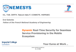
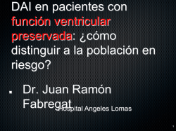
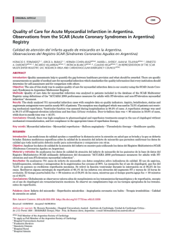
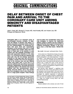
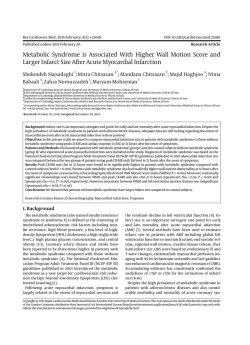
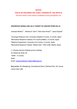
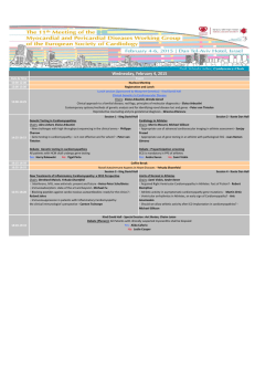
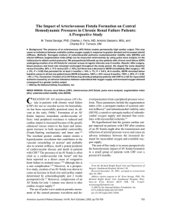
Review
Cardiovascular Research (2009) 82, 240–249
doi:10.1093/cvr/cvn346
Eicosanoid signalling pathways in the heart
Christopher M. Jenkins1, Ari Cedars1, and Richard W. Gross1,2,3*
1
Division of Bioorganic Chemistry and Molecular Pharmacology, Department of Medicine, Washington University School of
Medicine, 660 South Euclid Avenue, Campus Box 8020, St Louis, MO 63110, USA; 2Department of Developmental Biology,
Washington University School of Medicine, 660 South Euclid Avenue, Campus Box 8020, St Louis, MO 63110, USA; and
3
Department of Chemistry, Washington University, St Louis, MO 63130, USA
Received 8 October 2008; revised 21 November 2008; accepted 7 December 2008; online publish-ahead-of-print 14 December 2008
Time for primary review: 14 days
KEYWORDS
Myocardium;
Arachidonic acid;
Eicosanoid;
Phospholipase A2;
Ion channel;
Cyclooxygenase;
Lipoxygenase;
Cytochrome P450
Myocardial phospholipids serve as primary reservoirs of arachidonic acid (AA), which is liberated through
the rate-determining hydrolytic action of cardiac phospholipases A2 (PLA2s). A predominant PLA2 in myocardium is calcium-independent phospholipase A2b (iPLA2b), which, through its calmodulin (CaM) and
ATP-binding domains, is regulated by alterations in local cellular Ca2þ concentrations and cardiac bioenergetic status, respectively. Importantly, iPLA2b has been demonstrated to be activated by ischaemia
through elevation of the concentration of myocardial fatty acyl-CoA, which abrogates Ca2þ/CaMmediated inhibition of iPLA2b. AA released by PLA2-catalysed hydrolysis of phospholipids serves as a
precursor for eicosanoids generated by pathways dependent on cyclooxygenases (COX), lipoxygenases
(LOX), and cytochromes P450 (CYP). Eicosanoids initiate and propagate diverse signalling cascades,
primarily through their interaction with cellular receptors and ion channels. However, during pathologic
states such as ischaemia or congestive heart failure, eicosanoids contribute to multiple maladaptive
changes including inflammation, alterations of cellular growth programmes, and activation of multiple
transcriptional events leading to the deleterious sequelae of these pathologic states. This review summarizes the central roles of myocardial PLA2s in eicosanoid signalling in the heart, the major COX, LOX,
and CYP pathways of eicosanoid generation in the myocardium, and the effects of important eicosanoids
on receptor-, ion channel-, and transcription-mediated processes that facilitate cardiac hypertrophy,
mediate ischaemic preconditioning, and precipitate arrhythmogenesis in response to pathologic stimuli.
1. Introduction
Arachidonic acid (AA) and its eicosanoid metabolites occupy
central roles in the regulation of myocardial physiology,
bioenergetics, contractile function, and signalling pathways. The majority of AA in heart is esterified to the sn-2
position of myocardial phospholipids, in particular choline
and ethanolamine plasmalogens. Activation of intracellular
phospholipases that catalyse the release of AA from its
endogenous phospholipid storage depots is the ratedetermining step in the generation of eicosanoids in myocardium (Figure 1). The released non-esterified AA serves
as substrate for oxidation by multiple cyclooxygenases
(COXs), lipoxygenases (LOXs), and cytochrome P450 (CYP)
enzymes in the heart thereby producing a complex spectrum
of lipid second messengers. Specific eicosanoids have been
demonstrated to regulate a diverse array of critical cellular
processes, such as gene transcription, ion channel kinetics,
and haemodynamic function, that promote salutary
adaptive changes in myocardium during physiologic
perturbations. However, chronic or persistent induction
* Corresponding author. Tel: þ1 314 362 2690; fax: þ1 314 362 1402.
E-mail address: [email protected]
of these pathways, although initially adaptive in the
preservation of normal myocardial function (e.g. ischaemic
preconditioning), often eventually become maladaptive
leading to multiple deleterious sequelae including dysfunctional excitation–contraction coupling due to alterations in
ion channel kinetics, bioenergetic inefficiency, apoptosis,
and accelerated necrosis, which collectively promote the
development of congestive heart failure and tachyarrhythmias leading to sudden death.
The metabolism of AA in the heart is primarily determined
by three cell types (i.e. myocardial, endothelial, and vascular smooth muscle), which rely upon complex intercellular
communication through paracrine signalling to coordinate
blood flow, contractile state, and haemodynamic function.
In addition, inflammatory cells such as neutrophils or macrophages can acutely infiltrate myocardium following ischaemic damage or chronically participate in inflammatory
changes and the development of fibrosis during cardiomyopathic processes precipitating congestive heart failure.
These and other cell types (e.g. platelets and fibroblasts)
can contribute to the production of eicosanoids and other
lipid second messengers inducing wound repair programmes
that although designed to repair damaged tissue often have
Published on behalf of the European Society of Cardiology. All rights reserved. & The Author 2008.
For permissions please email: [email protected].
Eicosanoid signalling pathways in the heart
241
Figure 1 Myocardial phospholipase A2 (PLA2) mediated generation of arachidonic acid (AA). Arachidonate-containing phospholipids are hydrolysed by myocardial PLA2s resulting in the release of free AA. Disruption of the calcium-activated calmodulin (CaM)-calcium-independent PLA2b (iPLA2b) inhibitory complex by
increases in fatty acyl-CoA levels (e.g. in diabetic cardiomyopathy or ischaemia) leads to the activation of iPLA2b. Lipolysis of diacyl phospholipids by either
iPLA2b or cytosolic PLA2a (cPLA2a) results in the direct liberation of AA while iPLA2g predominantly catalyses the production of 2-arachidonoyl lysolipids. Subsequent action by lysophospholipase D (Lyso PLD) or lysophospholipases (LPLs) generates 2-arachidonoyl glycerol or AA from 2-arachidonoyl phospholipids,
respectively. 2-Arachidonoyl-glycerol can be further metabolized by lipoxygenase (LOX), cyclooxygenase (COX), or monoacylglycerol (MAG) lipase enzymes.
The vinyl ether linkage at the sn-1 position of plasmalogen phospholipids restricts iPLA2g (and other PLA2s) to directly release AA which is further metabolized
to the indicated eicosanoids. CYP, cytochrome P450 monooxygenases; EETs, epoxyeicosatrienoic acids; HETEs, hydroxyeicosatrienoic acids.
deleterious effects on atherosclerotic plaque formation and
stability during chronic and prolonged stimulation. This
review will first focus on the identification and characterization of the myocardial phospholipases A2 (PLA2s) that serve
as the major mediators of AA release in heart and thus function as the rate-determining step in eicosanoid production.
Secondly, the major metabolic nodes for myocardial eicosanoid signalling pathways as catalysed by COXs, LOXs, and
CYPs will be discussed with respect to ischaemia/reperfusion injury, cardiac hypertrophy, haemodynamic function,
and inflammation.
2. Regulation of calcium-independent
phospholipase A2-mediated release of
arachidonic acid for eicosanoid production
in myocardium
2.1 Calcium-independent phospholipase A2b
Mammalian myocardial membranes (sarcolemma and
sarcoplasmic reticulum) are electrophysiologically active
membranes that are highly enriched in choline and ethanolamine plasmalogens (as well as smaller amounts of
diacyl glycerophospholipids) containing AA esterified at the
sn-2 position.1,2 In addition to serving as an important
source of AA during cardiac myocyte activation, myocardial
plasmalogens have profound effects on membrane
dynamics, modulate the properties of membrane proteins
(e.g. ion channels and ion pumps), and promote membrane
fusion through the unique stereoelectronic structure
imposed by the vinyl ether linkage at the sn-1 aliphatic
chain.2–7 Phospholipases A2 (PLA2s) catalyse the hydrolysis
of the sn-2 ester linkage of glycerophospholipids to generate
free fatty acid (e.g. AA) and lysolipid products (Figure 2).
Substantial experimental evidence supports the importance
of myocardial PLA2 activation as the primary mediator of AA
release from cellular phospholipids in response to a diverse
array of physiologic and pathophysiologic stimuli.8–10 In
the mid-1980s, a novel type of calcium-independent phospholipase A2 (iPLA2, now designated iPLA2b) present in
canine and human myocardium was identified that did not
require Ca2þ for membrane binding or catalysis, and
selectively hydrolysed plasmenylcholine and plasmenylethanolamine substrates.11,12 Subsequent experiments demonstrated that myocardial iPLA2 activity was activated within
minutes of cardiac ischaemia and that purified iPLA2b
could produce alterations in both ion channel and mitochondrial function replicating those observed during ischaemia.8,9,13 Furthermore, mechanism-based inhibition of
iPLA2b activity by (E)-6-(bromomethylene)-3-(1-naphthalenyl)-2-H-tetrahydropyran-2-one (BEL) prior to the induction
of myocardial ischaemia was shown to have salutary effects
on infarct size, mitochondrial degeneration, and arrhythmogenesis.10,14 To further substantiate the importance of
iPLA2b activation during ischaemia and clarify its role in
the pathogenesis of ischaemic damage, transgenic (TG)
mice were generated that overexpressed iPLA2b in a
cardiac myocyte-restricted manner.10 Although normally
perfused TG iPLA2b myocardium possessed a lipid profile
similar to that of its non-TG counterpart, the induction of
myocardial ischaemia resulted in the robust activation of
iPLA2b activity as demonstrated by a massive release of
fatty acids into the venous effluent and the accumulation
of both fatty acid and lysophosphatidylcholine (LPC) in
ischaemic tissue.10 The ischaemia-mediated activation of
iPLA2b was accompanied by a dramatic induction of ventricular arrhythmias including multiple episodes of tachycardia
within minutes of ischaemia. Both premature ventricular
contraction and ventricular tachycardia were completely
suppressed by inhibition of iPLA2b activity with BEL. In
sharp contrast, non-TG murine myocardium, which has
virtually undetectable levels of iPLA2b activity, did not
242
C.M. Jenkins et al.
Figure 2 Myocardial eicosanoid signalling pathways. Phospholipase A2 (PLA2) catalysed release of arachidonic acid (AA) from cellular phospholipids [X ¼ polar
head group (e.g. choline, ethanolamine, serine, etc.)] results in the production of prostaglandin (PG) G2 (PGG2) and PGH2 by either COX-1 or COX-2. PGH2 is
further metabolized to PGE2, PGF2a, and PGI2, by their corresponding synthases. PGI2 is further metabolized to form 6-keto-PGF1a. Oxidation of AA to
12-hydroperoxyeicosatrienoic acid (12-HpETE) by 12-lipoxygenase (12-LOX), the most abundant LOX in myocardium, and subsequent conversion to
12-hydroxyeicosatrienoic acid (12-HETE) is shown as an example. Representative epoxidation and hydroxylation reactions as catalysed by cytochrome P450 epoxigenase and hydroxylase enzymes to form 14,15-epoxyeicosatrienoic acid (14,15-EET), 16-HETE, and 20-HETE are shown as indicated. Formation of
14,15-dihydroxyeicosatrienoic acid (14,15-DHET) from 14,15-EET is catalysed by epoxide hydrolase.
exhibit arrhythmias and neither released fatty acids into the
venous effluent nor accumulated LPC when subjected to
ischaemia. These results demonstrated that iPLA2b was activated during ischaemia resulting in the release of fatty acids
and the generation of lysolipids leading to dual signalling
pathways with multiple downstream effectors emanating
from a single reaction. Moreover, activation of myocardial
phospholipases during myocardial ischaemia modulates
membrane dynamics leading to alterations in the interactions of membrane-associated protein complexes that
modulate myocardial metabolism and bioenergetics.
During the course of these experiments, a protein factor
from myocardium was purified, which inhibited iPLA2b
activity in the presence, but not in the absence, of Ca2þ.
This protein factor was identified as calmodulin (CaM).15
Importantly, iPLA2b activity could be incrementally regulated within the physiological range of calcium ion concentrations (50 nM–1 mM Ca2þ) in the presence of CaM
(indicating a direct physical interaction between the two
proteins) and CaM-mediated inhibition was rapidly reversed
by either EGTA or CaM antagonists (e.g. W-7) in vitro. The
physiologic importance of this interaction was identified by
the demonstration that iPLA2b activity was tonically inhibited in intact cells and could be fully activated by cellpermeable CaM antagonists resulting in fatty acid
release.16 Thus, iPLA2b was tightly associated with CaM
in vivo and therefore constitutively inactive until released
from CaM-mediated inhibition.
In addition to its phospholipase activity, iPLA2b was
recently found to hydrolyse fatty acyl-CoAs.17 This was of
particular relevance since acyl-CoA accumulates dramatically within minutes of myocardial ischaemia. Accordingly,
it was hypothesized that the binding of acyl-CoA to iPLA2b
could potentially reverse the tonic inhibition of PLA2 activity
by CaM and was potentially responsible for the rapid activation of iPLA2b in ischaemic myocardium leading to membrane dysfunction. To test this hypothesis, real-time
kinetic analyses in conjunction with mass spectrometry
were used to demonstrate that low micromolar concentrations of acyl-CoA could reverse the CaM-mediated
inhibition of phospholipase activity.17 These results collectively identified acyl-CoA as both a substrate for and regulator of iPLA2b (Figure 1).
Although in vitro assays of purified iPLA2b and other
iPLA2s have not revealed greater than two- to three-fold
selectivity for arachidonate-containing phospholipids, ventricular myocytes have been demonstrated to robustly
release AA within minutes of hypoxia or agonist stimulation.
Work by McHowat et al. 18,19 has demonstrated that
both hypoxia and thrombin-induced AA release from
rabbit ventricular myocytes are sensitive to the iPLA2
selective inhibitor BEL. Furthermore, measurement of
Eicosanoid signalling pathways in the heart
membrane-associated and cytosolic PLA2 activities in vitro
revealed selective hydrolysis of plasmalogen- and
arachidonate-containing phospholipid substrates that were
predominantly calcium-independent and BEL sensitive.18–21
243
substantiating a predominant role of iPLA2g in the generation of 2-AA-LPC and its downstream bioactive metabolites
in myocardium.31
2.3 Cytosolic phospholipase A2a
2.2 Calcium-independent phospholipase A2g
In addition to iPLA2b, a second major phospholipase A2 in
myocardium, iPLA2g, has been identified which contains
both N-terminal mitochondrial localization and C-terminal
(-SKL) peroxisomal localization sequences.22,23 Multiple
mRNA splice and proteolytically processed variants of
iPLA2g have been identified, indicating the regulatory complexity of iPLA2g.23 Experiments with the recombinant purified 63 kDa peroxisomal isoform of iPLA2g in combination
with arachidonate-containing phospholipids (e.g. 1-palmitoyl-2-arachidonoyl-sn-glycero-3-phosphorylcholine, PAPC)
have revealed a novel role for this enzyme in eicosanoid
signalling (Figure 1). Mass spectral analyses demonstrated
that 2-arachidonoyl lysophosphatidylcholine (2-AA-LPC) was
the major hydrolytic product generated from PAPC substrate
following incubation with iPLA2g.24 Importantly, significant
amounts of 2-arachidonoyl-LPC were identified in human
myocardium.24 The sequential hydrolysis of diacyl
arachidonate-containing phospholipids by iPLA2g followed
by the rapid cPLA2a- or lysophospholipase (LPL)-mediated
hydrolysis of 2-AA-LPC would be predicted to generate
robust quantities of AA for eicosanoid production in myocardium.25 Alternatively, 2-arachidonoyl-LPC may serve as a
substrate for enzymes with lysophospholipase C activity,
such as nucleotide pyrophosphatase/phosphodiesterase 6,26
thereby generating the endocannabinoid 2-arachidonoylglycerol. Notably, 2-arachidonoyl-glycerol is a substrate
for COX-2 (but not COX-1),27 12-LOX,28 and 15-LOXs.29 In
addition, iPLA2g was demonstrated to directly release AA
from arachidonate-containing plamenylcholine.24 These
results identified a novel mechanism for eicosanoid generation which place 2-arachidonoyl LPC as a key metabolic
node in the cardiac myocyte signalling system that generates
multiple discrete lipid second messengers.
Recently, to determine the function of iPLA2g in myocardium, both TG mice overexpressing the full-length isoform
of iPLA2g in a cardiac myocyte-selective manner30 and
knockout (KO) mice null for the iPLA2g gene31 have been
generated and initially characterized. Analysis of the TG
iPLA2g mice revealed a central role of iPLA2g in myocardial
lipid metabolism. Specifically, myocardial phospholipid mass
was substantially depleted (35% in comparison to control
mice) and brief caloric restriction resulted in dramatic
triglyceride accumulation (50% of total lipid mass) in mice
selectively overexpressing iPLA2g in cardiac myocytes.
Immunohistochemical analysis localized iPLA2g to both
the peroxisomal and mitochondrial compartments. The presence of elevated levels of iPLA2g in mitochondria led to
marked morphological alterations (e.g. loosely packed and
disorganized cristae) and defects in mitochondrial bioenergetic efficiency. Importantly, TG iPLA2g myocardium contained significantly elevated levels of 2-arachidonoyl-LPC
and 2-docosahexaenoyl-LPC, demonstrating the ability of
iPLA2g to generate these signalling molecules in vivo. In
contrast, myocardial mitochondria from iPLA2g KO mice
possessed only 35% of the phospholipase A1 activity of
wild-type mitochondria utilizing PAPC as substrate further
Although present at lower levels relative to the iPLA2s,
cPLA2a may play an important role in myocardial eicosanoid
signalling. Cytosolic PLA2a displays approximately five-fold
selectivity for AA at the sn-2 position of phosphatidylcholine
relative to oleic acid.32 By virtue of its C2 calcium-binding
domain, cPLA2a is activated by increases in intracellular
Ca2þ which results in translocation of the enzyme to cellular
membranes.33,34 In addition, phosphorylation of cPLA2a by
various protein kinases, such as MAPKs and CaMKII, is
believed to be important for modulating cellular AA
release, although identification of the precise phosphorylation sites leading to its activation is still an area of active
investigation.35–37 At the present time, the roles of Ser505
and Ser727 have been most extensively studied and implicated in cPLA2a activation.38
In myocardium, genetic knockout of cPLA2a results in significant myocardial hypertrophy which is increased following
the stress of pressure overload in comparison to wild-type
control mice.39 In this study, cPLA2a was implicated as a
negative regulator of striated muscle growth due to enhanced
insulin growth factor (IGF)-1-mediated phosphorylation of
Akt, GSK-3b, and p38-MAPK in cPLA2a KO hearts (Figure 3).
The basis for the cPLA2a-mediated regulation of this
process was proposed to occur through PDK-1/PKCz (also a
negative regulatory pathway of striated muscle hypertrophic
growth) signalling. Specifically, the absence of cPLA2a was
shown to interfere with PDK-1 recruitment and activation of
PKCz which normally down-regulates IGF-1-mediated hypertrophic growth. AA supplementation restored IGF-1-mediated
PKCz phosphorylation, possibly through enhancement of
PDK-1/PKCz interactions.
Experiments by Pavoine and co-workers have implicated a
role for cPLA2a in the inhibition of cardiac b2-adrenergic
receptor-mediated activation of adenylate cyclase and
protein kinase A.40 Stimulation of cardiac myocytes with
the b2-adrenergic receptor selective agonist zinterol
results in the induction of [Ca2þ]i transients and fractional
shortening which were enhanced by pre-treatment with arachidonyl trifluoromethyl ketone (AACOCF3), indicating the
role of a PLA2 in the down-regulation of these processes.40
Although Pavoine et al. emphasize that AACOCF3 is a cPLA2
inhibitor, it should be recognized that AACOCF3 inhibits
both cPLA2 and iPLA2 enzymes with similar effectiveness.
2.4 Conclusions
Recent studies regarding genetic ablation and/or cardiac
specific overexpression of established myocardial intracellular PLA2s have provided important insight into the function
of these enzymes in myocardium. Various members of the
iPLA2 and cPLA2 families likely have central roles both in
the parallel processing and sequential integrated generation
of free AA for eicosanoid production as well as the generation of multiple lysolipid signalling molecules. Alterations
in membrane dynamics and resultant maladaptive changes
in membrane function are also likely mediators of the sequelae of myocardial ischaemia and congestive heart failure.
Future work will depend on multiple genetic approaches in
244
C.M. Jenkins et al.
Figure 3 Effects of eicosanoid mediated signalling on cardiac hypertrophy. G-protein (Gq)-coupled receptor and insulin growth factor (IGF) receptor pathways
are central to development of cardiac hypertrophy. Activation of Gq-coupled receptors results in IP3 generation triggering intracellular Ca2þ release [increasing
CaMKIId, calcineurin (CN), and NFAT (nuclear factor of activated T-cells) pathways] and DAG generation activating PKC. Stimulation of CaMKIId increases phosphorylation of the ryanodine receptor (RyR), phospholamban (PLB), and histone deacetylase (activating MEF2 and SRF transcription of hypertrophic genes).84 In
addition, Gq-coupled receptors increase hypertrophic signalling through PI3K, PDK-1, and Akt/PKB. Knockout of the IP receptor (Gas-coupled)57 or cPLA2a39 have
been found to result cardiac hypertrophy. Stimulation of the FP receptor through PGF2a mediates the hypertrophic response through increased phosphorylation of
Erk2 (through MEK) and JNK1 and activation of cytosolic tyrosine kinases (TKs).52,53 JNK1 may suppress hypertrophic signalling through phosphorylation of NFAT,
thereby preventing its nuclear translocation and activation. PGF2a has also been demonstrated to up-regulate GLUT-1 expression in cardiac myocytes.55
Increased levels of 12-HETE through overexpression of 12-LOX leads to a hypertrophic and fibrotic phenotype.66
conjunction with enantiospecific inhibition to mechanistically define the roles of each of these enzymes, their
splice variants, and post-translational modifications in myocardial signal transduction pathways during ischaemia and
congestive heart failure.
3. Myocardial cyclooxygenases and prostanoid
signalling
An abundance of recent pharmacologic evidence has demonstrated that selective COX-2 inhibition (e.g. administration
of celecoxib or rofecoxib) can be detrimental to myocardial
function, whereas non-selective COX inhibition can be
cardioprotective as in the case of aspirin.41,42 One possible
explanation for this observed effect has been that specific
COX-2 inhibition results in decreased prostacyclin (PGI2) synthesis (contributing to increased thrombogenesis, hypertension, and atherogenesis), while not reducing the production
of the pro-thrombotic eicosanoid thromboxane A2 (TXA2).43
Human myocardium has been demonstrated to express
both COX-1 and COX-2,44,45 each of which has been proposed
to have distinct functions. COXs catalyse the incorporation
of two molecular oxygens per AA molecule to form prostaglandin G2 which is then converted to the prostanoid
series-2 precursor prostaglandin H2 by the peroxidase
activity of the enzyme (Figure 2). COX-1 is believed to be
constitutively expressed and likely serves to maintain
normal cardiac homeostasis.45 In contrast, although COX-2
is induced in various pathophysiologic states as a mediator
of inflammatory responses which can lead to cardiac fibrosis,46–48 increasing evidence indicates that it may also
serve a cardioprotective role. In ischaemic preconditioning
of rabbit myocardium, COX-2 mRNA is rapidly (,1 h)
induced followed by increased COX-2 protein expression
and PGE2 and 6-keto-PGF1a production49 at 24 h following
ischaemia/reperfusion. Selective inhibition of COX-2 using
NS-398 or celecoxib abolished the cardioprotective effects
of ischaemic preconditioning, indicating that PGE2 and/or
PGI2 are the likely mediators of this process.49 COX-2
appears to be up-regulated during ischaemic preconditioning through PKC-, Src-PTK, and NF-kB pathways and requires
iNOS-generated NO in the heart.50,51
Multiple effects of downstream prostanoids generated by
myocardial COXs have been documented. PGF2a is produced
by both COX-1 and COX-2 and is involved in inflammation
and hypertrophy. In addition, PGF2a is known to activate
several signalling pathways in cardiac myocytes including
the PGF2a receptor (Gq coupled), JNK1, and c-Jun pathways,
although not all activated kinases (e.g. PKC, ERK, and p38)
were required for the hypertrophic response52–54
(Figure 3). Administration of PGF2a to rat ventricular myocytes has been demonstrated to result in a dramatic
increase in GLUT-1 expression and glucose uptake.55
However, glucose was not required for the hypertrophic
response induced by PGF2a. Targeted knockout of the receptors for PGF2a and TXA2 (FP and TP, respectively), and not
those for other prostanoids [DP, EP (isoforms 1–4), and IP],
has shown that their respective ligands mediate the tachycardia that accompanies inflammatory changes in myocardium.56 Notably, lipopolysaccharide-stimulated tachycardia
was diminished in FP and TP null mice and absent in FP, TP
double knockout animals. PGI2 has also been documented
to have substantial effects on myocardial physiology. Mice
null for the receptor for PGI2 (IP) have been demonstrated
to exhibit salt-sensitive hypertension, cardiac hypertrophy,
and severe cardiac fibrosis.57 IP KO mice also exhibited
Eicosanoid signalling pathways in the heart
exacerbated cardiac hypertrophy in response to pressure
overload in comparison to wild-type controls.58
3.1 Conclusions
The complexity of eicosanoid signalling downstream of the
COX enzymes, although necessary to balance and maintain
normal physiologic function in myocardium, has complicated
efforts to develop specific COX-2 inhibitors designed to
reduce inflammation and platelet aggregation in patients
with cardiovascular disease. Similarities between the detrimental effects of selective COX-2 inhibition and the phenotypes of mice null for different prostanoid receptors will
continue to provide valuable insight into the relative influence of eicosanoid-mediated signalling pathways involved
in vascular tone, hypertension, atherosclerosis, and thrombogenesis as well as ischaemic heart disease and congestive
heart failure.
4. Hydroxyeicosatrienoic acids mediated
myocardial signalling
Lipoxygenases catalyse the stereospecific oxidation of the
olefinic linkages of AA to produce hydroperoxyeicosatetraenoic acids (HpETEs), which are then reduced to form their
hydroxyeicosatrienoic (HETE) derivatives (Figure 2). LOX
activity has been previously detected in subcellular fractions of rat heart and cultured rat myocytes.59 The majority
of the myocardial LOX activity measured in this study was
determined to be 12-LOX (i.e. 12-HETE produced from AA)
with trace amounts of 15-LOX (15-HETE production).
Myocardial ischaemia results in LOX activation in myocytes as well as recruitment of leucocytes to damaged
areas, thereby leading to an increased production of a
wide variety of oxidized biologically active eicosanoids.
For example, 5-HETE and 12-HETE levels were found to dramatically increase in cultured canine myocytes following
45 min of hypoxia and 5 h re-oxygenation.60 Similarly,
ischaemic rabbit myocardium produced greater amounts of
leukotriene B4, 5-HETE, and 12-HETE than non-ischaemic
controls, with the latter being the major product.61
Recently, 12-HETE has been demonstrated to increase mitochondrial Ca2þ concentration and mtNOS activity (NO production) in isolated rat heart mitochondria and in HL-1
cardiac myocytes which underwent apoptosis following
treatment with 12-HETE.62 Elevated levels of NO under
these conditions were found to decrease mitochondrial respiratory efficiency and transmembrane potential, thereby
leading to cell death. In addition, 15(S)-HETE, 11(S)-HETE,
and 5(S),15(S)-diHETE have been shown to increase the sensitivity of the isoproterenol-mediated b-adrenergic response
in cardiomyocytes.63 Of particular interest in this study was
the selective incorporation of 15-HETE into cellular phosphatidylinositols and the activation of PKC in 15-HETEmediated enhancement of b-adrenergic signalling.
Lipoxygenase-mediated production of HETEs likely participates in maintaining normal myocardial function and its
alteration in multiple disease states. Inhibition of LOXs inhibits insulin-stimulated GLUT-4-mediated glucose uptake by
cultured ventricular myocytes through prevention of actin
cytoskeletal rearrangement, but not through alterations in
Akt phosphorylation or association of phosphoinositide
245
3-kinase with IRS-1,64,65 suggesting a potential role of
HETEs in maintaining sensitivity to insulin. Separately, eicosanoid products of 12-LOX have been implicated in the
development of cardiac fibrosis and hypertrophy.66,67 Overexpression of 12-LOX in cardiac fibroblasts resulted in
increased incorporation of leucine, thymidine, and uridine,
increased cell size, and a reduction in the rate of cell division in comparison to control cells. The presence of
12-LOX also resulted in a significant elevation of collagen
and fibronectin levels, indicative of a fibrotic phenotype.
4.1 Conclusions
The roles of LOXs and their eicosanoid products in myocardial function remain relatively unexplored. Initial investigations have revealed evidence of the up-regulation of
LOX enzymes and/or LOX products following myocardial
ischaemia/infarction and may contribute to cardiac hypertrophy, myocyte apoptosis, and fibrosis. Alternatively, LOXs
and their eicosanoid products may facilitate b-adrenergic
signalling and glucose transport. Future studies using
genetic models in conjunction with specific pharmacologic
agents will help deconvolute the role of each specific
HETE moiety in physiologic adaptation in addition to their
contribution to compromised myocardial function in pathologic states.
5. Epoxyeicosatrienoic and
hydroxyeicosatrienoic acids produced
by myocardial cytochromes P450
Multiple members of the CYP superfamily (e.g. CYP1B,
CYP2A, CYP2B, CYP2C, CYP2E, CYP2J, CYP4A, and CYP11)
have been identified in human heart and their expression
and resultant AA metabolites have been found to be generally increased during cardiac hypertrophy and heart
failure.68,69 Myocardial CYPs which metabolize AA are
haem protein monooxygenases which require O2, CYP
reductase, and NADPH for catalysis. Notably, CYP inhibitors
(chloroamphenicol, cimetidine, and sulfaphenazole) have
been demonstrated to decrease myocardial ischaemia/
reperfusion damage, presumably through attenuation of
CYP-mediated reactive oxygen species (ROS) generation
although other mechanisms are possible.70 Members of the
CYP 2C and 2J subfamilies are believed to be the predominant P450 isoforms involved in eicosanoid signalling in the
heart. Murine myocardium contains at least three CYP 2C
members (CYP2C29, CYP2C40, and CYP2C50) that predominantly catalyse the production of epoxyeicosatrienoic acids
[EETs, primarily 8(S),9(R)-EET and 14(R),15(S)-EET], HETEs
(primarily 16-HETE with minor amounts of 20-HETE), as
well as a variety of lower abundance EETs and HETEs,
respectively.71,72 The epoxide linkage of EETs can be
further metabolized by soluble epoxide hydrolases to dihydroxyeicosatrienoic acids which generally possess less
potent biologic effects. Zeldin and co-workers73 identified
a novel CYP epoxygenase, CYP2J2, abundantly expressed
in human myocardium which was highly enantioselective
for the production of 14(R),15(S)-EET from AA. Other EET
enantiomeric pairs (e.g. 8,9-EET and 11,12-EET) were produced in nearly equal racemic distributions. Interestingly,
the EET composition of human myocardium paralleled that
of AA metabolites generated with purified recombinant
246
CYP2J2, suggesting that this is a predominant pathway in
eicosanoid generation.73 However, it should be noted that
other eicosanoids, although present in smaller amounts,
may be of equal or greater importance in signalling depending on their relative affinities for target proteins and subsequent mechanisms used for signal amplification.
Endogenous generation of EETs has been demonstrated to
be cardioprotective by reduction of ischaemia–reperfusion
damage and rescue of myocardial function. Selective TG
overexpression of CYP2J2 in heart results in improved
post-myocardial infarction (MI) recovery of left ventricular
developed pressure, an effect that was reversed by
administration of the CYP2J2 inhibitor N-methanesulfonyl6-(2-propargyloxyphenyl)hexanamide (MS-PPOH).74 The
CYP2J2-dependent recovery of heart function was also
blocked by either the sarcolemmal KATP channel inhibitor
glibenclamide or the mitochondrial KATP channel inhibitor
5-hydroxydecanoate. Mitochondrial KATP channel activity
was found to be higher in TG CYP2J2 cardiomyocytes in
comparison to their non-TG controls and low micromolar
concentrations of P450 2J2 generated EETs increased mitochondrial redox potential as measured by flavoprotein
fluorescence (indicative of KATP channel activity) in non-TG
animals. Although the phosphorylation state of MAPK p42/
p44 was similar between CYP2J2 TG and non-TG hearts
under basal and ischaemic conditions, reperfusion induced
increased levels of MAPK p42/p44 phosphorylation in TG
vs. non-TG controls. Importantly, selective inhibition of
MEK with PD98059 abolished the CYP2J2-dependent
post-MI recovery of left ventricular developed pressure.74
In contrast to EETs, 20-HETE has been found to be
detrimental to cardiac function. The CYP v-hydroxylases
CYP4A1, CYP4A2, and CYP4F have been demonstrated to
be expressed in canine heart75 and produce 20-HETE, a
potent vasoconstrictor that acts through inhibition of Ca2þsensitive Kþ channels.76 High concentrations of 20-HETE are
released in the coronary venous effluent during reperfusion
of canine hearts rendered ischaemic.75 Pre-treatment with
the specific P450 v-hydroxylase inhibitors, 17-octadecanoic
acid (17-ODYA) and N-methylsulfonyl-12,12-dibromododec11-enamide (DDMS), significantly inhibited 20-HETE production and reduced infarct size compared with the total
area at risk. Consistent with this, infusion of exogenous
20-HETE prior to left anterior descending coronary artery
occlusion resulted in a significant increase in infarct size.
Further studies demonstrated that the putative 20-HETE
antagonist 20-hydroxyeicosa-6(Z),15(Z)-dienoic acid also
decreased infarct size similar to DDMS which was augmented
by ischaemic preconditioning.77 Additional experiments
utilizing ischaemic/reperfused rat hearts confirmed that
17-ODYA and DDMS dose-dependently reduced ischaemic
damage, but that prior treatment with the P450 epoxygenase inhibitor MS-PPOH before induction of ischaemia or
reperfusion was without effect.78
5.1 Conclusions
Cytochrome P450 monooxygenases represent important
participants in myocardial eicosanoid signalling. Increased
expression of the epoxygenase CYP2J2 in mouse myocardium
decreased ischaemia–reperfusion damage through production of EETs which modulate KATP channel function
and MAPK signalling. Conversely, production of HETEs (in
C.M. Jenkins et al.
particular 20-HETE) during ischaemia by CYP4 family
members likely promotes ischaemic damage either through
elevated vasoconstriction and/or potentially increased ROS
generation, although this remains to be determined.
6. Arachidonic acid and eicosanoid-mediated
regulation of myocardial ion channel function
Disruption of normal ion channel function represents a
primary mechanism for induction of the lethal ventricular
arrhythmias which occur following myocardial ischaemia.
Definition of the influence of AA on this process is complicated by differential pro- and anti-arrhythmogenic effects
of AA and other polyunsaturated fatty acids as well as their
oxidized metabolites on ion channel function. In addition, it
has been difficult to differentiate the direct effects of AA
and its oxidized metabolites on ion channel function from
those mediated through receptor-activated pathways.
Specific AA-mediated effects have been demonstrated in
the case of the human delayed rectifier Kþ channel, Kv 1.1,
which undergoes significant increases in both macroscopic
voltage-dependent activation and inactivation kinetics
following administration of micromolar concentrations of
AA13 (Figure 4). In addition, the effects of free AA on Kv
1.1 function could be recapitulated by the intracellular introduction of iPLA2b and were dependent upon the presence of
AA-containing membrane phospholipids. Importantly, these
effects were observed in Sf9 cells which are not known to
oxidize the olefinic linkages of AA (as determined by autoradiographic thin layer chromatography), thereby ruling out
the potential influence of downstream eicosanoids.
Epoxyeicosatrienoic acids have been demonstrated to
have varying effects on different cardiac ion channels
(Figure 4). Addition of low nanomolar concentrations of
exogenous 5,6- and 11,12-EET significantly increased shortening of isolated ventricular myocytes and intracellular
Ca2þ concentrations, while 8,9- and 14,15-EETs were
without effect.79 Treatment of rat ventricular myocytes
with the imidazole-based P450 inhibitors clotrimazole, econazole, or miconazole significantly blocked L-type Ca2þ
current (ICa), intracellular Ca2þ signalling, and cell shortening.80 Clotrimazole suppressed ICa was reversed by administration of the b-adrenergic agonist isoprenaline or by
incubation with 8-bromo-cAMP. Levels of cAMP in cells
treated with clotrimazole were found to be significantly
reduced while incubation of myocytes with 11,12-EET markedly increased the concentration of intracellular cAMP. From
these and other data, it was concluded that P450-generated
arachidonate metabolites modulated cAMP levels, Ca2þ
channel phosphorylation (presumably protein kinase A
mediated), Ca2þ signalling, and cellular contraction. In a
separate study, cardiac L-type Ca2þ channels reconstituted
into planar lipid bilayers were found to be inhibited by nanomolar concentrations of EETs which decreased channel open
probability, accelerated channel inactivation, and
decreased the unitary current amplitude of open channels.81
Interestingly, the same effects on L-type Ca2þ channel function were mediated by 11,12-EET when it was esterified at
the sn-2 position of surrounding phosphatidylcholine molecules, suggesting a direct EET/Ca2þ channel interaction
within the lipid phase of the bilayer. Utilizing an inside-out
patch clamp technique, Lu et al.82 demonstrated that low
Eicosanoid signalling pathways in the heart
247
Figure 4 Influence of arachidonic acid, EETs, and HETEs on myocardial ion channels/currents. Arachidonic acid generated by iPLA2b has been demonstrated to
increase activation and deactivation kinetics of Kv1.1 ion channels.13 Cytochrome P450 epoxygenases (CYPs) catalyse the production of 5,6-EET and 11,12-EET
regioisomers which elevate cAMP and intracellular Ca2þ levels through Ca2þ-mediated activation of the ryanodine receptor, resulting in increased fractional
shortening.79,80 L-type Ca2þ current may be enhanced by direct EET-ion channel interactions or through protein kinase A mediated Ca2þ channel phosphorylation.85 11,12-EET has been demonstrated to decrease KATP channel sensitivity to ATP,86 while several EETs have been shown to attenuate the gating of Naþ channels.83 The potent vasoconstrictive and pro-ischaemic effects of 20-HETE are proposed to occur through its inhibition of Ca2þ-sensitive Kþ (BK) channels.76
micromolar concentrations of 11,12-EET markedly increased
the activity (open channel probability) of ATP-sensitive Kþ
channels by reducing sensitivity of the channels to ATP in a
dose- and voltage-dependent manner. Intact myocyte Naþ
currents were found to be inhibited by low micromolar concentrations of 8,9-EET as well as 5,6-, 11,12-, and 14,15-EETs
in a channel-use and voltage-dependent manner.83 Single
channel recordings indicated that 8,9-EET markedly
reduced the duration and probability of Naþ channel
opening.
6.1 Conclusions
Arachidonic acid and its EET derivatives have been demonstrated to significantly alter myocardial ion channel
function. Although the precise mechanisms involved in
eiconsanoid regulation of myocardial ion channels have not
been demonstrated in molecular detail, they likely involve
both direct (e.g. binding and conformational alterations)
and indirect (e.g. elevation of cAMP levels followed by
PKA-mediated signalling) processes.
7. Concluding remarks
Elucidation of the cardiovascular effects of AA and its
eicosanoid derivatives has led to a new appreciation of
the importance of enzymes mediating their generation and
their influence on cardiac physiology in health and disease.
Multiple enzymes are involved in the release of AA (i.e.
PLA2s) and its stereospecific oxidation (i.e. COXs, LOXs,
and CYPs) to generate this diverse array of bioactive signalling molecules. Moreover, our understanding of the role
played by eicosanoids in altering ion channel function, transcriptional programming, and ventricular remodelling, and
in orchestrating the pleiotropic responses to cellular perturbations has begun to evolve. Prominent examples include
the roles of eicosanoids in activating protein kinase cascades
and ion channels and their resultant effects on cardiac
hypertrophy, myocardial preconditioning, infarction, and
arrhythmogenesis. Although considerable progress has
been made, much work remains to be done to identify the
complex interwoven pathways that integrate eicsosanoid
signalling pathways with finely tuned myocardial responses
to alterations in metabolic state, haemodynamic burden,
or nutritional history of the cardiac myocyte. Continuing
advances in pharmacologic and genetic approaches will
allow greater insight into these mechanisms, thereby providing a foundation for the development of targeted
pharmaceutical approaches for limiting ischaemic damage,
attenuating the deleterious sequelae of cardiac hypertrophy, and preventing arrhythmogenesis.
Acknowledgements
We thank David J. Mancuso and Harold F. Sims for their critical
reading of this review.
Conflict of interest: none declared.
Funding
This work was supported by the National Institutes of Health grants
2P01HL057278-11 and R01HL041250-14A1.
References
1. Gross RW. High plasmalogen and arachidonic acid content of canine myocardial sarcolemma: a fast atom bombardment mass spectroscopic and
gas chromatography-mass spectroscopic characterization. Biochemistry
1984;23:158–165.
2. Gross RW. Identification of plasmalogen as the major phospholipid
constituent of cardiac sarcoplasmic reticulum. Biochemistry 1985;24:
1662–1668.
3. Bick RJ, Youker KA, Pownall HJ, Van Winkle WB, Entman ML. Unsaturated
aminophospholipids are preferentially retained by the fast skeletal
muscle Ca2þATPase during detergent solubilization. Evidence for a
specific association between aminophospholipids and the calcium pump
protein. Arch Biochem Biophys 1991;286:346–352.
4. Chen X, Gross RW. Phospholipid subclass-specific alterations in the
kinetics of ion transport across biologic membranes. Biochemistry 1994;
33:13769–13774.
248
5. Chen X, Gross RW. Potassium flux through gramicidin ion channels is
augmented in vesicles comprised of plasmenylcholine: correlations
between gramicidin conformation and function in chemically distinct
host bilayer matrices. Biochemistry 1995;34:7356–7364.
6. Ford DA, Hale CC. Plasmalogen and anionic phospholipid dependence of
the cardiac sarcolemmal sodium-calcium exchanger. FEBS Lett 1996;
394:99–102.
7. Duhm J, Engelmann B, Schonthier UM, Streich S. Accelerated maximal
velocity of the red blood cell Naþ/Kþ pump in hyperlipidemia is related
to increase in 1-palmitoyl,2-arachidonoyl-plasmalogen phosphatidylethanolamine. Biochim Biophys Acta 1993;1149:185–188.
8. Hazen SL, Ford DA, Gross RW. Activation of a membrane-associated phospholipase A2 during rabbit myocardial ischemia which is highly selective
for plasmalogen substrate. J Biol Chem 1991;266:5629–5633.
9. Ford DA, Hazen SL, Saffitz JE, Gross RW. The rapid and reversible activation of a calcium-independent plasmalogen-selective phospholipase
A2 during myocardial ischemia. J Clin Invest 1991;88:331–335.
10. Mancuso DJ, Abendschein DR, Jenkins CM, Han X, Saffitz JE, Schuessler RB
et al. Cardiac ischemia activates calcium-independent phospholipase
A2b, precipitating ventricular tachyarrhythmias in transgenic mice:
rescue of the lethal electrophysiologic phenotype by mechanism-based
inhibition. J Biol Chem 2003;278:22231–22236.
11. Wolf RA, Gross RW. Identification of neutral active phospholipase C which
hydrolyzes choline glycerophospholipids and plasmalogen selective phospholipase A2 in canine myocardium. J Biol Chem 1985;260:7295–7303.
12. Hazen SL, Gross RW. Human myocardial cytosolic Ca2þ-independent phospholipase A2 is modulated by ATP. Concordant ATP-induced alterations in
enzyme kinetics and mechanism-based inhibition. Biochem J 1991;280:
581–587.
13. Gubitosi-Klug RA, Yu SP, Choi DW, Gross RW. Concomitant acceleration of
the activation and inactivation kinetics of the human delayed rectifier Kþ
channel (Kv1.1) by Ca2þ-independent phospholipase A2. J Biol Chem
1995;270:2885–2888.
14. Sargent CA, Wilde MW, Dzwonczyk S, Tuttle JG, Murray HN, Atwal K et al.
Glyburide-reversible cardioprotective effects of calcium-independent
phospholipase A2 inhibition in ischemic rat hearts. Cardiovasc Res 1996;
31:270–277.
15. Wolf MJ, Gross RW. The calcium-dependent association and functional
coupling of calmodulin with myocardial phospholipase A2. Implications
for cardiac cycle-dependent alterations in phospholipolysis. J Biol
Chem 1996;271:20989–20992.
16. Wolf MJ, Wang J, Turk J, Gross RW. Depletion of intracellular calcium
stores activates smooth muscle cell calcium-independent phospholipase
A2. A novel mechanism underlying arachidonic acid mobilization. J Biol
Chem 1997;272:1522–1526.
17. Jenkins CM, Yan W, Mancuso DJ, Gross RW. Highly selective hydrolysis of
fatty acyl-CoAs by calcium-independent phospholipase A2b. Enzyme
autoacylation and acyl-CoA-mediated reversal of calmodulin inhibition
of phospholipase A2 activity. J Biol Chem 2006;281:15615–15624.
18. McHowat J, Creer MH. Thrombin activates a membrane-associated
calcium-independent PLA2 in ventricular myocytes. Am J Physiol 1998;
274:C447–C454.
19. McHowat J, Liu S, Creer MH. Selective hydrolysis of plasmalogen phospholipids by Ca2þ-independent PLA2 in hypoxic ventricular myocytes.
Am J Physiol 1998;274:C1727–C1737.
20. McHowat J, Creer MH. Calcium-independent phospholipase A2 in isolated
rabbit ventricular myocytes. Lipids. 1998;33:1203–1212.
21. McHowat J, Creer MH. Selective plasmalogen substrate utilization by
thrombin-stimulated Ca2þ-independent PLA2 in cardiomyocytes. Am J
Physiol Heart Circ Physiol 2000;278:H1933–H1940.
22. Mancuso DJ, Jenkins CM, Gross RW. The genomic organization, complete
mRNA sequence, cloning, and expression of a novel human intracellular
membrane-associated calcium-independent phospholipase A2. J Biol
Chem 2000;275:9937–9945.
23. Mancuso DJ, Jenkins CM, Sims HF, Cohen JM, Yang J, Gross RW. Complex
transcriptional and translational regulation of iPLA2g resulting in multiple
gene products containing dual competing sites for mitochondrial or peroxisomal localization. Eur J Biochem 2004;271:4709–4724.
24. Yan W, Jenkins CM, Han X, Mancuso DJ, Sims HF, Yang K et al. The highly
selective production of 2-arachidonoyl lysophosphatidylcholine catalyzed
by purified calcium-independent phospholipase A2g: identification of a
novel enzymatic mediator for the generation of a key branch point intermediate in eicosanoid signaling. J Biol Chem 2005;280:26669–26679.
25. Pete MJ, Exton JH. Purification of a lysophospholipase from bovine brain
that selectively deacylates arachidonoyl-substituted lysophosphatidylcholine. J Biol Chem 1996;271:18114–18121.
C.M. Jenkins et al.
26. Sakagami H, Aoki J, Natori Y, Nishikawa K, Kakehi Y, Natori Y et al. Biochemical and molecular characterization of a novel choline-specific glycerophosphodiester phosphodiesterase belonging to the nucleotide
pyrophosphatase/phosphodiesterase family. J Biol Chem 2005;280:
23084–23093.
27. Kozak KR, Rowlinson SW, Marnett LJ. Oxygenation of the endocannabinoid,
2-arachidonylglycerol,
to
glyceryl
prostaglandins
by
cyclooxygenase-2. J Biol Chem 2000;275:33744–33749.
28. Moody JS, Kozak KR, Ji C, Marnett LJ. Selective oxygenation of the endocannabinoid 2-arachidonylglycerol by leukocyte-type 12-lipoxygenase.
Biochemistry 2001;40:861–866.
29. Kozak KR, Gupta RA, Moody JS, Ji C, Boeglin WE, DuBois RN et al.
15-Lipoxygenase metabolism of 2-arachidonylglycerol. Generation of a
peroxisome proliferator-activated receptor alpha agonist. J Biol Chem
2002;277:23278–23286.
30. Mancuso DJ, Han X, Jenkins CM, Lehman JJ, Sambandam N, Sims HF et al.
Dramatic accumulation of triglycerides and precipitation of cardiac
hemodynamic dysfunction during brief caloric restriction in transgenic
myocardium expressing human calcium-independent phospholipase A2g.
J Biol Chem 2007;282:9216–9227.
31. Mancuso DJ, Sims HF, Han X, Jenkins CM, Guan SP, Yang K et al. Genetic
ablation of calcium-independent phospholipase A2g leads to alterations
in mitochondrial lipid metabolism and function resulting in a deficient
mitochondrial bioenergetic phenotype. J Biol Chem 2007;282:
34611–34622.
32. Clark JD, Lin LL, Kriz RW, Ramesha CS, Sultzman LA, Lin AY et al. A novel
arachidonic acid-selective cytosolic PLA2 contains a Ca2þ-dependent
translocation domain with homology to PKC and GAP. Cell 1991;65:
1043–1051.
33. Channon JY, Leslie CC. A calcium-dependent mechanism for associating a
soluble arachidonoyl-hydrolyzing phospholipase A2 with membrane in the
macrophage cell line RAW 264.7. J Biol Chem 1990;265:5409–5413.
34. Nalefski EA, Sultzman LA, Martin DM, Kriz RW, Towler PS, Knopf JL et al.
Delineation of two functionally distinct domains of cytosolic phospholipase A2, a regulatory Ca2þ-dependent lipid-binding domain and a Ca2þindependent catalytic domain. J Biol Chem 1994;269:18239–18249.
35. Gijon MA, Spencer DM, Siddiqi AR, Bonventre JV, Leslie CC. Cytosolic
phospholipase A2 is required for macrophage arachidonic acid release
by agonists that Do and Do not mobilize calcium. Novel role of mitogenactivated protein kinase pathways in cytosolic phospholipase A2 regulation. J Biol Chem 2000;275:20146–20156.
36. Shimizu M, Nakamura H, Hirabayashi T, Suganami A, Tamura Y,
Murayama T. Ser515 phosphorylation-independent regulation of cytosolic
phospholipase A2a (cPLA2a) by calmodulin-dependent protein kinase:
possible interaction with catalytic domain A of cPLA2a. Cell Signal
2008;20:815–824.
37. Pavicevic Z, Leslie CC, Malik KU. cPLA2 phosphorylation at serine-515 and
serine-505 is required for arachidonic acid release in vascular smooth
muscle cells. J Lipid Res 2008;49:724–737.
38. Ghosh M, Tucker DE, Burchett SA, Leslie CC. Properties of the Group IV
phospholipase A2 family. Prog Lipid Res 2006;45:487–510.
39. Haq S, Kilter H, Michael A, Tao J, O’Leary E, Sun XM et al. Deletion of
cytosolic phospholipase A2 promotes striated muscle growth. Nat Med
2003;9:944–951.
40. Ait-Mamar B, Cailleret M, Rucker-Martin C, Bouabdallah A, Candiani G,
Adamy C et al. The cytosolic phospholipase A2 pathway, a safeguard of
b2-adrenergic cardiac effects in rat. J Biol Chem 2005;280:18881–18890.
41. Grosser T, Fries S, FitzGerald GA. Biological basis for the cardiovascular
consequences of COX-2 inhibition: therapeutic challenges and opportunities. J Clin Invest 2006;116:4–15.
42. Funk CD, FitzGerald GA. COX-2 inhibitors and cardiovascular risk.
J Cardiovasc Pharmacol 2007;50:470–479.
43. McAdam BF, Catella-Lawson F, Mardini IA, Kapoor S, Lawson JA,
FitzGerald GA. Systemic biosynthesis of prostacyclin by cyclooxygenase
(COX)-2: the human pharmacology of a selective inhibitor of COX-2.
Proc Natl Acad Sci USA 1999;96:272–277.
44. Testa M, Rocca B, Spath L, Ranelletti FO, Petrucci G, Ciabattoni G et al.
Expression and activity of cyclooxygenase isoforms in skeletal muscles
and myocardium of humans and rodents. J Appl Physiol 2007;103:
1412–1418.
45. Zidar N, Dolenc-Strazar Z, Jeruc J, Jerse M, Balazic J, Gartner U et al.
Expression of cyclooxygenase-1 and cyclooxygenase-2 in the normal
human heart and in myocardial infarction. Cardiovasc Pathol 2007;16:
300–304.
Eicosanoid signalling pathways in the heart
46. Wong SC, Fukuchi M, Melnyk P, Rodger I, Giaid A. Induction of
cyclooxygenase-2 and activation of nuclear factor-kB in myocardium of
patients with congestive heart failure. Circulation 1998;98:100–103.
47. Saito T, Rodger IW, Hu F, Robinson R, Huynh T, Giaid A. Inhibition of COX
pathway in experimental myocardial infarction. J Mol Cell Cardiol 2004;
37:71–77.
48. Zhang Z, Vezza R, Plappert T, McNamara P, Lawson JA, Austin S et al.
COX-2-dependent cardiac failure in Gh/tTG transgenic mice. Circ Res
2003;92:1153–1161.
49. Shinmura K, Tang XL, Wang Y, Xuan YT, Liu SQ, Takano H et al.
Cyclooxygenase-2 mediates the cardioprotective effects of the late
phase of ischemic preconditioning in conscious rabbits. Proc Natl Acad
Sci USA 2000;97:10197–10202.
50. Shinmura K, Xuan YT, Tang XL, Kodani E, Han H, Zhu Y et al. Inducible
nitric oxide synthase modulates cyclooxygenase-2 activity in the heart
of conscious rabbits during the late phase of ischemic preconditioning.
Circ Res 2002;90:602–608.
51. Xuan YT, Guo Y, Zhu Y, Han H, Langenbach R, Dawn B et al. Mechanism of
cyclooxygenase-2 upregulation in late preconditioning. J Mol Cell Cardiol
2003;35:525–537.
52. Kunapuli P, Lawson JA, Rokach JA, Meinkoth JL, FitzGerald GA. Prostaglandin F2a (PGF2a) and the isoprostane, 8, 12-iso-isoprostane F2a-III,
induce cardiomyocyte hypertrophy. Differential activation of downstream signaling pathways. J Biol Chem 1998;273:22442–22452.
53. Adams JW, Sah VP, Henderson SA, Brown JH. Tyrosine kinase and c-Jun
NH2-terminal kinase mediate hypertrophic responses to prostaglandin
F2a in cultured neonatal rat ventricular myocytes. Circ Res 1998;83:
167–178.
54. Adams JW, Migita DS, Yu MK, Young R, Hellickson MS, Castro-Vargas FE
et al. Prostaglandin F2a stimulates hypertrophic growth of cultured neonatal rat ventricular myocytes. J Biol Chem 1996;271:1179–1186.
55. Morissette MR, Howes AL, Zhang T, Heller Brown J. Upregulation of
GLUT1 expression is necessary for hypertrophy and survival of neonatal
rat cardiomyocytes. J Mol Cell Cardiol 2003;35:1217–1227.
56. Takayama K, Yuhki K, Ono K, Fujino T, Hara A, Yamada T et al. Thromboxane A2 and prostaglandin F2a mediate inflammatory tachycardia. Nat Med
2005;11:562–566.
57. Francois H, Athirakul K, Howell D, Dash R, Mao L, Kim HS et al. Prostacyclin protects against elevated blood pressure and cardiac fibrosis. Cell
Metab 2005;2:201–207.
58. Hara A, Yuhki K, Fujino T, Yamada T, Takayama K, Kuriyama S et al. Augmented cardiac hypertrophy in response to pressure overload in mice
lacking the prostaglandin I2 receptor. Circulation 2005;112:84–92.
59. Breitbart E, Sofer Y, Shainberg A, Grossman S. Lipoxygenase activity in
heart cells. FEBS Lett 1996;395:148–152.
60. Kuzuya T, Hoshida S, Kim Y, Oe H, Hori M, Kamada T et al. Free radical
generation coupled with arachidonate lipoxygenase reaction relates to
reoxygenation induced myocardial cell injury. Cardiovasc Res 1993;27:
1056–1060.
61. Hughes H, Gentry DL, McGuire GM, Taylor AA. Gas chromatographic-mass
spectrometric analysis of lipoxygenase products in post-ischemic rabbit
myocardium. Prostaglandins Leukot Essent Fatty Acids 1991;42:225–231.
62. Nazarewicz RR, Zenebe WJ, Parihar A, Parihar MS, Vaccaro M, Rink C
et al. 12(S)-hydroperoxyeicosatetraenoic acid (12-HETE) increases mitochondrial nitric oxide by increasing intramitochondrial calcium. Arch
Biochem Biophys 2007;468:114–120.
63. Wallukat G, Morwinski R, Kuhn H. Modulation of the b-adrenergic
response of cardiomyocytes by specific lipoxygenase products involves
their incorporation into phosphatidylinositol and activation of protein
kinase C. J Biol Chem 1994;269:29055–29060.
64. Dransfeld O, Rakatzi I, Sasson S, Eckel J. Eicosanoids and the regulation
of cardiac glucose transport. Ann N Y Acad Sci 2002;967:208–216.
65. Dransfeld O, Rakatzi I, Sasson S, Gruzman A, Schmitt M, Haussinger D
et al. Eicosanoids participate in the regulation of cardiac glucose transport by contribution to a rearrangement of actin cytoskeletal elements.
Biochem J 2001;359:47–54.
66. Wen Y, Gu J, Liu Y, Wang PH, Sun Y, Nadler JL. Overexpression of
12-lipoxygenase causes cardiac fibroblast cell growth. Circ Res 2001;
88:70–76.
249
67. Wen Y, Gu J, Peng X, Zhang G, Nadler J. Overexpression of
12-lipoxygenase and cardiac fibroblast hypertrophy. Trends Cardiovasc
Med 2003;13:129–136.
68. Zordoky BN, El-Kadi AO. Modulation of cardiac and hepatic cytochrome
P450 enzymes during heart failure. Curr Drug Metab 2008;9:122–128.
69. Delozier TC, Kissling GE, Coulter SJ, Dai D, Foley JF, Bradbury JA et al.
Detection of human CYP2C8, CYP2C9, and CYP2J2 in cardiovascular
tissues. Drug Metab Dispos 2007;35:682–688.
70. Granville DJ, Tashakkor B, Takeuchi C, Gustafsson AB, Huang C, Sayen MR
et al. Reduction of ischemia and reperfusion-induced myocardial damage
by cytochrome P450 inhibitors. Proc Natl Acad Sci USA 2004;101:
1321–1326.
71. Tsao CC, Coulter SJ, Chien A, Luo G, Clayton NP, Maronpot R et al. Identification and localization of five CYP2Cs in murine extrahepatic tissues and
their metabolism of arachidonic acid to regio- and stereoselective products. J Pharmacol Exp Ther 2001;299:39–47.
72. Wang H, Zhao Y, Bradbury JA, Graves JP, Foley J, Blaisdell JA et al.
Cloning, expression, and characterization of three new mouse cytochrome p450 enzymes and partial characterization of their fatty acid oxidation activities. Mol Pharmacol 2004;65:1148–1158.
73. Wu S, Moomaw CR, Tomer KB, Falck JR, Zeldin DC. Molecular cloning and
expression of CYP2J2, a human cytochrome P450 arachidonic acid epoxygenase highly expressed in heart. J Biol Chem 1996;271:3460–3468.
74. Seubert J, Yang B, Bradbury JA, Graves J, Degraff LM, Gabel S et al.
Enhanced postischemic functional recovery in CYP2J2 transgenic hearts
involves mitochondrial ATP-sensitive Kþ channels and p42/p44 MAPK
pathway. Circ Res 2004;95:506–514.
75. Nithipatikom K, Gross ER, Endsley MP, Moore JM, Isbell MA, Falck JR et al.
Inhibition of cytochrome P450v-hydroxylase: a novel endogenous
cardioprotective pathway. Circ Res 2004;95:e65–e71.
76. Seubert JM, Zeldin DC, Nithipatikom K, Gross GJ. Role of epoxyeicosatrienoic acids in protecting the myocardium following ischemia/reperfusion injury. Prostaglandins Other Lipid Mediat 2007;82:50–59.
77. Nithipatikom K, Endsley MP, Moore JM, Isbell MA, Falck JR, Campbell WB
et al. Effects of selective inhibition of cytochrome P-450 v-hydroxylases
and ischemic preconditioning in myocardial protection. Am J Physiol
Heart Circ Physiol 2006;290:H500–H505.
78. Gross ER, Nithipatikom K, Hsu AK, Peart JN, Falck JR, Campbell WB et al.
Cytochrome P450 v-hydroxylase inhibition reduces infarct size during
reperfusion via the sarcolemmal KATP channel. J Mol Cell Cardiol 2004;
37:1245–1249.
79. Moffat MP, Ward CA, Bend JR, Mock T, Farhangkhoee P, Karmazyn M.
Effects of epoxyeicosatrienoic acids on isolated hearts and ventricular
myocytes. Am J Physiol 1993;264:H1154–H1160.
80. Xiao YF, Huang L, Morgan JP. Cytochrome P450: a novel system modulating Ca2þ channels and contraction in mammalian heart cells. J Physiol
1998;508:777–792.
81. Chen J, Capdevila JH, Zeldin DC, Rosenberg RL. Inhibition of cardiac
L-type calcium channels by epoxyeicosatrienoic acids. Mol Pharmacol
1999;55:288–295.
82. Lu T, Hoshi T, Weintraub NL, Spector AA, Lee HC. Activation of ATPsensitive Kþ channels by epoxyeicosatrienoic acids in rat cardiac ventricular myocytes. J Physiol 2001;537:811–827.
83. Lee HC, Lu T, Weintraub NL, VanRollins M, Spector AA, Shibata EF. Effects
of epoxyeicosatrienoic acids on the cardiac sodium channels in isolated
rat ventricular myocytes. J Physiol 1999;519:153–168.
84. Zhang T, Kohlhaas M, Backs J, Mishra S, Phillips W, Dybkova N et al.
CaMKIId isoforms differentially affect calcium handling but similarly
regulate HDAC/MEF2 transcriptional responses. J Biol Chem 2007;282:
35078–35087.
85. Xiao YF, Ke Q, Seubert JM, Bradbury JA, Graves J, Degraff LM et al.
Enhancement of cardiac L-type Ca2þ currents in transgenic mice with
cardiac-specific overexpression of CYP2J2. Mol Pharmacol 2004;66:
1607–1616.
86. Lu T, Ye D, Wang X, Seubert JM, Graves JP, Bradbury JA et al. Cardiac and
vascular KATP channels in rats are activated by endogenous epoxyeicosatrienoic acids through different mechanisms. J Physiol 2006;575:
627–644.


© Copyright 2026