
adaptation to deep-sea hydrothermal vents
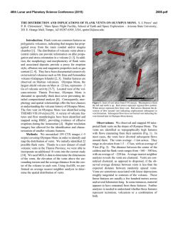
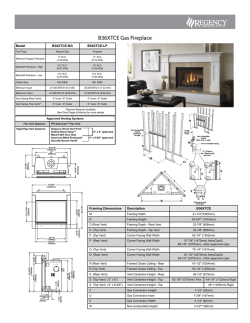
Special Issue, pp. 37-53 (2007)
37
ADAPTATION TO DEEP-SEA HYDROTHERMAL
VENTS: SOME MOLECULAR AND
DEVELOPMENTAL ASPECTS
Florence Pradillon* and Françoise Gaill*
Key words: Annelids, molecular adaptations, ECM, development,
reproduction.
ABSTRACT
Alvinella pompejana is a polychaetous annelid inhabiting the
surface of deep sea hydrothermal chimneys along the ridge of the east
part of the Pacific ocean. The main characteristic of this emblematic
species is its habitat, which is very aggressive considering its
temperature. The exceptional thermotolerance of this species (up to
80°C) has been the subject of much controversy. This review is
focused on the thermal adaptation of this worm regarding molecular
data relative to its extracellular matrix and life history traits.
at the smoker surface [27]. These exoskeletons allow
the Alvinella juveniles to colonize the mineral substrate
of the smokers.
In the eighties, deep-sea biologists have come up
with spectacular observations which strongly suggested
that this species could withstand unusually high
a
INTRODUCTION
Alvinella pompejana, the so-called Pompeii worm
[15], is one of the emblematic animals living in the
extreme environment of deep-sea hydrothermal vents.
This tubicolous animal is found exclusively in association with high temperature venting, at the surface of
hydrothermal chimneys on the East Pacific Rise (Figure
1). The strong gradient from the chimney wall to the
surrounding seawater, depicted in Desbruyères et al.
[16], has been more finely assessed with high resolution
surveys (review in [44]). While the 2°C background
seawater temperature is generally recorded less than ten
centimeters above tube openings, temperatures largely
above 100°C are measured in contact with the mineral
substrate directly beneath the tubes (as in Figure 1b).
The reported temperature maxima for Alvinella colonies on different chimneys range from 125°C in [45] to
175°C in [18]. In contrast to these temperature extremes,
moderately warm conditions were reported at tube
openings, ranging from 6°C to 45°C on average [45].
Alvinella pompejana are living inside tubes they secrete
Author for Correspondence: Florence Pradillon.
E-mail: [email protected]
*Adaptation et Evolution en Milieux Extrêmes, SAE UMR 7138 CNRS IRD
MNHN UPMC, 7 Quai Saint Bernard, 75005 Paris, France.
© AMEX / Phare
b
Fig. 1. (a) Alvinella pompejana out of its tube after recovery. The
animal is about 10 cm long; (b) Alvinella pompejana crawling
on bare mineral surface at vent sites.
38
Special Issue (2007)
temperatures, (up to more than 100°C [10]), and which
triggered an on-going debate on its upper thermal limits
[12]. Temperature inside A. pompejana tubes was initially determined by [7]. An average temperature of 68
±6°C monitored inside a tube over 2 hours, with spikes
as high as 81°C was reported. The relevance of this
value was strongly debated and several artifacts were
suggested, including disturbance of the animal behavior
or piercing the tube when inserting the probe [12].
Since that date, Cary and co-workers have confirmed
the ability to reproduce hour-long temperature monitoring inside tubes [18].
Not only the extreme temperatures encountered in
some tubes, but also the variable temperature over time
support the idea that the medium is not necessarily in
thermal equilibrium with the worm body. The 2 to 4
hours long temperature records presented in [7] and [18]
exhibit frequent temperature changes of 10 to 20°C over
a few minutes and frequent sharp spikes of up to 40°C
in amplitude. The modulation of mean temperature
over more than two hours in one of the hottest tubes was
shown to range between 60°C and 100°C [18]. Furthermore, the large external temperature gradient along a
tube length of c.a. 15 cm suggests a substantial longitudinal gradient inside the tube supporting the idea that
the pompeii worms are also the most eurythermal animals ever known in the oceans.
Besides in situ measurements conducted on and
within colonies, in vivo experiments using pressure
aquaria confirmed the exceptional thermotolerance of
representatives of the alvinellid family. A northern
Pacific relative of A. pompejana, Paralvinella sulfincola,
was very recently shown to be tolerant to temperature of
50 to 55°C [35], the highest ever found for a marine
metazoan. Even more surprising, this worm was shown
to prefer temperature in the range 40°C to 50°C.
Several reviews devoted to A. pompejana have
been published in the past 20 years, assessing current
knowledge in its ecology [16], biology [27] or providing a general overview of the various ecological, physiological and biochemical studies related to this organism [17]. New tools have been used in the last decade
to precise the adaptation strategies of A. pompejana to
the extreme environmental conditions of its habitat and
these recent data have been reviewed by [44]. Part of
this paper will use data which were discussed in previous reviews including the most recent one [44], but will
focus on the thermal adaptation of A. pompejana through
the description of its extra-cellular matrix characteristics and life history traits.
After a brief review of the thermal behavior of the
animal, the thermotolerance of Alvinella pompejana
will be considered in the light of the findings concerning the properties of its exoskeleton, the tube, and of its
collagen molecules. Extra-cellular biopolymers are
supposed to protect the animals from the harsh surroundings and would give indication about the upper
temperature limit the animal may support. In this
respect comparison with extra-cellular matrix (ECM)
from the giant tube worm Riftia pachyptila will allow us
to precise what is specific to the alvinellid family from
what is more widely found in annelids. We will start
with ECM covering the animal body, i.e. the tubes and/
or mucus, and further deal with the main components of
the ECM, which are the collagen molecules. We will
see that data obtained on the biopolymers are in favor of
a worm body of less than 50°C, even though this animal
can withstand up to 65°C. How such a high thermostability may be reached by structural proteins like
collagens? Part of the review will be dedicated to the
thermal adaptation at the molecular level.
ECM data were obtained on adult tissues.
However, we still do not know to which extent the
larvae may face high temperature, since early steps of
development of Alvinella have been shown to occur in
rather cold environment, i.e. less than 20°C [57]. So, a
specific description of the life history traits of the
pompeii worms and sister species will help us in the
understanding of the life cycle of this unusual thermophilic metazoan. We will first review current knowledge on the reproduction mode of the alvinellids which
exhibit specific reproduction strategies [56, 59] and the
development process of the animal [57, 60].
1. Thermal tolerance of alvinellids and related species
New types of high-pressure aquaria [35, 58, 64]
have allowed to examine in vivo the thermal limits of
several hydrothermal vents species inhabiting chimney
walls [35, 46, 61, 62, 65, 66]. Although very promising
high-pressure experiments have demonstrated the capacity to maintain A. pompejana alive up to 24 hours after
recovery [64], its limited survival after collection did not
allowed, to date, in vivo experimentation on this species.
In an effort to characterize the thermal tolerance of
various species living in close proximity to high temperature emissions, Shillito et al. [65] considered the
hesionid worm, Hesiolyra bergi, that was observed
crawling at the surface of A. pompejana colonies and
often entering their tube for a few second to several
minutes. These authors have demonstrated an escape
behavior at 35°C and a lethal limit at 41°C for this
congener, which indicated that high thermal tolerance is
not a prerequisite to live among A. pompejana colonies.
The large thermal heterogeneity characterizing these
colonies over space and time, however, precludes considering this mobile species as a biological ‘thermometer’, as it was suggested by [65]. As reviewed in
F. Pradillon & F. Gaill: Adaptation to Deep-Sea Vents
[44], it is now well established that the surface of the
colony is exposed to temperatures ranging from a few
degrees to 45°C, and that the highest temperatures in
this range are restricted to the vicinity of localized fluid
outflows. Another example with the alvinellids supports the idea that non-thermotolerant species can in
fact inhabit high temperature chimney wall. Paralvinella
sulfincola and Paralvinella palmiformis, the two
alvinellids species of the Juan de Fuca Ridge, were
shown to have very different temperature tolerances
while sharing the same habitat [35]. While P. sulfincola
preference to temperature in range 40-50°C was established in vivo, the later consistently avoided temperatures above 35°C.
P. sulfincola is the only animal that is now firmly
identified to prefer chronic exposure to temperature as
high as 50-56°C. Although lower than for some terrestrial animals (55 to 65°C; see examples in [10, 35]), the
temperature preference and tolerance of this species
stand at the upper limited of the accepted range for
metazoans. While most hydrothermal vent animal species were indeed observed to live at ‘room temperature’
(c.a. about 20°C) (see review in [77]), some species,
such as P. sulfincola, reveal outstanding temperature
preference in in vivo experimental settings offering
them the possibility to chose their thermal environment.
In this regard, in vivo experimentation should now
allow us to investigate thermal behavior of vent
organisms. The association of Alvinella pompejana to
extremely hot substrates, comparable in this respect to
the environment of P. sulfincola [41], supports the idea
that this species would have a similarly high
thermotolerance. If the limits of its thermal preference
and tolerance remain to be empirically defined, its
exceptional thermal adaptation is now firmly assessed
from molecular markers.
39
tube originates from an anterior-ventral glandular shield
[79]. The bulk of the material is formed from homogeneous granules secreted by the deep main cells of the
shield, while an accessory component is provided by
mucocytes secreting sulfated glycosaminoglycans. The
granules are proteinaceous but also contain minerals (e.
g., phosphorus, calcium, iron, and lesser amounts of
magnesium and zinc, see next section). Although the
organic material of the tube is mainly protein, about 7%
of the tube is hexose sugar, presumably as poly- or
oligosaccharide material bound to the protein.
The tube is a concentrically multilayered, fibrous
structure in which the superimposed layers of parallel
fibrils vary in direction from one sheet to the next.
Neither the number nor the thickness of the layers is
constant and varies between different parts of the same
tube. Old and mineralized tubes tend to have thinner
walls than those that have been secreted more recently
[26]. The inner surface of alvinellid tubes is covered by
filamentous bacteria, which, as the tube is being laid
down, become trapped under consecutive layers of
material. Each layer of tube is composed of sub-layers
and individual layers are separated by entrapped bacteria.
Within the layers of secreted protein, a novel distribution of fibrils is displayed, reminiscent of the arrangement of the polymeric units in a cholesteric liquid
crystal. Observation of the Alvinella behaviour during
the Phare cruise, allowed us to infer the role of the worm
motion in this twist variation [44]. Mucous secretion,
and bacteria associated with that secretion, may provide
the initial structure-dictating constraint and is later
modified by the fibrous structure itself as it is laid down
or by pulses of additional mucus production and bacterial growth. Such biopolymeric organisation is thought
to provide a specific thermal resistant to the worm [27].
(b) Mineral content
2. Alvinellid extracellular matrix
Alvinella extra-cellular matrices are composed of
two different tissues: the tube, which is the exoskeleton
of the animal, allowing the worm to settle on the chimney wall, and the collagen which is the main molecular
component of the tissues covering the worm body.
Other alvinellid species such as Paralvinella grasslei
dot not have solid exoskeleton and secrete soft mucus,
which allow their adhesion to organic or mineral substrate [27].
(1) Alvinellid tubes
(a) Composition and structure
The material which A. pompejana uses to build its
The mineral content of the A. pompejana tubes, as
reflected by the ash content, is high (29%). In addition
to this involatile inorganic content, there is also between 12 and 25% of elemental free sulphur, the amount
depending upon the age and the area where the tube
comes from [26]. The mineral seems to be present as a
mixture of sulfides, phosphates, and carbonates.
Minerals show specific patterns of association
with tubes. Zinc-iron sulphide nanocrystals grouped in
submicrometer-sized clusters were described between
proteinaceous tube layers [84]. These minerals show a
specific zinc-iron signature, and have a conserved size
contrary to mineral precipitations found on the outside
of the tubes. The nanometer size of individual minerals
within tubes and their specific constant composition
suggested the biological origin of these crystals, most
40
Special Issue (2007)
probably induced by bacteria associated to the tube
[84]. Mineral particles were also seen as useful markers
for evaluating the chemical characteristics of the microenvironment [85]. Gradients in mineral crystals size
and composition were described and hypothesized to
reflect gradients in chemical characteristics between
the inside and the outside of the tubes, as well as
decimetre-scale gradients between tubes located more
or less deep in the thickness of the alvinella colony.
From these results, the authors hypothesized that the
tube acts as an efficient barrier to the external environment [85].
(c) Associated bacteria and sulphur
The dense covering of filamentous bacteria on the
internal face of the tube is not uniform. Some region
may be free of bacteria, perhaps reflecting differences
in secretory activity of the epidermis of the worm. The
bacteria can generate iron oxydes from pyrite within the
tube and can become embedded in amorphous silica.
Crystals of pure sulphur have been observed by us on
the inside surfaces of the tubes in association with
filamentous bacteria, and free amorphous sulphur is
present in mucus of Paralvinella which has been laid
down for some time and which is in the process of
mineralizing [27].
(d) Specificity of the Alvinella tube composition
Unlike vestimentiferan tubes [25], Alvinella tubes
do not contain chitin. There is no indication from X-ray
diffraction studies of an ordered secondary structure of
proteins, although the amino acid composition, with its
high glycine, alanine, and serine levels, is typical of the
type of beta-pleated fibrous proteins such as silk fibroins [27]. Polychaete worms exhibit a peculiar versatility in regard to the composition of their tube materials
and, therefore, the amino acid composition of the
alvinellid tube is probably of little use in establishing
comparison with other polychaetes. However one characteristic has to be retained: it is its highly hydrophobic
nature.
(e) Chemical and physical stability
The material of the Alvinella tube has considerable
chemical stability. While it is not unusual for invertebrate structural materials to be very chemically stable,
usually as a consequence of extensive cross-linking,
most will swell eventually disrupt at room temperature
in strongly acidic or alkaline solutions or else in
chaotropic agents such as anhydrous formic and
haloacetic acids or lithium thiocyanate. The Alvinella
tube shows little response to these or to disulfide bondbreaking agents, although a cycle of concentrated hydrochloric acid and potassium hydroxide treatments
will cause delamination, swelling, and a little solubilization [26]. Thermal stability is great also, with little
swelling or shrinkage taking place over the 0 to 100°C
temperature range. This too probably reflects a high
degree of cross-linking.
(2) Alvinellid Mucus
Tubes are not secreted by the genus Paralvinella,
but extensive mucus production helps to serve a similar
protective purpose. This material eventually can form
permanent structures as mineralization takes place.
Freshly secreted mucus is transparent with no visible
deposits in it but then becomes yellowed due to elemental sulphur at levels as high as 80% of the dry weight (5).
Sulphur found in mucus is probably a product of bacterial metabolism since mucus is extensively colonized by
sulfide-oxidizing bacteria.
(a) Amino acid composition
The amino acid composition of the freshly secreted mucin of P. grasslei is quite different from that
of the tube of Alvinella, discounting the view that the
latter might simply represent a cross-linked from of
mucin secretion (5). Instead the aspartic and glutamic
acid contents are high, the glycine content lower, and
the alanine and serine contents appreciably less.
However, as the deposited mucus ages and mineralizes,
the proteins degrade and leave a core of very hydrophobic proteins, which is richer in glycine, alanine,
and valine.
(b) Mineral composition
The secretion of mucins may fulfil many roles,
among which protection against a spectrum of environmental affronts, feeding, and detoxification. Thiolic
metal-binding proteins are detectable in the mucins, and
iron, zinc, copper, and uranium have all been detected,
with concentration of uranium ranging from less than 0.
45 to 3.0 µg Kg -1. These concentrations exceed that of
seawater itself. Uranium enrichment in worm tubes is
not confined to animals living on the white smokers, but
has also been found in tubes from the black smokers and
the Galapagos hydrothermal mounds.
(3) Alvinellid collagens
The collagen molecules belong to a family of
extra-cellular proteins, which are characterized by a
F. Pradillon & F. Gaill: Adaptation to Deep-Sea Vents
triple helical domain. This domain is formed by association of 3 similar peptides, called α chains, which are
composed of a succession of Gly-X-Y amino acid triplets.
Usually the Y position is occupied by a proline aminoacid that is often hydroxylated. The collagen is
synthetized inside the cell, and secreted outside the cell
in the extra-cellular compartment. The intracellular
collagen is composed of a central triple helical domain,
which is conserved during the whole life time of the
molecule. The carboxy- and amino-propeptides (C-pro
and N-pro respectively), ending the triple helical central part, are removed when the molecule is secreted in
the extra-cellular matrix.
The collagen characteristics may provide a relevant set of information relative to the characteristics of
the surroundings. This molecule is one of the most well
known extra-cellular proteins of the animal kingdom
and is a relevant marker of thermal adaptation [25].
This molecule has been well characterized in Alvinella
pompejana [29, 30, 32, 33] and vent species [48] and the
origin of the thermostability of the Alvinella collagen
has been determined by [68].
(a) Interstitial and cuticular collagens
Vent annelid species possess two abundant collagen types, which differ in composition, size, domain structure [33] and immunological properties
[30]. Whereas the interstitial collagen is similar in
morphology to the fibrillar collagen of vertebrates,
the cuticular collagen is rather unusual. The length
of the triple helix is of 280 to 300 nm for all interstitial collagens of the alvinellid studied. A similar
length is reported for the interstitial collagen of
another vent endemic annelid, Riftia pachyptila. In
contrast, the cuticle collagens of annelids, with
lengths of up to 2.5 µm in the alvinellid species, are
the longest collagenous protein known [25]. They
possess a terminal globular domain and no comparable counterpart has so far been identified in other
invertebrates or in vertebrates. The cuticular collagen of R. pachyptila is not so long as its length is
about 1.5 µm. It has been shown that the cuticular
collagen of tube worms from various chemosynthetic environments has a similar length and that
such a characteristic is phylogenetic.
Except for the thermostability of the molecule,
cuticular collagens from coastal and vent species shared
similar structural characteristics. This is also true for
the interstitial collagen of annelids from various habitats.
These characteristics were apparently conserved in various annelid families including a substantial and not very
variable level of 4-hydroxyprolinee in the Y position of
the Gly-X-Y sequence triplets.
41
(b) C-propeptide
The C-propeptides (C-pro) from the annelids collagens share overall feature of the mammalian fibrillar
collagen C-pro. They all present a potential cleavage
site, similar in type I, II and III collagen chains, leaving
in the collagen molecule a telopeptide of about 30
amino acid long, which is consistent with the length
found in other fibrillar collagen chains [68]. Specific
residues are also conserved such as the cyclic ones (F,
Y,W), the charged ones (D,N,E,Q,R,K) and proline
residues as well as the glycosylation site (NXT/S).
The main difference between annelids and mammalian C-pro is related to the number of cystein residues.
So far C-pro contained 7 or 8 cysteins depending on the
type of association, hetero- or homo-trimer, of the α
chains. The reduced number of cysteins (6) observed in
the Arenicola collagen [67] is common to all worms
collagen. The missing cysteins (2 and 3 in the α 1(I)
chain) are thought to form intermolecular bonds but
have also been shown of minor importance in realizing
triple helix [5]. The alvinellid and Riftia C-pro are a
natural example of the Bulleid observations [5] that the
molecular mechanism for chain recognition does not
solely rely on the number of cystein residues, but also
on the divergent regions of the C-propeptides.
(c) Thermal stability
Alvinella has the most thermostable protein ever
known [32] and [1] have shown that pressure is not
involved in such a characteristic. The temperature at
which the collagen molecule is denatured (Tm) is 46°C
for the cuticular collagen covering the animal epidermis
and 45°C for the interstitial one which is found in the
worm tissue [32]. Among the fibrillar collagens of 40
other vertebrates and invertebrates, the A. pompejana
collagen is positioned at the upper limit for melting
temperature, only before that of thermostable synthetic
collagens (review in [44]).
The level of thermal stability of Alvinella
pompejana cuticular interstitial collagen is significantly
higher than that of other vent annelids. In comparison,
Riftia pachyptila molecular collagen stability only
reaches 29°C and the collagen of Paralvinella grasslei
has a denaturation temperature of only 35°C [48].
(d) Thermal stability process
The origin of the collagen stability is not well
understood but it is obvious that the rate of proline
hydroxylation is an important factor. In collagen-like
peptides that form triple helices, the substitution of
hydroxyproline (Hyp) for proline in the Y position of
42
Special Issue (2007)
the repeating Gly-X-Y triplets provides additional sites
for hydrogen bonding of water molecules in crystals of
the peptides (review in [68]). In fact the water bridges
would only contribute in part to stability. Different
explanations have been proposed, involving the entropy
state of the chain and an electron withdrawing inductive
effect of the hydroxyl group. No one knows today the
exact correlation between the thermal stability score
and the different processes involved in the molecular
stability.
In Alvinella, as almost all the collagen proline
residues in the Y position of the Gly-X-Y triplets have
been shown to be fully hydroxylated [32], the collagen
stability process would be the same than what is known
in vertebrate and human fibrillar collagen. Sicot et al.
[68] have demonstrated that there is a clear correlation
between the thermal stability and the proline content in
Y position. This would indicate that in living organisms,
proline in the Y position of the GXY triplet would be a
decisive factor involved in the collagen thermal stability.
The relative percentage of proline in the Y position of the triplets is 3 times higher in Alvinella than in
Riftia, which has the lowest relative percentage. The
frequency of the double-P triplets (GPP) relies only on
the frequency of the overall proline content among the
various aminoacids present at the second and third
position of the triplets. Hence, an increase in proline
content would automatically lead to an increase in GPP
triplets. Since GPP triplets are among the most stabilizing triplets, an increase in proline content would result
in an increase in the thermal stability of the triple helix.
Bachinger and Davis [2] have proposed the concept of sequence specific relative stability of the collagen triple helix to quantify the molecule stability.
Considering this point, Alvinella has also the highest
score of the fibrillar collagens analyzed so far, with the
highest stabilizing triplets and the highest GPP
frequency. Moreover, all the stabilizing factors known
today are amplified in the Alvinella collagen including
the percentage of stabilizing triplets, the proline content
and the frequency of hydroxyproline in the Y position of
the Gly-X-Y triplets.
(e) Diversity of stability process
Results obtained on the second type of fibrillar
collagen, the cuticular collagen, observed in the
alvinellid and siboglinid worms [30, 48], indicate that
the same type of protein family would exhibit two
different strategies of thermal stability. One which had
a great success in the course of evolution is that found
in the alvinellid interstitial collagen, relying on the
proline hydroxylation of the residues in the Y position
of the GXY triplets; and one which seems to be original
up to now, which relies on the glycosylation of the
threonine in the same position of the triplet [48]. The
latter is found in the cuticular collagen of R. pachyptila.
Both these examples underline the importance of posttranslational processes in the molecular stability. No
one knows today if these differences are phylogenetically related or collagen type specific and additional
data are needed on the cuticular collagen sequence to
answer this question.
Another potential stability process is the number
and distribution of Gly-X-Y triplets in α chains. In
theory, there are more than 400 possible Gly-X-Y triplets,
but analysis of sequences from fibrillar and non fibrillar
collagens shows that only a limited set of triplets are
found in significant numbers and many are never observed (review in [68]). The distribution of the triplets
through the chain length of these collagens has not
been yet precisely analysed. It would be of interest to
determine the frequency and distribution of the triplets
through the chain length of the available collagen
molecules.
(f) Thermostability and collagen evolution
The alvinellid collagen sequences obtained by [68]
have confirmed the monophyly of the annelid interstitial collagens [67]. However, depending on the considered collagen domain, the triple helix or the C-pro, the
resulting phylogenetic trees are different. If the C-pro
is solely considered in the phylogenetic analysis, the
obtained tree is in accordance with the species classification published in [67]. A. pompejana and coastal
annelid A. marina collagen domains are grouped whereas
R. pachyptila has a distinct location. In contrast, the R.
pachyptila collagen groups with that of A. marina when
the triple helical domain is analysed. This result indicates that different selective constraints have been applied on the 2 domains during evolution (Table 1). This
has been hypothesized to be related to the temperature
the worms inhabit, A. marina and R. pachyptila living in
a colder habitat than the thermophilic worm Alvinella.
The collagen is a modular protein where the triple
helical domain has a longest life time than the Cpropeptide, which is removed once the collagen is secreted outside the cell. The triple helix and the C-Pro
domains would have evolved independently, the selective pressure affecting more the triple helical domain
which is exposed to high temperature in A. pompejana
ECM. The collagen evolution being not neutral, the
conclusion of Sicot et al. [68] leads to the hypothesis
that the collagen triple helix part would have evolved in
different direction according to the living temperature
of the animal, the evolution of the C-propeptide remaining constant.
F. Pradillon & F. Gaill: Adaptation to Deep-Sea Vents
43
Table 1. Evolutionary distances between annelids’ fibrillar collagens (from [68]). Evolutionary distances between
Alvinella pompejana, Riftia pachyptila and Arenicola marina collagen chains were computed. Distances
computed from the helical domain are always greater than distances computed from the C-propeptide. This
means that the selective constraints on the two domains of the same molecule are different. Indeed, the helix
only is maintained in the extracellular matrix, while the role of the C-propeptide is to catalyze the formation
of the triple helix. It can be assumed that the selective constraints exerted on the C-pro would not differ from
one species to the other, while the helix might be more prone to environmental influences. Hence, the Cpropeptide domain was taken as an internal control measuring the part of evolutionary drift after speciation
in the evolution of the molecules. The rate of evolution of the helical domain is about twice the rate of evolution
of the C-propeptide along the distance from Riftia to Arenicola and along the distance from Riftia to Alvinella.
This means that changes in the helical domains after the divergence of Alvinella and Arenicola from their
common ancestor are more numerous than expected. This was taken as circumstantial evidence for an adaptive
effect.
Distance between
Helix
C-pro
Relative rate Helix/C-pro
Total sites
Variable sites
Alvinella /Riftia
Arenicola/Riftia
Arenicola/Alvinella
354
246
237
114
98.21
54.33
1.81
98.88
49.82
1.98
77.62
31.43
2.47
(g) Molecular thermostability and animal thermotolerance
Since the thermal stability of the collagen molecule is 45°C, it can be assume that synthesis of the
collagen would be stopped at a higher temperature, and
the Alvinella pompejana maximum body temperature
would be less than 45°C. However, the collagen is not
found in tissues in a molecular form, but in a supramolecular state. Fibrillar structure is the physiological
state of the collagen in metazoan tissues. Once
synthesized, the collagen molecules assemble in fibrils
and such a polymeric organization has a thermal stability which can exceed by 20°C that of collagen molecules
[28]. A. pompejana fibrillar collagen assemblage may
thus resist up to 65°C, which is consistent with experimental data (review in [44]). If molecular data let us
think that above 45°C, the collagen cannot be synthesized by the epidermal cells, the worm could still sustain
such a high temperature without any damage in its
collagen assemblage. This means that thermal fluctuations between 40 and 60°C could be easily supported by
this animal. This would also support the idea of a
thermal gradient along the animal body length [6],
which is consistent with the observation that the cuticle
of the oldest worms disappears on their posterior part
[31]. Additional data on the Alvinella pompejana
prolylhydroxylase [42] indicate that the worm is not
only facing the highest temperature ever known for
marine invertebrates but would have a metabolic machinery adapted for working in low oxygen environments (review in [44]).
3. Reproduction and development in Alvinellid species
Hydrothermal vents are highly unstable ecosystems both on a spatial and on a temporal scale. For this
reason, the question of the maintain of species and of
population distribution has immediately been raised
and is still not answered. Genetic studies carried out on
most vent taxa evidenced exchanges between populations over distances greater than the average intervals
that occur between vent sites. Fairly wide dispersal
capabilities were thus inferred for most species inhabiting vent sites.
Many vent species are sessile and cannot survive
as adults outside of vent sites. Dispersal was thus
mainly attributed to the larval phase. However, this
phase of the life cycle of vent organisms remains unknown for most species. In Alvinellids, reproductive
strategies have now been investigated in several species,
and early developmental stages were obtained in
Alvinella pompejana [57].
A. pompejana has been described as a pioneer on
newly formed chimneys [17]. Colonization experiments have further confirmed the settlement of the first
individuals within a few days on colonization devices
deployed over a smoker wall, following the formation
of filamentous microbial mats [71]. The formation of
several centimeter-thick assemblages of tubes (cf Figure 2) within two months, partly encrusted in mineral
precipitates, underlines a fast colonization process in
response to rapid production of newly available substrates [85]. Among the processes that can govern the
formation of these new settlements, we [59] have con-
Special Issue (2007)
44
(a) Reproductive modes in Terebellida
Sea-water
Alvinellid colony
Chimney wall
Fig. 2-3 D diagram of the Alvinella colony. In the upper sea-water layer,
temperature and pH vary according to local fluid outflows.
Within the thickness of the colony, tubes are surrounded by a
matrix of mineral and organic deposits. Non-diluted acidic
hydrothermal fluids circulate through this matrix. The inside
of tubes is mainly sea-water entering tubes through their
opening, heated by thermal conduction. From [45].
sidered two alternative possibilities: the recruitment of
larvae or the migration of post-larvae stages. Almost
nothing is known on the life cycle and dispersal strategies of the major vent species. To date, embryos of only
two vent species (Riftia pachyptila and A. pompejana)
have been studied for temperature and pressure tolerance [49, 57]. This is mainly limited by the fact that
catching larvae directly in situ remains highly
challenging. As an alternative to sampling, in vitro
fertilization methods combined to in vivo experiments
revealed to be a very pertinent approach to obtain essential information on the ability of early stages to deal
with the extreme environment of adult colonies [57].
(1) Reproductive strategy
Because vent systems are unstable, vent species
were expected to exhibit efficient reproductive strategies allowing them to survive unpredictable environmental changes. That means an early sexual maturity,
and the quick production of a large number of offsprings
and an efficient fertilization mode. If environmental
constraints may have influenced the evolution of reproductive strategies on one hand, on the other hand, in
several vent taxa, reproductive strategies were found to
be similar to those of non-hydrothermal relatives, suggesting that phylogenetic constraints may be stronger
than environmental constraints in some cases [20, 76,
78]. Then, although polychaete species exhibit a great
plasticity in their reproductive strategies [34, 81], most
hypotheses originally emitted concerning reproduction
in the Alvinellidae were based on known reproductive
characteristics from species of the Terebellida group to
which the Alvinellidae family belongs.
The closest families to the Alvinellidae are
Ampharetidae, Terebellidae and Trichobranchidae [22,
63]. Different reproductive modes are found in these
families : continuous or discontinuous reproduction,
species that reproduce once in their life time (monotelic)
or several times (polytelic), free spawners or species
incubating the embryos inside their tube or even inside
the female body. Some species such as the Terebellidae
Eupolymnia nebulosa Montagu, may even exhibit 2
types of spawning, with free spawning in Atlantic
populations, and spawning in a gelatinous cocoon in
Mediterranean populations [47]. Some characteristics
appear to be relatively conserved. Oocyte size often lay
between 100 and 300 µm in diameter. Development is
for most species direct (i.e. without a free larval phase)
or lecithotrophic (i.e. free larval phase that do not feed
in the plankton but exclusively on oocyte reserves), but
rarely planktotrophic (free living phase that feeds in the
plankton).
(b) Morphological characteristics
All Alvinellid species are gonochoric and exhibit
sexual dimorphism (Table 2). In all species examined,
males display a pair of modified peribucal tentacles,
which are lacking in females [17, 40, 82, 86]. Males of
Paralvinella grasslei have a pair of small blind cavities on the peristomium [82], and in two Paralvinella
species, females have papillae at the base of the gills
which are absent in males [82, 86]. Finally, genital
pore dimorphism was also observed in several species
[56, 86].
The organisation of the reproductive apparatus
is similar in all species analyzed so far [17, 39, 51,
56, 82, 86]. Females have a single pair of oviducts
that extends trough the anterior part of the coelomic
cavity. The oviducts are connected to a pair of
spermathecea located in the dorsal part of the most
anterior setigerous segment. The spermathecae communicate with the exterior by a short common canal,
located medially at the base of the most posterior
gills. In males, the reproductive apparatus is similar
with one pair of spermiducts connected to seminal
vesicles, which open to a unique genital pore also
located at the base of the posterior gills. The presence of only one pair of gonoducts was described in
the Ampharetidae Ampharete grubei [21]. This is a
rather unusual feature in Terebellida where several
pairs of gonoducts are found for the majority of the
species [69, 70]. The Alvinellidae is, to date, the
only taxon in the order Terebellida known to possess
spermathecae.
F. Pradillon & F. Gaill: Adaptation to Deep-Sea Vents
45
Table 2. Reproductive characteristics of alvinellid species.
Species
Sexual
dimorphism
Fecundity
(average)
Oocyte
maximum
diameter
Spermatozoa
Fertilization
Reproductive
synchrony
Development
References
Alvinella
pompejana
• Pair of modified
buccal tentacles
in ♂
• Morphological
difference in ♂
/♀ genital pore
80000
200
• No acrosome
• No midpiece
• Small size (4 µm)
• Conical shape
• Short flagellum
• Sperm transfert
to spermathecae
• External
fertilization
No
• Lecitotrophic or
direct
• Temperature
sensitive early
development
• Negative egg
buoyancy
[17, 39, 40,
56, 57, 60]
Alvinella
caudata
• Pair of modified
buccal tentacles
in ♂
• Morphological
difference in ♂
/ ♀ genital pore
?
?
• No acrosome
• No midpiece
• Small size (4.5 µm)
• Conical shape
• Short flagellum
• Sperm transfert
to spermathecae
?
• Lecitotrophic or
direct
[39]
(autors
personal
obs.)
Paralvinella
grasslei
• Pair of modified
buccal tentacles
in ♂
• Pair of blind
cavities on
peristonium
in ♂
• Papillae on the
stem of posterior
gills in ♀
3900
275
• No acrosome
• No midpiece
• Oval “head” (10
µm)
• No flagellum
• Long caudal
process.
• Sperm transfert
to spermathecae
Yes, at the
scale of a
single vent
• Lecitotrophic or
direct (in situ
observation of
erpochaete larva)
[39, 82, 83]
Paralvinella
pandorae
pandorae
• Pair of modified
buccal tentacles
in ♂
4500
215
• Elongated “head”
(19 µm)
• Atypical midpiece
• Long flagellum
inserted poteriorily
but directed
anteriorily
• Sperm transfert
to spermatheca
• Spermatozoa
attached to the
spermathecal
walls
No
• Lecitotrophic or
direct
[39, 51, 83]
Paralvinella
pandorae
irlandei
?
?
?
• Elongated “head”
(19 µm)
• Atypical midpiece
• Long flagellum
inserted poteriorily
but directed
anteriorily
• Sperm transfert
to spermatheca
• Spermatozoa
attached to the
spermathecal
walls
?
• Lecitotrophic or
direct (in situ
observation of
erpochaete
[15, 39]
Paralvinella
palmiformis
• Pair of modified
buccal tentacles
in ♂
• Morphological
difference in ♂
/♀ genital pore
• Papillae on the
stem of posterior
gills in ♀
18000
260
• No acrosome
• No midpiece
• Oval “head” (10
µm)
• No flagellum
• Short process.
• Sperm transfert
to spermatheca
Yes, over
several vents
for colonies at
a similar stage.
• Lecitotrophic or
direct
[13, 14, 39,
51, 86]
Paralvinella
sulfincola
Paralvinella
dela
?
?
250
No
[13]
• Pair of modified
buccal tentacles
in ♂
?
?
• Lecitotrophic or
direct
?
?
(c) Gametogenesis
Early steps of gametes development occur in the
coelomic cavity. To date, the gonades, where the repro-
?
?
[86]
ductive cells start their differenciation, have not been
identified in any Alvinellid species. In P. grasslei,
glands located on both sides of the medio ventral nervous chain from setigerous segment 5 (S5) to S35-40
46
Special Issue (2007)
were first described as gonades [82]. However, later on,
very similar glands were observed in P. palmiformis and
P. dela, but interpreted as mucus glands [86].
The earliest stages of spermatozoa development
were described in the coelomic cavity of P. palmiformis
and P. pandorae pandorae [51]. The spermatogonia
divide into spematocytes rosettes. Spermatocytes then
mature into groups of spermatocytes with flagellae called
sperm morulae. Spermatozoa are further stored into the
spermiducts.
Ultrastructural investigations on spermatozoa revealed modified structures in all studied species, being
flagellated (P. pandorae) or not (A. pompejana, A.
caudata, P. grasslei, P. palmiformis), having no
acrosome and an atypical or absent midpiece (Table 2).
Other special features were also described and thought
to be involved in sperm storage into the female spermathecae and/or fertilization process. In A. pompejana
and A. caudata spermatozoa exhibit a flat vesicular
surface that could be involved in adhesiveness in the
spermathecae. Spermatozoa of P. palmiformis also
possess a vesicular surface that could produce some
adhesive material favouring clustering and storage in
the spermathecae. In P. p. irlandei and P. p. pandorae,
spermatozoa are implanted in the spermathecal walls
after the sperm transfer [39].
During vitellogenesis oocytes float freely in the
coelomic cavity [17, 51, 56, 82]. Oocytes have a
flattened discoid shape, and in P. grasslei and A.
pomejana they exhibit a micropyle , which was related
to the absence of acrosome in spermatozoa [17, 82].
Maximal diameter in coelomic ocytes vary between
Alvinellid species from 200 to 275 (m [13, 14, 17, 51,
56, 82].
In A. pompejana, ultrastructural analyses showed
that coelomic oocytes do not rely on any type of helper
cells for nutrition, but may use autosynthetic mechanisms for yolk production [56]. Combination of
extraovarian vitellogenesis and autosynthesis of yolk
suggest a rather slow oogenesis process, which is contradictory with the original hypothesis of fast egg
production. After completing yolk synthesis, grown
oocytes enter the oviduts through funels opening into
the coelomic cavity. At this stage, a selection process
based on oocytes size or membrane characteristics would
occur (Figure 3) [56]. Such selection mechanism of ripe
gametes by gonoducts was already suggested in other
Terebellida species [50, 69]. Thus, the organisation of
the genital tract seems to allow the storage of a distinct
pool of ripe oocyte. This pool could then be spawn at
any time, possibly in response to specific environmental cues (Figure 3). Such mechanism would give spawning processes enough flexibility to face chaotic environmental conditions, despite a slow oocyte production.
Oogenesis
Storage Gametes
mature
oocytes mixing
Size/shape Spawning
selection? signal?
Fertilization
Spermatozoa
Oocyte
Coelomic cavity
Oviducts
Spermathecae
Fig. 3. Diagram of the gametogenesis, fertilization and spawning
mechanisms in Alvinella pompejana.
(d) Fecundity
Fecundity is highly variable in Alvinellids. Average values range from 3900 oocytes per female in P.
grasslei, up to 80 000 in A. pompejana [8, 17, 59]. Such
high fecundity values can not, however, be compared
with fecundities of other Terebellida species, since
studies in these species report the number of eggs per
spawning event, whereas estimates for Alvinellid species were calculated from the total amount of coelomic
oocytes. Spawning in Alvinellids will most probably
involve only mature oocytes stored in the oviducts. In
A. pompejana, full oviducts may contain about 3000
oocytes, which would be the size of a single spawning
event [60].
(e) Fertilization
In several Alvinellid species, spermatozoa were
observed inside the spermathecae, and a transfer of
sperm from males to females during a probable
pseudocopulation process was suggested [17, 39, 82].
In situ observations show that Alvinellids frequently
leave their tubes to enter tubes of other individuals [9,
24] and sperm transfer may occur during these events.
In P. pandorae irlandei and P. grasslei, pairs individuals were observed in mucus cocoons at the base of the
tubes of vestimentiferen tubeworms, suggesting that an
appariement of sexes might take place in Alvinellids
during reproduction [38, 83]. Sperm transfer to the
spermathecae is interpreted as a way to avoid gamete
losses and increase fertilization efficiency in the highly
dynamic environment [83].
However, sperm transfer to the spermathecae does
not necessarily mean that fertilization is internal as
suggested before. In A. pompejana, fertilized eggs were
never found in the spermathecae, although it was filled
F. Pradillon & F. Gaill: Adaptation to Deep-Sea Vents
with spermatozoa, and the oviducts of the same female
were packed with ripe oocytes. This suggests that
oocytes are not incubated in the spermathecae, but
rather go through the spermathecae during spawning,
where spermatozoa could possibly attach to their surface through their vesicular surface, and fertilization
would take place outside after spawning [56] (Figure 3).
Such strategy has now also been suggested in Siboglinid
species [36].
Newly-formed
surface
Colonisation
Dispersal,
recruitment
(f) Synchrony in reproductive processes
In a number of marine invertebrates from coastal
environments, gametogenesis processes are controlled
by endogenous rhythms of hormones production, which
are under the influence of external factors such as
temperature, photoperiod or moon phases [3, 4, 43, 53,
54, 80]. In the abyssal environment, where food abundance is limited, and environmental variations almost
non-existent, the initial hypothesis was that of a continuous reproduction [55]. A number of abyssal species
nevertheless exhibit clearly synchronised reproduction
with a periodic maturation of reproductive tissues, followed by spawning events [73]. Such periodicity was
explained by the arrival of seasonal phytoplankton
blooms on the oceanic seafloor [72]. At hydrothermal
vents, photoperiod cannot be detected, and organic matter
input from the surface is negligible compared to the
high local production rates. The prevailing hypothesis
was therefore that of a continuous reproduction for vent
species.
Most vent species display continuous reproduction [74, 75]. Alvinellids, however, seem to have evolved
diverse strategies. In the alvinellid family, asynchronous reproduction was suggested in Paralvinella
sulfincola [13] and P. pandorae [51]. In contrast, P.
grasslei [68] and P. palmiformis [14] were suggested to
reproduce synchronously at vent scale, responding to
periodic variation linked to tidal regime in environmental factors such as temperature [11, 37]. In P. palmiformis, however, spatial variation in reproductive patterns was found at vent scale, which may reflect the
successional mosaic of the vent community, with immature individuals in earlier successional stages [14]. In
A. pompejana, the dynamic disturbance/colonisation
process similarly results in a mosaic of patches
harbouring individuals at different reproductive stages
[60]. New surfaces are colonised within a few days by
juveniles and non-reproductive individuals (Figure 4).
In such patches all individual are non-reproductive,
similarly to what was observed in early successional
stages in P. palmiformis [14]. In older colonies, reproductive females were found, but females of a same patch
did not show synchronism in reproductive stages. On
47
Maturation
Migration of
non-reproductive
individuals
Fertilisation,
spawning
Migration
Active surface
Non-reproductive individual
Inactive surface, or
decreasing activity
Reproductive individual
Fig. 4. Reproduction and colonization patterns in Alvinella pompejana,
at th escale of local patches on a vent chimney. From [59].
the contrary, the diversity of reproductive stages suggested that spawning episodes would occur repeatedly
and would concern only a fraction of the adult population [59].
If physico-chemical gradients may be steep on the
interface between the sea-water and the chimney wall,
Alvinella colonies modify fluid circulations by building
tubes [45] (Figure 2). Then well-established colonies
form an isolating layer that may greatly reduce temperature gradients [45]. Since A. pompejana females are
only found in such colonies, this might reflect their
preference for milder environment during reproduction.
(2) Development
Developmental characteristics such as the length
of the cells cycles during embryonic cleavages, the
developmental mode, the embryos buoyancy or physiological tolerance to physical environmental parameters have a strong influence on dispersal capabilities of
larvae. However, deciphering these characteristics require first to be able to obtain the embryos. In the
abyssal environment, collecting larvae has been
challenging, and in the cases where larvae were indeed
collected, they could not be easily identified.
Special Issue (2007)
48
Alvinellid embryos were never identified in situ so
far, and development was suggested to be direct or
lecithotophic (i.e. with no or limited dispersal
capabilities) because of hypotheses based on the oocyte
size [51, 83] and observation of larval stages in in situ
collection which were similar to those of ampharetid
polychaetes [16, 83].
Embryos of A. pompejana were obtained using in
vitro fertilization methods [57], which were also used to
study embryos of the Siboglinid tubeworm Riftia
pachyptila [49]. Since A. pompejana naturally experience pressure of 250 atmospheres (250 atm. ≈ 2500 m.
depth) at vent sites, specific pressure equipment including a microscopy imaging system were designed for
developmental studies. In A. pompejana, first divisions
are asymmetrical, the mechanism of this pattern being
the formation of polar lobe [60]. Early embryos have a
typical spiral development as found in most polychaete
[19] (Figure 5).
thermotolerant species as an adult, and for embryos, two
main hypotheses were first analyzed : (1) either embryos are able to develop in the abyssal sea-water with
temperature typically around 2°C, (2) or they are also
thermotolerant, and are able to develop on the vent
chimney walls within adult colonies (>20°C). In the
first hypothesis, dispersal could occur through transportation with marine currents over tens to hundreds of
kilometres, allowing colonization of new distant sites.
In the second hypothesis, embryos would develop without dispersal.
In vitro, embryos of A. pompejana exhibit low
temperature tolerance, being unable to survive above
20°C. At optimal temperature (around 10°C), cleavage
rates are very slow with approximately 1 division every
24h. In polychaete species living in the coastal
environment, larvae may already be obtained after 24h
[84]. In addition, the developmental process was shown
to be arrested at 2°C, but a transient temperature increase could trigger development of arrested embryos
[57].
(b) Thermal tolerance of the embryos
(c) What is the in situ embryo behaviour?
One of the objectives of these developmental studies were to determine physiological tolerance of embryos to physico-chemical parameters, which would
give an idea of the favourable developmental conditions
in situ, and could be used to deduce possible development area and dispersal capabilities. A. pompejana is a
The thermal tolerance window determined for early
embryos of A. pompejana is therefore restricted to
temperature lower than those encountered most of the
time in adult colonies. This would suggest that embryos
cannot develop there. However, recent studies evidenced that a great diversity of habitat with various
hydrothermal influence [45]. Diffuse flow areas, with
mild temperatures would be compatible with development of embryos of A. pompejana. Also, part of the
embryos could be entrained with bottom currents where
they would be exposed to very low temperature that
would arrest their development. This kind of dormancy
would then be stopped by temperature increase if the
embryos happen to arrive close to a vent. This mechanism can potentially allow wide dispersal capabilities.
However, the potential duration of this dormancy is still
an open question.
To test these hypotheses, incubators containing
embryos of A. pompejana were deployed in different
habitats of a single edifice, along a gradient of hydrothermal influence [60]. Only 10% of the embryos
incubated above an Alvinella colony survived after 5
days, among the surviving embryos, none of them had
developed. On the contrary, 70% of the embryos incubated in milder area (Riftia clump 1 meter below the
Alvinella colony, and on the bare mineral seafloor) (4 to
6°C), developed [60]. These results supported the idea
that development outside of the colony is possible,
while it would not be viable in the adult colony. Temperature measurement close to the incubation device in
(a) Development mode of Alvinellids
a
b
c
d
Fig. 5. Embryos of A. pompejana obtained by in vitro fertilization and
incubated under pressure. (a) fertilized egg; (b) 2-cell embryos
exhibiting asymmetric division; (c) 4-cell embryo; (d) 96h
embryo. Modified from [60].
F. Pradillon & F. Gaill: Adaptation to Deep-Sea Vents
the Alvinella colony indicated an average of 13°C during the 5 days of the experiment, but frequent burst
above 20°C were also recorded. In addition, sulphide
levels were up to 10 fold higher in the Alvinella colony
than in the 2 other habitats. These experiments, however,
did not allow us to decipher which parameters predominantly affect embryos survival and development.
(d) Larvae dispersal
Both in vitro and in situ studies indicate that A.
pompejana embryos could disperse through abyssal
seawater and develop when they find conditions around
10°C. Development in the shallow part of the ocean can
be excluded since embryos can not develop at atmospheric pressure [60]. Since pressure tolerance has not
been precisely determined, the range of possible vertical movement is still matter of speculation, and embryos might be entrained by the hydrothermal plume far
enough above the sea bottom to be further entrained by
upper layers of currents with possibly different regimes
than those running on the bottom [52]. Recent studies
showed that low temperatures may be found at the
surface of adult colonies because the tubes build by this
species may isolate the surface of the colony from the
hot chimney wall [45]. This could provide suitable
habitat for embryos, and lead to reconsider the assumption that early development is excluded from the adult
environment.
CONCLUSIONS
The ability of Alvinella pompejana to colonize
high temperature substrates is far from being fully
understood, but the exceptional properties of its extracellular biopolymers and the behavior of the worm can
be now considered as major clues in the colonization
process. A. pompejana could thus stand at the limits
authorized for its biological machinery in a highly
dynamic environment where temperature can readily
reach lethal limits, but where temperature regulation by
the animal itself would prevent exposure to deleterious
thermal spikes. The animal has a specific cellular
machinery which has been selected during the course of
evolution. Alvinella collagen is the most thermostable
protein ever known (Tm 46°C). This species exhibits
enzymes able to synthesize these unique extra-cellular
proteins in an anoxic environment. Sicot et al. [68] have
demonstrated that the collagen stabilization process
would be the same than that known in vertebrate and
human fibrillar collagen. Moreover, all the stabilizing
factors known today are amplified in the Alvinella collagen including the percentage of stabilizing triplets,
proline content and the frequency of hydroxyproline in
49
the Y position of the Gly-X-Y triplets.
Such a thermostability results from an adaptation
process to high temperature. This thermophilic metazoan worm occupies a very specific niche being a pioneer at the surface of the vent smoker. Once recruited
at the surface of the smoker, the animal is able to secrete
very specific biopolymers, allowing it to colonize new
warm mineral surfaces and to optimize the interactions
with the hydrothermal fluids. If we know now that this
animal is thermophilic in its adult stage, the worm
would prefer the cold abyssal sea-water in its early steps
of development [57, 60]. This would be a good strategy
to survive in the deep sea-water far away from the vents
while dispersing. However, what we do not know yet,
is the mechanism of the larval recruitment. It is possible
that the larvae travel in between vent sites using the
currents. However, how these larvae are able to find a
new vent site is still mysterious. What are the signals
indicating that a new smoker surface is available? How
larvae find them and what are the signals indicating that
it is time to settle on a new smoker surface? This is one
aspect of the future of the research on this worm.
Proteomic and genomic data will be available in the
future and will bring new insights in the thermotolerance
process involved in the biology of this unique deep-sea
vent animal.
REFERENCES
1. Auerbach, G., Gaill, F., Jaenicke, R., Schulthess, T.,
Timpl, R., and Engel, J., “Pressure Dependence of Collagen Melting,” Matrix Biology, Vol. 14, pp. 589-592
(1995).
2. Bächinger, H.P. and Davis, J.M., “Sequence-Specific
Thermal Stability of the Collagen Triple Helix,” international Journal of Biological Macromolecules, Vol. 13,
pp. 331-338 (1991).
3. Bentley, G.M. and Pacey, A.A., “Physiological and Environmental Control of Reproduction,” Polychaetes
Oceanography and Marine Biology: An Annual Review,
Vol. 30, pp. 443-481 (1992).
4. Bentley, M.G., Boyle, J., and Pacey, A.A., “Environmental influences on Endocrine Systems Controlling
Reproduction,” Polychaetes. in 4ème Conférence
Internationale des Polychètes, Paris (1994).
5. Bulleid, N.J., “Novel Approach to Study the initial Events
in the Folding and Assembly Procollagen,” Seminars in
Cell and Developmental Biology, Vol. 7, pp. 667-672
(1996).
6. Cary, C.S. and Stein, J.L., “Spanning the Thermal Limits:
an Extreme Eurythermal Symbiosis,” Cahiers De Biologie
Marine, Vol. 39, pp. 275-278 (1998).
7. Cary, C.S., Shank, T., and Stein, J.L., “Worms Bask in
Extreme Temperatures,” Nature, Vol. 391, pp. 545-546
50
Special Issue (2007)
(1998).
8. Chevaldonné, P., “Ecologie des Cheminées
Hydrothermales Actives,” In Océanologie, Université
de la Méditerranée, Marseille, p. 257 (1996).
9. Chevaldonné, P. and Jollivet, D., “Videoscopic Study of
Deep-Sea Hydrothermal Vent Alvinellid Polychaete
Populations: Biomass Estimation and Behaviour,” Marine Ecology Progress Series, Vol. 95, pp. 251-262
(1993).
10. Chevaldonné, P., Desbruyères, D., and Childress, J.J.
“Some Like It Hot and Some Even Hotter,” Nature, Vol.
359, pp. 593-594 (1992).
11. Chevaldonné, P., Desbruyères, D. and Le Haître, M.,
“Time-Series of Temperature from Three Deep-Sea Hydrothermal Vent Sites,” Deep-Sea Research, Vol. 38,
No.11, pp. 1417-1430 (1991).
12. Chevaldonne, P., Fisher, C.R., Childress, J.J.,
Desbruyeres, D., Jollivet, D., Zal, F., and Toulmond, A.,
“Thermotolerance and the Pompeii Worms,” Marine
Ecology Progress Series, Vol. 208, pp. 293-295 (2000).
13. Copley, J.T.P., “Ecology of Deep-Sea Hydrothermal
Vents,” (Report of Department of Oceanography, University of Southampton), Southampton, p. 204 (1998).
14. Copley, J.T.P., Tyler, P.A., Van Dover, C.L., and Philp,
S.J., “Spatial Variation in the Reproductive Biology of
Paralvinella Palmiformis (Polychaeta: Alvinellidae) from
a Vent Field on the Juan de Fuca Ridge,” Marine Ecology
Progress Series, Vol. 255, pp. 171-181 (2003).
15. Desbruyères, D. and Laubier, L., “Les Alvinellidae, Une
Famille D’annélides Polychètes inféodées Aux Sources
Hydrothermales Sous-Marines: Systématique, Biologie
et Écologie,” Canadian Journal of Zoology, Vol. 64, pp.
2227-2245 (1986).
16. Desbruyères, D., Gaill, F., Laubier, L., and Fouquet, Y.,
“Polychaetous Annelids from Hydrothermal Vent
Ecosystems: an Ecological Overview,” The Biological
Society of Washington Bulletin, Vol. 6, pp. 103-116
(1985).
17. Desbruyères, D., Chevaldonne, P., Alayse, A.M., Jollivet,
D., Lallier, F.H., Jouin-Toulmond, C., Zal, F., Sarradin,
P.M., Cosson, R., Caprais, J.C., and Arndt, C., “Biology
and Ecology of the “Pompeï Worm” (Alvinella
Pompejana, Desbruyères et Laubier), a Normal Dweller
of an Extreme Deep-Sea Environment : a Synthesis of
Current Knowledge and Recent Developments,” DeepSea Research II, Vol. 45, pp. 383-422 (1998).
18. Di Meo-Savoie, C., Luther III, G.W., and Cary, C.S.,
“Physicochemical Characterization of the Microhabitat
of the Epibionts Associated with Alvinella Pompejana, A
Hydrothermal Vent Annelid,” Geochimica et
Cosmochimica Acta, Vol. 68, No. 9, pp. 2055-2066
(2004).
19. Dorresteijn, A., “Cell Lineage and Gene Expression in
the Development of Polychaetes,” Hydrobiologia, Vol.
535/536, pp. 1-22 (2005).
20. Eckelbarger, K.J. and Watling, L., “Role of Phylogenetic
Constraints in Determining Reproductive Patterns in
Deep-Sea invertebrates,” Invertebrate Biology, Vol. 114,
pp. 256-269 (1995).
21. Fauvel, P., “Recherches sur les Ampharétiens,” Bulletin
Scientifique France Belgique, Vol. 30, pp. 277-489 (1897).
22. Féral, J.-P. and Philippe, H., “Phylogénie Moléculaire de
Polychètes Alvinellidae des Sources Hydrothermales
Actives de L’océan Pacifique,” Comptes Rendus de
l’Académie des Sciences de Paris, Sciences de la Vie,
Vol. 317, pp. 771-779 (1994).
23. Fischer, A. and Dorresteijn, A., “The Polychaete
Platynereis Dumerilii (Annelida): A Laboratory Animal
with Spiralian Cleavage, Lifelong Segment Proliferation
and a Mixed Benthic/Pelagic Life Cycle,” Bioessays,
Vol. 26, No. 3, pp. 314-325 (2004).
24. Fustec, A., Desbruyères, D., and Juniper, S.K., “DeepSea Hydrothermal Vent Communities at 13°N on the Est
Pacific Rise: Microdistribution and Temporal Variations,” Biological Oceanography, Vol. 4, pp. 121-164
(1987).
25. Gaill, F., “Aspects of Life Development At Deep-Sea
Hydrothermal Vents,” The FASEB Journal, Vol. 7, pp.
558-565 (1993).
26. Gaill, F. and Hunt, S., “Tubes of Deep Sea Hydrothermal
Vent Worms Riftia Pachyptila (Vestimentifera) and
Alvinella Pompejana (Annelida),” Marine Ecology
Progress Series, Vol. 34, pp. 267-274 (1986).
27. Gaill, F. and Hunt, S., “The Biology of Annelid Worms
from High Temperature Hydrothermal Vent Regions,”
Review in Aquatic Sciences, Vol. 4, No. 2-3, pp. 107-137
(1991).
28. Gaill, F., Herbage, D., and Lepescheux, L., “Cuticle
Structure and Composition of Two Hydrothermal Vents
invertebrates,” Oceanologica Acta, Vol. 8, pp. 155-159
(1988).
29. Gaill, F., Zbinden, M., and Pradillon, F., “Adaptations of
Hydrothermal Vent Organisms to Their Environment,”
Proceedings of 18th international Zoology: The New
Panorama of Animal Evolution, pp. 513-517 (2003).
30. Gaill, F., Hamraoui, L., Sicot, F.X., and Timpl, R.,
“Immunological Properties and Tissue Localization of
Two Different Collagen Types in Annelid and
Vestimentifera Species,” European Journal of Cell
Biology, Vol. 65, pp. 392-401 (1994).
31. Gaill, F., Desbruyères, D., Prieur, D., and Gourret, J.P.,
“Mise en Évidence de Communautés Bactériennes
Épibiontes Du “Ver de Pompéï” (Alvinella Pompejana),” Comptes Rendus de l’Académie des Sciences de
Paris, Série III, Vol. 298, pp. 553-558 (1984).
32. Gaill, F., Mann, K., Wiedemann, H., Engel, J., and
Timpl, R., “Structural Comparison of Cuticule and intersticial Collagens from Annelids Living in Shallow Sea-
F. Pradillon & F. Gaill: Adaptation to Deep-Sea Vents
Water and at Deep-Sea Hydrothermal Vents,” Journal of
Molecular Biology, Vol. 246, pp. 284-294 (1995).
33. Gaill, F., Wiedemann, H., Mann, K., Kühn, K., Timpl, R.,
and Engel, J., “Molecular Characterization of Cuticular
and interstitial Collagens from Worms Collected at DeepSea Hydrothermal Vents,” Journal of Molecular Biology,
Vol. 221, pp. 157-163 (1991).
34. Giangrande, A., “Polychaete Reproductive Patterns, Life
Cycles And Life Histories: an Overview,” Oceanography and Marine Biology: An Annual Review, Vol. 35, pp.
323-386 (1997).
35. Girguis, P.R. and Lee, R.W., “Thermal Preference and
Tolerance of Alvinellids,” Science, Vol. 312, pp. 231
(2006).
36. Hilario, A., Young, C.M., and Tyler, P.A., “Sperm Storage,
internal Fertilization, and Embryonic Dispersal in Vent
and Seep Tubeworms (Polychaeta: Siboglinidae:
Vestimentifera),” The Biological Bulletin, Vol. 208, pp.
20-28 (2005).
37. Johnson, K.S., Childress, J.J., Beehler, C.L., and
Sakamoto, C.M., “Biogeochemistry of Hydrothermal
Vent Mussel Communities: The Deep-Sea Analogue to
the Intertidal Zone,” Deep-Sea Research, Vol. 41, pp.
993-1011 (1994).
38. Jollivet, D., “Distribution et Évolution de la Faune
Associée Aux Sources Hydrothermales À 13°N sur la
Dorsale Du Pacifique Oriental: le Cas Particulier des
Polychètes Alvinellidae,” Ph.D. Thesis, Université de
Bretagne Occidentale (1993).
39. Jouin-Toulmond, C., Mozzo, M., and Hourdez, S., “Ultrastructure of Spermatozoa in Four Species of
Alvinellidae (Annelida: Polychaeta),” Cahiers de
Biologie Marine, Vol. 43, pp. 391-394 (2002).
40. Jouin-Toulmond, C., Zal, F., and Hourdez, S., “Genital
Apparatus and Ultrastructure of the Spermatozoa in
Alvinella Pompejana (Annelida: Polychaeta),” Cahiers
de Biologie Marine, Vol. 38, pp. 128-129 (1997).
41. Juniper, S.K., Jonasson, I.R., Tunnicliffe, V., and
Southward, A.J., “Influence of a Tube-Building Polychaete on Hydrothermal Chimney Mineralisation,”
Geology, Vol. 20, pp. 895-898 (1992).
42. Kaule, G., Timpl, R., Gaill, F., and Günzler, V., “Prolyl
Activity in Tissue Homogenates of Annelids from DeepSea Hydrothermal Vents,” Matrix Biology, Vol. 17, pp.
205-212 (1998).
43. Lawrence, A.J., “Environmental and Endocrine Control
of Reproduction in Two Species of Polychaete: Potential
Bio-Indicators for Global Climate Change,” Journal of
the Marine Biological Association of the United Kingdom,
Vol. 76, pp. 247-250 (1996).
44. Le Bris, N. and Gaill, F., “How Does the Annelid Alvinella
Pompejana Deal with an Extreme Hydrothermal
Environment?” Reviews in Environmental Science and
Biotechnology, Vol. 6, pp. 197-221 (2007).
51
45. Le Bris, N., Zbinden, M., and Gaill, F., “Processes
Controlling the Physico-Chemical Micro-Environments
Associated with Pompeii Worms,” Deep-Sea Research I,
Vol. 52, pp. 1071-1083 (2005).
46. Lee, R.W., “Thermal Tolerance of Deep-Sea Hydrothermal Vent Animals for the Northeast Pacific,” Biological
Bulletin, Vol. 205, pp. 98-101 (2003).
47. Lenaers, G. and Bhaud, M.R., “Molecular Phylogeny of
Some Polychaete Annelids: an initial Approach to the
Atlantic-Mediterranean Speciation Problem,” Journal of
Molecular Evolution, Vol. 35, pp. 429-435 (1992).
48. Mann, K., Mechling, D.E., Bächinger, H.P., Eckerskorn,
C., Gaill, F., and Timpl, R., “Glycosylated Threonine But
Not 4-Hydroxyproline Dominates the Triple Helix Stabilizing Positions in the Sequence of a Hydrothermal Vent
Worm Cuticule Collagen,” Journal of Molecular Biology,
Vol. 261, pp. 255-266 (1996).
49. Marsh, A.G., Mullineaux, L.S., Young, C.M., and
Manahan, D.T., “Larval Dispersal Potential of the
Tubeworm Riftia Pachyptila at Deep-Sea Hydrothermal
Vents,” Nature, Vol. 411, pp. 77-80 (2001).
50. Martin, D., Cha, J.H., and Bhaud, M., “Consequences of
Oocyte form Modifications in Eupolymnia Nebulosa
(Annelida; Polychaeta),” invertebrate Reproduction and
Development, Vol. 29, pp. 27-36 (1996).
51. Mchugh, D., “Population Structure and Reproductive
Biology of Two Sympatric Hydrothermal Vent
Polychaetes, Paralvinella Pandorae and Paralvinella
Palmiformis,” Marine Biology, Vol. 103, pp. 95-106
(1989).
52. Neubert, M.G., Mullineaux, L.S., and Hill, M.F., “A
Metapopulation Approach to interpreting Diversity at
Deep-Sea Hydrothermal Vents,” In Kritzer, J.P. and
Sale, P.F. (Eds), Marine Metapopulations, pp. 321-350
(2006).
53. Olive, P.J.W., “Annual Breeding Cycles in Marine Invertebrates and Environmental Temperature: Probing
the Proximate and Ultimate Causes of Reproduction
Synchrony,” Journal of Thermal Biology, Vol. 20, No.
1/2, pp. 79-90 (1995).
54. Olive, P.J.W., Lewis, C., and Beardall, V., “Fitness
Components of Seasonal Reproduction: an Analysis Using
Nereis Virens as a Life History Model,” Oceanologica
Acta, Vol. 23, No. 4, pp. 377-389 (2000).
55. Orton, J.H., “Sea Temperature, Breeding, and Distribution in Marine Animals,” Journal of the Marine Biological Association of the United Kingdom, Vol. 12, pp. 339366 (1920).
56. Pradillon, F. and Gaill, F., “Oogenesis Characteristics in
the Hydrothermal Vent Polychaete Alvinella Pompejana,” Invertebrate Reproduction and Development, Vol.
3, No. 3, pp. 223-235 (2003).
57. Pradillon, F., Shillito, B., Young, C.M., and Gaill, F.,
“Developmental Arrest in Vent Worm Embryos,” Nature,
52
Special Issue (2007)
Vol. 413, pp. 698-699 (2001).
58. Pradillon, F., Shillito, B., Young, C.M., and Gaill, F.,
“Pressure Vessels for in Vitro Studies of Deep-Sea
Fauna,” High Pressure Research, Vol. 24, No. 2, pp.
237-246 (2004).
59. Pradillon, F., Zbinden, M., Mullineaux, L.S., and Gaill,
F., “Colonisation of Newly-Opened Habitat by a Pioneer
Species, Alvinella Pompejana (Polychaeta: Alvinellidae),
at East Pacific Rise Vent Sites,” Marine Ecology Progress
Series, Vol. 302, pp. 147-157 (2005).
60. Pradillon, F., Le Bris, N., Shillito, B., Young, C., and
Gaill, F., “Influence of Environmental Conditions on
Early Development of The Hydrothermal Vent Polychaete Alvinella Pompejana,” Journal of Experimental
Biology, Vol. 208, pp. 1551-1561 (2005).
61. Ravaux, J., Gaill, F., Bris, N.L., Sarradin, P.M., Jollivet,
D., and Shillito, B., “Heat-Shock Response and Temperature Resistance in the Deep-Sea Vent Shrimp
Rimicaris Exoculata,” The Journal of Experimental
Biology, Vol. 206, pp. 2345-2354 (2003).
62. Ravaux, J., Toullec, J.Y., Léger, N., Lopez, P., Gaill, F.,
and Shillito, B., “First Hsp70 from Two Hydrothermal
Vent Shrimps, Mirocaris Fortunata and Rimicaris
Exoculata: Characterization and Sequence Analysis,”
Gene, (2007) (in Press).
63. Rousset, V., Rouse, G.W., Féral, J.-P., Desbruyères, D.,
and Pleijel, F., “Molecular and Morphological Evidence
of Alvinellidae Relationships (Terebelliformia,
Polychaeta, Annelida),” Zoologica Scripta, Vol. 32, No.
2, pp. 185-197 (2003).
64. Shillito, B., Le Bris, N., Gaill, F., Rees, J.-F., and Zal, F.,
“First Access to Live Alvinellas,” High Pressure
Research, Vol. 24, pp. 169-172 (2004).
65. Shillito, B., Jollivet, D., Sarradin, P.-M., Rodier, P.,
Lallier, F.H., Desbruyères, D., and Gaill, F., “Temperature Resistance of Hesiolyra Bergi, A Polychaetous Annelid Living on Deep-Sea Vent Smoker Walls,” Marine
Ecology Progress Series, Vol. 216, pp. 141-149 (2001).
66. Shillito, B., Le Bris, N., Hourdez, S., Ravaux, J., Cottin,
D., Caprais, J.C., Jollivet, D., and Gaill, F., “Temperature
Resistance Studies on the Deep-Sea Vent Shrimp
Mirocaris Fortunata,” Journal of Experimental Biology,
Vol. 209, No. 5, pp. 945-955 (2006).
67. Sicot, F.-X., Exposito, J.-Y., Masselot, M., Garrone, R.,
Deutsch, J., and Gaill, F., “Cloning of an Annelid Fibrillar Collagen Gene and Phylogenetic Analysis of Vertebrate and invertebrate Collagens in Nucleic Acids, Protein Synthesis and Molecular Genetics,” European Journal of Biochemistry, Vol. 590, pp. 1-9 (1997).
68. Sicot, F.-X., Mesnage, M., Masselot, M., Exposito, J.Y.,
Garrone, R., Deutsch, J., and Gaill, F., “Molecular Adaptation to an Extreme Environment: Origin of the Thermal
Stability of the Pompeii Worm Collagen,” Journal of
Molecular Biology, Vol. 302, pp. 811-820 (2000).
69. Smith, R.I., “Mixonephridia or Nephromixia in Terebellid
Polychaetes? A Clarification,” Comparative Biochemistry and Physiology, Vol. 91C, pp. 265-272 (1989).
70. Smith, R.I., “Diversity of Reproductive Nephromixia in
Terebellid Polychaetes,” Bulletin of Marine Science,
Vol. 48, pp. 594-595 (1991).
71. Taylor, C.D., Wirsen, C.O., and Gaill, F., “Rapid Microbial Production of Filamentous Sulfur Mats at Hydrothermal Vents,” Applied and Environmental Microbiology, Vol. 65, No. 5, pp. 2253-2255 (1999).
72. Tyler, P.A., “Seasonality in the Deep Sea,” Oceanography and Marine Biology: an Annual Review, Vol. 26, pp.
227-258 (1988).
73. Tyler, P.A. and Young, C.M., “Reproduction of Marine
Invertebrates in Stable Environments: The Deep-Sea
Model,” Invertebrate Reproduction and Development,
Vol. 22, pp. 185-192 (1992).
74. Tyler, P.A. and Young, C.M., “Reproduction and Dispersal at Vents and Cold Seeps,” Journal of the Marine
Biological Association of the United Kingdom, Vol. 79,
pp. 193-208 (1999).
75. Tyler, P.A. and Young, C.M., “Dispersal at Hydrothermal Vents: A Summary of Recent Progress,” Hydrobiologia, Vol. 503, pp. 9-19 (2003).
76. Turner, R.D., Lutz, R.A., and Jablonski, D., “Modes of
Molluscan Larval Development at Deep-Sea Hydrothermal Vents,” Biological Society of Washington Bulletin,
Vol. 6, pp. 167-184 (1985).
77. Van Dover, C.L. and Lutz, R.A., “Experimental Ecology
at Deep-Sea Hydrothermal Vents: A Perspective,” Journal of Experimental Marine Biology and Ecology, Vol.
300, pp. 273-307 (2004).
78. Van Dover, C.L., Factor, J.R., Williams, A.B., and Berg,
Jr. C.J., “Reproductive Patterns of Decapod Crustaceans
from Hydrothermal Vents,” Proceedings of The Biological Society of Washington, Vol. 6, pp. 223-227 (1985).
79. Vovelle, J. and Gaill, F., “Données Morphologiques,
Histochimiques et Microanalytiques sur L’élaboration
du Tube Organominéral d’Alvinella Pompejana,
Polychète des Sources Hydrothermales, et leur Implications Phylogénétiques,” Zoologica Scripta, Vol. 15, pp.
33-43 (1986).
80. Watson, G.J., Williams, M.E., and Bentley, M.G., “Can
Synchronous Spawning Be Predicted from Environmental Parameters? A Case Study of the Lugworm Arenicola
Marina,” Marine Biology, Vol. 136, pp. 1003-1017 (2000).
81. Wilson, W.H., “Sexual Reproductive Modes in
Polychaetes: Classification and Diversity,” Bulletin of
Marine Science, Vol. 48, No. 2, pp. 500-516 (1991).
82. Zal, F., Desbruyères, D., and Jouin-Toulmond, C., “Sexual
Dimorphism in Paralvinella Grasslei, A Polychaete Annelid from Deep-Sea Hydrothermal Vents,” Comptes
Rendus de l’Académie des Sciences de Paris, Sciences de
la Vie, Vol. 317, pp. 42-48 (1994).
F. Pradillon & F. Gaill: Adaptation to Deep-Sea Vents
83. Zal, F., Jollivet, D., Chevaldonne, P., and Desbruyères,
D., “Reproductive Biology and Population Structure of
The Deep-Sea Hydrothermal Vent Worm Paralvinella
Grasslei (Polychaeta: Alvinellidae) at 13°N on the East
Pacific Rise,” Marine Biology, Vol. 122, pp. 637-648
(1995).
84. Zbinden, M., Martinez, I., Guyot, F., Cambon-Bonavita,
M.A., Gaill, F., “Zinc-Iron Sulphide Mineralization in
Tubes of Hydrothermal Vents Worms,” European Journal of Mineralogy, Vol. 13, pp. 653-658 (2001).
53
85. Zbinden, M., Le Bris, N., Compere, P., Martinez, I.,
Guyot, F., and Gaill, F., “Mineralogical Gradients Associated With Alvinellids at Deep-Sea Hydrothermal
Vents,” Deep-Sea Research Part I, Vol. 50, pp. 269-280
(2003).
86. Zhadan, A.E., Tsetlin, A.B., and Safronova, M.A.,
“Anatomy of Some Representatives from the Family
Alvinellidae (Polychaeta, Terebellida) from the Pacific
Hydrothermal Habitats,” Zoologiceskij Zurnal, Vol. 79,
No. 2, pp. 141-160 (2000).
© Copyright 2026