
Adjunctive Treatment with Asenapine Augments the Escitalopram
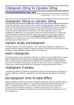
International Journal of Neuropsychopharmacology Advance Access published January 30, 2015 International Journal of Neuropsychopharmacology, 2015, 1–12 doi:10.1093/ijnp/pyu068 Research Article research article Adjunctive Treatment with Asenapine Augments the Escitalopram-Induced Effects on Monoaminergic Outflow and Glutamatergic Neurotransmission in the Medial Prefrontal Cortex of the Rat Carl Björkholm, PhD; Olivia Frånberg, PhD; Anna Malmerfelt, BSc; Monica M. Marcus, PhD; Åsa Konradsson-Geuken, PhD; Björn Schilström, PhD; Kent Jardemark, PhD; Torgny H. Svensson, MD, PhD Department of Physiology and Pharmacology, Section of Neuropsychopharmacology, Karolinska Institutet, Stockholm, Sweden. Correspondence: Torgny H. Svensson, MD, PhD, Professor of Pharmacology, Department of Physiology and Pharmacology, Section of Neuropsychopharmacology, Karolinska Institutet, SE-171 77 Stockholm, Sweden ([email protected]). Abstract Background: Substantial clinical data support the addition of low doses of atypical antipsychotic drugs to selective serotonin reuptake inhibitors (SSRIs) to rapidly enhance the antidepressant effect in treatment-resistant depression. Preclinical studies suggest that this effect is at least partly explained by an increased catecholamine outflow in the medial prefrontal cortex (mPFC). Methods: In the present study we used in vivo microdialysis in freely moving rats and in vitro intracellular recordings of pyramidal cells of the rat mPFC to investigate the effects of adding the novel atypical antipsychotic drug asenapine to the SSRI escitalopram with regards to monoamine outflow in the mPFC and dopamine outflow in nucleus accumbens as well as glutamatergic transmission in the mPFC. Results: The present study shows that addition of low doses (0.05 and 0.1 mg/kg) of asenapine to escitalopram (5 mg/kg) markedly enhances dopamine, noradrenaline, and serotonin release in the rat mPFC as well as dopamine release in the nucleus accumbens. Moreover, this drug combination facilitated both N-methyl-d-Aspartate (NMDA)– and α-amino-3hydroxy-5-methyl-4-isoxazolepropionic acid (AMPA)–induced currents as well as electrically evoked excitatory postsynaptic potentials in pyramidal cells of the rat mPFC. Conclusions: Our results support the notion that the augmentation of SSRIs by atypical antipsychotic drugs in treatmentresistant depression may, at least in part, be related to enhanced catecholamine output in the prefrontal cortex and that asenapine may be clinically used to achieve this end. In particular, the subsequent activation of the D1 receptor may be of importance for the augmented antidepressant effect, as this mechanism facilitated both NMDA and AMPA receptor-mediated transmission in the mPFC. Our novel observation that the drug combination, like ketamine, facilitates glutamatergic transmission in the mPFC may contribute to explain the rapid and potent antidepressant effect obtained when atypical antipsychotic drugs are added to SSRIs. Keywords: depression, atypical antipsychotics, selective serotonin reuptake inhibitor, dopamine-glutamate interactions, prefrontal cortex Received: April 16, 2014; Revised: July 2, 2014; Accepted: July 21, 2014 © The Author 2015. Published by Oxford University Press on behalf of CINP. This is an Open Access article distributed under the terms of the Creative Commons Attribution Non-Commercial License (http://creativecommons.org/licenses/by-nc/4.0/), which permits non-commercial re-use, distribution, and reproduction in any medium, provided the original work is properly cited. For commercial re-use, please contact [email protected] 1 2 | International Journal of Neuropsychopharmacology, 2015 Introduction Major depressive disorder (MDD) is a common psychiatric disorder associated with high disability, mortality, and socioeconomic cost (Ösby et al., 2001; Kessler et al., 2003). Inadequate and delayed response to treatment is a major problem in MDD, and only about 50% of the patients respond adequately to treatment with the most commonly prescribed antidepressant drugs, the selective serotonin reuptake inhibitors (SSRIs) (Rush et al., 2006; Trivedi et al., 2006). Even when the antidepressant treatment effectively improves affective symptoms, residual cognitive symptoms may persist (Boeker et al., 2012) and the degree of cognitive impairment may determine treatment outcome, as for example schizophrenia and bipolar disorder (BPD) (Green, 1996; McCall and Dunn, 2003; Martinez-Aràn et al., 2004). Substantial clinical data support the adjunctive use of low to moderate doses of atypical antipsychotic drugs (APDs) in combination with SSRIs to rapidly enhance the antidepressant effect in treatment-resistant MDD and bipolar depression (see, e.g., Nelson and Papakostas, 2009; Cruz et al., 2010; Tohen et al., 2010). Preclinical studies have demonstrated increased outflow of both dopamine and noradrenaline in the medial prefrontal cortex (mPFC) by the combination of olanzapine or quetiapine with the noradrenaline reuptake inhibitor reboxetine or the SSRI fluoxetine (Zhang et al., 2000; Marcus et al., 2010; Björkholm et al., 2013), which has been suggested to contribute to the potent antidepressant effect observed with such drug combinations (Zhang et al., 2000). Asenapine is a novel atypical APD with a multi-receptor binding profile and a structure similar to that of the antidepressant drug mirtazapine. In binding studies, asenapine exhibits higher affinity for 5-HT2A, 5-HT2c, 5-HT6, 5-HT7, and α2-adrenoceptors than for dopamine D2 receptors (D2Rs) (Shahid et al., 2009), which all may contribute to its clinical efficacy. Moreover, asenapine acts as a partial agonist at 5-HT1Areceptors (5-HT1AR; Ghanbari et al., 2009; Shahid et al., 2009). The receptor binding profile of asenapine differs slightly from that of other APDs, for example, risperidone (which has weaker 5-HT1A and 5-HT6 affinity), olanzapine (weaker 5-HT1A and α2 affinity), quetiapine (weaker 5-HT2C and 5-HT6), and clozapine (stronger M1-4 and α2C affinity; Shahid et al., 2009). In clinical studies, asenapine has been found to be effective in schizophrenia and bipolar mania as well as to reduce depressive symptoms in BPD and schizophrenia (McIntyre et al., 2009; Kane et al., 2010; Szegedi et al., 2011). Asenapine is generally well tolerated with low propensity to induce extrapyramidal or anticholinergic side effects, substantial weight gain, or change in metabolic parameters (Kane et al., 2010). Preclinical studies have shown that asenapine increases dopamine, noradrenaline, and serotonin output in the mPFC, partly via activity at 5-HT2A receptors (5-HT2ARs) and α2adrenoceptors (Frånberg et al., 2008 2009 2012; Huang et al., 2008), which may indicate an inherent antidepressant activity in addition to the amelioration of positive and negative symptoms in schizophrenia as well as cognitive impairment. Furthermore, asenapine has also been shown to facilitate N-methyl-daspartate (NMDA)-induced currents in pyramidal cells of the mPFC via dopamine D1 receptor (D1R)-mediated mechanisms (Jardemark et al., 2010). The functional relevance of this mechanism was shown in behavioral studies where asenapine via D1Rs was found to reverse cognitive impairment induced by NMDA receptor (NMDAR) antagonists (Snigdha et al., 2011). Because the receptor binding profile of asenapine as well as its clinical profile indicates that it may be effective in augmenting antidepressant drugs in depression, we investigated here whether adjunctive treatment with asenapine, at subeffective doses not yielding full antipsychotic-like activity (Frånberg et al., 2008), may enhance the effect of the SSRI escitalopram on extracellular levels of dopamine, noradrenaline, and serotonin in the mPFC as well as dopamine in nucleus accumbens (NAc) using in vivo microdialysis in awake and freely moving animals. Moreover, the effects of asenapine and escitalopram, given alone and in combination, on NMDAR- and AMPA receptor (AMPAR)-mediated glutamatergic transmission as well as on electrically evoked excitatory postsynaptic potentials (EPSPs) were studied using intracellular electrophysiological recordings in vitro in pyramidal cells of the mPFC. Materials and methods Animals Male Wistar rats (Charles River Laboratories, Germany) weighing ~250 g upon arrival were used for the microdialysis experiments. For the in vitro electrophysiological experiments, male Sprague Dawley rats with a mean weight of 190.1 ± 7.8 g were used. The animals were housed under standard laboratory conditions (21.0 ± 0.4°C; relative humidity of 55–65%). Food (R34, Ewos, Södertälje, Sweden) and tap water were available ad libitum. The animals were kept on a 12/12 h light/dark cycle (lights on at 6:00 am) and were acclimatized for at least 5 days before the experiments. Experiments were performed between 8:00 am and 6:00 pm. All experiments were approved by the local Animal Ethics Committee, Stockholm North, and the Karolinska Institutet, Sweden. In Vivo Microdialysis Rats were anesthetized with Hypnorm (0.315 mg/mL fentanyl citrate and 10 mg/mL fluanisone; Janssen-Cilag, UK) and Dormicum (5 mg/mL midazolam; Roche AB, Sweden) diluted in distilled water (1:1:2; 5 mL/kg, intraperitoneal injection) and sterotactically implanted with concentric dialysis probes in the mPFC (at a 12° angle) or NAc (anteroposterior +2.5, +1.6; mediolateral −1.4, −1.4; dorsoventral −6.0, −8.2), respectively, relative to bregma and dural surface (in mm) (Paxinos and Watson,1998). Dialysis probes were manufactured in-house with a semipermeable membrane (Filtral AN69, Hospal Industrie, France) with an active surface length of 5.5 mm (mPFC) or 2 mm (NAc). Dialysis experiments were conducted approximately 48 hours after surgery in awake and freely moving rats. The dialysis probe was perfused with a physiological perfusion solution (in mM: 147 NaCl, 3.0 KCl, 1.3 CaCl2, 1.0 MgCl2, 1.0 Na2HPO4, and 0.2 NaH2PO4, pH 7.4) at a rate of 2.5 μL/min. Dialysate samples were collected during 30-minute (mPFC) or 15-minute (NAc) intervals and automatically injected into a high performance liquid chromatography system and quantified by electrochemical detection (ESA Bioscience) with a detection limit of approximately 0.08 nM. The injector (Valco Instruments) was directed by Totalchrom WS 6.3 software (Perkin Elmer). Separation of neurotransmitters and metabolites was achieved by reversed-phase liquid chromatography on a C-18 column (Kinetex 150 × 4.6 mm, 2.6 µm, Phenomenex). The mobile phase used for separation of catecholamines (NAc or mPFC) or serotonin alone (mPFC) consisted of a 55-mM sodium acetate buffer, pH 4.0, with 12% or 18% methanol and 0.55 or 0.81 mM octanesulfonic acid, respectively. Samples were quantified by sequential oxidation and reduction in a high sensitive analytical cell (model 5011; ESA Bioscience) that was controlled by a potentiostat (Coulochem II model 5200; ESA Björkholm et al. | 3 Bioscience) with applied potentials of 400 and −200 mV for detection of metabolites and monoamines, respectively. Injection of drug was performed when a stable outflow (<10% variation) of the neurotransmitters was established. Baseline (100%) was calculated as the average of the last 2 (mPFC) or 4 (NAc) preinjection values. At the end of the experiment, tetrodotoxin (3 µM) was infused into the probe to further verify peak identification. Placement of the probes was verified under microscope in Nissl stained sections. Preparation of Brain Slices and Electrophysiological Experiments in Vitro The preparation of brain slices and electrophysiological experiments in vitro were performed as previously described (Jardemark et al., 2012). In short, rats were anesthetized (halothane; Astra AB, Sweden), and the brain was quickly removed and cooled in ice-cold Ringer’s solution (in mM: 126 NaCl, 18 NaHCO3, 10 d-glucose, 2.5 KCl, 2.4 CaCl2, 1.3 MgCl2, 1.2 NaH2PO4, pH 7.4) aerated with 95% O2:5% CO2. The brains were sectioned coronally to 450-µm slices using a Vibroslice (Campden model MA752, World Precision Instruments). The slices were kept in aerated Ringer’s solution at room temperature for >1 hour to allow for recovery. A slice containing the mPFC was fixed in the recording chamber between 2 nylon nets and was continually perfused with aerated Ringer’s solution (30°C; flow rate of 1–2 mL/min). Recording electrodes were pulled from borosilicate glass capillaries (i.d. 0.58 mm; Clark Electromedical Instruments) and filled with 2 M potassium acetate (resistance: 55–140 MΩ) and used for recording with an Axoclamp 2B amplifier (Molecular Devices). Penetration of layer V or VI cells with sharp electrodes was performed blindly. Single electrode voltage-clamp (holding potential: – 60 mV) was performed in the discontinuous mode (sampling rate of 5–6.2 kHz). Voltage-clamp recordings were acquired using digital/analogue sampling and acquisition software (Clampex version 9.2 Molecular Devices). Tetrodotoxin (0.5 µM, to block action potentials), bicuculline (5 µM, to block the GABAA receptor), and glycine (1 µM, to enhance the NMDAinduced currents) were routinely included in the Ringer’s solution during the recordings. All drugs, including AMPA (2.5 µM) and NMDA (10–15 µM), were diluted in Ringer’s solution and applied via bath perfusion. NMDA and AMPA applications were performed at 5 and 30 minutes of drug treatment. In experiments in which the dopamine D1/5 receptor antagonist SCH23390 was included, the slice was pretreated with SCH23390 (1 µM) for 5 minutes before the administration of asenapine and escitalopram commenced. To calculate the effects of the drugs or drug combinations on the prefrontal glutamatergic transmission, the recorded amplitude of AMPA- or NMDA-induced current after administration of a drug or a drug combination was divided by the amplitude of the control AMPA- or NMDA-induced current. To elicit EPSPs in pyramidal cells, a stimulation electrode consisting of 2 stainless steel tips was placed medially in the forceps minor (white matter) close to the recording electrode, and a square pulse (0.3-millisecond duration, 11–31 V) was passed between the tips of the stimulation electrode. The evoked change in membrane potential (ie, the EPSP) of a layer V or VI pyramidal cell of the mPFC was then recorded in the current clamp mode. To evaluate the effect of the drugs or drug combination on the electrically evoked EPSPs, a submaximal potential was chosen, and the evoked response was recorded before and after 5, 15, 25, and 35 minutes of drug application. Bicuculline (2 µM) was routinely included in the perfusion solution (Ringer’s) to inhibit GABAA receptor-mediated inhibitory postsynaptic potentials. The effect of the drugs and the drug combination on electrically evoked EPSPs was evaluated both qualitatively for their ability to facilitate the induction of action potentials as well as their effect on the total area of the EPSP. Drugs Asenapine was generously obtained from Schering-Plough, UK and Merck Sharp & Dohme Corp (MSD) and escitalopram was generously obtained from Lundbeck A/S, Denmark. Tetrodotoxin, bicuculline, and (RS)-AMPA were purchased from Ascent Scientific, Bristol, UK, and glycine and NMDA were purchased from Sigma-Aldrich, St. Louis, MO. For the in vivo microdialysis experiments, asenapine and escitalopram were dissolved in physiological saline (0.9% NaCl) and subcutaneously (s.c.) injected at a volume of 1.0 mL/kg. For the in vitro electrophysiological experiments, stock solutions of asenapine (dissolved in purified water) and escitalopram (dissolved in dimethyl sulfoxine and diluted to stock concentration with purified water) were prepared and then diluted in Ringer’s solution to reach the final drug concentration. Statistics The effect of the drug treatments on the mean transmitter output during intervals of 60 to 240 min for mPFC and 45 to 240 min for NAc was statistically analyzed using 1-way ANOVA, followed by planned comparisons of Least Squares means. In vitro electrophysiological data of the NMDA and AMPA applications were analyzed using paired Student’s t test and, for multiple comparisons, 1-way ANOVA followed by the Newman-Keul’s multiple comparison test. The drug effect on the total EPSP area was analyzed using repeated-measures 2-way ANOVA followed by Fisher’s Least Significant Difference test. Statistical evaluation of microdialysis data and the EPSP area was performed using Statistica version 10 software (StatSoft, Tulsa, OK), and the effect on NMDA- and AMPA-induced currents was analyzed using Prism 5.02 (Graphpad Software Inc.). In all statistical assessments, P < .05 was considered significant. Results Asenapine 0.05 mg/kg Potentiates the Effect of Escitalopram on Dopamine Output in the mPFC and NAc The basal dopamine, noradrenaline, and serotonin output in the mPFC were 0.54 ± 0.06 (mean ± SEM, n = 24), 1.17 ± 0.14 (n = 23), and 0.56 ± 0.05 (n = 26) fmol/min, respectively, and for dopamine in NAc 3.23 ± 0.33 (n = 25) fmol/min. There were no statistically significant differences between mean baseline concentrations of each neurotransmitter between the different treatment groups. To assess the effects of the drugs alone and in combination, we analyzed the mean transmitter output after the second injection, that is, 60 to 240 minutes for mPFC and 45 to 240 minutes for NAc. Analysis of the dopamine output showed an overall effect in the mPFC (1-way ANOVA; F3,20 = 16.32, P < .001) (Figure 1b). Asenapine, alone and in combination with escitalopram, increased dopamine output in the mPFC compared with control (P < .01-.001), whereas escitalopram had no effect. The combination of escitalopram and asenapine significantly increased dopamine output compared with either drug alone (P < .05-.001). 4 | International Journal of Neuropsychopharmacology, 2015 Figure 1. The effects of escitalopram (5 mg/kg, subcutaneously [s.c.]), asenapine (0.05 mg/kg, s.c.) alone, and the combination of escitalopram and asenapine on dopamine (a-b), noradrenaline (c-d), and serotonin (e-f) output in the medial prefrontal cortex (mPFC) and dopamine (g-h) output in nucleus accumbens (NAc). Left panels show the effects over time, whereas right panels show the effects calculated as mean transmitter output during 60 to 240 minutes for the mPFC data and 45 to 240 minutes for NAc data, ie, after the second injection. Arrows indicate injections of escitalopram/ saline and asenapine/saline, respectively. The dotted line represents baseline (100%). The results are presented as mean ± SEM. The number in each bar indicates group size. *P < .05, **P < .01, *** P < .001 vs. control, ie, saline + saline. #P < .05, ###P < .001 as indicated in the figures. Björkholm et al. | 5 Analysis of noradrenaline output in the mPFC showed an overall effect (F3,19 = 4.35, P < .05) (Figure 1d). Asenapine treatment enhanced noradrenaline outflow in the mPFC compared with control (P < .05); this effect was, however, not further enhanced when asenapine was combined with escitalopram. Escitalopram treatment alone did not affect noradrenaline outflow. Analysis of serotonin output in the mPFC showed an overall effect (F3,22 = 16.08, P < .001) (Figure 1f). Escitalopram given alone and in combination with asenapine significantly increased serotonin output in the mPFC compared with control (P < .001). However, asenapine treatment alone did not affect serotonin outflow, nor did addition of asenapine further enhance the effect of escitalopram on serotonin output. Analysis of dopamine output in NAc showed an overall effect (F3,21 = 6.27, P < .001) (Figure 1h). Escitalopram, asenapine, and the combination of escitalopram and asenapine significantly increased dopamine output compared with control (P < .05- P < .001). However, there was no further enhancement by the combination of escitalopram and asenapine compared with either drug alone. The effect of escitalopram (5 mg/kg) on accumbal dopamine output has previously been reported (Marcus et al., 2012); however, it is now analyzed within the same time interval as our novel data, that is, between 45 and 240 minutes. Asenapine 0.1 mg/kg Potentiates the Effects of Escitalopram on Serotonin and Noradrenaline Output in the mPFC The effect of a higher dose of asenapine (0.1 mg/kg) alone and in combination with escitalopram (5 mg/kg) was also investigated. In this group, the mean ± SEM basal outflow of dopamine, noradrenaline, and serotonin in mPFC were 0.47 ± 0.05 (n = 27), 0.91 ± 0.10 (n = 26), and 0.39 ± 0.03 (n = 28) fmol/min, respectively, and for dopamine in NAc 2.95 ± 0.31 (n = 29) fmol/min. There were no statistically significant differences in the mean baseline concentrations of each neurotransmitter between different treatment groups. The effects of asenapine (0.1 mg/kg) on dopamine and noradrenaline output have also previously been reported (Frånberg et al., 2008 2009); however, the data are now analyzed within the same time interval as our novel data. Analysis of the dopamine output showed an overall effect in the mPFC (60–240 minutes; 1-way ANOVA; F3,23 = 22.15, P < .001) (Figure 2b). Asenapine treatment (0.1 mg/kg) significantly increased dopamine output compared with control (P < .001), whereas escitalopram administration did not. In contrast with the effect of the lower dose of asenapine (0.05 mg/ kg), the effect of addition of the higher dose of asenapine to escitalopram on dopamine output did not reach statistical significance. Analysis of noradrenaline output showed an overall effect in the mPFC (F3,22 = 20.88, P < .001) (Figure 2d). Although there was no effect of either asenapine or escitalopram on noradrenaline output when given separately, the combination of escitalopram and asenapine significantly increased noradrenaline outflow when compared either with vehicle or with each drug given alone (P < .001). Analysis of serotonin output showed an overall effect in the mPFC (F3,24 = 24.73, P < .001) (Figure 2f). Escitalopram administered alone increased the cortical serotonin concentrations, an effect that was further augmented by adjunctive asenapine, and the combined effect was significantly higher than the effect of either drug given alone (P < .01-.001). In NAc, the overall effect for dopamine output was statistically significant (45–240 minutes; F3,25 = 11.72; P < .001) (Figure 2h). Compared with control, escitalopram, asenapine, and the combination of escitalopram and asenapine all significantly increased dopamine output (P < .05-0.001). Addition of asenapine significantly increased the escitalopram-induced dopamine outflow (P < .01), but the effect was not larger than that of asenapine given alone. Electrophysiological Characterizations of Pyramidal Cells of the mPFC The electrophysiological criteria for distinguishing presumed pyramidal from nonpyramidal cells were previously described (Arvanov et al., 1997). In short, the presumed pyramidal neurons of the mPFC have a relatively long spike duration (1–3 milliseconds at half-maximum spike amplitude) and, in addition, show a pronounced spike frequency adaptation in response to constant-current depolarization pulses. In the present study, the presumed pyramidal cells of layers V and VI of the rat mPFC exhibited a mean membrane potential of −79.8 ± 1 mV (n = 60), action potential amplitude of 108.0 ± 2.8 mV (n = 60), a spike half width of 4.0 ± 0.2 milliseconds (n = 30), and an after-hyperpolarization potential of 4.4 ± 0.4 mV (n = 60) in Ringer’s solution. These results are similar to previously published results (Arvanov et al., 1997; Konradsson et al., 2006; Björkholm et al., 2013). Add-on Asenapine to Escitalopram Facilitates NMDA-Induced Currents in Pyramidal Cells of the Rat mPFC via D1 Receptor Activation Asenapine produces a biphasic concentration response curve with a maximum response at 5 nM (Frånberg et al., 2008). Also, escitalopram has been found to potentiate NMDA-induced currents in pyramidal cells of the mPFC at both 5 and 100 nM (Schilström et al., 2011). A combination of submaximal concentrations of asenapine (1 nM) and escitalopram (3 nM) significantly facilitated the NMDA-induced currents after 30 minutes (182.0 ± 17.5 %, n = 5, paired t test P < .05) compared with control, as well as each drug given alone (1-way ANOVA [F3,17 = 8.1, P < .01], Newman-Keuls multiple comparison test P < .01) (Figure 3f). The facilitating effect of the combination of asenapine and escitalopram was blocked by the addition of the D1R-antagonist SCH23390 (1 µM; 95.3 ± 20.6%, n = 4) (Figure 3f). Add-on Asenapine to Escitalopram Facilitates AMPAInduced Currents in Pyramidal Cells of the rat mPFC via D1 Receptor Activation Neither asenapine nor escitalopram affected the AMPA-induced currents at any concentration tested (Figure 4a-b). However, a combination of low concentrations of asenapine (1 nM) and escitalopram (3 nM) significantly potentiated AMPA-induced currents at both 5 minutes (173.6 ± 15.9%, n = 5, paired t test, P < .01) (Figure 5e) and 30 minutes (153.0 ± 7.0%, n = 5, P < .05) (Figure 5f). Between-groups comparison showed that the combination of asenapine and escitalopram facilitated the AMPAinduced current at both 5 minutes (1-way ANOVA [F3,19 = 11.2, P < .001], Newman-Keuls multiple comparison test, P < .001) (Figure 5e) and 30 minutes (F3,17 = 9.0, P < .01, P < .05-.01) (Figure 5f) compared with either drug given alone. Interestingly, also the potentiating effect of asenapine combined with escitalopram on the AMPA-induced currents was blocked by SCH23390 (1 µM; Figure 5e, 88.0 ± 13.1%, n = 4; Figure 5f, 84.3 ± 14.0%, n = 4), although SCH23390 (1 µM) treatment did not significantly affect 6 | International Journal of Neuropsychopharmacology, 2015 Figure 2. The effects of escitalopram (5 mg/kg, subcutaneously [s.c.]), asenapine (0.1 mg/kg, s.c.) alone, and the combination of escitalopram and asenapine dopamine (a-b), noradrenaline (c-d), and serotonin (e-f) output in the medial prefrontal cortex (mPFC) and dopamine output in nucleus accumbens (NAc) (g-h). Left panels show the effects over time, whereas right panels show the effects calculated as mean transmitter output during 60 to 240 minutes for the mPFC and 45 to 240 minutes for NAc, ie, after the second injection. Arrows indicate injections of escitalopram/saline and asenapine/saline, respectively. The dotted line represents baseline (100%). The results are presented as mean ± SEM. The number in each bar indicates group size. *P < .05, **P < .01, ***P < .001 vs. control, ie, saline + saline. ##P < .01, ###P < .001 as indicated in the figures. Note: The data for saline + saline and escitalopram (5 mg/kg) are the same as in Figure 1. Björkholm et al. | 7 Figure 3. Effects on N-methyl-d-aspartate (NMDA)-induced currents in pyramidal cells of the rat medial prefrontal cortex (mPFC). Representative electrophysiological traces showing the effect of NMDA application before (grey trace) and after application (black trace) of 3 nM escitalopram (a), 1 nM asenapine (b), 3 nM escitalopram + 1 nM asenapine (c), and 3 nM escitalopram + 1 nM asenapine + 1 µM SCH23390 (d). The grey and black horizontal bars indicate time of NMDA application for control and test trace, respectively. Data is summarized in bar charts 5 minutes (e) and 30 minutes (f) after drug application. The results are presented in percent as mean ± SEM. *P < .05 vs. control response. ##P < .01 as indicated in the figure (n = 4–6). The holding potential was −60 mV. b. 200 Escitalopram Asenapine 150 100 50 AMPA-Induced Currents (% of control) AMPA-Induced Currents (% of control) a. 200 Escitalopram Asenapine 150 100 50 0 0 1 10 Concentration [nM] 100 1 10 Concentration [nM] 100 Figure 4. Concentration-response curves for both asenapine and escitalopram of α-amino-3-hydroxy-5-methyl-4-isoxazolepropionic acid (AMPA)-induced response at (a) 5 and (b) 30 minutes after drug application. Each point represents the mean ± SEM percent of control (n = 3–7). The holding potential was −60 mV. AMPA-induced currents when administered alone (5 minutes, 119.0 ± 23.7%, n = 5; 30 minutes, 86.75 ± 7.6%, n = 4). One experiment in the asenapine 1 nM group was detected as an outlier according to the Grubbs test and therefore excluded. Add-on Asenapine to Escitalopram Potentiates Electrically Evoked EPSPs in Pyramidal Cells of the Rat mPFC Asenapine (1 nM) treatment facilitated the electrically evoked EPSP and induced action potentials in 1 of 4 cells tested, whereas escitalopram (3 nM) had no effect in any cell tested (for representative traces, see Figure 6a-b). However, the addition of asenapine (1 nM) to escitalopram (3 nM) facilitated the evoked EPSPs and induced bursts of action potentials overriding the EPSP in all 4 cells tested (Figure 6c). The effect of the combination of asenapine and escitalopram gradually increased over time, and the time to onset of the first spike varied between cells from 5 to 35 min. The area of the evoked EPSP was quantified using Clampfit 9.2 with the baseline set manually. Due to the large variation of the EPSP area (expressed as mV*ms), the data were log transformed before statistical analysis. There was no difference in the control EPSP area between the different groups, that is, the EPSP area assessed before drug treatment (Figure 6d). However, the combination of asenapine and escitalopram significantly enhanced the EPSP area compared with both escitalopram- and 8 | International Journal of Neuropsychopharmacology, 2015 Figure 5. Effects on α-amino-3-hydroxy-5-methyl-4-isoxazolepropionic acid (AMPA)-induced currents in pyramidal cells of the rat medial prefrontal cortex (mPFC). Representative electrophysiological traces showing the effect of AMPA application before (grey trace) and after application (black trace) of 3 nM escitalopram (a), 1 nM asenapine (b), 3 nM escitalopram + 1 nM asenapine (c), and 3 nM escitalopram + 1 nM asenapine + 1 µM SCH23390 (d). The grey and black horizontal bars indicate time of AMPA application for control and test trace, respectively. Data is summarized in bar charts 5 (e) and 30 (f) minutes after drug application. The results are presented in percent as mean ± SEM. *P < .05, **P < .01 vs. control response. #P < .05, ##P < .01, ###P < .001 as indicated in figure (n = 4–7). The holding potential was −60 mV. b. Asenapine 1 nM c. Escitalopram 3 nM+ Asenapine 1nM ¤¤¤ d. 10 mV 0.1 s Mean EPSP Area (log10 mV*ms) a. Escitalopram 3 nM ## *** 4 ¤¤¤ ¤¤¤ ## ## *** *** ** 3 2 Control 5 min 15 min 25 min 35 min Escitalopram 3 nM Asenapine 1 nM Asenapine 1nM + Escitalopram 3 nM Figure 6. Effects of asenapine, escitalopram, and the combination of asenapine and escitalopram on electrically evoked excitatory postsynaptic potentials (EPSPs) in pyramidal cells of the rat medial prefrontal cortex (mPFC). Representative electrophysiological traces showing the EPSPs before (grey) and after (black) application of 3 nM escitalopram (a), 1 nM asenapine (b), and 3 nM escitalopram + 1 nM asenapine (c). Arrows indicate time of electrical stimulation. The logarithm of the mean EPSP area (log10 mV*ms) is summarized in d. The results are presented as mean ± SEM (n = 4). The combination of asenapine and escitalopram significantly enhanced the EPSP area compared with both escitalopram (**P < .01, *** P < .001) and asenapine (##P < .01) as well as its own control EPSP area (ie, the EPSP area before drug application; ¤¤¤ P < .001). asenapine-treated groups as well as compared with its own control response (Figure 6d; 2-way repeated-measures ANOVA [F8,36 = 3.14, P < 0.01]; Fisher’s Least Significant Difference test P < 0.01-.001). Discussion The present study demonstrates that low doses of the atypical APD asenapine in combination with escitalopram generate a marked enhancement of extracellular dopamine, noradrenaline, and serotonin outflow in the mPFC as well as increases dopamine outflow in NAc. Furthermore, the combination of low, clinically relevant concentrations of asenapine and escitalopram facilitated glutamatergic transmission in the rat mPFC. Interestingly, the observed effect on AMPAR-mediated transmission was not attainable by each drug given alone, even at higher concentrations, indicating that the two drugs work synergistically to generate this effect. The effects on both NMDAR- and AMPAR-mediated transmission were, at least in part, mediated via activation of the dopamine D1R, as the facilitatory effects of the drug combination on both NMDAR- and AMPAR-mediated transmission were antagonized by a D1R antagonist. Importantly, the combination of asenapine and escitalopram also facilitated electrically evoked EPSPs and induced bursts of action potentials in pyramidal cells, further supporting a physiologically relevant facilitation of prefrontal glutamatergic transmission. Björkholm et al. | 9 Our results generally support the notion that the augmentation of the antidepressant effect of SSRIs by atypical APDs in treatment-resistant MDD may be related to enhanced catecholamine output in prefrontal cortical areas (c.f. Introduction). The enhanced prefrontal catecholamine outflow induced by addition of asenapine to escitalopram with ensuing activation of NMDAR-mediated transmission, following D1R activation, may be of clinical significance, since the degree of cognitive impairment is a critical determinant of treatment outcome in MDD and BPD as well as in schizophrenia (c.f. Introduction). Given that the addition of asenapine to escitalopram significantly facilitated NMDAR-mediated transmission via activation of D1R, the present results propose that add-on low doses of asenapine to SSRIs such as escitalopram may be used clinically to ameliorate certain aspects of cognitive impairment, since the D1R and NMDAR interaction is a mechanism highly implicated in cognitive functioning (Castner and Williams, 2007). Moreover, recent clinical and preclinical studies have shown that drugs mediating their effect via the co-agonist site of the NMDAR may possess an antidepressant effect (Malkesman et al., 2012; Huang et al., 2013), indicating that in addition to an effect on cognition, activation of NMDARs may contribute to an antidepressant effect per se. The effect of addition of asenapine to escitalopram on mono amine release was dose dependent, since addition of the lower dose of asenapine (0.05 mg/kg) to escitalopram induced a synergistic increase in dopamine release, whereas the higher dose (0.1 mg/kg) potentiated serotonin and noradrenaline output but did not further enhance dopamine output compared with asenapine given alone. The higher dose may block postsynaptic receptors to a higher extent than the lower dose and potentially reduce feed-back inhibition. The mechanisms by which the addition of asenapine to escitalopram produced these effects on monoaminergic transmission may involve action on several receptors. Previous studies have shown that SSRI-induced serotonin release acting on 5-HT2Rs on GABAergic interneurons inhibits locus coeruleus cell firing activity, and simultaneous treatment with an SSRI and a 5-HT2AR antagonist enhances both serotonergic and noradrenergic neuronal activity (Szabo and Blier, 2001 2002). Indeed, 5-HT2AR antagonists have been found to potentiate the antidepressant-like effect of an SSRI (Marek et al., 2005). Moreover, 5-HT2AR blockage appears to augment D2R antagonist-induced dopamine release via a 5-HT1AR-mediated mechanism (Ichikawa et al., 2001), and the 5-HT1AR agonist 8-OH-DPAT increases burst firing and firing rate of prefrontally projecting neurons in the ventral tegmental area as well as preferentially increase dopamine output in the mPFC (Arborelius et al., 1993a 1993b). A more recent study found that activation of postsynaptic 5-HT1ARs in the PFC enhanced dopamine cell firing in the ventral tegmental area and increased dopamine release in the same brain region (Diaz-Mataix et al., 2005). It seems possible that at the higher dose of asenapine (0.1 mg/kg), the postsynaptic stimulation of 5-HT1ARs in the PFC is governed by the intrinsic activity of asenapine at 5-HT1ARs, which may thus attenuate the stimulatory effect on dopamine cell firing of enhanced serotonin in the PFC induced by serotonin transporter blockade blockade and consequently attenuate dopamine release in the mPFC, which would contribute to explaining why dopamine release is not further enhanced by the higher dose of asenapine. Adjunctive treatment with an α2-adrenoceptor antagonist increases firing activity of noradrenergic neurons originating in the locus coeruleus following administration of antidepressant drugs (Svensson and Usdin, 1978) and potentiates cortical monoamine release induced by SSRIs (Gobert et al., 1997). Previous studies show that adjunctive treatment with α2-adrenoceptor antagonists may potentiate the effect of antidepressant drugs and generate a more rapid onset of action (Sanacora et al., 2004; Dhir and Kulkarni, 2007; Yanpallewar et al., 2010). Consequently, the antagonistic action of asenapine at α2adrenoceptors, 5-HT2ARs and partial agonistic effect at 5-HT1ARs may all provide mechanisms contributing to an enhanced monoamine release and allow for substantial improvement of the efficacy of SSRIs. In addition, as dopamine release in NAc is involved in reward-related behaviors (Dunlop and Nemeroff, 2007), enhanced mesolimbic dopamine release induced by asenapine and escitalopram may contribute to ameliorate anhedonia in MDD. In the present study, a combination of asenapine and escitalopram facilitated AMPA-induced currents in pyramidal cells of the mPFC. The observation that this drug combination, like ketamine and scopolamine, markedly facilitates glutamatergic transmission, particularly AMPAR-mediated transmission in the rat mPFC (Maeng et al., 2008; Li et al., 2010; Voleti et al., 2013), may thus contribute to explaining the rapid onset of the enhanced antidepressant effect obtained by adjunctive treatment with atypical APDs to SSRIs (c.f. Introduction). Given the complex interaction between the monoaminergic systems and the glutamatergic system in the mPFC, where, for example, dopamine and serotonin can either increase or decrease glutamatergic transmission (for reviews, see, eg, Puig and Gulledge, 2011; Tritsch and Sabatini, 2012) and the complex pharmacology of the asenapine and escitalopram combination, the precise mechanisms by which the combination, but not either drug when given separately, facilitates AMPAR-mediated transmission remains to be fully understood. Previous electrophysiological studies did not reveal any effect of D1R activation on AMPAR-mediated transmission in the mPFC (Seamans et al., 2001; Tseng and O’Donnell, 2004), whereas D2R activation seems to decrease AMPAR-mediated transmission (Tseng and O’Donnell, 2004). In cell cultures, however, D1R activation has been found to increase the surface expression of AMPARs on prefrontal pyramidal cells, whereas D2R had the opposite effect, although D1R activation by itself may not be sufficient to induce translocation of AMPARs to synaptic sites (Sun et al., 2005). Thus, the facilitating effect of the asenapine and escitalopram combination may, at least in part, be explained by increased dopamine output in the mPFC induced by the combination of asenapine and escitalopram in combination with a concomitant blockade of postsynaptic D2Rs by asenapine, resulting in a preferential activation of D1Rs enhancing the surface expression of AMPARs. However, it is likely that D1R activation is not the sole mechanism involved, since neither asenapine nor escitalopram facilitated AMPAR-mediated transmission when given alone, even at concentrations where these drugs have previously been found to increase NMDAR-mediated transmission via this mechanism (Jardemark et al., 2010, Schilström et al., 2011). Taken together, we propose that D1R activation by asenapine and escitalopram may well be necessary, but probably not entirely sufficient, to facilitate AMPAR-mediated transmission. In addition to the effects of D1R activation, serotonin has been found to induce glutamate release (Aghajanian and Marek, 1997). Moreover, serotonin may activate postsynaptically located 5-HT1ARs (Cai et al., 2002) or 5-HT1B receptors (Cai et al., 2013) which may contribute to modulate AMPAR-mediated transmission, thus suggesting that the enhanced AMPAR-mediated transmission may well be mediated by a combination of these mechanisms. The facilitatory effect of the combination of asenapine and escitalopram on the EPSPs is in analogy with the corresponding effect previously observed with the clozapine, which was found to be both NMDAR- and D1R-mediated (Chen and Yang, 2002). The latency to the onset of 10 | International Journal of Neuropsychopharmacology, 2015 the spikes varied, indicating that the bursts of action potentials may be the result of polysynaptic input from lateral interconnected pyramidal cells (Chen and Yang, 2002). This polysynaptic input could also contribute to the increased EPSP area by inducing several EPSPs with different latencies, superimposed on the initial EPSP. Clinically, the increased pyramidal cell excitability in the mPFC may thus probably serve to ameliorate both cognitive and depressive symptoms. In conclusion, our results demonstrate that a low dose of asenapine in combination with escitalopram can produce a marked activation of prefrontal monoamine output with an ensuing facilitation of glutamatergic transmission in the mPFC, neurobiological effects that may generate both a procognitive effect and an enhanced antidepressant activity in mood disorders such as MDD and BPD as well as schizophrenia. Our data also suggest that activation of the dopamine D1R may be crucial for this effect. Importantly, the results propose that adding asenapine to SSRIs such as escitalopram may produce a faster onset of action of the antidepressant effect compared with an SSRI alone, a low risk of conversion to mania in BPD, and probably with only modest weight gain and low EPS liability in mood disorders. Funding This work was supported by the Swedish Research Council (grant no. 4747), Karolinska Institutet, Torsten Soderbergs Stiftelse, Swedish Brain Foundation and Ahlen-stiftelsen. Acknowledgments We wish to thank Schering-Plough and Merck Sharp & Dohme Corp (MSD) for generous supply of asenapine as well as Lundbeck for escitalopram. Interest Statement Torgny H. Svensson has recived grants/support from AstraZeneca, Schering-Plough, Merck Sharp & Dohme, Lundbeck, and Astellas and has served as a consultant and on the advisory boards of AstraZeneca, Janssen, Lundbeck, Otsuka, Merck Sharp and Dohme, Organon, Pfizer and Carnegie Health Care Funds (Sweden). References Aghajanian GK, Marek GJ (1997) Serotonin induces excitatory postsynaptic potentials in apical dendrites of neocortical pyramidal cells. Neuropharmacology 36:589–599. Arborelius L, Chergui K, Murase S, Nomikos GG, Hook BB, Chouvet G, Hacksell U, Svensson TH (1993a) The 5-HT1A receptor selective ligands, (R)-8-OH-DPAT and (S)-UH-301, differentially affect the activity of midbrain dopamine neurons. Naunyn Schmiedebergs Arch Pharmacol 347:353–362. Arborelius L, Nomikos GG, Hacksell U, Svensson TH (1993b) (R)8-OH-DPAT preferentially increases dopamine release in rat medial prefrontal cortex. Acta Physiol Scand 148:465–466. Arvanov VL, Liang X, Schwartz J, Grossman S, Wang RY (1997) Clozapine and haloperidol modulate N-methyl-D-aspartateand non-N-methyl-D-aspartate receptor-mediated neurotransmission in rat prefrontal cortical neurons in vitro. J Pharmacol Exp Ther 283:226–234. Björkholm C, Jardemark K, Marcus MM, Malmerfelt A, Nyberg S, Schilström B, Svensson TH (2013) Role of concomitant inhibition of the norepinephrine transporter for the antipsychotic effect of quetiapine. Eur Neuropsychopharmacol 23:709–720. Boeker H, Schulze J, Richter A, Nikisch G, Schuepbach D, Grimm S (2012) Sustained cognitive impairments after clinical recovery of severe depression. J Nerv Ment Dis 200:773–776. Cai X, Gu Z, Zhong P, Ren Y, Yan Z (2002) Serotonin 5-HT1A receptors regulate AMPA receptor channels through inhibiting Ca2+/calmodulin-dependent kinase II in prefrontal cortical pyramidal neurons. J Biol Chem 277:36553–36562. Cai X, Kallarackal AJ, Kvarta MD, Goluskin S, Gaylor K, Bailey AM, Lee HK, Huganir RL, Thompson SM (2013) Local potentiation of excitatory synapses by serotonin and its alteration in rodent models of depression. Nature neuroscience 16:464–472. Castner SA, Williams GV (2007) Tuning the engine of cognition: a focus on NMDA/D1 receptor interactions in prefrontal cortex. Brain Cogn 63:94–122. Chen L, Yang CR (2002) Interaction of dopamine D1 and NMDA receptors mediates acute clozapine potentiation of glutamate EPSPs in rat prefrontal cortex. J Neurophysiol 87:2324–2336. Cruz N, Sanchez-Moreno J, Torres F, Goikolea JM, Valentí M, Vieta E (2010) Efficacy of modern antipsychotics in placebocontrolled trials in bipolar depression: a meta-analysis. Int J Neuropsychopharmacol 13:5–14. Dhir A, Kulkarni SK (2007) Effect of addition of yohimbine (alpha-2-receptor antagonist) to the antidepressant activity of fluoxetine or venlafaxine in the mouse forced swim test. Pharmacology 80:239–243. Díaz-Mataix L1, Scorza MC, Bortolozzi A, Toth M, Celada P, Artigas F (2005) Involvement of 5-HT1A receptors in prefrontal cortex in the modulation of dopaminergic activity: role in atypical antipsychotic action. J Neurosci. 25:10831–10843. Dunlop BW, Nemeroff CB (2007) The role of dopamine in the pathophysiology of depression. Arch Gen Psychiatry 64:327– 337. Frånberg O, Wiker C, Marcus MM, Konradsson A, Jardemark K, Schilström B, Shahid M, Wong EH, Svensson TH (2008) Asenapine, a novel psychopharmacologic agent: preclinical evidence for clinical effects in schizophrenia. Psychopharmacology (Berl) 196:417–429. Frånberg O, Marcus MM, Ivanov V, Schilström B, Shahid M, Svensson TH (2009) Asenapine elevates cortical dopamine, noradrenaline and serotonin release. Evidence for activation of cortical and subcortical dopamine systems by different mechanisms. Psychopharmacology (Berl) 204:251–264. Frånberg O, Marcus MM, Svensson TH (2012) Involvement of 5-HT(2A) receptor and α(2) -adrenoceptor blockade in the asenapine-induced elevation of prefrontal cortical monoamine outflow. Synapse 66:650–660. Ghanbari R, El Mansari M, Shahid M, Blier P (2009) Electrophysiological characterization of the effects of asenapine at 5-HT(1A), 5-HT(2A), alpha(2)-adrenergic and D(2) receptors in the rat brain. Eur Neuropsychopharmacol. 19:177–187. Gobert A, Rivet JM, Cistarelli L, Melon C, Millan MJ (1997) Alpha2adrenergic receptor blockade markedly potentiates duloxetine- and fluoxetine-induced increases in noradrenaline, dopamine, and serotonin levels in the frontal cortex of freely moving rats. J Neurochem 69:2616–2619. Green MF (1996) What are the functional consequences of neurocognitive deficits in schizophrenia? Am J Psychiatry 153:321–330. Huang M, Li Z, Dai J, Shahid M, Wong EH, Meltzer HY (2008) Asenapine increases dopamine, norepinephrine, and acetylcholine efflux in the rat medial prefrontal cortex and hippocampus. Neuropsychopharmacology 33:2934–2945. Huang CC, Wei IH, Huang CL, Chen KT, Tsai MH, Tsai P, Tun R, Huang KH, Chang YC, Lane HY, Tsai GE (2013) Inhibition of Björkholm et al. | 11 glycine transporter-I as a novel mechanism for the treatment of depression. Biol Psychiatry. 74:734–741. Ichikawa J, Ishii H, Bonaccorso S, Fowler WL, O’Laughlin IA, Meltzer HY (2001) 5-HT(2A) and D(2) receptor blockade increases cortical DA release via 5-HT(1A) receptor activation: a possible mechanism of atypical antipsychotic-induced cortical dopamine release. J Neurochem 76:1521–1531. Jardemark K, Marcus MM, Shahid M, Svensson TH (2010) Effects of asenapine on prefrontal N-methyl-D-aspartate receptormediated transmission: involvement of dopamine D1 receptors. Synapse 64:870–874. Jardemark K, Marcus MM, Malmerfelt A, Shahid M, Svensson TH (2012) Differential effects of AMPA receptor potentiators and glycine reuptake inhibitors on antipsychotic efficacy and prefrontal glutamatergic transmission. Psychopharmacology 221:115–131. Kane JM, Cohen M, Zhao J, Alphs L, Panagides J (2010) Efficacy and safety of asenapine in a placebo- and haloperidol-controlled trial in patients with acute exacerbation of schizophrenia. J Clin Psychopharmacol 30:106–115. Kessler RC, Berglund P, Demler O, Jin R, Koretz D, Merikangas KR, Rush AJ, Walters EE, Wang PS; National Comorbidity Survey Replication (2003) The epidemiology of major depressive disorder: results from the National Comorbidity Survey Replication (NCS-R). JAMA 289:3095–3105. Konradsson A, Marcus MM, Hertel P, Svensson TH, Jardemark KE (2006) Inhibition of the glycine transporter GlyT-1 potentiates the effect of risperidone, but not clozapine, on glutamatergic transmission in the rat medial prefrontal cortex. Synapse 60:102–108. Li N, Lee B, Liu RJ, Banasr M, Dwyer JM, Iwata M, Li XY, Aghajanian G, Duman RS (2010) mTOR-dependent synapse formation underlies the rapid antidepressant effects of NMDA antagonists. Science 329:959–964. Maeng S, Zarate CA Jr, Du J, Schloesser RJ, McCammon J, Chen G, Manji HK (2008) Cellular mechanisms underlying the antidepressant effects of ketamine: role of alpha-amino-3-hydroxy5-methylisoxazole-4-propionic acid receptors. Biol Psychiatry 63:349–352. Malkesman O, Austin DR, Tragon T, Wang G, Rompala G, Hamidi AB, Cui Z, Young WS, Nakazawa K, Zarate CA, Manji HK, Chen G (2012) Acute D-serine treatment produces antidepressantlike effects in rodents. Int J Neuropsychopharmacol 15:1135– 1148. Marcus MM, Jardemark K, Malmerfelt A, Björkholm C, Svensson TH (2010) Reboxetine enhances the olanzapine-induced antipsychotic-like effect, cortical dopamine outflow and NMDA receptor-mediated transmission. Neuropsychopharmacology 35:1952–1561. Marcus MM, Jardemark K, Malmerfelt A, Gertow J, KonradssonGeuken A, Svensson TH (2012) Augmentation by escitalopram, but not citalopram or R-citalopram, of the effects of low-dose risperidone: behavioral, biochemical, and electrophysiological evidence. Synapse 66:277–290. Marek GJ, Martin-Ruiz R, Abo A, Artigas F (2005) The selective 5-HT2A receptor antagonist M100907 enhances antidepressant-like behavioral effects of the SSRI fluoxetine. Neuropsychopharmacology 30:2205–2215. Martínez-Arán A, Vieta E, Colom F, Torrent C, Sánchez-Moreno J, Reinares M, Benabarre A, Goikolea JM, Brugué E, Daban C, Salamero M (2004) Cognitive impairment in euthymic bipolar patients: implications for clinical and functional outcome. Bipolar Disord 6:224–232. McCall WV, Dunn AG (2003) Cognitive deficits are associated with functional impairment in severely depressed patients. Psychiatry Res 121:179–184. McIntyre RS, Cohen M, Zhao J, Alphs L, Macek TA, Panagides J (2009) A 3-week, randomized, placebo-controlled trial of asenapine in the treatment of acute mania in bipolar mania and mixed states. Bipolar Disord 11:673–686. Nelson JC, Papakostas GI (2009) Atypical antipsychotic augmentation in major depressive disorder: a meta-analysis of placebo-controlled randomized trials. Am J Psychiatry 166:980–991. Ösby U, Brandt L, Correia N, Ekbom A, Sparén P (2001) Excess mortality in bipolar and unipolar disorder in Sweden. Arch Gen Psychiatry 58:844–850. Paxinos G, Watson C (1998) The rat brain in stereotaxic coordinates. 4th ed. San Diego: Academic Press. Puig MV, Gulledge AT (2011) Serotonin and prefrontal cortex function: neurons, networks, and circuits. Mol Neurobiol 44:449–464. Rush AJ, Trivedi MH, Wisniewski SR, Nierenberg AA, Stewart JW, Warden D, Niederehe G, Thase ME, Lavori PW, Lebowitz BD, McGrath PJ, Rosenbaum JF, Sackeim HA, Kupfer DJ, Luther J, Fava M (2006) Acute and longer-term outcomes in depressed outpatients requiring one or several treatment steps: a STAR*D report. Am J Psychiatry 163:1905–1917. Sanacora G, Berman RM, Cappiello A, Oren DA, Kugaya A, Liu N, Gueorguieva R, Fasula D, Charney DS (2004) Addition of the alpha2-antagonist yohimbine to fluoxetine: effects on rate of antidepressant response. Neuropsychopharmacology 29:1166–1171. Schilström B, Konradsson-Geuken A, Ivanov V, Gertow J, Feltmann K, Marcus MM, Jardemark K, Svensson TH (2011) Effects of S-citalopram, citalopram, and R-citalopram on the firing patterns of dopamine neurons in the ventral tegmental area, N-methyl-D-aspartate receptor-mediated transmission in the medial prefrontal cortex and cognitive function in the rat. Synapse 65:357–367. Seamans JK, Durstewitz D, Christie BR, Stevens CF, Sejnowski TJ (2001) Dopamine D1/D5 receptor modulation of excitatory synaptic inputs to layer V prefrontal cortex neurons. Proc Natl Acad Sci U S A 98:301–306. Shahid M, Walker GB, Zorn SH, Wong EH (2009) Asenapine: a novel psychopharmacologic agent with a unique human receptor signature. J Psychopharmacol 23:65–73. Snigdha S, Idris N, Grayson B, Shahid M, Neill JC (2011) Asenapine improves phencyclidine-induced object recognition deficits in the rat: evidence for engagement of a dopamine D(1) receptor mechanism. Psychopharmacology (Berl) 214:843– 853. Sun X, Zhao Y, Wolf ME (2005) Dopamine receptor stimulation modulates AMPA receptor synaptic insertion in prefrontal cortex neurons. J Neurosci 25:7342–7351. Svensson TH, Usdin T (1978) Feedback inhibition of brain noradrenaline neurons by tricyclic antidepressants: alphareceptor mediation. Science 202:1089–1091. Szabo ST, Blier P (2001) Serotonin (1A) receptor ligands act on norepinephrine neuron firing through excitatory amino acid and GABA(A) receptors: a microiontophoretic study in the rat locus coeruleus. Synapse 42:203–212. Szabo ST, Blier P (2002) Effects of serotonin (5-hydroxytryptamine, 5-HT) reuptake inhibition plus 5-HT(2A) receptor antagonism on the firing activity of norepinephrine neurons. J Pharmacol Exp Ther 302:983–991. 12 | International Journal of Neuropsychopharmacology, 2015 Szegedi A, Zhao J, van Willigenburg A, Nations KR, Mackle M, Panagides J (2011) Effects of asenapine on depressive symptoms in patients with bipolar I disorder experiencing acute manic or mixed episodes: a post hoc analysis of two 3-week clinical trials. BMC Psychiatry 11:101. Tohen M, Case M, Trivedi MH, Thase ME, Burke SJ, Durell TM (2010) Olanzapine/fluoxetine combination in patients with treatment-resistant depression: rapid onset of therapeutic response and its predictive value for subsequent overall response in a pooled analysis of 5 studies. J Clin Psychiatry 71:451–462. Tritsch NX, Sabatini BL (2012) Dopaminergic modulation of synaptic transmission in cortex and striatum. Neuron 76:33–50. Trivedi MH, Rush AJ, Wisniewski SR, Nierenberg AA, Warden D, Ritz L, Norquist G, Howland RH, Lebowitz B, McGrath PJ, ShoresWilson K, Biggs MM, Balasubramani GK, Fava M; STAR*D Study Team. (2006) Evaluation of outcomes with citalopram for depression using measurement-based care in STAR*D: implications for clinical practice. Am J Psychiatry 163:28–40. Tseng KY, O’Donnell P (2004) Dopamine-glutamate interactions controlling prefrontal cortical pyramidal cell excitability involve multiple signaling mechanisms. J Neurosci 24:5131– 5139. Voleti B, Navarria A, Liu RJ, Banasr M, Li N, Terwilliger R, Sanacora G, Eid T, Aghajanian G, Duman RS (2013) Scopolamine rapidly increases mammalian target of rapamycin complex 1 signaling, synaptogenesis, and antidepressant behavioral responses. Biol Psychiatry 74:742–749. Yanpallewar SU, Fernandes K, Marathe SV, Vadodaria KC, Jhaveri D, Rommelfanger K, Ladiwala U, Jha S, Muthig V, Hein L, Bartlett P, Weinshenker D, Vaidya VA (2010) Alpha2-adrenoceptor blockade accelerates the neurogenic, neurotrophic, and behavioral effects of chronic antidepressant treatment. J Neurosci 30:1096–1109. Zhang W, Perry KW, Wong DT, Potts BD, Bao J, Tollefson GD, Bymaster FP (2000) Synergistic effects of olanzapine and other antipsychotic agents in combination with fluoxetine on norepinephrine and dopamine release in rat prefrontal cortex. Neuropsychopharmacology 23:250–262.
© Copyright 2026