
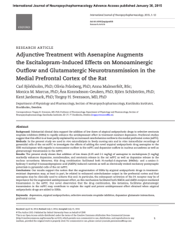
Figure 1c - International Journal of Neuropsychopharmacology
International Journal of Neuropsychopharmacology Advance Access published January 31, 2015
International Journal of Neuropsychopharmacology, 2015, 1–11
doi:10.1093/ijnp/pyu085
Research Article
research article
Chronic Desipramine Prevents Acute StressInduced Reorganization of Medial Prefrontal
Cortex Architecture by Blocking Glutamate Vesicle
Accumulation and Excitatory Synapse Increase
Nicoletta Nava, PhD-student; Giulia Treccani, PhD; Nico Liebenberg, PhD;
Fenghua Chen, PhD; Maurizio Popoli, PhD; Gregers Wegener, PhD;
Jens Randel Nyengaard, DMSc
Stereology and Electron Microscopy Laboratory, Centre for Stochastic Geometry and Advanced Bioimaging,
Aarhus University Hospital, Aarhus, Denmark (Drs Nava, Chen, and Nyengaard); Translational Neuropsychiatry
Unit, Department of Clinical Medicine, Aarhus University, Risskov, Denmark (Drs Nava, Treccani, Liebenberg,
Chen, and Wegener); Pharmaceutical Research Center of Excellence, School of Pharmacy, North-West
University, Potchefstroom, South Africa (Dr Wegener); Laboratory of Neuropsychopharmacology and Functional
Neurogenomics, Dipartimento di Scienze Farmacologiche e Biomolecolari and Center of Excellence on
Neurodegenerative Diseases, Università degli Studi di Milano, Milano, Italy (Drs Treccani and Popoli).
Correspondence: Nicoletta Nava, PhD, Translational Neuropsychiatry Unit, Aarhus University, Skovagervej 2, 8240 Risskov, Denmark ([email protected]).
Abstract
Background: Although a clear negative influence of chronic exposure to stressful experiences has been repeatedly demonstrated,
the outcome of acute stress on key brain regions has only just started to be elucidated. Although it has been proposed that acute
stress may produce enhancement of brain plasticity and that antidepressants may prevent such changes, we still lack ultrastructural
evidence that acute stress-induced changes in neurotransmitter physiology are coupled with structural synaptic modifications.
Methods: Rats were pretreated chronically (14 days) with desipramine (10 mg/kg) and then subjected to acute foot-shock
stress. By means of serial section electron microscopy, the structural remodeling of medial prefrontal cortex glutamate
synapses was assessed soon after acute stressor cessation and stress hormone levels were measured.
Results: Foot-shock stress induced a remarkable increase in the number of docked vesicles and small excitatory synapses,
partially and strongly prevented by desipramine pretreatment, respectively. Acute stress-induced corticosterone elevation
was not affected by drug treatment.
Conclusions: Since desipramine pretreatment prevented the stress-induced structural plasticity but not the hormone level
increase, we hypothesize that the preventing action of desipramine is located on pathways downstream of this process
and/or other pathways. Moreover, because enhancement of glutamate system remodeling may contribute to overexcitation
dysfunctions, this aspect could represent a crucial component in the pathophysiology of stress-related disorders.
Keywords: stress, glutamate, synapse, desipramine
Received: May 8, 2014; Revised: July 24, 2014; Accepted: July 26, 2014
© The Author 2015. Published by Oxford University Press on behalf of CINP.
This is an Open Access article distributed under the terms of the Creative Commons Attribution Non-Commercial License
(http://creativecommons.org/licenses/by-nc/4.0/), which permits non-commercial re-use, distribution, and reproduction in any
medium, provided the original work is properly cited. For commercial re-use, please contact [email protected]
1
2 | International Journal of Neuropsychopharmacology, 2015
Introduction
Emotional arousing and stressful experiences represent constitutive parts of our daily life; tight and efficient regulation of the
systems in charge of mediating the stress response is therefore
required to ensure adaptation (Sterling and Eyer, 1988). However,
when dysregulation of the stress response network or too much
stress output occur, an overload of the system and its physical and psychological manifestations such as depressed mood
and inflammation may be promoted (McEwen and Stellar, 1993).
Among the key targets for stress hormones is the prefrontal
cortex (PFC) (Meaney and Aitken, 1985; McEwen, 2007), a limbic
region mediating highly evolved executive functions, including working memory and cognitive flexibility (McEwen and
Morrison, 2013). Depending on the severity and duration of the
stimulus, stressful episodes have been reported to exert differential effects on the PFC (Popoli et al., 2012). Preclinical studies
in animal models of stress have shown that chronic exposure
to repeated stressors produces a number of physiological, cognitive, and structural deficits within medial PFC (mPFC); these
changes cover suppression of glutamate receptor expression and
function, reduced glial metabolism, and decreased expression
of synaptic proteins as well as impaired decision-making and
working memory (Dias-Ferreira et al., 2009; Banasr et al., 2010; Li
et al., 2011; Yuen et al., 2012). Chronic stress has also been shown
to induce profound structural remodeling of mPFC layer II-III
pyramidal neurons, including dendritic shortening, spine loss,
and neuronal atrophy (Cook and Wellman, 2004; Radley et al.,
2004). In contrast, short-term activation of the stress response
systems, as produced by acute stressors, has been reported to
cause plasticity-enhancing effects, including potentiation of
glutamate transmission in conjunction with enhanced working
memory and increase of excitatory aminoacid levels and of glutamate release in parallel with increased release probability in
layer III pyramidal neurons of mPFC (Moghaddam, 1993; Yuen
et al., 2009; Musazzi et al., 2010). Less, however, is known about
the effects of acute stress on mPFC structural remodeling. It is
well established that excitatory synapses play a major role in
mediating synaptic transmission, plasticity, and memory; on
the other hand, it has recently been demonstrated that stress
and glucocorticoids, the major stress hormones, can actively
and dynamically regulate these synapses (Krugers et al., 2012;
Timmermans et al., 2013). Thus, in the present study we aimed
at investigating the immediate effects of acute stress on presynaptic and synaptic remodeling of layer II-III pyramidal neurons
within the mPFC. It has recently been demonstrated that a single
foot-shock (FS) stress exposure may produce hippocampal synapse loss 24 hours after stress (Hajszan et al., 2009). Moreover,
we have recently shown that acute FS stress may strongly and
rapidly increase the number of glutamate vesicles available for
release within mPFC layer II-III (Treccani et al., 2014); however,
whether the previously observed rearrangement of synaptic
vesicles towards the presynaptic membrane may be coupled
with changes in the number of mPFC synapses has never been
explicitly demonstrated soon after acute stress. In this study, we
also aimed to determine whether pretreatment with the traditional antidepressant desipramine (DMI) may affect any such
change. It has recently been demonstrated that pretreatment
with the norepinephrine reuptake inhibitor DMI dampens acute
FS stress-induced increase of depolarization-evoked glutamate
release from synaptic terminals of frontal and PFC cortex, and
enhancement of the release probability in layer III pyramidal
neurons within mPFC (Musazzi et al., 2010). However, it has
never been explicitly demonstrated whether the preventive
action of DMI on FS stress-induced enhancement of glutamate
transmission may be coupled with any structural changes of
mPFC after acute FS stress.
Materials and Methods
Animals and Drug Treatment
Male Sprague-Dawley rats (175–200 g, Charles River, Calco, Italy)
were housed 2 per cage in a light-controlled room (under a
12-h-light/-dark cycle; lights on at 7:00 am) at room temperature
(22ºC) with food and water ad libitum. After 5 days of housing,
one-half of the animals were subjected to chronic treatment
(14 days) with DMI (10 mg/kg) delivered in drinking water. Drug
solutions were changed every 2 days according to the animals’
weight and water intake. All experimental procedures involving animals were performed in accordance with the European
Community Council Directive 86/609/EEC and approved
by Italian legislation on animal experimentation (Decreto
Ministeriale 116/1992).
Stress Paradigms and Corticosterone Levels
Twenty-four hours after the last drug or vehicle administration,
animals were subjected to 2 different acute stressors. For the FS
stress, rats were placed in a plastic chamber for 40 minutes, and
20 minutes of total actual shock (0.8 mA) was delivered with a
random inter-shock length between 2 and 8 seconds (Vollmayr
and Henn, 2001). Control animals were kept in the stress apparatus for the same interval of time (40 minutes) without delivering
the shocks. For the acute restraint (RS) stress, rats were immobilized in air-accessible cylinders for 1 hour (Deolindo et al., 2013).
Stress exposure was conducted between 8:00 am and 12:00 am in
a room different from that used for housing.
Trunk blood corticosterone (CORT) levels were estimated
using the IDS ELISA kit (Immunodiagnostic System, Herlev,
Denmark). Inter- and intra-assay coefficient of variation was
<10% (supplementary Material).
Confocal Microscopy of Synapses
Four male Sprague-Dawley rats (175–200 g) were treated for
14 days with either vehicle or DMI, as described above. Following
a 24-hour wash-out, rats were deeply anesthetized with pentobarbital sodium (Unikem A/S, Copenhagen, Denmark; intraperitoneal, 75 mg/kg) and transcardially perfused with 100 mL
heparinized (10 mg/mL) saline (NaCl 0.9%, pH = 7.4) for 5 minutes
followed by 2% fresh paraformaldehyde in 0.1 M phosphate buffered saline (pH = 7.4, 4°C). Brains were paraffin-embedded and
processed for immunofluorescence staining against Piccolo and
glutamate vesicular transport 1 protein (VGLUT1); for quantitative analysis, sections were examined in a 2-photon Zeiss
Confocal LSM 510 META microscope (supplementary Material).
Total Number of Synapses and Synaptic Vesicles
Using Electron Microscopy
Directly after the end of acute stress, animals were anesthetized and transcardially perfused with heparinized (10 mg/mL)
saline (NaCl 0.9%, pH = 7.4) for 5 minutes followed by fixative
containing 2.5% glutaraldehyde and 2% fresh paraformaldehyde in 0.1 M cacodylate buffer (pH 7.4). Series of 100-µm-thick
sections were cut coronally on a vibratome 3000 (Vibratome, St.
Louis, MO); based on a systematic, uniform random sampling
Nava et al. | 3
principle and a sampling fraction of one-sixth, 2 series were
chosen. Given their exquisite sensitivity to chronic and acute
stressors (Radley et al., 2004; Musazzi et al., 2010), mPFC layer
II-III containing the apical dendrites of the vast majority of cortical pyramidal neurons were unambiguously identified and
processed for electron microscopy (supplementary Material)
(Nava et al., 2014).
Synaptic vesicle quantification was performed on a series
of 45-nm-thick sections sampled from 2 regional areas per
animal. Briefly, neurotransmitter-containing vesicles were
classified into docked vesicles, when overlapping with the presynaptic membrane; all nondocked vesicles were defined as
reserve pools. Synaptic vesicles were quantified in perforated
(PSs) (Figure 1a) and nonperforated (NPSs) (Figure 1b) synapses.
Quantification was performed as previously described (supplementary Material) (Nava et al., 2014).
For synapse number estimation, four epon blocks were sliced
into series of 65-μm-thick serial sections with a MC MT-7 ultramicrotome (Tucson, AZ), and for each block two regions were
sampled and imaged in a FEI Morgagni transmission electron
microscope at final magnification of 18 000x. Synapses were
classified into axo-spinous, including PSs (Figure 1a) and NPSs
(Figure 1b) synapses, based on the presence of a perforation on
their postsynaptic density (black arrow in 1a), and axo-dendritic,
namely axo-shafts (AXSs) (Figure 1c-d), synapsing directly onto
the dendritic shaft (asterisk in Figure 1c-d) (Geinisman et al.,
2001). For quantification, a modified physical disector was
adopted (Sterio, 1984) (supplementary material). All animals
were assigned code numbers and decoded only after completion of the quantification.
mPFC Volume Estimation
mPFC total volume was obtained by applying the Cavalieri estimator in combination with the 2D-nucleator (6 half-lines; newCAST software) on thionin-stained sections (supplementary
Material) (Gundersen and Jensen, 1987).
Preparation of Figures
Electron micrographs, as shown in the figures, were adjusted for
contrast and brightness using Adobe Photoshop (San Jose, CA).
Three-dimensional reconstructions were performed by applying
of the RECONSTRUCTT softare (Fiala, 2005).
Statistical Analysis
All results were expressed as mean and relative coefficient of
variation and calculated as the standard deviation of the estimates, divided by the mean. N represented the number of animals per group, and n represented the number of sections per
animal analyzed for immunofluorescence study.
Coefficients of error (CEs) for synapse and vesicle number estimation were estimated as previously described (Nava
et al., 2014) (supplementary Tables S1 and S2). Data were analyzed using 2-way (stress x treatment) and 1-way (stress subtypes) analysis of variance. When analysis of variance revealed
significant group differences, the Bonferroni posthoc test was
employed. For the immunofluorescence study, an unpaired t
test was used. Statistical analysis and graphical representation
was carried out using Systat 12.0 and GraphPad Prism 5.0. The
degrees of significance were considered *P < .05, ** P < .01, and
***P < .001.
Results
Glutamatergic Synaptic Terminal Distribution
within mPFC
Figure 1. Electron micrographs identifying subpopulations of synapses analyzed. Axo-spinous synapses were classified into perforated (a) and nonperforated (b) based on the presence of a discontinuity (black arrow in a) in the postsynaptic density. Axo-dendritic synapses (c-d) were identified by the presence of
a mitochondrion on the postsynaptic compartment (asterisk in c and d). Scale
bar = 500 nm.
It is believed that excitatory synapses represent the majority of
synapses within neocortex (Sanacora et al., 2012). However, to
verify this, we assessed the relative distribution of mPFC excitatory terminals in rats subjected to chronic treatment with vehicle
or DMI. A presynaptic active zone was visualized with antibody
to Piccolo protein (Figure 2a) (Dondzillo et al., 2010); glutamatespecific synaptic terminals were identified using antibody to
VGLUT1 (Figure 2b), regarded as expressed primarily in glutamatergic neurons and also in glial cells within cortical areas
(Sanacora et al., 2012). In line with this, transporter VGLUT1 and
Piccolo showed a high level of colocalization (Figure 2c) in both
4 | International Journal of Neuropsychopharmacology, 2015
Figure 2. Relative distribution of excitatory synaptic terminals within medial prefrontal cortex (mPFC). Dual staining for active-zone marker Piccolo (white arrow) (a)
and transporter glutamate vesicular transport 1 protein (VGLUT1) (white arrow) (b) shows high degree of colocalization (white arrow) (c). Scale bar = 10 μm. (d) Summary
of quantitative data of colocalized punctate on dual-stained mPFC sections from rats treated with either vehicle (Veh) or chronic desipramine (DMI), obtained using VIS
Image Analysis Software (Visiopharm, Hørsholm, Denmark). P value, ns. (N = 4; n = 5).
vehicle- and DMI-treated groups, with an average of 92.3% and
92.1%. Unpaired t test revealed no significant difference in the
degree of colocalization of the 2 proteins between the 2 groups
(Figure 2d). Taken together, these results confirm that the large
majority of mPFC synaptic contacts are excitatory.
Acute Stress-Induced Increase of Docked Vesicles
Is Partially Prevented by Chronic Pretreatment
with DMI
At the synaptic level, presynaptic terminals (blue region in
Figure 3a) are enriched with neurotransmitter-containing vesicles of varying release competence (Dobrunz, 2002), so that vesicles docked to the active zone (cyan in Figure 3a) are thought as
ready to be released (white spheres in Figure 3a) (Schikorski and
Stevens, 2001); instead, nondocked vesicles (purple spheres in
Figure 3a) comprise the reserve pool.
Acute FS stress has been reported to induce a strong and rapid
increase of glutamate release from frontal and PFC synaptic
terminals. This effect was paralleled by increased release probability and changes of excitatory postsynaptic current (EPSC)
kinetics in layer III pyramidal neurons, two electrophysiological measurements of pre- and postsynaptic activity, respectively
(Musazzi et al., 2010). Moreover, we have recently shown that an
FS stress-induced increase of glutamate release occurs in parallel with an increased number of glutamate vesicles available for
release within mPFC (Treccani et al., 2014). Chronic treatment
with DMI dampened the stress-induced increase of glutamate
release and release probability, but not the kinetics of EPSC, suggesting a mechanism of action limited at the presynaptic level
(Musazzi et al., 2010). Thus, to evaluate the effect of chronic DMI
on stress-induced glutamate vesicle redistribution within the
presynaptic compartment, we quantified the number of docked
and reserve pool vesicles in stressed animals pretreated with
chronic DMI. At the end of acute FS stress exposure, the average number of docked vesicles was increased almost 2-fold in
PSs (+90.3%; N = 6; P < .001), and this increase was partially prevented by chronic DMI (−6.9%; N = 6; P < .05) (Figure 3b, left panel).
No effect of either stress or DMI was observed in NPSs (N = 6; P,
ns) (Figure 3b, right panel). In contrast, the average number of
reserve pool vesicles significantly decreased after stress in NPSs
(−16.0%; N = 6; P < .05) (Figure 3c, right panel) with no effect on
PSs (N = 6; P, ns) (Figure 3c, left panel).
These results confirm that acute FS stress induces a dramatic
and selective increase in the number of docked vesicles available for release in PSs, as previously reported (Treccani et al.,
Nava et al. | 5
Figure 3. Foot shock stress-induced presynaptic ultrastructural plasticity is partially prevented by chronic desipramine (DMI). (a) A reconstructed synapse showing
a typical spine contact. Presynaptic terminal (blue) is enriched with neurotransmitter-containing vesicles, divided into docked vesicles (white spheres) overlapping
with the presynaptic membrane at the level of the active zone (cyan) and into reserve pool vesicles (purple spheres). Red spheres represent large, dense core vesicles,
which were not included in the quantification. Cube side = 50 nm. (b) Acute FS-stressed rats showed significantly higher number of vesicles docked to the presynaptic
membrane in synapses with perforation of postsynaptic density (PSs) (P < .001); treatment with chronic desipramine (DMI) prevented the stress-induced increase of
the number of docked vesicles (left panel) (P < .05). Conversely, the number of docked vesicles in small nonperforated synapses (NPSs) was shown to be stable (right
panel). (c) The total number of reserve pool vesicles was stable among experimental groups in PSs (left panel). The number of reserve pool vesicles in NPSs was slightly
decreased after acute FS stress (right panel) (P < .05).
2014). Moreover, the results show that, in PSs, the stress-induced
major redistribution of synaptic vesicles towards the presynaptic membrane is attenuated by DMI pretreatment. Overall, these
results suggest that, by reducing the stress-induced increase
of docked vesicles, chronic DMI acts at the presynaptic level by
preventing the stress-induced potentiation of a morphological
correlate of excitatory synaptic strength.
Acute Stress Induces a Rapid Increase of mPFC
Excitatory Synapse Number, Which Is Prevented by
Chronic DMI
An altered number of docked vesicles has been observed during long-term potentiation in conjunction with fast synaptic
remodeling, as shown by an increased number of synapses in
hippocampal Cornu Ammonis 1 as soon as 30 minutes after the
stimulus induction (Bourne and Harris, 2011; Bourne et al., 2013).
Moreover, high vulnerability of synapses to acute stressful events
has previously been reported in studies showing hippocampal
synaptic marker increase and synapse loss, respectively, shortly
and 24 hours after stress termination (Hajszan et al., 2009;
Sebastian et al., 2013).
Hence, to investigate the effects of acute FS stress and DMI
treatment on mPFC excitatory terminal remodeling, we quantitated the number of mPFC asymmetric synapses regarded as
excitatory by means of physical disector and electron microscopy. Interestingly, the total number of excitatory synapses was
found to be rapidly and dramatically increased by 42.6% (N = 6;
P < .001) after 40 minutes of FS stress compared with the sham
group, whereas chronic administration of DMI partially prevented the stress-induced increase (+13.3% compared with control group; N = 6; P < .01) (Figure 4a).
To further verify whether the observed synaptogenic effect
was specific for FS stress or common to other acute stress models, we included rats previously subjected to 1 hour of RS stress
in our analysis. When comparing the total number of asymmetric synapses across different stress subtypes, acute RS stress
also increased asymmetric synapses by 22% compared with the
sham group (N = 6; P < .05) (Figure 4a).
We have recently shown that the majority of mPFC asymmetric spine synapses, namely PSs and NPSs, are endowed with
heterogeneous structural features that may reflect differences
in synaptic efficacy. For example, NPSs have shown smaller bouton volume and lower numbers of neurotransmitter-containing
vesicles compared with PSs (Nava et al., 2014). Moreover, asymmetric synapses may undergo differential activity-dependent
remodeling, depending on the subtype. For example, long-term
potentiation, a synaptic model of memory, has been shown to
selectively increase the number of PSs only (Geinisman et al.,
1991, 1993).
Therefore, to clarify whether the synaptogenic effect of acute
stress is selective for a specific synaptic population, we further
classified asymmetric synapses between axospinous, including NPSs and PSs, and axo-dendritic contacts, namely AXSs
(Figure 4c), in our serial section electron microscopy analysis
(Figure 4b). Small NPSs were increased after FS stress compared
with the sham group (+39.6%; N = 6; P < .001) regardless of treatment with DMI. Comparison across stressor subtypes showed
6 | International Journal of Neuropsychopharmacology, 2015
Figure 4. Total number of medial prefrontal cortex (mPFC) excitatory synapses is rapidly and selectively increased after 40 minutes of acute foot shock (FS) stress and
after 1 hour of restraint (RS) stress, whereas treatment with chronic desipramine (DMI) partially prevents FS stress-induced synaptogenesis. (a) Total number of mPFC
synapses was found to be rapidly increased at the end of 40 minutes of FS stress exposure (P < .001); however, FS stress-induced synaptogenesis was partially prevented
by treatment with chronic DMI (P < .01). RS stress also increased the total number of synapses compared with sham vehicle-treated (Veh) group (P < .05). (b) For synapse
quantification, a modified physical disector was applied (Tang et al., 2001); a grid was superimposed over 15 serial sections; section 1 was look-up section, sections
from 2 to 10 were used as the reference section of the disectors, whereas the last 5 sections were used as look-up sections to ensure that all counted postsynaptic
densities were included in the section series. Synaptic profiles touching the inclusion (green) lines were included in the analysis, whereas those touching the exclusion
(red) lines were excluded (scale bar = 1 μm). Synapses were classified between axo-spinous, synapsing onto spines, including nonperforated (blue dots) and perforated
(orange dots). Contacts synapsing directly onto the dendritic shaft were classified as axo-shaft (AXSs) (yellow dots). (c) Application of disector technique allowed unbiased estimates of the number of neocortical synapses, reconstructed here as spheres, within neuropil volume. Blue spheres represent nonperforated synapses (NPSs),
orange spheres perforated synapses (PSs), and yellow spheres AXSs. Representative estimates of the number of synapses are reported as the number of 3D spheres for
each experimental group: Sham+Veh (n = 21); FS+Veh (n = 37); Sham+DMI (n = 18); FS+DMI (n = 26); RS+Veh (n = 35). (d) Acute FS stress led to a strong significant increase
in small NPSs (P < .001) regardless of DMI treatment. The number of NPSs was also increased in RS animals compared with the sham group (P < .01). (e) The total number
of great PSs was shown to be stable among experimental groups. (f) AXSs were found strongly increased after acute FS stress (P < .001), whereas treatment with chronic
DMI prevented the stress-induced increase (P < .05). (g) mPFC volume estimated by the Cavalieri estimator did not show any significant difference among experimental
groups. (h) Corticosterone concentration was found significantly increased after both FS and RS stress, regardless of previous DMI treatment (P < .001). Compared with
RS-stressed animals, rats subjected to FS stress showed higher levels of CORT (P < .05).
Nava et al. | 7
that RS stress also significantly increased the number of NPSs
(+41.2%; N = 6; P < .01) (Figure 4d). Interestingly, big PSs were stable across groups (N = 6; P, ns) (Figure 4e). Moreover, only acute
FS stress, but not RS stress, induced a strong increase in AXSs
(+51.2%; N = 6; P < .001), and this effect was partially prevented
by treatment with chronic DMI (+6.01%; N = 6; P < .05) (Figure 4f).
When estimating overall mPFC volume, no significant effect of
stress-induced new synapse formation was observed (Figure 4g).
To determine whether the increase of mPFC asymmetric
synapses in acutely stressed animals is correlated with the elevated levels of adrenal corticosteroid hormones, we performed
enzyme-linked immunosorbent assay to measure CORT levels.
As shown in Figure 4h, compared with unstressed animals, rats
exposed to acute stressors had significantly higher blood concentration of CORT (FS stress: +532%, P < .001; RS stress: +390%,
P < .001) regardless of previous treatment with DMI (Musazzi
et al., 2010). When comparing among stressor subtypes, CORT
levels were found to be significantly higher after FS compared
with RS stress animals (+29.1%, P < .05) (Figure 4h).
Thus, an acute severe stressor rapidly induces strong sprouting of asymmetric synaptic contacts, mainly by increasing the
number of small and less efficacious NPSs; on the other hand,
the number of glutamate synapses is only modestly increased
after exposure to a less severe stressor. Moreover, DMI pretreatment, ineffective in the absence of stress, partially prevents
the stress-induced increase. These results suggest that an
acute stressor can rapidly boost the mPFC glutamate system by
enhancing the number of glutamate release sites and apposed
postsynaptic specializations. On the other hand, the preventive
effect of DMI on stress-induced synapse sprouting, but not on
CORT increase, suggests that the site of action of DMI in the
previously observed modulation of stress-induced glutamate
release (Musazzi et al., 2010) is downstream of CORT release, as
previously found (Conti et al., 2004; Musazzi et al., 2010), and
may directly involve the blockade of new pre- and postsynaptic
element formation.
Acute FS Stress Induces a Generalized Increase of
Glutamate Vesicles Available for Release within
mPFC, Which Is Prevented by DMI Pretreatment
The observed effects of FS stress and DMI pretreatment on synaptogenesis and presynaptic ultrastructural plasticity showed a
high degree of selectivity towards different synaptic subtypes.
Thus, acute stress increased the number of docked vesicles in
PSs only and increased the number of NPSs and AXSs. Therefore,
we examined how the stress-induced increase in number of
NPSs could affect the total number of docked vesicles and,
possibly, excitatory synaptic strength within mPFC by combining the data obtained on docked vesicles and synapse number
estimation.
The total number of docked vesicles, calculated by multiplying the total number of synapses within mPFC with the average number of vesicles, was significantly increased after FS
stress in both PSs (+78.1%; N = 6; P < .01) and NPSs (+145.4%; N = 6;
P < .001) compared with the control group, with a more remarkable effect on the latter subtype. Treatment with DMI, ineffective in the absence of stress, fully and partially normalized the
stress-induced increase in PSs (−11.9%; N = 6; P < .001) and NPSs
(+31.4%; N = 6; P < .01), respectively (Figure 5a, left and right
Figure 5. Acute foot shock (FS) stress dramatically increases morphological correlates of synaptic strength by increasing the number of vesicles available for release
in medial prefrontal cortex (mPFC) perforated (PSs) and nonperforated (NPSs) synaptic population. (a) The total number of docked vesicles in PSs within mPFC was
significantly increased by almost 2-fold after acute FS stress (P < .01); treatment with chronic desipramine (DMI) completely prevented the stress-induced increase in
the number of docked vesicles within mPFC PSs (left panel) (P < .001). (b) Similarly, in mPFC NPSs, FS stress significantly increased the total number of docked vesicles
(P < .001), whereas treatment with DMI partially prevented the stress-induced effects (right panel) (P < .01). In PSs, the total number of mPFC reserve pool vesicles was
not affected by stress or DMI treatment in either PSs or NPSs. (c) Three-dimensional representation of synaptic vesicles (white spheres) docked to the active zone (cyan)
for each experimental group. Cube side = 50 nm.
8 | International Journal of Neuropsychopharmacology, 2015
panels). No effect of either stress or treatment was observed on
reserve-pool vesicles in PSs and NPSs (Figure 5b, left and right
panels). These results suggest that, following the stress-induced
increase of synapses, acute FS stress produces a strong and generalized increase in the number of vesicles available for release
in both synapse subtypes, which is strongly normalized by DMI
pretreatment (Figure 5c).
Discussion
The results from the present study suggest that a single brief
exposure to stress potentiates excitatory neurotransmission
by inducing presynaptic structural plasticity, as shown by an
increased number of docked vesicles and large sprouting of
excitatory synapses. Moreover, these changes were partially
blocked by DMI pretreatment. Our ultrastructural findings were
localized in layers II-III of mPFC, which contains predominantly
somata of pyramidal neurons, and is known to be particularly
sensitive to chronic stress insults (McEwen and Morrison, 2013).
With regards to the effects of acute stress on mPFC structural
plasticity, the first result of the present study showed that acute
FS stress strongly increases the number of docked vesicles in
PSs only, confirming our previous observation on the selective
effect of acute FS stress (Treccani et al., 2014). These results
suggest that following acute FS stress, more vesicles from the
reserve pool become competent to fuse with the presynaptic
membrane and empty their content by being released from
the cytoskeletal matrix (Schikorski and Stevens, 2001; Sudhof,
2004). In line with this hypothesis, acute FS stress has shown to
increase presynaptic release probability (Musazzi et al., 2010).
We also observed for the first time that a single severe stressor
can rapidly induce a strong increase in the number of asymmetric synapses, whereas synapse increase is only modest after
a less severe stressor. This finding deserves particular attention considering that the general synapse increase was differentially provided for the 2 stressors by the counting of AXSs
and NPSs. Interestingly, AXSs were found increased only after
FS (+51.2%) but not RS stress. Most likely due to their relatively
higher strength through their strategic location directly on dendritic shafts rather than on spines (Figure 1c-d), we postulate
that potentiation of AXSs only after FS stress may be linked to
significantly higher levels of CORT found after FS compared
with RS stress, as well as to a different degree of complexity
among the 2 stressors (Maras et al., 2014). Small NPSs were
increased after both FS (+39.6%) and RS stress (+41.2%), with
greater stability being observed among PSs. Hippocampal PSs
mediate greater synaptic efficacy compared with small NPSs by
expressing a larger number of α-amino-3-hydroxy-5-methyl4-isoxazole propionate receptors (AMPARs) (Ganeshina et al.,
2004b). Because NPSs express fewer AMPARs and are regarded as
less efficacious (Ganeshina et al., 2004a, 2004b), we hypothesize
that the observed stress-induced increase of NPSs is consistent
with an increase in the so-called silent synapses, containing
mainly N-methyl-d-aspartate receptors (NMDARs) and few or
no AMPARs (Isaac et al., 1995). Despite being functionally silent
at resting potentials, these synapses represent a new substrate
for the formation, by insertion of AMPARs, of new functional
synapses more likely to be potentiated (Suvrathan et al., 2014).
In line with this hypothesis, a number of recent studies strongly
suggest postsynaptic changes in glutamate receptors, as shown
by an increased level of surface NMDARs and AMPARs in PFC
slices examined 1 to 4 hours poststress and altered EPSC kinetics likely occurring at the postsynaptic level (Yuen et al., 2009;
Musazzi et al., 2010).
According to a well-accepted model of synaptic remodeling following long-term potentiation, an increased number
of synapses is provided by splitting small NPSs into large PSs
(Geinisman, 2000). However, in the present study, the stressinduced increase in small NPSs and AXSs was not accompanied by a corresponding decrease in PSs. Therefore, we
postulate that additional AXSs and NPSs observed after acute
stress are formed mostly ex novo rather than being converted
from another synapse subtype. Although a number of in vitro
studies have shown that activity-dependent synaptogenesis
can occur within 2 hours from the application of the stimulus
(Bourne et al., 2013), only few studies investigated the formation of new synaptic contacts ex vivo, reporting 24 hours as the
shortest interval required for the formation of large efficacious
boutons (Medvedev et al., 2012). Interestingly, our findings provide the first ex vivo evidence that activity-dependent synaptogenesis of small and less efficacious NPSs can occur locally
ex novo as early as 40 minutes after the application of a severe
stressful stimulus. This shows remarkable dynamics of synapse
structure observed in a short time-frame following application
of the stressor. Given that the effects of acute stress on synapse
number were observed immediately after the end of the stress
protocol, it is conceivable that these changes may be accounted
for by changes in local protein translation in dendrites. One of
the crucial mediators of activity-dependent synaptic plasticity
is the neurotrophin brain derived neurotrophic factor involved
in the translation of a subset of dendritic mRNAs at synapses,
including cytoskeletal proteins involved in synaptic rearrangement (Leal et al., 2014). This new knowledge can be of great
importance in clarifying the dynamics leading to the long-term
behavioral and structural impairments that exposure to CORT
may produce (Gourley et al., 2013).
A number of previous studies have shown that tricyclic
antidepressants, including DMI, can reduce glutamate release
and transmission under both basal conditions in hippocampus
(Bonanno et al., 2005; Tokarski et al., 2008) and in the presence
of an acute stressful challenge in prefrontal and frontal cortex (Musazzi et al., 2010). In the last study, electrophysiological
recordings showed that DMI exerts its effect mainly by acting at
the presynaptic level, with little or no effect at the postsynaptic
side (Musazzi et al., 2010). In the present study, we examined
whether DMI may also dampen the enhanced structural plasticity induced by acute FS stress; this was done by investigating the size of synaptic vesicle pools, as presynaptic markers,
and the number of glutamate synapses as an overall measure of
synaptic changes. Here we showed that DMI treatment prevents
the effects of acute FS stress on structural plasticity. Although
subchronic treatment (5–7 days) with DMI has been shown to
reverse the detrimental effects of inescapable FS on hippocampal spine synapse number (Hajszan et al., 2009), the present
results suggest that DMI may also have a preventive action on
the effects of stress on structural plasticity, as shown by normalized vesicle docking and small synapse sprouting.
On the other hand, the dampening action of DMI on the
stress-induced structural potentiation was not accompanied by
a reduction of CORT levels. Thus, we suggest that the potential
mechanism underlying the blockade of stress effects by chronic
DMI must be located on pathways downstream of CORT release
or on alternative pathways. Several mechanisms may account
for the DMI-induced prevention of the stress-induced elevation
of structural plasticity. As previously discussed (Musazzi et al.,
2010), by increasing noradrenaline availability, DMI can induce
downregulation of chronically activated autoreceptors inhibiting noradrenaline release and leading in turn to disinhibition of
Nava et al. | 9
noradrenaline exocytosis (Raiteri et al., 1986; Starke et al., 1989).
In parallel, activation of α2-adrenergic heteroreceptors located
on glutamatergic neurons does not seem to lead to their downregulation, thereby inhibiting glutamate release (Raiteri et al.,
1983; Kamisaki et al., 1992; Pittaluga et al., 2007). In particular,
a major role in these processes may be played by synapsin I,
which is known to control the trafficking of synaptic vesicles
between the reserve and the readily releasable pool, depending on phosphorylation state (Greengard et al., 1993; Ceccaldi
et al., 1995). Because we have recently shown that acute FS
stress increases levels of Phospho-Ser9 synapsin I in synaptic
membranes (Treccani et al., 2014), it will be interesting to analyze the effects of DMI pretreatment on the acute stress-induced
increase of Phospho-Ser9 synapsin I. Overall, this effect of DMI
on stress-induced vesicle accumulation may account for the
previously described blockade of acute stress-induced enhancement of glutamate release and transmission by antidepressants
(Musazzi et al., 2010).
Moreover, the effect of DMI on stress-induced structural
plasticity was found double-sided. Besides limiting the accumulation of presynaptic-located docked vesicles, DMI was found to
act at the postsynaptic level by limiting the number of highly
electron-dense PSD, regarded as containing postsynaptic receptors (Gray, 1959). Antidepressants have long been known to regulate glutamate receptors by limiting NMDAR function through
decreased protein expression level of its subunits (Pittaluga
et al., 2007) and, conversely, potentiating AMPAR-mediated
transmission (Barbon et al., 2006). Therefore, it will be interesting to assess the effect of DMI pretreatment on the postsynaptic
surface level of glutamate receptors.
By limiting the number of glutamate vesicles available for
release and the number of excitatory synapses as potential glutamate release sites, DMI may help prevent FS stress-induced
boosting of the mPFC glutamate system and subsequent hyperglutamate–induced noxious effects (Sanacora et al., 2008; Popoli
et al., 2012).
In conclusion, by increasing vesicle docking and forming
new small synapses, acute stress promotes an immediate form
of ultrastructural plasticity of mPFC pyramidal neurons. This
heterogeneous population of neurons tends to segregate inputs
with extracortical afferents (from the mediodorsal nucleus of
the thalamus and hippocampal CA3) clustering on distal dendrites (layer I) (Swanson and Cowan, 1977; Groenewegen, 1988)
and synapses of local cortical circuits clustering on proximal
apical and basal dendrites (Scheibel and Scheibel, 1970). The
acute FS stress-induced enhancement of structural plasticity as
observed here was circumscribed to mPFC layer II-III containing
primarily the proximal portions of the apical and basilar arbor:
in turn, this may suggest a shift in emphasis from subcortical
to intracortical information, with important implications for the
functioning of mPFC and cognitive behaviors mediated by it.
Although further research is needed to assess how long the
stress-induced synaptogenesis and presynaptic potentiation
can be sustained and which pathways mediate the preventive
effects of DMI, these results support our initial hypothesis that
acute stress-induced presynaptic plasticity occurs in conjunction with increased synaptic remodeling, suggesting in turn an
overall FS stress-induced enhancement of mPFC structural plasticity. In addition to the previously observed preventive effect on
stress-induced increase of glutamate release, pretreatment with
classic antidepressant DMI can strongly counteract FS stressinduced mPFC structural plasticity. This preventing action of
DMI may be directly linked with its therapeutic effect in the
treatment of mood and anxiety disorders.
While our observations on stress-induced structural plasticity confirms the high vulnerability of mPFC to even a single stress exposure, our data on the preventive effects of DMI
suggest synapse remodeling as a neuronanatomical marker
predicting the effectiveness of antidepressant compounds on
preventing long-term noxious consequences.
Acknowledgments
We thank Herdis Krunderup, Lone Lysgaard, and Anette Larsen
for skillful technical assistance. Laura Musazzi is gratefully
acknowledged for advice and support during the study.
Statement of Interest
N.N. is supported by Aarhus University Mobility Fellowship. The
Centre for Stochastic Geometry and Advanced Bioimaging is
supported by the Villum Foundation.
References
Banasr M, Chowdhury GM, Terwilliger R, Newton SS, Duman RS,
Behar KL, Sanacora G (2010) Glial Pathology in an animal model
of depression: reversal of stress-induced cellular, metabolic
and behavioral deficits by the glutamate-modulating drug riluzole. Mol Psychiatry 15:501–511.
Barbon A, Popoli M, La Via L, Moraschi S, Vallini I, Tardito D, Tiraboschi E, Musazzi L, Giambelli R, Gennarelli M, Racagni G,
Barlati S (2006) Regulation of editing and expression of glutamate alpha-amino-propionic-acid (AMPA)/kainate receptors
by antidepressant drugs. Biol Psychiatry 59:713–720.
Bonanno G, Giambelli R, Raiteri L, Tiraboschi E, Zappettini S,
Musazzi L, Raiteri M, Racagni G, Popoli M (2005) Chronic antidepressants reduce depolarization-evoked glutamate release
and protein interactions favoring formation of SNARE complex in hippocampus. J Neurosci 25:3270–3279.
Bourne JN, Harris KM (2011) Coordination of size and number
of excitatory and inhibitory synapses results in a balanced
structural plasticity along mature hippocampal CA1 dendrites during LTP. Hippocampus 21:354–373.
Bourne JN, Chirillo MA, Harris KM (2013) Presynaptic ultrastructural plasticity along CA3-->CA1 axons during LTP in mature
hippocampus. J Comp Neurol 521:3898–3912.
Ceccaldi PE, Grohovaz F, Benfenati F, Chieregatti E, Greengard P,
Valtorta F (1995) Dephosphorylated synapsin I anchors synaptic vesicles to actin cytoskeleton: an analysis by videomicroscopy. J Cell Biol 128:905–912.
Chen F, Madsen TM, Wegener G, Nyengaard JR (2010) Imipramine
treatment increases the number of hippocampal synapses
and neurons in a genetic animal model of depression. Hippocampus 20:1376–1384.
Conti AC, Kuo YC, Valentino RJ, Blendy JA (2004) Inducible camp
early repressor regulates corticosterone suppression after
tricyclic antidepressant treatment. J Neurosci 24:1967–1975.
Cook SC, Wellman CL (2004) Chronic stress alters dendritic morphology in rat medial prefrontal cortex. J Neurobiol 60:236–248.
Deolindo MV, Reis DG, Crestani CC, Tavares RF, Resstel LB, Correa
FM (2013) NMDA receptors in the lateral hypothalamus have
an inhibitory influence on the tachycardiac response to acute
restraint stress in rats. Eur J Neurosci 38:2374–2381.
Dias-Ferreira E, Sousa JC, Melo I, Morgado P, Mesquita AR, Cerqueira JJ, Costa RM, Sousa N (2009) Chronic stress causes
frontostriatal reorganization and affects decision-making.
Science 325:621–625.
10 | International Journal of Neuropsychopharmacology, 2015
Dobrunz LE (2002) Release probability is regulated by the size of
the readily releasable vesicle pool at excitatory synapses in
hippocampus. Int J Dev Neurosci 20:225–236.
Dondzillo A, Satzler K, Horstmann H, Altrock WD, Gundelfinger
ED, Kuner T (2010) Targeted three-dimensional immunohistochemistry reveals localization of presynaptic proteins bassoon and piccolo in the rat calyx of held before and after the
onset of hearing. J Comp Neurol 518:1008–1029.
Fiala JC (2005) Reconstruct: a free editor for serial section microscopy. J Microsc 218:52–61.
Ganeshina O, Berry RW, Petralia RS, Nicholson DA, Geinisman
Y (2004a) Differences in the expression of AMPA and NMDA
receptors between axospinous perforated and nonperforated
synapses are related to the configuration and size of postsynaptic densities. J Comp Neurol 468:86–95.
Ganeshina O, Berry RW, Petralia RS, Nicholson DA, Geinisman Y
(2004b) Synapses with a segmented, completely partitioned
postsynaptic density express more AMPA receptors than
other axospinous synaptic junctions. Neuroscience 125:615–
623.
Geinisman Y (2000) Structural synaptic modifications associated
with hippocampal LTP and behavioral learning. Cerebral Cortex 10:952–962.
Geinisman Y, Detoledo-Morrell L, Morrell F (1991) Induction of
long-term potentiation is associated with an increase in the
number of axospinous synapses with segmented postsynaptic densities. Brain Res 566:77–88.
Geinisman Y, Detoledo-Morrell L, Morrell F, Heller RE, Rossi M,
Parshall RF (1993) Structural synaptic correlate of long-term
potentiation: formation of axospinous synapses with multiple, completely partitioned transmission zones. Hippocampus 3:435–445.
Geinisman Y, Berry RW, Disterhoft JF, Power JM, Van Der Zee EA
(2001) Associative learning elicits the formation of multiplesynapse boutons. J Neurosci 21:5568–5573.
Gourley SL, Swanson AM, Koleske AJ (2013) Corticosteroidinduced neural remodeling predicts behavioral vulnerability
and resilience. J Neurosci 33:3107–3112.
Gray EG (1959) Electron microscopy of synaptic contacts on dendrite spines of the cerebral cortex. Nature 183:1592–1593.
Greengard P, Valtorta F, Czernik AJ, Benfenati F (1993) Synaptic
vesicle phosphoproteins and regulation of synaptic function.
Science 259:780–785.
Groenewegen HJ (1988) Organization of the afferent connections
of the mediodorsal thalamic nucleus in the rat, related to the
mediodorsal-prefrontal topography. Neuroscience 24:379–
431.
Gundersen HJ, Jensen EB (1987) The efficiency of systematic sampling in stereology and its prediction. J Microsc 147:229–263.
Hajszan T, Dow A, Warner-Schmidt JL, Szigeti-Buck K, Sallam NL,
Parducz A, Leranth C, Duman RS (2009) Remodeling of hippocampal spine synapses in the rat learned helplessness
model of depression. Biol Psychiatry 65:392–400.
Isaac JT, Nicoll RA, Malenka RC (1995) Evidence for silent synapses: implications for the expression of LTP. Neuron 15:427–
434.
Kamisaki Y, Hamahashi T, Hamada T, Maeda K, Itoh T (1992) Presynaptic inhibition by clonidine of neurotransmitter amino
acid release in various brain regions. Eur J Pharmacol 217:57–
63.
Krugers HJ, Karst H, Joels M (2012) Interactions between
noradrenaline and corticosteroids in the brain: from electrical activity to cognitive performance. Front Cell Neurosci
6:15.
Leal G, Comprido D, Duarte CB (2014) BDNF-induced local protein synthesis and synaptic plasticity. Neuropharmacology
76 Pt C:639–656.
Li N, Liu RJ, Dwyer JM, Banasr M, Lee B, Son H, Li XY, Aghajanian G,
Duman RS (2011) Glutamate N-methyl-D-aspartate receptor
antagonists rapidly reverse behavioral and synaptic deficits
caused by chronic stress exposure. Biol Psychiatry 69:754–761.
Maras PM, Molet J, Chen Y, Rice C, Ji SG, Solodkin A, Baram TZ
(2014) Preferential loss of dorsal-hippocampus synapses
underlies memory impairments provoked by short, multimodal stress. Mol Psychiatry 19:811–822.
Mcewen BS (2007) Physiology and neurobiology of stress and
adaptation: central role of the brain. Physiol Rev 87:873–904.
Mcewen BS, Morrison JH (2013) The brain on stress: vulnerability and plasticity of the prefrontal cortex over the life course.
Neuron 79:16–29.
Mcewen BS, Stellar E (1993) Stress and the individual. Mechanisms leading to disease. Arch Intern Med 153:2093–2101.
Meaney MJ, Aitken DH (1985) [3H]Dexamethasone binding in rat
frontal cortex. Brain Res 328:176–180.
Medvedev NI, Dallerac G, Popov VI, Rodriguez Arellano JJ, Davies
HA, Kraev IV, Doyere V, Stewart MG (2012) Multiple spine boutons are formed after long-lasting LTP in the awake rat. Brain
Struct Funct 219:407–414.
Moghaddam B (1993) Stress preferentially increases extraneuronal levels of excitatory amino acids in the prefrontal cortex:
comparison to hippocampus and basal ganglia. J Neurochem
60:1650–1657.
Musazzi L, Milanese M, Farisello P, Zappettini S, Tardito D, Barbiero VS, Bonifacino T, Mallei A, Baldelli P, Racagni G, Raiteri M, Benfenati F, Bonanno G, Popoli M (2010) Acute stress
increases depolarization-evoked glutamate release in the rat
prefrontal/frontal cortex: the dampening action of antidepressants. Plos One 5:E8566.
Nava N, Chen F, Wegener G, Popoli M, Nyengaard JR (2014) A new
efficient method for synaptic vesicle quantification reveals
differences between medial prefrontal cortex perforated and
nonperforated synapses. J Comp Neurol 522:284–297.
Pittaluga A, Raiteri L, Longordo F, Luccini E, Barbiero VS, Racagni
G, Popoli M, Raiteri M (2007) Antidepressant treatments and
function of glutamate ionotropic receptors mediating amine
release in hippocampus. Neuropharmacology 53:27–36.
Popoli M, Yan Z, Mcewen BS, Sanacora G (2012) The stressed synapse: the impact of stress and glucocorticoids on glutamate
transmission. Nat Rev Neurosci 13:22–37.
Radley JJ, Sisti HM, Hao J, Rocher AB, Mccall T, Hof PR, Mcewen BS,
Morrison JH (2004) Chronic behavioral stress induces apical
dendritic reorganization in pyramidal neurons of the medial
prefrontal cortex. Neuroscience 125:1–6.
Raiteri M, Marchi M, Maura G (1983) Chronic drug treatments
induce changes in the sensitivity of presynaptic autoreceptors but not of presynaptic heteroreceptors. Eur J Pharmacol
91:141–143.
Raiteri M, Bonanno G, Maura G (1986) Changes of sensititvity of
presynaptic Α2-adrenoreceptors induced by chronic clonidine in rat brain. J Hyperthens 4:S122–S124.
Ribic A, Zhang M, Schlumbohm C, Matz-Rensing K, UchanskaZiegler B, Flugge G, Zhang W, Walter L, Fuchs E (2010) Neuronal MHC class I molecules are involved in excitatory
synaptic transmission at the hippocampal mossy fiber synapses of marmoset monkeys. Cell Mol Neurobiol 30:827–839.
Sanacora G, Zarate CA, Krystal JH, Manji HK (2008) Targeting the
glutamatergic system to develop novel, improved therapeutics for mood disorders. Nat Rev Drug Discov 7:426–437.
Nava et al. | 11
Sanacora G, Treccani G, Popoli M (2012) Towards a glutamate hypothesis of depression: an emerging frontier of neuropsychopharmacology for mood disorders. Neuropharmacology 62:63–77.
Scheibel ME, Scheibel AB (1970) Of pattern and place in dendrites. Int Rev Neurobiol 13:1–26.
Schikorski T, Stevens CF (2001) Morphological correlates of functionally defined synaptic vesicle populations. Nat Neurosci 4:391–395.
Sebastian V, Estil JB, Chen D, Schrott LM, Serrano PA (2013) Acute
Physiological stress promotes clustering of synaptic markers and
alters spine morphology in the hippocampus. Plos One 8:E79077.
Starke K, Gothert M, Kilbinger H (1989) Modulation of neurotransmitter release by presynaptic autoreceptors. Physiol Rev 69:864–989.
Sterio DC (1984) The unbiased estimation of number and sizes of
arbitrary particles using the disector. J Microsc 134:127–136.
Sterling P, Eyer J (1988) Allostasis: a new paradigm to explain
arousal pathology. In: Handbook of life stress, cognition, and
health. (Fisher S, Reason JT, eds). Chicester, NY: Wiley.
Sudhof TC (2004) The synaptic vesicle cycle. Annu Rev Neurosci
27:509–547.
Suvrathan A, Bennur S, Ghosh S, Tomar A, Anilkumar S, Chattarji
S (2014) Stress enhances fear by forming new synapses with
greater capacity for long-term potentiation in the amygdala.
Philos Trans R Soc Lond B Biol Sci 369:20130151.
Swanson LW, Cowan WM (1977) An autoradiographic study of the
organization of the efferent connections of the hippocampal
formation in the rat. J Comp Neurol 172:49–84.
Tafoya LC, Mameli M, Miyashita T, Guzowski JF, Valenzuela CF,
Wilson MC (2006) Expression and function of SNAP-25 as a
universal SNARE component in gabaergic neurons. J Neurosci
26:7826–7838.
Tang Y, Nyengaard JR, De Groot DM, Gundersen HJ (2001) Total
regional and global number of synapses in the human brain
neocortex. Synapse 41:258–273.
Timmermans W, Xiong H, Hoogenraad CC, Krugers HJ (2013)
Stress and excitatory synapses: from health to disease. Neuroscience 248:626–636.
Tokarski K, Bobula B, Wabno J, Hess G (2008) Repeated administration of imipramine attenuates glutamatergic transmission
in rat frontal cortex. Neuroscience 153:789–795.
Treccani G, Musazzi L, Perego C, Milanese M, Nava N, Bonifacino
T, Lamanna J, Malgaroli A, Drago F, Racagni G, Nyengaard JR,
Wegener G, Bonanno G, Popoli M (2014) Stress and corticosterone increase the readily releasable pool of glutamate vesicles in synaptic terminals of prefrontal and frontal cortex.
Mol Psychiatry 19:433–443.
Vollmayr B, Henn FA (2001) Learned helplessness in the rat:
improvements in validity and reliability. Brain Res Brain Res
Protoc 8:1–7.
Yuen EY, Liu W, Karatsoreos IN, Feng J, Mcewen BS, Yan Z (2009)
Acute stress enhances glutamatergic transmission in prefrontal cortex and facilitates working memory. Proc Natl
Acad Sci U S A 106:14075–14079.
Yuen EY, Wei J, Liu W, Zhong P, Li X, Yan Z (2012) Repeated stress
causes cognitive impairment by suppressing glutamate
receptor expression and function in prefrontal cortex. Neuron 73:962–977.


© Copyright 2026