
Laser vestibuloplasty. Case report - edigraphic.com
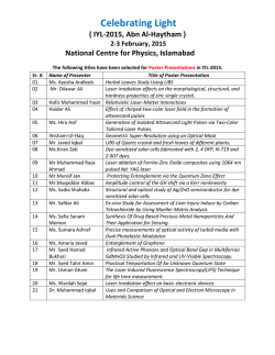
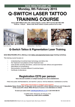
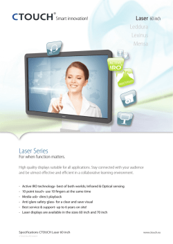
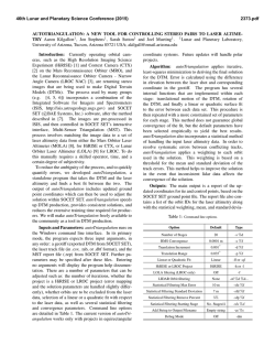
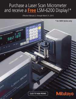
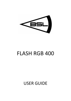
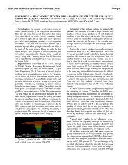
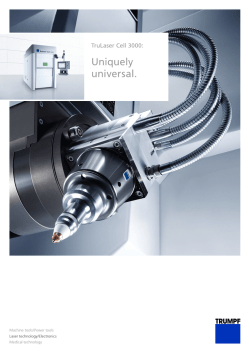
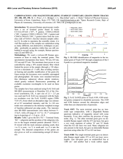
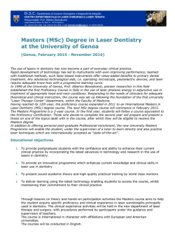
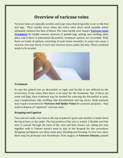
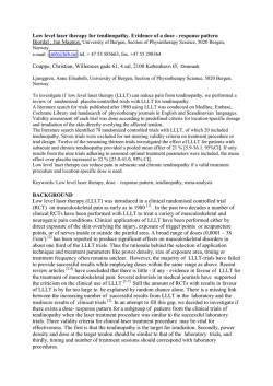
www.medigraphic.org.mx
Revista Odontológica Mexicana
Vol. 18, No. 4
Facultad de Odontología
October-December 2014
CASE REPORT
pp 259-262
Laser vestibuloplasty. Case report
Vestibuloplastia con láser. Reporte de caso
Myriam Amparo Pulido Rozo,* Meisser Vidal Madera Anaya,§ Lesbia Rosa Tirado AmadorII
ABSTRACT
RESUMEN
One of the greatest challenges when rehabilitating fully edentulous
patients, is to manufacture a denture to be placed on an alveolar
ridge with advanced bone resorption. This requires performing preprosthetic surgery and prepare groundwork bone and surrounding
soft tissues to receive a tissue-borne prosthesis, as well as providing
suitable retention and support. This will elicit lesser trauma and thus
allow prompt tissue healing. The case here presented is that of a 56
year old female, with non-contributory medical history. The patient
had been wearing dentures for the last 14 years; her dentures were
ill-adapted. Intra-oral clinical examination revealed collapsed and
re-absorbed ridges. Due to a lack of retention in the lower ridges, a
laser surgery was performed to deepen the vestibule. 18 days after
surgery tissues were completely healed, the vestibule had recovered
6 mm depth in the anterior section and 4 mm in the posterior section.
Rehabilitation was completed with the manufacture of full bimaxillary dentures. It was thus concluded that laser vestibuloplasty
confers depth to the vestibule and offers the advantages of laser
incision, which decreases hemorrhage, provides better operating
field and reduces post-operative inflammation and pain.
En pacientes edéntulos totales, uno de los desafíos para rehabilitación bucal es la confección de prótesis en un reborde alveolar con
avanzada resorción ósea, lo que hace necesario realizar cirugías
preprotésicas que preparen las bases óseas y los tejidos blandos
circundantes para recibir la prótesis mucosoportada, brindando retención y adecuado soporte; además de generar menor cantidad
de trauma posible, permitiendo la pronta cicatrización tisular. Se
presenta el caso de mujer de 56 años, sin antecedentes médicos
relevantes. Refiere portar prótesis totales desde hace 14 años, las
cuales se encuentran desadaptadas. Al examen clínico intraoral se
observan rebordes colapsados y reabsorbidos. Debido a la falta de
retención en los rebordes inferiores, se realizó cirugía de profundización del vestíbulo con láser. Se obtiene a los 18 días tejido completamente cicatrizado, con una recuperación en profundidad del vestíbulo de 6 mm en sector anterior y 4 mm en posterior. Finalmente se
rehabilita con prótesis total bimaxilar. Se concluye que la vestibuloplastia láser permite profundizar el vestíbulo y ofrece las ventajas de
la incisión láser que disminuye la hemorragia, brinda mejor campo
operatorio, disminuye la inflamación y el dolor postoperatorio.
Key words: Laser therapy, low intensity laser therapy, vestibuloplasty.
Palabras clave: Terapia por láser, terapia por láser de baja intensidad, vestibuloplastia.
INTRODUCTION
In fully edentulous patients, maxillary atrophy can
be defined as the advanced physiological reduction of
alveolar apophyses. This can be due to multiple factors
among which we can count pre-existent periodontal
disease, endocrine and systemic disorders, diet factors,
anatomical and mechanical considerations, gender,
and facial morphology.1 It might be one of the most
incapacitating oral conditions; the reason for this lies in
its nature which is chronic, progressive, accumulative
and irreversible. It represents one of the main problems
when trying to rehabilitate a totally edentulous patient
with a tissue-borne full denture, since this conventional
treatment is acceptable when there is sufficient alveolar
ridge to support the denture.1-3 Nevertheless, in cases
when alveolar atrophy is advanced, patients afflicted
with severe retention problems can benefit from preprosthetic surgery procedures in order to obtain a wider
bony base.1,4
One pre-prosthetic alternative would be vestibule
deepening, or vestibuloplasty following Edlan
Mejchar technique. This technique was described in
1963 and particularly applies to cases where there
is scarce or no remaining attached gum tissue.
This technique is additionally adopted in an effort
to overcome complications of post-operative loss of
vestibule depth, which occurs in other techniques.
This technique purports a successful prognosis, since
it assesses the patient’s comfort and maintenance
www.medigraphic.org.mx
*
§
II
Master in Public Health, Periodontist, DDS, Professor at the
School of Dentistry, University of Cartagena, Colombia.
DDS, Research Assistant, School of Dentistry, University of Cartagena, Colombia.
DDS, University of Cartagena, Colombia.
This article can be read in its full version in the following page:
http://www.medigraphic.com/facultadodontologiaunam
Pulido RMA et al. Laser vestibuloplasty
260
of the prosthesis in the surgically created vestibular
extension.
Laser application of the Edlan Mejchar technique
offers the possibility of deepening the vestibule,
facilitating thus post-operative procedures and
allowing to reduce healing time in the tissues and so
achieve prompt rehabilitation.5
Currently use of laser is widely accepted in the field
of dentistry. Laser is applied in multiple procedures,
according to their intensity. Low intensity lasers are
mainly used to achieve bio-stimulating, analgesic and
anti-inflammatory effects. High intensity lasers are
used to prepare cavities, eliminate old fillings, seal pits
and fissures, tooth hyper-sensitivity in order to prepare
root canals in endodontics, in esthetic dentistry to
manufacture dental veneers, tooth whitening, dental
prostheses to prepare the tooth to receive crowns, in
oral surgery to extract impacted teeth, apical surgery,
osteotomy, and exeresis of pre-malignant lesions in
soft tissues.6,7
Case report
A 56 year old female patient attended our services
dissatisfied with esthetic appearance as well as
function. The patient’s history was non-contributory,
she informed of wearing full dentures for the last 14
years; dentures were ill adapted and in poor esthetic
circumstances.
Stomatological examination revealed clinical
absence of all teeth, thick upper alveolar ridge, with
optimum height, collapsed lower ridges with advanced
resorption (Figure 1). Palate mucosa was swollen,
of an intensely red color and papillary appearance.
Clinical impressions were false anodontia and type 3
sub-prosthetic stomatitis.
Treatment plan suggested was oral rehabilitation
with upper and lower full dentures. Lack of retention
present due to resorption of lower ridges prompted
pre-prosthetic surgery in order to deepen the vestibule
with the help of a laser beam.
Surgical procedure
Infiltrative local anesthesia was applied in the
intervention area, between teeth 36 and 46. An
incision was then performed, using diode surgical
laser AsGaAL 4 w, with equipment Pocket laser,
Orotig Med aiming beam class 3R Laser 3mW CW
(MAX) 635 nm power beam class 4 Laser 6 w cw
(MAX) with 915 nm beam engagement. Central
and lateral frena were subjected to frenilectomies,
the vestibule was deepened. At the same time
refrigeration was achieved with triple syringe, six
gauzes were used in all the procedure to induce
hemostasis. The low amount of bleeding present
obviated the need to suture (Figures 2 and 3). To
complete the procedure, a low intensity laser was
used during 3 minutes (AsGaAL, 685 nm) in order
to stimulate healing (Figure 4). Surgical time was
approximately 10 minutes, when procedure was
completed, nimeluside-type analgesic was prescribed
(100 mg every 24 hours), no antibiotic coverage was
used. Extra-oral ice applications were recommended
to be used in the first day.
The first control took place 24 hours after
procedure. No edema could be observed. Upon
applying pain, assessment with EVA scale (0 to
www.medigraphic.org.mx
w
ww.medig phic.org.mx
Figure 1. Lower alveolar ridges with advanced bone
resorption.
Figure 2. Use of surgical laser in the lower vestibule.
Revista Odontológica Mexicana 2014;18 (4): 259-262
261
Figure 5. Control image 24 hours after surgical procedure.
Figura 3. Results observed after surgical laser use.
EEste
Es
ste
te documento
doc
ocum
meennto
to eess el
eelaborado
lab
la
aboorrad
ado po
por Me
por
M
Medigraphic
eddiigr
grap
aphi
hic
Figure 4. Use of low-intensity laser application to achieve
bio-stimulating effect.
10), the patient reported a number three score. At
that point a second therapeutic laser application
was performed (Figure 5). Second control was
undertaken eight days after procedure. Remarkable
recovery and absence of pain were observed,
after this, a third therapeutic laser application was
performed.
After 18 days, the third control was undertaken. Fully
healed tissue could be observed, tissue was in optimum
conditions to take preliminary impressions as well as for
full denture recording. In anterior and posterior areas of
the vestibule a respectively 6 and 4 mm depth recovery
could be observed (Figure 6). Finally, 28 days after
surgery, the patient was rehabilitated with bi-maxillary
full dentures. At later control assessments, the patient
informed to be free of discomfort.
Figure 6. Recovery of depth in lower alveolar ridges.
DISCUSSION
Bilateral anesthesia of the mandibular nerve is
required when performing pre-prosthetic surgery with
Edlan Mejchar technique and using a scalpel. When
using laser technique lesser amounts of infiltrative
local anesthesia can be used.8
Sutures are needed after many surgical
procedures. It is suggested to preserve the epithelium
sutures from 8 to 10 days, this is normally the time
required by the periosteum to recover. When using
laser, in many surgical procedures sutures are no
longer needed, this will impact in the patient’s postoperative comfort.9
Accelerated tissue recovery achieved with laser
therapy decreases prosthetic rehabilitation time,
www.medigraphic.org.mx
Pulido RMA et al. Laser vestibuloplasty
262
when compared with scalpel surgery. According
to Martin, 10 a period of 4 to 6 months is frequently
allowed to elapse to allow healing and alveolar
mucosa remodeling, to then be able to take an
impression which will yield working models to
manufacture the denture.
Other advantages offered by a laser vestibuloplasty
procedure are the following: intervention time of
approximately five minutes, precision in the incision,
asepsis, analgesia, hemorrhage decrease, little
inflammation and edema since tissues and blood
vessels with a diameter smaller than the laser beam
are vaporized, and nerves, upon being sectioned, are
sealed off with the heat of the laser beam.11-13 One of
the limitations encountered when using laser beam
therapy is the high cost of therapeutic laser equipment,
nevertheless, its purchase is justified in view of its
many applications in the dental practice.
After surgery, use of low intensity laser helps in the
favorable evolution of the patient. Its bio-stimulating
properties might accelerate tissue regeneration and
favor wound healing, achieving thus less pain and
swelling.14-16
CONCLUSION
Pre-prosthetic surgery of the vestibule accomplished
with laser beam offers favorable clinical results during
and after the operation which become evident with
the following: a relatively short operative period, small
amounts of anesthesia required, suitable hemostasis,
anti-inflammatory and analgesic effect, not requiring
sutures and a short healing time which facilitates
prompt impression taking in order to obtain work
models and thus manufacture dentures.
REFERENCES
1. García O, Arredondo M. Evolución en el tratamiento de la atrofia
alveolar. Rev Cubana Estomatol. 2002; 39 (2). HTML.
2. Xie Q, Wolf J, Tilvis R, Ainamo A. Resorption of mandibular
canal wall in the edentulous aged population. J Prosthet Dent.
1997; 77 (6): 596-600.
3. Catillo E, García M. Rehabilitación implantoprotésica:
sobredentadura. Rev Cubana Ortod. 2000; 15 (2): 75-81.
4. Hillerup S. Preprosthetic vestibular sulcus extension by the
operation of Edlan and Mejchar. Int J Oral Surg. 1979; 8: 333-339.
5. Gupta H, Kinra P. Vestibular extension by Edlan-Mejchar
technique followed by permanent fibre splinting-a case report.
Indian Journal of Dental Sciences. 2010; 2 (2): 17-19.
6. Arnabat J, España A, Berini L. Aplicaciones del láser en
odontología. RCOE. 2004; 9 (5): 497-511.
7. Henning H, Deppe H. New aspects of lasers in oral and
craniomaxillofacial surgery. Medical Laser Application. 2005; 20: 7-11.
8. García F, España A, Berini L et al. Aplicaciones del láser de CO2
en odontología. RCOE. 2004; 9 (5): 567-576.
9. Edlan A, Mejchar B. Plastic surgery of the vestibulum in
periodontal 194 therapy. Int Dent J. 1963; 13: 593-596.
10. Martin J, Lemon J, Shusterman M. Oral and dental rehabilitation
after mandible reconstruction. Operative Techniques in Plastic
and Reconstructive Surgery. 1996; 3 (4): 264-271.
11. Pogrel MA. The carbon dioxide laser in soft tissue preprosthetic
surgery. J Prosthetd Dent. 1989; 61: 203-208.
12. Cavalcanti TM, Almeida-Barros RQ, Catão MH, Feitosa AP, Lins
RD. Knowledge of the physical properties and interaction of laser
with biological tissue in dentistry. An Bras Dermatol. 2011; 86
(5): 955-960.
13. Benjamín SD. Soft tissue lasers: it’s the wavelength, power,
ergonomics, and control that matter! Dent Econ. 2009; 99.
14. Oltra D, España A, Berini L et al. Aplicaciones del láser de baja
potencia en odontología. RCOE. 2004; 9 (5): 517-524.
15. Alves R, Dantas E, Reposo K, Chaves M, Granville A, Carvalho
L. Biostimulation effects of low-power laser in the repair process.
An Bras Dermatol. 2010; 85 (6): 849-855.
16. Gomes A, Cazal C, Lisboa J. Ação da laserterapia no processo
de proliferação e diferenciação celular. Revisão da literatura.
Rev Col Bras Cir. 2010; 37 (4): 295-302.
Mailing address:
Dr. Meisser Vidal Madera Anaya
E-mail: [email protected]
www.medigraphic.org.mx
© Copyright 2026