
COMPLETE TEM-TOMOGRAPHY: 3D - USRA
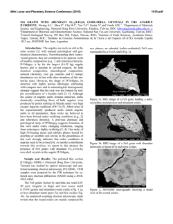
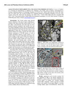
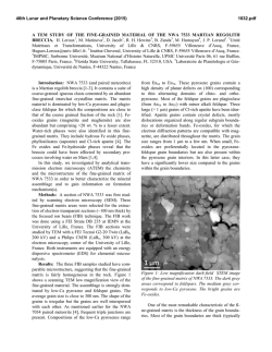
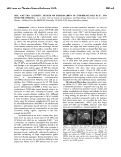
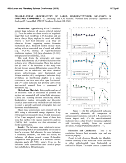
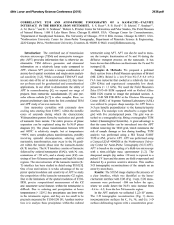
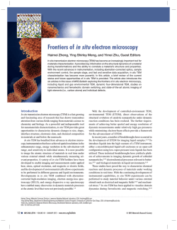
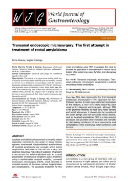
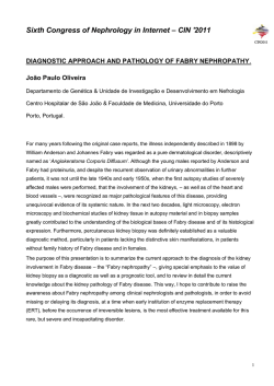
46th Lunar and Planetary Science Conference (2015)
2177.pdf
COMPLETE TEM-TOMOGRAPHY: 3D STRUCTURE OF GEMS CLUSTER. J. Matsuno1, A. Miyake1, A.
Tsuchiyama1, S. Messenger2, and K. Nakamura-Messenger2, 1Dep. of Geology and Mineralogy, Kyoto University,
Kitashirakawa, Oiwake-cho, Sakyou-ku, Kyoto, 606-8502, JAPAN ([email protected]), 2Robert M.
Walker Laboratory for Space Science, EISD, ARES, NASA Johnson Space Center, Houston TX 77058, USA
Introduction: GEMS (glass with embedded metal
and sulfide) grains in interplanetary dust particles
(IDPs) are considered to be one of the ubiquitous and
fundamental building blocks of solids in the Solar System. They have been considered to be interstellar silicate dust that survived various metamorphism or alteration processes in the protoplanetary disk [e.g., 1−3]
but the elemental and isotopic composition measurements suggest that most of them have been formed in
the protoplanetary disk as condensates from high temperature gas [e.g., 4]. This formation model is also
supported by the formation of GEMS-like grains with
respect to the size, mineral assemblage, texture and
infrared spectrum by condensation experiments from
mean GEMS composition materials [5].
Previous GEMS studies [1−4] were performed only
with 2D observation by transmission electron microscopy (TEM) or scanning TEM (STEM). However, the
3D shape and structure of GEMS grains [3] and the
spatial distribution of Fe/FeS’s [4] has critical information about their formation and origin. Recently, the 3D
structure of GEMS grains in ultrathin sections of cluster IDPs was revealed by electron tomography using a
TEM/STEM (JEM-2100F, JEOL) [6]. However, CT
images of thin sections mounted on Cu grids acquired
by conventional TEM-tomography are limited to low
tilt angles (e. g., < |75°|). In fact, previous 3D TEM
observations of GEMS were affected by some artifacts
related to the limited tilt range in the TEM used [6].
Complete tomographic images should be acquired by
rotating the sample tilt angle over a range of more than
|80|° otherwise the CT images lose their correct structures [7].
In order to constrain the origin and formation process of GEMS grains more clearly, we performed
complete electron tomography for GEMS grains. Here
we report the sample preparation method we have developed for this study, and the preliminary results.
Sample and Methods: In order to obtain transmission images upon tilting at large angles, we processed a rod-shaped specimen fabricated by a focused
ion beam (FIB), the details described in [7,8]. The
sample was placed on a TEM holder which had been
modified by cutting the top of the retainer (Fig. 1).
Fine rod-shaped specimens were also prepared for atom-probe tomography (APT) analysis [e. g., 9,10],
which makes it possible to obtain 3D distribution of
elements in atomic scale. In the preparation, the spec-
imen was mounted on the top of a pointed tungsten
needle and sharpened the top by an annular FIB [9].
Such rod-shaped samples are also suitable for TEMtomography as well as APT. The present sampling
methods to acquire the complete TEM-tomography
was applied by using the FIB processes for APT analysis.
A potted butt of a cluster IDP (L2036AA10 cluster
4) prepared by an ultramicrotome was used for the
present observation. After a short survey of the potted
butt by FE-SEM (JSM-7001F, JEOL), a portion composed of fine GEMS grains was excavated by Ga+ ion
beam by FIB (Quanta 200 3DS, FEI) and lifted up with
a tungsten needle (Fig. 1A). Before the sampling, the
surface of the potted butt was covered with platinum
deposition to protect it against ion beam damage. Another tungsten needle (0.1−0.2 mm in diameter and
~7.5 mm in length) attached to the bottom of a Cu pipe
(2 mm in diameter) was mounted on a FIB stage and
Fig 1: Preparation for a rod sample in this study. (A) A target
area in a potted butt was pulled out by FIB. The specimen
was attached to a W needle. Pt deposition covered the surface to protect from the beam damages. (SEM image) (B)
The specimen was mounted on another W needle and
trimmed (secondary ion micrographic (SIM) image). (C) A
back scattered electron (BSE) image of the sample rod after
sharped by Ga+ beam. The top is capped by Pt.
46th Lunar and Planetary Science Conference (2015)
the specimen with platinum deposition was remounted
on the top of the tungsten needle (Fig. 1B). It was
trimmed and sharpened to be rod-shaped by the FIB.
The final diameter of the rod is ~400 nm and the length
is ~4 µm (Fig. 1C). The rod was put on a single tilting
holder with an improved TEM retainer whose top was
cut and a V-shaped notch with a groove was fabricated
in order to keep the needle on the retainer. The sample
was observed by BF-TEM (bright field-TEM) and
HAADF-STEM (high angle annular dark fieldscanning TEM) modes using FE-TEM (JEM-2100F,
JEOL) at Kyoto University. The spot size of the STEM
mode was 0.7 nm, the convergence angle of HAADF
mode was 13 mrad, and the acceptable angle was from
35 (inside diameter) to 92 (outside diameter) mrad
(HAADF1 condition).
Fig 2: (A) A CT image acquired by HAADF-STEMtomography. GEMS grains embedded in epoxy exist as a
cluster while the grain boundaries are obscure. White particles correspond to Fe and FeS inclusions. Some GEMS
grains have abundant Fe and FeS inclusions (blue arrow)
while others seem to have only one inclusion (white arrow).
(B) A slice image at yz plane. An iron or iron sulfide has a
void-like inclusion (white arrow).
2177.pdf
Before performing tomography, 2D elemental mapping
was performed to estimate mineral phases by using
EDS (energy dispersive X-ray spectroscopy) system
(JED-2300T, JEOL) equipped in the TEM/STEM.
However, the fine texture could not be obtained due to
the thick rod. A series of tilting images was obtained
almost completely in the tilting range from -90° to
86.5° in 1° intervals automatically using a software
(TEMography, System In Frontier Inc.) by HAADFSTEM mode. The alignment of the rotating axis was
performed manually by tracking fine Fe and FeS inclusions as ground control points. The voxel size of acquired slice image is 2.44 nm and the fitting error average of the rotation axis is 0.0698 pixel. CT images
were reconstructed by a filtered back-projection method.
Results and discussions: A series of slice images
were successfully obtained (Fig. 2). Artifacts due to
the incomplete tomography, such as tails extending
from Fe and FeS inclusions observed in the previous
method [6] weren’t detected. However, mismatching
of the rotation axis due to the manual alignment caused
some image distortion. Many GEMS grains were observed to be aggregated into clusters, as proposed by
prior 2D TEM observation [4] (Fig. 2A) although the
grain surfaces are too obscure to trace them clearly.
Bright particles typically 5−20 nm in size corresponding to Fe and FeS inclusions were observed in
each GEMS grain (Fig. 2A). Some of them have voidlike inclusions (Fig. 2B) as observed in the previous
report [6]. Some GEMS grains have abundant Fe and
FeS inclusions while others seem to have only one
inclusion or no inclusions (Fig. 2A) at least within the
present spatial resolution. This may reflect that GEMS
grains (or the primary grains) formed in various environments and aggregated with each other as the observed cluster. We also emphasized that the present
study is the first successful example adapting the complete TEM-tomography to the IDPs. This type of analysis will be also important for planetary material sciences in the future.
References: [1] J. P. Bradley et al. (1994) Science.
265, 925–929. [2] J. P. Bradley et al. (1999) Science,
285, 1716–1718. [3] J. P. Bradley and Z. R. Dai (2004)
Astrophys. J. 617, 650–655. [4] L. P. Keller and S.
Messenger (2011) Geochim. Cosmochim. Acta 75,
5336–5365. [5] Matsuno et al. (2014) Lunar & Planetary Science, 45, #1335 (abstr.). [6] Matsuno et al.
(2014) Meteorit. Planet. Sci., 77, A5079 (abstr.). [7]
Kawase et al. (2007) Ultramicroscopy 107, 8–15. [8]
Miyake et al. (2014) Microscopy, 63, i24–25. [9] Chen
et al. (2009) Scr. Mater. 61, 693–696. [10] Valley et al.
(2014) Nat. Geosci. 7, 219–223.


© Copyright 2026