
The Kidney Pathology of Fabry Disease
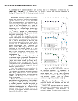
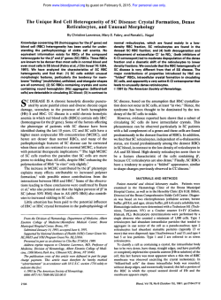
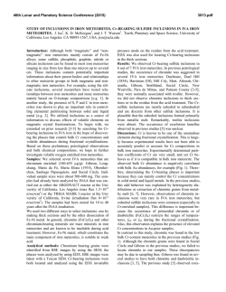
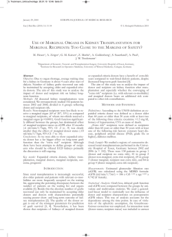
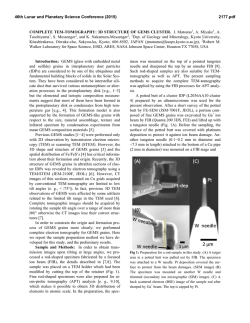
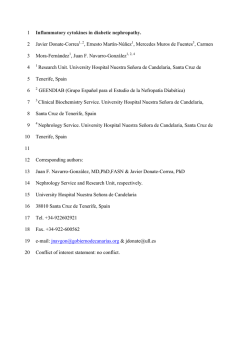
Sixth Congress of Nephrology in Internet – CIN '2011
CIN2011
DIAGNOSTIC APPROACH AND PATHOLOGY OF FABRY NEPHROPATHY.
João Paulo Oliveira
Departamento de Genética & Unidade de Investigação e Desenvolvimento em Nefrologia
Centro Hospitalar de São João & Faculdade de Medicina, Universidade do Porto
Porto, Portugal.
For many years following the original case reports, the illness independently described in 1898 by
William Anderson and Johannes Fabry was regarded as a pure dermatological disorder, descriptively
named as „Angiokeratoma Corporis Diffusum’. Although the young males reported by Anderson and
Fabry had proteinuria, and despite the recurrent observation of urinary abnormalities in further
patients, it was not until the late 1940s and early 1950s, when the first autopsy studies of severely
affected males were performed, that the involvement of the kidneys, – as well as of the heart and
blood vessels –, were recognized as major pathological features of this disease, providing
unequivocal evidence of its systemic nature. In the next two decades, light microscopy, electron
microscopy and biochemical studies of kidney tissue in autopsy material and in biopsy samples
greatly contributed to the understanding of the biological bases of Fabry disease and of its histological
expression. Furthermore, percutaneous kidney biopsy was definitely established as a valuable
diagnostic method, particularly in patients lacking the distinctive skin manifestations, in patients
without family history of Fabry disease and in females.
The purpose of this presentation is to summarize the current approach to the diagnosis of the kidney
involvement in Fabry disease – the “Fabry nephropathy” –, giving special emphasis to the value of
kidney biopsy as a diagnostic as well as a prognostic tool, and to review in detail the current
knowledge about the kidney pathology of Fabry disease. This way, I hope to contribute to raise the
awareness about Fabry nephropathy among clinical nephrologists and pathologists, in order to avoid
missing or delaying its diagnosis, at a time when early institution of enzyme replacement therapy
(ERT), before the occurrence of irreversible lesions, is the most effective treatment available for this
rare, but severe and incapacitating disorder.
1
Overview of Fabry disease – biochemical, genetic, clinical and epidemiological
aspects
CIN2011
Fabry disease is the lysosomal storage disorder that results from an inborn deficiency of the activity of
alpha-galactosidase, a glycoside hydrolase involved in the catabolic processing of
globotriaosylceramide (Gb3Cer) and of other minor neutral glycosphingolipids that contain terminal,
non-reducing alpha-D-galactose residues. Progressive accumulation of Gb 3Cer in vascular endothelia
leading to obstruction of the microvasculature and ischemic tissue damage, particularly to the brain,
heart and kidneys, has been advocated as the major pathogenic mechanism underlying the systemic
complications of the disease.
Since the locus of the alpha-galactosidase gene (GLA) is on the X-chromosome, band Xq22.1,
affected males are hemizygous for a pathogenic GLA mutation while almost all affected females are
heterozygous for the normal allele and a pathogenic mutation. The in vitro demonstration of deficient
alpha-galactosidase activity in cell protein extracts (usually from leukocytes) or in biological fluids
(usually plasma), utilizing a synthetic fluorogenic substrate, is a straightforward diagnostic test for
Fabry disease in males. However, by the effect of random X-chromosome inactivation, females
carrying pathogenic GLA mutations in heterozygosity are mosaics of cells, the large majority of which
express either the normal or the mutated enzyme. The relative proportion of these two cell
populations in specific organs and tissues is a major determinant of the ultimate pathological and
clinical phenotype, including of the level of enzyme activity measured in vitro. For this reason, the
enzymatic diagnosis of Fabry disease is not reliable in females since carriers of pathogenic GLA
mutations may have normal enzyme activity, as conventionally assayed in leukocytes or plasma, and
the finding of pathogenic GLA mutations is the gold-standard diagnostic test for Fabry disease in
females.
In males, the precocity and severity of the clinical manifestations of Fabry disease are related to the
degree of alpha-galactosidase deficiency. Patients carrying GLA mutations associated with absent or
very low levels (usually ≤1% normal) of residual enzyme activity, who have the so-called „classical
phenotype‟, typically present in childhood or adolescence with acroparesthesias, hypohidrosis,
abdominal pain, diarrhea, angiokeratomas and cornea verticillata. The early symptoms experienced
by the classically affected children and teenagers largely reflect the involvement of the peripheral and
autonomic nervous systems but, with advancing age, these patients eventually develop major renal,
cardiac and cerebrovascular complications. Indeed, in young- and middle-aged adults, end-stage
renal disease (ESRD), arrhythmias, left ventricular hypertrophy and myocardial infarction, transient
ischemic attacks and stroke are major causes of the morbidity and premature mortality associated
with Fabry disease. The late complications of Fabry disease are thought to result from as yet elusive
non-specific secondary pathogenic processes, most likely related to the continuous accumulation of
Gb3Cer in those organs. When the residual enzyme activity is higher (usually <5-30% normal),
patients present later in life with renal disease and/or cardiac disease, lacking the early, distinctive
skin and neuropathic manifestations of the „classical phenotype‟. The expression of renal disease in
2
patients with the „renal variant‟ is clinically indistinguishable from that of the „classical phenotype‟.
Although patients with the „renal variant‟ also have cardiac complications, patients with the „cardiac
CIN2011
variant‟ usually do not develop ESRD. As the residual enzyme activity in patients with the „cardiac
variant‟ is significantly higher than in patients with the „renal variant‟, the alpha-galactosidase activity
threshold for progressive kidney disease seems to be lower than for heart disease.
Presumably as a beneficial effect of the cellular mosaicism created by X-chromosome inactivation,
females commonly have less severe forms of Fabry disease as compared to males carrying the same
GLA mutations, although a significant number of heterozygotes may ultimately experience major
cardiac, cerebrovascular, or renal clinical events.
Because of its rarity, the diagnosis of classical Fabry disease has been frequently overlooked in
clinical practice, particularly in patients not previously known to be at genetic risk. The diagnosis of
the incomplete, later-onset variants of Fabry disease is even more difficult to recognize, requiring a
high index of suspicion. Recently, large-scale newborn screening programs have estimated the
population frequency of GLA mutations associated with the severe phenotype as ~1:35.000-40.000;
however, the overall allelic frequency of mutations associated with the later-onset phenotypes was
estimated as at least >10-fold higher.
Until a decade ago, the treatment of patients with Fabry disease was limited to the non-specific
supportive management of disease complications. Although the available preventive and symptomatic
therapies permitted to alleviate the patients‟ morbidity and improved life expectation, they were of
limited success and did not address the underlying metabolic cause of Fabry disease. The recent
advent of ERT with genetically engineered human alpha-galactosidases (agalsidase alfa, agalsidase
beta) was a major advance, but its clinical efficacy seems to be critically dependent on early initiation,
before irreversible lesions occur. Therefore, and despite its rarity, clinicians are now challenged with
the new responsibility of having to be more aware of Fabry disease, in order to expedite its diagnosis.
Since patients with Fabry disease may seek care from a variety of medical specialties, due to the
involvement of multiple organ systems, this applies to many different specialists, including clinical
nephrologists.
Clinical manifestations of Fabry nephropathy
Chronic kidney disease (CKD) with proteinuria and progressive azotemia is one of the prominent
features of classical Fabry disease and of the later-onset renal variant. As the renal function
deteriorates blood pressure increases in many patients but, at comparable CKD stages, the
prevalence of hypertension in patients with Fabry disease is lower than in the general CKD
population. Proteinuria is of glomerular origin in almost all cases, albumin representing its major
component (≥50%). The prevalence of proteinuria increases with age, being relatively uncommon in
children and teenagers. However, by the age of 35 years, ~50% of the affected males are estimated
to have proteinuria, and all patients who survive into the 6th decade of life eventually develop
proteinuria. Nephrotic range proteinuria occurs in <20% of male Fabry patients with CKD, but the full3
blown presentation of nephrotic syndrome is relatively unusual. Death from ESRD on the 4th or early
on the 5th decade of life was a frequent outcome of affected males before the advent of chronic
CIN2011
dialysis and kidney transplantation, but since effective renal replacement therapies became available
the median age of death has increased by 10-15 years. At early adulthood, most males with Fabry
disease have normal glomerular filtration rate (GFR), as estimated from the serum creatinine level
(eGFR), but cases of ESRD have exceptionally been reported in teenagers. In affected males, the risk
and rate of CKD progression are related to the level of residual alpha-galactosidase activity. Early on
their 40s, about half the patients with ≤1% residual enzyme activity already have serum creatinine
levels ≥1.5 mg/dl (corresponding to estimated CKD stage 3 or higher), while those with higher
enzyme activity levels seldom reach stage 3 CKD before the age of 45 years. The rate of decline of
the eGFR is more than twofold higher in patients with advanced CKD (stage 3 or higher) as compared
to patients with eGFR >60 ml/min/1.73m2 at baseline. Time of progression from CKD stage 3 to
ESRD is quite variable, ranging from ~1 year to more than 12 years. In patients with advanced CKD,
typical natural history eGFR average decline rates range between –6.5 to –10.5 ml/min/1.73m2.
Higher levels of baseline proteinuria are also positively associated with more rapid progression to
ESRD. Virtually all males with the classical phenotype who survive to their mid-50s develop ESRD.
Only a minority of patients reach advanced CKD stages without developing overt proteinuria.
Heterozygous females have a significantly lower risk of ESRD as compared to the hemizygous males,
and in both the European and the United States ESRD registries only ~12% of the patients with the
diagnosis of Fabry disease were women. However, females with Fabry disease who have progressive
CKD, reach ESRD at similar age ranges than the Fabry male patients.
Additional laboratory manifestations of Fabry nephropathy include various defects of tubular function
and abnormalities of the urine sediment. The most commonly reported functional tubular defect is an
impairment of the urinary concentration mechanisms in the distal tubule, which has been
demonstrated even in patients with normal GFR. The urine concentrating defect may lead to
isosthenuria, causing symptoms of polyuria, nocturia, and polidipsia. Since early in the natural history
of Fabry nephropathy, red blood cells, white blood cells, oval fat bodies, hyaline and granular casts
may be present in the urine sediment. About 75% of the exfoliated cells in the urine of classically
affected Fabry patients are glycosphingolipid-laden renal tubular cells, which can be recognized as
oval fat bodies in the urine sediment. Glycosphingolipid globules may also be seen free in the urine,
as fat droplets. Like the oval fat bodies commonly seen in the context of heavy proteinuria, these
droplets, as well as the glycosphingolipid inclusions in tubular cells, are birefringent, displaying a
„Maltese cross‟ pattern when viewed under polarized light microscopy. However, the oval fat bodies
that correspond to glycosphingolipid-laden renal tubular cells in Fabry disease additionally have a
characteristic internal lamellar appearance that allows its specific identification.
Parapelvic renal cysts are more prevalent among adult males with Fabry disease than among
individuals in the general population and their finding the diagnostic workup of a patient with CKD
should lead to the consideration of Fabry disease in the differential diagnosis.
4
Diagnosis of Fabry nephropathy
Non-invasive diagnostic approach
CIN2011
In patients diagnosed with Fabry disease, or in individuals at genetic risk for Fabry disease,
proteinuria and progressive CKD are generally assumed to be evidence of Fabry nephropathy. This
clinical assumption can be non-invasively corroborated by assaying specific metabolic biomarkers in
the urine and/or by targeted urine microscopy examination.
Increased urinary excretion of Gb 3Cer and/or the finding of lysoGb 3 (a deacylated metabolite of
Gb3Cer) in the urine support the non-invasive diagnosis of Fabry nephropathy in patients both
genders, including in children. Urinary Gb 3Cer is consistently elevated in virtually all Fabry patients,
including heterozygotes, when measured in the sediment of a 24-hour urine collection, and in most
patients when measured in a random sample of whole urine or in urine collected on a filter paper.
Urine Gb3Cer levels are highest in patients with null GLA mutations and no residual enzyme activity
and lower in patients with significant residual alpha-galactosidase activity and in heterozygotes. In
these latter cases, urine Gb3Cer may even be within the normal range when measured in random
samples of whole urine. Urinary excretion of Gb 3Cer does not correlate with patient age. The lysoGb3
assay in the urine is technically more demanding than the Gb 3Cer assay but is more specific as a
diagnostic test for Fabry nephropathy, because lysoGb 3 is not excreted in the urine of healthy
subjects whereas low amounts of Gb 3Cer (≤25 μg/mmol creatinine) are normally found in the urine. In
patients who have never received ERT, the lysoGb3/creatinine ratios in the urine are ~6000 and
~1000 times less than the corresponding Gb 3Cer/creatinine ratios, respectively in males and females.
The urinary lysoGb3/creatinine and Gb3Cer/creatinine ratios are strongly correlated. Like the urine
levels of Gb3Cer, the urinary lysoGb3/creatinine ratio correlates with gender and the type of GLA
mutation, which is a surrogate for residual alpha-galactosidase activity.
Fresh urine sediment examination under phase-contrast microscopy with polarized light, together with
immunocytochemistry for Gb3Cer, have recently been proposed as a cheap, sensitive and specific
diagnostic test for Fabry disease. Patients with Fabry nephropathy excrete birefringent oval fat bodies
which have characteristic internal lamellation and irregular surface protrusions, ranging in shape from
hook-like to spherical. Under polarized light, separate „Maltese crosses‟ are visible in the main body of
these particles as well as in the larger protrusions. Such morphological features, which are best
appreciated with minor adjustments in focal length, allow to specifically discriminating these particles
from the birefringent oval fat bodies commonly found in heavy proteinuric patients. Furthermore,
immunocytochemistry of the urine sediment using a standard immunoperoxidase technique with a
monoclonal anti-Gb3Cer primary antibody positively stains large vacuolated mononuclear epithelial
cells that are reportedly specific for Fabry disease. The lamellated „Maltese cross‟ particles, as well as
the Gb3Cer-positive cells, are detectable both in male hemizygotes and female heterozygotes,
including before the development of any clinical signs of Fabry nephropathy.
5
Diagnostic kidney biopsy
Atypical clinical presentations for Fabry nephropathy, including acute nephritic syndrome, acute onset
of severe nephrotic syndrome, severe hypertension early in the course of progressive CKD, rapidly
progressive loss of renal function, and macroscopic hematuria, are indications for diagnostic kidney
biopsy, even in patients with the diagnosis of Fabry disease. Indeed, minimal change nephropathy,
IgA nephropathy, crescentic glomerulonephritis, granulomatous interstitial nephritis and lupus
nephritis have been described as coexisting diseases in patients with Fabry nephropathy. Besides to
confirm the diagnosis of Fabry nephropathy and to exclude other kidney disorders in patients with
atypical presentations, a kidney biopsy is also useful for evaluating the superimposed pathology of
concomitant diseases (e.g. diabetes mellitus or malignant hypertension) and for assessing the
histological severity of Fabry nephropathy. Kidney biopsy is additionally recommended in adult Fabry
patients with GLA mutations associated with a high residual enzyme activity, if they present with low
GFR and proteinuria. It is not uncommon that a kidney biopsy reveals Fabry nephropathy in
previously undiagnosed patients who are being evaluated for proteinuric CKD, including heterozygous
females. Histomorphological evidence of Fabry nephropathy is a very robust clue to the diagnosis of
Fabry disease and justifies a thorough molecular genetics diagnostic approach in those cases where
a pathogenic GLA mutation is not identified after routine gene sequencing studies. Conversely, the
clinical diagnosis of Fabry nephropathy has been excluded by kidney biopsy in a proteinuric female
with typical angiokeratomas, who carried a GLA mutation associated with high residual enzyme
activity. However, the diagnosis of Fabry disease may be missed in incidental kidney biopsies if only
routine light microscopy is done. Identification of glycosphingolipid deposits in kidney tissue requires
that the biopsy sample be adequately processed with an appropriate fixative (e.g. glutaraldehyde) for
light microscopy and electron microscopy. Under illumination in a stereomicroscope, the glomeruli of
patients with Fabry nephropathy, even children with minimal albuminuria, have a striking white color
that contrasts with the usual red color of the glomeruli. Therefore, bedside stereomicroscopic
inspection of the kidney biopsy material in patients with renal disease of unknown cause may
immediately raise the suspicion of Fabry nephropathy and can be useful to guide the histological
processing of the biopsy sample.
The pathogenesis and the pathology of Fabry nephropathy
The kidneys are one of the major sites of glycosphingolipid accumulation in Fabry disease.
Globotriaosylceramide and galabiosylceramide (Ga 2Cer) are the glycosphingolipid compounds that
are extractable in highest amounts from the kidney tissue of adult Fabry patients, although the latter
at threefold lower concentration. The Gb3Cer and the Ga2Cer deposited in the kidney cells of Fabry
patients are believed to be locally generated by normal physiologic processes and metabolic
pathways. Both Gb3Cer and its major catabolic precursor, globoside, are normal constituents of the
human kidney, and the synthase of Gb 3Cer is strongly expressed in the kidneys.
Globotriaosylceramide is the membrane antigen CD77, expressed in the kidneys by many different
6
CIN2011
cell types, particularly in the cortical tubular epithelium. CD77 is involved in cellular signaling in CD19mediated cell adhesion and interferon-alpha-induced growth inhibition and apoptosis but also
CIN2011
functions as a receptor for the Shiga-like toxins (verotoxins) of Escherichia Coli, which cause the
hemolytic-uremic syndrome. Furthermore, the inhibition of GLA gene expression in cultured human
tubular epithelial cells (HK2) and primary cultured tubular epithelial cells by RNA interference led to an
increase in membrane Gb3Cer expression and to the formation of osmiophilic intracellular lamellar
inclusions resembling those observed in patients with Fabry disease.
Rudimentary storage of Gb3Cer in the kidney has been noted as early as the second trimester of
gestation, a time when only myenteric plexuses cells were also affected. In adults, the concentration
of Gb3Cer extracted from kidney tissue is significantly higher in patients with no residual alphagalactosidase activity as compared to patients with detectable residual enzyme activity, indirectly
suggesting that the metabolic overload of glycosphingolipids in kidney cells is one of the factors that
determine the natural history of Fabry nephropathy. Progressive accumulation of Gb 3Cer in the
glomerular and interstitial microvascular endothelia of the kidney, eventually leading to luminal
obstruction and tissue ischemia, has been regarded as a prominent factor in the pathogenesis of the
nonspecific degenerative glomerular (segmental and global sclerosis/hyalinosis) and tubulointerstitial
(tubular atrophy, interstitial fibrosis) changes associated with CKD progression in patients with Fabry
disease. For this reason, the clearance of Gb3Cer deposits from the renal microvascular endothelium
was selected as the primary efficacy end point in a major randomized, placebo-controlled, doubleblind trial of ERT with agalsidase beta. Another putative mechanism of vascular disease in Fabry
nephropathy is the necrosis of severely involved smooth muscle cells in the arteriolar and arterial
vessel walls. Lethal injury to Gb3Cer overloaded podocytes, as well as necrosis of mesangial cells,
may be additional mechanisms contributing to the pathogenesis of glomerular sclerosis. Because
podocytes are highly differentiated, postmitotic cells they generally are not replaced when they are
lethally injured. As a result of podocyte loss, parietal epithelial cells of Bowman‟s capsule may gain
access to bare areas of the glomerular basement membrane, forming focal adhesions and thereby
starting the cascade of pathogenic events that lead to segmental glomerular sclerosis. Moreover,
endothelial cells in the vicinity of damaged podocytes are submitted to additional hydraulic forces that
may also lead to segmental glomerular collapse and sclerosis. Finally, direct toxic injury to the tubules
from the Gb3Cer storage within the tubular epithelial cells may be a factor involved in the
pathogenesis of focal tubular atrophy and interstitial fibrosis.
Glycosphingolipids consist of a hydrophobic ceramide moiety linked to a hydrophilic carbohydrate.
Because of their amphiphilic solubility properties, glycosphingolipids are almost entirely extracted
from tissue sections by the non-polar solvents (e.g., ethanol and xylene) that are used in the standard
clearing and paraffin-embedding histological procedures for light microscopy examination. For this
reason, the most distinctive histopathological feature of Fabry disease on routine light microscopy is
the vacuolization of cells in affected tissues. Staining of formalin-fixed paraffin-embedded tissue
sections with lipid-soluble dyes is also negative. Glycosphingolipids can be identified in frozen tissue
7
sections by any of the currently available histochemical lipid-staining methods but the staining
reactions are not specific for glycosphingolipids and the freezing of tissue leads to loss of fine cellular
CIN2011
details. Intracellular glycosphingolipid deposits can be also easily identified as dark-blue or grey-blue
inclusions on toluidine blue- or methylene blue-stained semi-thin (0.5–1.0 µm) plastic-embedded
electron microscopy scout sections. However, as frozen specimens are not routinely obtained and/or
processing of samples for electron microscopy are not performed in many clinical settings, it is
imperative that pathologists promptly recognize the light microscopy features of Fabry nephropathy.
Indeed, the diagnosis of Fabry disease has been overlooked on kidney biopsies exclusively assessed
by standard light microscopy study, while re-examination by electron microscopy readily allowed the
recognition of the typical ultrastructural features of Gb3Cer deposits. It has recently been
demonstrated that the amount of Gb3Cer remaining in paraffin-embedded kidney tissue sections
allows its specific recognition by immunohistochemistry. Therefore, immunohistochemical staining of
Gb3Cer is a relatively simple and specific diagnostic method for Fabry disease in routine light
microscopy of kidney biopsies, which can be used even for retrospective diagnosis of Fabry
nephropathy in archive paraffin blocks, particularly when cell vacuolization is a prominent finding.
Furthermore, it allows the differential diagnosis of concurrent kidney disease in patients carrying GLA
mutations associated with high residual enzyme activity or with atypical clinical presentations of Fabry
nephropathy.
Light microscopy
The most characteristic finding on routine light microscopy of kidney biopsies of patients with Fabry
disease is the vacuolization of the cytoplasm of affected cells. The pattern of cell type involvement
and the prevalence of vacuolated cells of each type are roughly dependent on the age and gender of
the patient and on the clinical severity of Fabry nephropathy. In children, teenagers and young adult
male patients, the cells most prominently affected are the podocytes which are diffusely enlarged,
showing abundant cytoplasm filled with numerous clear vacuoles, giving a “honeycomb” appearance
to the glomeruli [Fig.1]. Cytoplasm enlargement and
vacuolization is also remarkable in the distal tubular epithelial
cells, including those of Henle‟s loop and the collecting duct,
particularly intercalated cells. The morphological changes are
of mild to moderate degree in the epithelial cells of Bowman's
capsule, and in the endothelial and smooth muscle cells of the
arteries. The glomerular endothelium and the mesangial cells
are minimally affected and most proximal tubules look
histologically normal. The involvement of the epithelial cells of
Bowman‟s capsule, mesangial cells and endothelial cells is
more pronounced as the patients get older. With the
Figure 1. Extensive vacuolization of the
glomerular tuft.
Reproduced from: Faria V, Doença de
Fabry, tesaurismose rara, Jornal do
Médico 1970, LXXII (1423).
methenamine silver stain, argyrophilic inclusions may be identified in some affected cells.
Heterozygous females have similar morphological abnormalities, but of milder degree as compared to
8
males. Although vacuolization is a light microscopy feature of a number of other kidney diseases,
either genetic or non-genetic, the age of the patient at the onset of renal symptoms, the specific
CIN2011
clinical phenotype, particularly of the extra-renal manifestations, and the histological pattern of
involvement of the kidney cells should allow the differential diagnosis with Fabry nephropathy.
Light microscopy examination of toluidine blue- or methylene blue-stained semi-thin sections allows a
more accurate identification of the stored glycosphingolipids and can disclose the presence of
cytoplasmic Gb3Cer deposits even within cell types which apparently are not vacuolated on the
conventional paraffin-embedded sections. At higher magnification (objective: x40–x100), Gb3Cer
inclusions appear as small, dark, dense beaded granules, or as larger, more structured, laminated
bodies, located either in the cytoplasm around the nucleus or clustering in the peripheral cytoplasm.
The laminated inclusions are typically seen in the podocytes and in the epithelium of distal convoluted
tubules and collecting ducts. Podocyte nuclei are often eccentrically positioned, pushed aside by the
mass of Gb3Cer inclusions. In small capillaries, the cytoplasm of heavily loaded endothelial cells may
protrude into the lumen, to the point of causing microvascular obstruction. In studies using this
histological technique, glomerular parietal epithelial cells were the most extensively affected in kidney
biopsies of middle-aged heterozygous women with CKD stage 2 and no overt proteinuria, and
glycosphingolipid inclusions were abundant in the enlarged podocytes of an older male with a cardiac
variant of Fabry disease, while the vascular endothelia and smooth muscle cells, and the tubular
epithelium looked unremarkable.
Immunohistochemistry of paraffin-embedded kidney tissue sections of patients with Fabry
nephropathy using an anti-Gb3Cer primary antibody allows the specific identification of residual
Gb3Cer in all types of glomerular, tubular, interstitial and vascular kidney cells, although at relatively
lower scorings than the corresponding histological scorings on semi-thin plastic embedded sections.
The Gb3Cer immunostaining pattern in Fabry nephropathy is distinct from that observed in controls,
which have mild Gb3Cer expression limited to tubular cells.
Immunofluorescence is important in the differential diagnosis of other kidney diseases that may occur
concurrently in patients with Fabry disease. Asymptomatic IgA deposits have been repeatedly
identified in kidney biopsies of patients with Fabry disease, with a frequency that seems to be higher
than in non-selected autopsy cases. Segmental IgM deposits, typical of focal glomerular sclerosis,
may also be identified in some cases.
Non-specific changes
Non-specific degenerative glomerular and vascular changes are present even before progression to
overt proteinuria and decreased glomerular filtration rate. Round deposits of hyaline-like material
within the media of arteries are the most prevalent non-specific vascular lesion in kidney biopsies of
patients with Fabry disease. These deposits are observed whatever the caliber of the vessels and are
especially numerous in the afferent arterioles. They have been described as early as age 11 years, in
both affected boys and girls. As the disease progresses, increased mesangial matrix and mesangial
widening, segmental and global glomerular sclerosis, vascular intimal and medial thickening, tubular
9
atrophy, interstitial fibrosis and chronic inflammatory cell infiltrates are variably recognized by light
microscopy. In a study of children and adolescents with Fabry disease and minimal albuminuria,
CIN2011
glomerular hyaline was detected as early as in a 7-year-old boy; incipient signs of focal segmental
glomerulosclerosis were present in a 14-year-old girl; and most of the boys aged 16–18 years already
had a globally sclerotic glomerulus in the biopsy section. Interstitial fibrosis, of mild degree (~5% of
the cut section), was seen exclusively in these latter patients. However, in young adult males aged
25-29 years old, presenting with the classical phenotype of Fabry disease, the degree of tubular
atrophy and interstitial fibrosis was moderate to severe and diffuse in all cases. Expansion of the
mesangial area, by reducing the surface of glomerular filtration, might contribute to the pathogenesis
of segmental glomerular sclerosis. The proportion of segmentally or globally sclerotic glomeruli
increases with age in both genders. Most of the progressive non-specific degenerative lesions
described in Fabry nephropathy are thought to be related to ischemic damage.
Clinicopathological correlations and gender differences
In a study of 9 young males with Fabry disease, aged 11–29 years old, with normal creatinine
clearance, four of them with overt proteinuria (range: 0.7–2.8 g/day), segmental and/or global
glomerulosclerosis were the only histopathological correlates of overt proteinuria.
Light microscopy glomerular and tubulointerstitial pathology scores, as well as a glycosphingolipid
inclusion score, were computed for kidney biopsies of 25 adult male Fabry patients aged 20–49
years, with residual alpha-galactosidase activity <15% of the control values, as baseline assessment
for a double-blind placebo-controlled trial of ERT with agalsidase alfa. Six patients had advanced
CKD (stages 3 or 4) and 16 presented overt proteinuria (>300mg/day). The glomerular pathology
score was based on the mesangial morphology of each glomerulus, scored into one of the following
categories, of increasing histopathological severity: normal; diffuse mesangial widening; segmental
sclerosis or solidification; and global sclerosis or glomerular obsolescence. The final glomerular
pathology score was the average of the individual glomerular scorings in each biopsy. The
tubulointerstitial pathology score was determined as the average of the individual scorings for the
degrees of tubular atrophy, interstitial inflammation, interstitial fibrosis, vascular medial thickening and
vascular hyalinosis. Glycosphingolipid inclusions were assessed on toluidine blue-stained semi-thin
plastic sections, and their amounts rated in glomerular epithelial cells, glomerular
endothelial/mesangial cells, proximal tubular epithelial cells, distal tubular epithelial cells, vascular
endothelial cells and vascular medial cells. The overall glycosphingolipid inclusion score was the
average of the individual cell type scorings. Both the glomerular and the tubulointerstitial pathology
scores showed highly significant inverse correlation with GFR at baseline, as estimated by inulin
clearance, and direct correlation with proteinuria. Age at kidney biopsy was inversely correlated with
inulin clearance and directly correlated with the glomerular pathology score, but did not correlate
significantly with the tubulointerstitial pathology score. As it might have been expected, the glomerular
and the tubulointerstitial pathology scores were significantly correlated but in stepwise linear
regression analysis the glomerular pathology score emerged as the major predictor of both the inulin
10
clearance and the proteinuria levels. Yet, a wide range of variation in individual glomerular pathology
scores was noted among patients with inulin clearances below 90 ml/min/1.73m 2. Contrastingly, the
CIN2011
glycosphingolipid inclusion score did not correlate with GFR, proteinuria or the other pathology
scores. The composite nature of these kidney pathology scorings may have limited their usefulness to
recognize additional, particularly cell-type dependent clinicopathological correlations.
A different approach was used for the scoring of baseline and post-treatment kidney biopsies
obtained from patients enrolled in the phase 3 double-blind, randomized, placebo-controlled trial and
open label extension studies of ERT with agalsidase beta. The initial cohort comprised 58 patients
(mean age: 30.2 years; range: 16–61 years), including two females, with severe deficiency of alphagalactosidase activity and a serum creatinine level not exceeding 2.2 mg/dl. Mean serum creatinine
was 0.8 mg/dl and mean inulin clearance was 89.8 ml/min. Glycosphingolipid accumulation was semiquantitatively scored on semi-thin (1 µm) epoxy-embedded plastic sections, stained with methylene
blue/Azure II and examined by light microscopy. Glycosphingolipid inclusion scores were determined
for each of the following cell types: peritubular (interstitial) capillary endothelial cells, glomerular
endothelial cells, mesangial cells, arterial/arteriolar endothelial cells, vascular smooth muscle cells,
interstitial cells (a mixed population of fibroblasts and phagocytic cells), podocytes, and the epithelium
of distal convoluted tubules and collecting ducts. In addition, the mesangial matrix of individual
glomeruli was also evaluated for pathologic change. The primary endpoint of the trial was the
clearance of Gb3Cer deposits from the interstitial capillary endothelial cells.
At baseline, glycosphingolipid accumulation was much more concentrated and extensive in the
podocytes and the epithelial cells of distal convoluted tubules and collecting ducts than in vascular
endothelial and smooth muscle cells, mesangial cells and interstitial cells. Proximal tubular epithelial
cells were relatively unaffected. After 20–26 weeks of ERT, highly effective clearance of Gb3Cer
deposits from the peritubular capillary, glomerular and arterial/arteriolar endothelial cells, and from the
mesangial cells, was robustly demonstrated. Comparable levels of Gb3Cer clearance were reached
somewhat later in the interstitial cells. After 22 months of ERT, only moderate Gb3Cer clearance had
been obtained from the smooth muscle cells of arterioles and small arteries; clearance in podocytes
and in the distal tubular epithelium was much more limited than in the other cell types. In a small
number of cases (n = 8) assessed after 48–54 months on ERT, complete Gb3Cer deposits was noted
in the distal convoluted tubule and collecting duct cells but significant storage remained within
podocytes and in vascular smooth muscle cells. In general, clearance of Gb3Cer storage was the
most effective in cell types that have relatively rapid physiological turnover rates, like the endothelial,
mesangial and interstitial cells. In contrast, Gb3Cer clearance was much less effective in podocytes,
which are terminally differentiated cells with little resting turnover. Although the distal tubular epithelial
cells are regularly shed into the urine and replaced, and have a relatively high proliferation index, the
delayed Gb3Cer clearance response to ERT observed in the distal convoluted tubules and collecting
ducts might be the result of the exposure of these epithelia to high concentrations of Gb3Cer, leading
to active or passive uptake. The mesangial matrix was moderately enlarged at baseline and did not
11
change significantly after 20 weeks of ERT. Even in patients treated with agalsidase beta ERT for 11
months, the decrease of the mean score for mesangial matrix widening was negligible.
CIN2011
Six out of the 43 patients who had glomeruli available for scoring in both the kidney biopsies obtained
at baseline and after 6 months into the open label extension study, had focal segmental
glomerulosclerosis or global glomerulosclerosis in ≥50% of the glomeruli. All 3 patients who had a
>50% increase in their serum creatinine levels during the first 24-30 months of therapy with
agalsidase beta belonged to this group. During the first 48-54 months of treatment with agalsidase
beta, the mean rate of decline in eGFR for patients with baseline proteinuria >1 g/day, was
significantly higher as compared to patients with lower proteinuria levels (-7.4 vs. -1.0 ml/min per 1.73
m2/year). A similar finding was made for patients with baseline segmental or global glomerular
sclerosis in ≥50% of the glomeruli, as compared to those with lesser degrees of glomerulosclerosis (8.9 vs. -1.4 ml/min per 1.73 m2/year).
Combined analysis of light microscopy data of the kidney biopsies of 12 heterozygous females
(median age: 43.5 years; range: 8–73 years), with eGFR ranging from normal to CKD stage 5, half of
them presenting with proteinuria ≥1000 mg/day, demonstrated highly significant correlations between
proteinuria and glomerular sclerosis, between CKD stage and glomerular sclerosis and interstitial
fibrosis, as well as between glomerular sclerosis and interstitial fibrosis. On stepwise logistic
regression, glomerular sclerosis emerged as the most important predictor of proteinuria while
interstitial fibrosis was the most important predictor of CKD stage. A disproportionately higher degree
of interstitial fibrosis than tubular atrophy was seen in a middle-aged woman with hypertension.
Glycosphingolipid inclusions were observed in proximal tubules exclusively in the two patients
presenting with overt proteinuria.
The International Study Group of Fabry Nephropathy (ISGFN) recently validated a light microscopy
scoring system for Fabry nephropathy using standard light microscopy paraffin-embedded sections
and epoxy-embedded toluidine blue-stained semi-thin plastic sections. The degrees of interstitial
fibrosis and of global and segmental sclerosis could be robustly scored on the paraffin-embedded
sections, as well as the podocyte inclusions on the semi-thin sections. The ISGFN scored the kidney
biopsies of 59 patients with Fabry nephropathy (age range: 16–70 years), including 35 males (mean
age: 36.4 years) and 24 females (men age: 43.9 years); 13 patients, including one female, had
advanced CKD (stage 3 or 4); median urine protein to creatinine ratio was 420 mg/g (range: 10–5620
mg/g).
As compared to the females, males showed greater podocyte vacuolization scores on standard light
microscopy and glycosphingolipid inclusions scores on semi-thin sections. Even in the earliest CKD
stages, half of the males had large podocyte inclusions involving>50% of the glomerular tuft. Males
also had significantly more proximal tubule, peritubular capillary and vascular intimal inclusions.
Segmental and/or global sclerosis and interstitial fibrosis were seen even in patients with CKD stages
1–2 with minimal proteinuria. In patients with eGFR >60 ml/min/1.73 m2, the average proportion of
non-sclerotic glomeruli was 75–80%, and 63% of them already had glomerular sclerotic lesions. On
12
trend analyses, statistically significant direct correlations were identified between CKD stage and the
proportion of segmentally and/or globally sclerotic glomeruli, the arterial sclerosis score, and the
CIN2011
degree of interstitial fibrosis. The proportion of normal glomeruli correlated inversely with the CKD
stage. The relationship between CDK stage and interstitial fibrosis appeared to be non-linear, with
minimal fibrosis observed in patients with CKD stages 1–2. On regression analyses, the major
predictor of proteinuria in males was the proportion of non-sclerotic glomeruli, and in females were the
proportion of glomeruli with severe segmental sclerosis and the degree of interstitial fibrosis. A major
predictor of severe proteinuria in males, irrespective of CKD stage, was the presence of proximal
tubular inclusions. Although clinical disease was milder in the female cohort, there were no gender
difference in the scorings of segmental and global glomerulosclerosis, interstitial fibrosis, distal tubular
inclusions and vascular medial inclusions. The degree of arteriolar hyalinosis was similar in both
genders, but females had significantly more arterial hyalinosis.
Electron microscopy
On transmission electron microscopy, Gb3Cer deposits typically appear as intralysosomal dense
osmiophilic, coarsely
lamellated inclusions
composed of alternating
dark and clear layers.
These lamellae can be
seen arranged either in
concentric (variably
named “myelin figures”,
”myeloid bodies” or “onionskin structures”) [Fig.2] or in
parallel arrays (“zebra
Figure 2. Multiple “myelin figures”
within a podocyte (x11,500).
Reproduced from: Faria V, Doença de
Fabry, tesaurismose rara, Jornal do
Médico 1970, LXXII (1423).
Figure 3. Zebra body within a podocyte,
next to a myelin figure (x27,500).
Reproduced from: Faria V, Doença de
Fabry, tesaurismose rara, Jornal do
Médico 1970, LXXII (1423).
bodies”) [Fig.3], and have a periodicity ranging between 3 and 10 nm, when measured using routine
plastic thin sections. Smaller, amorphous dense osmiophilic deposits may also be seen in some cells,
but are less frequent and not as immediately reminiscent of the diagnosis of Fabry disease as the
myelin figures or the zebra bodies. However, at higher magnifications (x50.000–x100.000), these
amorphous inclusions also exhibit a regular pattern of lamellation. Even though the ultrastructural
features of the Gb3Cer inclusions are not pathognomonic, transmission electron microscopy is the
most reliable morphological method to identify glycosphingolipid deposits in tissue specimens of
patients with Fabry disease. In kidney biopsies, the finding of myelin figures in podocytes is generally
regarded as a diagnostic hallmark of Fabry nephropathy.
Electron microscopy study of adult kidney tissue specimens shows lamellar inclusions within all types
of glomerular, tubular, vascular and interstitial cells, even when they look unaffected by conventional
histology. The involvement of all types of kidney cells and the morphological features of the Gb3Cer
deposits are similar in both genders, but in the heterozygous females the pattern of storage has been
13
described as more irregular than in males, consistent with the expected morphological expression of
X-chromosome inactivation. The diameters of the myelin figures in kidney cells most commonly range
CIN2011
between 0.3–3.0 μm but larger inclusions may be seen in podocytes and in epithelial cells of the
Henle‟s loops and distal tubules. The tubular involvement is not homogenous being usually mild in the
proximal segments, where it is accompanied by changes of the brush-borders, and more marked and
affecting many more cells in Henle‟s loops of distal tubules. In the tubules, normal cells can lay side
by side with strikingly enlarged cells, containing giant lamellar inclusions. In the endothelial cells, the
glycosphingolipid inclusions are remarkably pleomorphic and may cause the cytoplasm to swollen
and protruded into the vascular lumen. Lamellated particles have also been reported in extracellular
locations, namely in the Bowman‟s space and in the proximal tubular lumen.
Incipient glycosphingolipid inclusions were limited to podocytes on the electron microscopy study of
an affected male fetus, aborted at 19 weeks of gestational age, but in children and young teenagers,
irrespective of their gender, the pattern of glycosphingolipid storage in kidney cells is already similar
to the adult Fabry patients, even in children with normal renal function and no proteinuria. In children,
adolescents and young adults with Fabry nephropathy, the largest Gb3Cer inclusions, measuring up
to 6–10 μm in diameter, are seen in podocytes and in epithelial cells of the Henle‟s loops and distal
tubules. Inclusions are also numerous in the epithelial cells of the Bowman's capsule, but smaller
(0.5–3.0 μm) than those in podocytes. Within the glomeruli, the smallest inclusions (0.3–2.0 μm) are
seen in mesangial and in endothelial cells. The Gb3Cer deposits in the endothelial cells of peritubular
capillaries, pericytes, endothelial and smooth muscle cells of small- or medium-sized arteries are
smaller (0.3-3.0 μm) than those in the epithelial distal tubular cells. In this age group, the highest
prevalence of Gb3Cer deposits per cell type is seen in the podocytes, followed by the epithelium of
the Henle‟s loops and distal tubules, and the vascular smooth muscle cells. Glycosphingolipid storage
is also quite prevalent in parietal epithelial cells and interstitial cells (e.g., fibroblasts). At least in
middle-aged heterozygous women with Fabry nephropathy, the prevalence of Gb 3Cer deposits in
cells of the Bowman‟s capsule may be higher than in podocytes. The electron microcopy study of the
kidney biopsy of a 75-year-old hypertensive male with a cardiac variant of Fabry disease (residual
enzyme activity estimated as ~8-10% of the normal), who presented with CKD stage 3 (creatinine
clearance = 49 ml/min) and severe proteinuria (2.18 g/day), showed abundant Gb3Cer inclusions only
within the podocytes. Glycosphingolipid deposition was scanty in the epithelium of Bowman‟s
capsules and in atrophic distal tubular segments, rare in mesangial cells, and absent in interstitial,
and in vascular endothelial and smooth muscle cells. There was mild expansion of the mesangial
matrix and segmental effacement of the podocyte foot processes, involving an estimated 45% of the
glomerular basement membrane circumference.
A recent quantitative study of the glomerular ultrastructural lesions found in kidney biopsies of young
Fabry male and female patients aged 4–19 years, using unbiased stereological methods,
demonstrated a significant age-related increase of the volume fraction of glycosphingolipid deposits
within podocytes, but not in the mesangial or endothelial cells. Podocyte foot process width also
14
increased significantly with age and correlated directly with the degree of proteinuria. There were
highly significantly direct correlations between the volume fraction of glycosphingolipid inclusions per
CIN2011
podocyte and both the degree of proteinuria and the width of the podocyte foot processes. Significant
direct correlations were also found between the volume fraction of glycosphingolipid inclusions per
mesangial cell and the volume fraction of glycosphingolipid deposits within podocytes, the podocyte
foot process width and the degree of proteinuria. There was a significant direct correlation between
the volume fraction of glycosphingolipid inclusions per mesangial cell and the volume fraction of
glycosphingolipid inclusions per endothelial cell, as well as a trend for a direct relationship between
the volume fraction of glycosphingolipid inclusions in endothelial cells and in podocytes. However,
there was no relationship between the volume fraction of glycosphingolipid inclusions per endothelial
cell and proteinuria. The volume fractions of glycosphingolipid inclusions in endothelial cells and in
podocytes were significantly greater in the male than in the female patients, this gender difference
being particularly striking for the endothelial cells. There was a trend for greater volume fraction of
glycosphingolipid inclusions in mesangial cells in the males. Podocyte foot process width was
significantly higher in the Fabry male patients than in the healthy controls, but the differences between
the male and female Fabry patients and between the Fabry females and the healthy controls were not
significant.
Non-specific changes
Mesangial stalks may be diffusely enlarged, with a marked increase in mesangial matrix. The
ultrastructure of the slit diaphragms of the foot processes may be spared despite the evidence of
massive glycosphingolipid deposits in the cytoplasm of podocytes. Effacement of the podocyte foot
processes correlates with the presence of moderate to nephrotic proteinuria, both in male and in
female patients. However, using a stereological quantitative approach to study electron
microphotographs of kidney biopsies, segmental effacement of foot processes was noted in all the
glomeruli of children and preadolescent boys and girls who had urinary protein excretion rates within
the normal range for age, but detachment of podocytes from the glomerular basement membrane was
very rare. The ultrastructure of the glomerular basement membrane is usually normal, even alongside
heavily affected podocytes, but focal thickening and wrinkling of the glomerular basement membrane
and extensive fusion of podocyte foot processes are associated with tortuosity, wrinkling and collapse
of the capillary walls. The basement membranes of atrophic tubules also appear thickened and
wrinkled.
Extracellular deposits of striated membranous structures intermingled with finely granular ground
material have been described in the walls of interstitial arteries, in areas of focal and segmental
hyalinosis, in association with glomerular basement membrane duplications and in the mesangium.
The origin of these deposits, some of which contain residues of dense laminated bodies, is not known
but they might represent remnants of damaged vascular smooth muscle cells or of endothelial, and/or
residual glycosphingolipids from ruptured cells. Intravascular platelets and platelet aggregates,
platelet adhesion to endothelial cells and platelet adhesion to the basement membrane in sites of
15
capillary endothelial microlesions are frequently observed in glomerular and interstitial vessels are
possible morphological features of the prothrombotic state described in Fabry disease.
CIN2011
Electron-microscopy phenocopies of Fabry nephropathy
Cytoplasmic inclusions with ultrastructural features identical to those of Fabry disease have been
described on kidney biopsies of patients with silicosis or with drug-induced phospholipidosis, a
nephrotoxic condition associated with the therapeutic use of some drugs, including amiodarone,
antimalarial agents (chloroquin) and aminoglycosides. The pattern of cell involvement in amiodaroneand chloroquin-induced renal phospholipidosis, with lamellated inclusions visible in virtually all types
of kidney cells and dominant involvement of podocytes and epithelial cells of distal tubules, may lead
to the erroneous diagnosis of Fabry nephropathy, if the iatrogenic phospholipidosis is not considered
in the differential diagnosis. In the case of aminoglycoside-induced renal phospholipidosis, the
lamellar deposits are typically restricted to epithelial tubular cells, a pattern of distribution that does
not mimic Fabry nephropathy.
Discussion, conclusions and future perspectives
In patients with Fabry disease, kidney biopsy provides important information that is not available from
routine assessment of kidney function and proteinuria. Like in other progressive kidney diseases, nonspecific glomerular sclerosis and interstitial fibrosis, rather than the direct (i.e., glycosphingolipid
inclusions) and indirect (i.e., cell vacuolization) signs of glycosphingolipid storage, are the major
histopathological correlates of CKD stage and degree of proteinuria. However, the pathogenesis of
the glomerular and interstitial degenerative lesions underlying progressive CKD in patients with Fabry
disease is not well understood and taking into consideration the recent data from systematic
histological and ultrastructural studies of kidney biopsies of Fabry patients, mechanisms other than
the ischemic tissue damage due to microvascular endothelial involvement will have to be additionally
considered. In contrast to patients with Fabry disease, alpha-galactosidase deficient mice do not
manifest significant proteinuria or progressive CKD, even at advanced ages. Histologically, they
accumulate Gb3Cer in kidney cells with a similar pattern to that observed in Fabry patients but do not
develop glomerular sclerosis or interstitial fibrosis. The biological mechanisms underlying this major
difference between the animal model and human Fabry disease are not known and their elucidation
might give some clues to the pathogenesis of Fabry nephropathy.
Glycosphingolipids accumulated in the kidney cells of patients with Fabry disease are probably of
endogenous production within the kidney, and the varied storage loads in the different kidney cell
types, as assessed by light microscopy and electron microscopy techniques, most likely reflect the
balance between the local synthesis and residual catabolism of Gb3Cer, the turnover rate of the
different kidney cell populations and the exposure to high concentrations of Gb3Cer in the urine.
Histological studies of repeat kidney biopsies in patients receiving long-term ERT suggest that its
therapeutic success, in addition to or rather than Gb3Cer clearance from affected cells, might be due
to the physiological replacement of these cells by new cells that are prevented from accumulating
16
abnormal amounts of glycosphingolipids by ERT. Podocytes are the kidney cells most vulnerable to
glycosphingolipid accumulation in Fabry disease, showing extensive storage even in patients with
CIN2011
residual levels of alpha-galactosidase activity that are high enough to prevent significant Gb3Cer
storage in other cell types. Low grade podocyte loss into the urine is a frequent finding in urine
samples of patients with Fabry disease, whether or not they have clinical Fabry nephropathy or are
treated with ERT. Like in other progressive glomerular diseases, podocyte shedding into the urine
may be involved in the development of progressive glomerular sclerosis also in Fabry nephropathy.
The parietal epithelial cells of the Bowman‟s capsule have the slowest turnover rate of all kidney cell
populations, which could be the explanation for their increasingly extensive pathological involvement
with age. However, the pathogenic implications of Gb3Cer storage in the Bowman‟s capsule have so
far been ignored. The reasons for the difference in susceptibility to glycosphingolipid accumulation
between the proximal and the distal tubular epithelia are not known and the finding that Gb3Cer
accumulation within proximal tubular cells is related to higher levels of protein excretion rates in the
urine is worth investigating in more detail, as it may help to elucidate the pathophysiology of
proteinuria in Fabry nephropathy.
Although Gb3Cer accumulation within kidney cells is regarded as the critical pathogenic event of
Fabry nephropathy, the biological links between intralysosomal Gb3Cer storage and cellular
dysfunction are mostly unknown. On the basis of available electron microscopy data, lethal injury to
cells following mechanical disruption of Gb3Cer overloaded lysosomes might be a factor in some
cells. Many secondary biochemical processes have been identified in recent years that might be
involved in the pathogenesis of Fabry disease. These include lysoGb3-induced vascular smooth
muscle cells proliferation, altered lipid composition of membranes causing abnormalities in the
trafficking and sorting of rafts-associated proteins, and compromised cellular energy metabolism. The
latter two mechanisms, for example, might provide a pathophysiologic link between Gb3Cer storage
in proximal tubular cells and increased urinary protein excretion rates, by interfering with the
reabsorption of proteins by the proximal tubules.
Despite the major progresses made in recent years in the understanding and treatment of Fabry
disease, the elucidation of the biological mechanisms underlying progressive CKD in Fabry
nephropathy should remain in the investigation agenda as a priority as it may provide important clues
to novel, additional therapeutic approaches.
17
Further reading:
1. Alroy J, Sabnis S, Kopp JB. Renal pathology in Fabry disease. J Am Soc Nephrol 2002;
13: S134–138.
2. Askari H, Kaneski CR, Semino-Mora C et al. Cellular and tissue localization of
globotriaosylceramide in Fabry disease. Virchows Arch 2007; 451: 823–834.
3. Branton MH, Schiffmann R, Sabnis SG et al. Natural history of Fabry renal disease:
influence of alpha-galactosidase A activity and genetic mutations on clinical course.
Medicine (Baltimore) 2002; 81: 122–138.
4. Desnick RJ, Ioannou YA, Eng CM. Alpha-Galactosidase A deficiency: Fabry disease. In
Scriver C, Beaudet A, Sly W, Valle D (eds), The metabolic bases of inherited disease, 8th
edn. McGraw-Hill, New York, 2001; pp 3733–3774.
5. Desnick RJ, Wasserstein MP, Banikazemi M. Fabry disease (alpha-galactosidase A
deficiency): renal involvement and enzyme replacement therapy. Contrib Nephrol 2001;
136: 174–192.
6. Desnick RJ, Brady R, Barranger J et al. Fabry disease, an under-recognized
multisystemic disorder: expert recommendations for diagnosis, management, and
enzyme replacement therapy. Ann Intern Med 2003; 138: 338–346.
7. Eng CM, Guffon N, Wilcox WR et al. Safety and efficacy of recombinant human alphagalactosidase A–replacement therapy in Fabry‟s disease. N Engl J Med 2001; 345: 9–16.
8. Faraggiana T, Churg J, Grishman E et al. Light- and electron-microscopic histochemistry
of Fabry's Disease. Am J Pathol 1981; 103: 247–262.
9. Fischer EG, Moore MJ, Lager DJ. Fabry disease: a morphologic study of 11 cases. Mod
Pathol 2006; 19: 1295–1301.
10. Fogo AB, Bostad L, Svarstad E et al. Scoring system for renal pathology in Fabry
disease: report of the International Study Group of Fabry Nephropathy (ISGFN) Nephrol
Dial Transplant 2010; 25: 2168–2177.
11. Germain DP, Waldek S, Banikazemi M et al. Sustained, long-term renal stabilization after
54 months of agalsidase beta therapy in patients with Fabry disease. J Am Soc Nephrol
2007; 18: 1547–1557.
12. Gubler MC, Lenoir G, Grunfeld JP et al. Early renal changes in hemizygous and
heterozygous patients with Fabry‟s disease. Kidney Int 1978; 13: 223–235.
13. Meehan SM, Junsanto T, Rydel JJ, Desnick RJ. Fabry disease: renal involvement limited
to podocyte pathology and proteinuria in a septuagenarian cardiac variant. Pathologic
and therapeutic implications. Am J Kidney Dis 2004; 43: 164–171.
18
CIN2011
14. Najafian B, Svarstad E, Bostad L et al. Progressive podocyte injury and
globotriaosylceramide (GL-3) accumulation in young patients with Fabry disease. Kidney
Int 2011; 79: 663–670.
15. Nakao S, Kodama C, Takenaka T et al. Fabry disease: detection of undiagnosed
hemodialysis patients and identification of a "renal variant" phenotype. Kidney Int 2003;
64: 801–807.
16. Oqvist B, Brenner BM, Oliveira JP et al. Nephropathy in Fabry disease: the importance of
early diagnosis and testing in high-risk populations. Nephrol Dial Transplant 2009; 24:
1736–1743.
17. Ortiz A, Oliveira JP, Wanner C et al. Recommendations and guidelines for the diagnosis
and treatment of Fabry nephropathy in adults. Nat Clin Pract Nephrol 2008; 4: 327–336.
18. Ortiz A, Cianciaruso B, Cizmarik M et al. End-stage renal disease in patients with Fabry
disease: natural history data from the Fabry Registry. Nephrol Dial Transplant 2010; 25:
769–775.
19. Schiffmann R, Kopp JB, Austin HA 3rd et al. Enzyme replacement therapy in Fabry
disease: a randomized controlled trial. JAMA 2001; 285: 2743–2749.
20. Schiffmann R, Warnock DG, Banikazemi M et al. Fabry disease: Progression of
nephropathy, and prevalence of cardiac and cerebrovascular events before enzyme
replacement therapy. Nephrol Dial Transplant 2009; 24: 2102–2111.
21. Schiffmann R, Waldek S, Benigni A, Auray-Blais C. Biomarkers of Fabry Disease
Nephropathy. Clin J Am Soc Nephrol 2010; 5: 360–364.
22. Selvarajah M, Nicholls K, Hewitson TD, Becker GJ. Targeted urine microscopy in
Anderson-Fabry Disease: a cheap, sensitive and specific diagnostic technique. Nephrol
Dial Transplant 2011; Mar 7 [Epub ahead of print].
23. Sessa A, Meroni M, Battini G et al. Renal involvement in Anderson-Fabry disease. J
Nephrol 2003; 16: 310–313.
24. Sessa A, Meroni M, Battini G et al. Evolution of renal pathology in Fabry disease. Acta
Paediatr Suppl 2003; 92(443): 6–8.
25. Thomaidis T , Relle M , Golbas M et al. Downregulation of -galactosidase A upregulates
CD77: functional impact for Fabry nephropathy. Kidney Int 2009; 75: 399–407.
26. Thurberg BL, Rennke H, Colvin RB et al. Globotriaosylceramide accumulation in the
Fabry kidney is cleared from multiple cell types after enzyme replacement therapy.
Kidney Int 2002; 62: 1933–1946.
19
CIN2011
27. Tøndel C, Bostad L, Hirth A, Svarstad E. Renal biopsy findings in children and
adolescents with Fabry disease and minimal albuminuria. Am J Kidney Dis 2008; 51:
CIN2011
767–776.
28. Tosoni A, Nebuloni M, Zerbi P et al. Ultrastructural study of renal involvement in two
females with Anderson-Fabry disease. Ultrastruct Pathol 2005; 29: 203–207.
29. Valbuena C, Carvalho E, Bustorff M et al. Kidney biopsy findings in heterozygous Fabry
disease females with early nephropathy. Virchows Arch 2008; 453: 329–338.
30. Valbuena C, Oliveira JP, Carneiro F et al. Kidney histologic alterations in αgalactosidase-deficient mice. Virchows Arch 2011; 458: 477–486.
31. Wanner C, Oliveira JP, Ortiz A et al. Prognostic indicators of renal disease progression in
adults with Fabry disease: natural history data from the Fabry Registry. Clin J Am Soc
Nephrol 2010; 5: 2220–2228.
32. Warnock DG. Fabry disease: diagnosis and management, with emphasis on the renal
manifestations. Curr Opin Nephrol Hypertens 2005; 14: 87–95.
33. Warnock DG, Valbuena C, West M, Oliveira JP. Renal manifestations of Fabry Disease.
In Elstein D, Altarescu G, Beck M (eds), Fabry Disease, 1 st edn. Springer, Dordrecht,
2010; pp 211–243.
34. Warnock DG, Ortiz A, Mauer M et al. Renal outcomes of agalsidase beta treatment for
Fabry disease: role of proteinuria and timing of treatment initiation. Nephrol Dial
Transplant 2011; Jul 29 [Epub ahead of print].
35. Wilcox WR, Banikazemi M, Guffon N et al. Long-term safety and efficacy of enzyme
replacement therapy for Fabry disease. Am J Hum Genet 2004; 75: 65–74.
20
Acknowledgements
The illustrations included in this presentation were reproduced from an article published in “Jornal do
Médico”, a historical Portuguese medical journal that has ceased its publication a long time ago. The
author of that article, Vítor Faria, was the pioneer of histological and ultrastructural studies of the
pathology of Fabry disease in Portugal.
21
CIN2011

© Copyright 2026