
Frontiers of in situ electron microscopy
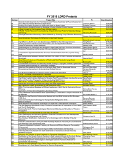
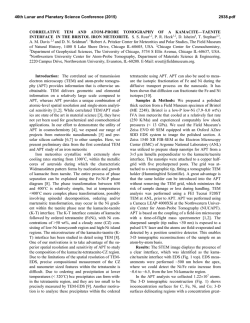
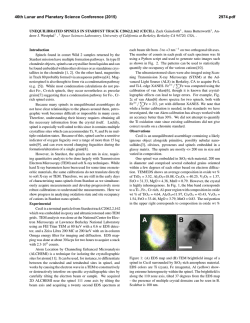
Frontiers of in situ electron microscopy Haimei Zheng, Ying Shirley Meng, and Yimei Zhu, Guest Editors In situ transmission electron microscopy (TEM) has become an increasingly important tool for materials characterization. It provides key information on the structural dynamics of a material during transformations and the ability to correlate a material’s structure and properties. With the recent advances in instrumentation, including aberration-corrected optics, sample environment control, the sample stage, and fast and sensitive data acquisition, in situ TEM characterization has become more powerful. In this article, a brief review of the current status and future opportunities of in situ TEM is provided. The article also introduces the six articles in this issue of MRS Bulletin exploring the frontiers of in situ electron microscopy, including liquid and gas environmental TEM, dynamic four-dimensional TEM, studies on nanomechanics and ferroelectric domain switching, and state-of-the-art atomic imaging of light elements (i.e., carbon atoms) and individual defects. Introduction In situ transmission electron microscopy (TEM) is a fast-growing and fascinating area of research that has drawn tremendous attention from various fields ranging from materials science to chemistry and biology. As a powerful and indispensable tool for nanomaterials characterization, in situ TEM provides great opportunities to characterize dynamic changes in size, shape, interface structure, electronic state, and chemical composition in materials at and below the nanoscale. In situ TEM has benefited from advances in electron microscopy instrumentation that have achieved spatial resolutions in the subnanometer range, energy resolution in the sub-electron-volt range, and sensitivity to individual atoms. It is now possible to image the atomic structure of materials in real time under various external stimuli while simultaneously measuring relevant properties. A variety of in situ TEM holders have been developed to enable imaging and measurements under applied heat, stress, optical excitation, and magnetic or electric fields, and the development of environmental cells allows experiments to be performed in different gaseous and liquid environments. Developments in in situ TEM combined with aberrationcorrected high-resolution imaging, electron energy-loss spectroscopy (EELS), and energy dispersive x-ray spectroscopy have enabled many discoveries in dynamic materials processes at the atomic level that were not previously possible.1–6 With the development of controlled-environment TEM, environmental TEM (ETEM), direct observations of the structural evolution of catalytic nanoparticles under dynamic reaction conditions has been realized. The further requirements of achieving better spatial and energy resolution of dynamic measurements under relatively high gas pressures while minimizing electron-beam effects provide a framework for the advancement of ETEM. In recent years, a number of breakthroughs have occurred in the development of ETEM for imaging liquid samples.2,7,8 To introduce liquids into the high vacuum of a TEM instrument, either a microfabricated liquid-cell enclosure or an open-cell configuration using low-vapor-pressure ionic liquids has been utilized. These technical breakthroughs have yielded a plethora of achievements in imaging dynamic growth of colloidal nanoparticles,2,7,9 electrochemical processes relevant to batteries,10,11 and biological materials in liquid environments.8,12 These studies have paved the way to characterize chemical reactions and dynamic processes of materials under working conditions in real time. With the continuing development of instrumental capabilities, in situ TEM experiments can be performed to study material behavior under various external stimuli such as electrical and magnetic fields13,14 and mechanical stress.15 In situ TEM has been applied to visualize domain dynamics during ferroelectric and magnetic switching,14,16 Haimei Zheng, Materials Sciences Division, Lawrence Berkeley National Laboratory, USA; [email protected] Ying Shirley Meng, Department of NanoEngineering and Materials Science Program, University of California–San Diego, USA; [email protected] Yimei Zhu, Institute for Advanced Electron Microscopy, Brookhaven National Laboratory, USA; [email protected] DOI: 10.1557/mrs.2014.305 12 MRS BULLETIN • VOLUME 40 • JANUARY 2015 • www.mrs.org/bulletin © 2015 Materials Research Society FRONTIERS OF IN SITU ELECTRON MICROSCOPY shedding light on the switching mechanism of these fundamental processes in ferroelectric and magnetic materials. Mechanical TEM holders enabling quantitative measurements of structural evolution under an applied compressive or tensile strain have unfolded the relationship between material microstructure such as defects and mechanical properties. The recent development of ultrafast imaging and diffraction has made time-resolved four-dimensional (4D) measurement of materials a reality. In their article in this issue of MRS Bulletin, LaGrange et al. offer an overview of state-of-the-art dynamic transmission electron microscopy, in particular the principle and instrumentation of the single-short movie mode that has set a benchmark for practical ultrafast electron microscopy for ultrafast science and applications. The imaging of changes in the atomic or electronic structure of materials, including size and shape evolution during chemical reactions, requires that perturbations from the electron beam be limited. Low-dose and sometimes low-kilovolt imaging are preferred, and highly efficient data acquisition is necessary. Here, we provide an overview of recent advances in the in situ TEM study of dynamic processes in materials. We discuss opportunities for the future development of in situ TEM. This article also highlights the topics of the six articles featured in this issue of MRS Bulletin, where liquid and gas environmental TEM, dynamic 4D TEM, nanomechanics, and ferroelectric domain switching studied by in situ TEM are reported. Also in this issue, Sun et al. discuss state-of-the-art atomic imaging of light elements (i.e., carbon atoms) and single defects. In situ electron microscopy was featured in a recent workshop.17 The intent of this issue is to boost in situ TEM research globally and advance the forefront of in situ characterization of materials. in situ characterization that enables the probing of dynamic catalytic processes under real reaction conditions is necessary. In situ ETEM studies of gas–solid reactions under controlled reaction conditions can provide great opportunities for the characterization of heterogeneous catalysts. A local gaseous environment can be created by a number of approaches, including the use of a closed-gas TEM cell and gas injection into an open sample area with differential pumping in the TEM column.20–22 There have been many studies on the use of ETEM to visualize nanoparticle catalysts with atomic resolution during gas reactions. Local structural and chemical information about a catalyst particle can also be obtained through a combination of imaging, diffraction, and spectroscopy.23,24 Key insights have been acquired on catalytic active sites, defect structural evolution, the nature of bonding in redox reactions, and the correlation between microstructure and catalytic performance.6,25–28 Some examples of the ETEM study of nanoparticle catalysts are as follows: Formation of a subsurface oxide on a metal catalyst was identified during the catalytic oxidation of carbon monoxide due to the incorporation of oxygen into the metal at elevated temperatures.29 Copper nanoparticle catalysts were found to exhibit remarkable restructuring in various gaseous environments, including reversible surface faceting, which was a result of preferential adsorption of reactant molecules on different crystalline facets.25 Another recent study of an Au/CeO2 catalyst by aberration-corrected ETEM visualized the restructuring of {100} facets of gold nanoparticles during CO oxidation at room temperature.6 The CO molecules adsorbed onto the on-top sites of gold atoms in the undulating hexagonal lattice, and the restructured {100} facets could sustain CO adsorption at higher surface coverages (Figure 1). Advances in the imaging of dynamic materials processes Gas-ETEM study of catalysis Heterogeneous nanoparticle catalysts that catalyze reduction or oxidation reactions at the solid–gas interface are of paramount importance in a wide range of chemical and energy applications. Heterogeneous catalysis is a complex process that involves a number of critical steps, including diffusion, reactant adsorption, surface reaction, and product desorption. The interaction of the active catalyst with reactant gases varies with temperature, gas pressure, and surface structure of the catalyst. Transformation and restructuring of a catalytic particle often occur under reaction conditions.18,19 Understanding and elucidating the mechanism of catalysis requires knowledge of the structural and chemical evolution of the nanoparticle catalysts during the reaction. Because such information is difficult to access through ex situ characterization in a vacuum, Figure 1. In situ environmental transmission electron microscope study of catalytic Au nanoparticles (NPs) supported on CeO2. Au NP on CeO2 (a) in ultrahigh vacuum (UHV) and (b) in a gas environment that contained 1 vol% CO in air at 45 Pa at room temperature. Higher magnification images of these regions are shown at the bottom of the corresponding panels. The difference between (a) and (b) shows that the interlayer distance of Au NPs in 1 Torr (∼133 Pa ∼1.3 mbar) CO increases in contrast to that in UHV. (c) High-magnification image of Au NPs with adsorbed CO. (d) Simulated image based on an energetically favorable model, which corresponds to the selected region in (c).6 Adapted from Reference 6. © 2012 American Association for the Advancement of Science. MRS BULLETIN • VOLUME 40 • JANUARY 2015 • www.mrs.org/bulletin 13 FRONTIERS OF IN SITU ELECTRON MICROSCOPY More recently, ETEM studies of nanoporous catalysts were performed to understand the dependence of catalytic activity on pore size and residual elements.26,30 In situ ETEM observations of dealloyed nanoporous gold provided compelling evidence that surface defects are active sites for the catalytic oxidation of CO, and residual silver stabilizes the atomic steps by suppressing {111} faceting kinetics.26 Another study on a nanoporous cobalt catalyst was performed in the presence of hydrogen at various temperatures using atomic imaging and EELS. The results revealed that during H2 reduction, the valence state of CoOx nanoporous particles changed from cobalt oxide to metallic cobalt.30 In their article in this issue, Crozier and Hansen provide additional examples on in situ and operando TEM of catalytic materials. Dynamic processes in liquids native environment at ambient temperature.12,40 Whole cells in liquids have also been captured at nanometer resolution with the assistance of Au nanoparticle labels.8,41 These studies provide the basis for future nanometer-scale dynamic imaging of biological materials in liquids using TEM with proper management of the electron dosage. The study of liquid-cell TEM is rapidly expanding. The development of electrochemical liquid cells allows monitoring of electrochemical processes in liquid electrolytes in real time.10 There have been many recent studies in this area, and this work is of great interest to electron microscopists as well as battery researchers. Solid-state batteries A solid electrolyte might be the ultimate solution for safe batteries. Brazier et al. reported the first cross-sectional ex situ TEM observations of an all solid-state lithium ion “nanobattery,”42 where they used a focused ion beam to make Wang et al. describe liquid-cell TEM, a new experimental platform that allows imaging through liquids with subnanometer resolution. The development of liquid-cell TEM is largely due to technical advances in nanofabrication and membrane technology. Recently, graphene liquid cells have opened new opportunities to study liquid reactions in situ in the transmission electron microscope with improved resolution (Figure 2).7 Liquid-cell TEM has been applied to the study of nanoparticle growth mechanisms, and important insights have been achieved by direct observations of nanoparticle growth trajectories.1,9,31–38 These studies illustrate that the growth of a nanowire involves attachment of nanoparticles grown from the solution, and recrystallization and rearrangement of nanoparticles are essential for nanoparticle coalescence.1,36 The direct observation of Pt nanocube facet development revealed that growth by following the conventional surface-energy-minimization law breaks down at the nanoscale.2 The application of liquid-cell TEM has become increasingly important for studying a wide range of other materials transformations in materials science, as well as in chemistry and biology. The dynamics of nanobubbles39 and water nanodroplets40 were revealed using liquid-cell TEM. The properties of water at interfaces (see the December 2014 issue of MRS Bulletin on “Water at Functional Interfaces”) play a crucial role in functional biological membranes, flow of liquids through pores and over surfaces, hydration of biomolecules, and chemical reactions in Figure 2. (a) Graphene liquid cell and in situ high-resolution transmission electron aqueous solutions. The ability to directly study microscope imaging of Pt nanoparticle growth.7 (b) The arrow indicates a small cluster attaching to the existing nanoparticle. A twinned Pt nanoparticle is achieved with water in contact with the substrate interface a (111) mirror plane, as shown in the fast Fourier transform pattern at the bottom could provide insights into the dynamic properright. Scale bar in the image sequence is 2 nm. Note: Z.A., zone axis. Adapted from ties of water. Liquid-cell TEM has also made it Reference 7. © 2012 American Association for the Advancement of Science. possible to directly image biomaterials in their 14 MRS BULLETIN • VOLUME 40 • JANUARY 2015 • www.mrs.org/bulletin FRONTIERS OF IN SITU ELECTRON MICROSCOPY a nanobattery from a pulsed-laser-deposited solid-state battery. For probing electrodes and electrolyte materials and their interfaces, ex situ experiments often provide only limited insights because of the sample preparation and transfer needed, which prevents the determination of the time constants of electrochemically induced phase transformations. More importantly, electrochemical systems often operate at states far from equilibrium, where a system tends to relax to its equilibrium state with time. To probe the kinetics in an electrochemical system, progress has been made with in situ techniques that are able to probe the structural, morphological, and chemical changes that take place during electrochemical processes at the nanoscale, particularly at the interfaces between an electrode and electrolyte. Yamamoto et al. reported on the in situ dynamic visualization of the electric potential in an all solid-state battery with electron holography and EELS.43 Interestingly, Ruzmetov et al. found that a substantial reduction in the electrolyte thickness, into the nanometer regime, can lead to rapid self-discharge of the battery even when the electrolyte layer is conformal and pinhole-free.44 A procedure for fabricating electrochemically active, electron-transparent solid-state batteries using a focused ion beam were developed, and compositional and structural changes have been observed at the interfaces between the LiCoO2 cathode and LiPON electrolyte as well as at the interfaces between the Si anode and Cu current collector, as shown in Figure 3.45 In situ TEM with EELS is becoming one of the most important tools for studying solid– solid interfaces in a variety of solid-state devices, including batteries, solar cells, and solid-oxide fuel cells. Ferroelectric and ferromagnetic switching and behavior The need to continuously decrease the size of magnetic-bit elements in magnetic-storage media, such as magnetoresistive random access memory, is perhaps the strongest driving force underlying studies of magnetic nanostructures. A similar trend occurred for ferroelectric nanostructures. Intriguing structures and properties emerge as the size of the structure contracts. Switchable spontaneous polarization, domain engineering, and strain control of ferroelectrics were recently targeted for energy-efficient nonvolatile memories and ferroelectric field-effect transistors. Nevertheless, these efforts were hindered by a lack of experimental methods, especially ones to characterize domain-defect interactions and their dynamics, as well as to directly link local atomic displacement to polarization.5,13,46–49 Lorentz microscopy has been widely used to study skyrmions (a hypothetical particle related originally to baryons; skyrmions as topological objects are important in solid-state physics), including magnetic switching and domain configuration evolution. The dynamic process of generation and annihilation of magnetic biskyrmions was observed by in situ TEM.48 As an alternative approach, a dedicated magnetizing stage can be used to generate a magnetic field at the specimen area. For example, a magnetizing stage can be built by adding Helmholtz coils on each side of the specimen,50,51 which can be used to study the field-induced motion of magnetic domain walls. Another possible design is to bring a piezo-driven sharp needle made of a permanent magnet close to the specimen.52,53 Furthermore, to obtain the relationship between magnetic structure and temperature, a thermal element also needs to integrate with the holder. A new design capable of applying gigahertz resonance electric current and pulsed excitations in situ was developed, as shown in Figure 4, to measure the nonadiabatic spin torque effect16 and to map strongly coupled coaxial vortex motion in the dipolar- and indirect exchange-coupled regimes.54 In this issue, Li et al. present details on in situ measurements of ferroelectric domain switching using in situ biasing, atomic imaging, and displacement mapping. Other methods for probing ferroelectric switching, including off-axis electron holography, are described in References 3 and 14. Figure 3. Electron energy-loss spectroscopy (EELS) mapping and scanning transmission electron microscopy (STEM) images of a solid-state Si/LiPON/LiCoO2 rechargeable battery. Reprinted with permission from Reference 45. © 2013 American Chemical Society. Mechanical properties Many fundamental assumptions of classical mechanics and continuum mechanics fail as the MRS BULLETIN • VOLUME 40 • JANUARY 2015 • www.mrs.org/bulletin 15 FRONTIERS OF IN SITU ELECTRON MICROSCOPY Figure 4. Visualizing vortex dynamics in a nanomagnet. Top: The spin precession measurement scheme. Yellow indicates two electrodes allowing GHz electric current (j(t), where t is time) to sweep across the Landau domain structure with a clockwise chirality (in-plane magnetization) and a counter-clockwise vortex-core polarity (out-of-plane magnetization). Bottom: Lorentz micrograph of the permalloy square with the Landau state showing the vortex core orbit under resonance excitation in a transmission electron microscope. The vortex core size is approximately 20 nm.16 system size is reduced to the nanoscale. Thus, mechanical testing in the transmission electron microscope has emerged. The development of a dedicated mechanical TEM holder enables the in situ investigation of mechanical behaviors and properties at the nanoscale and even the atomic scale. Throughout recent decades, the mechanical behavior of sub-10-nm structures has been studied inside the transmission electron microscope. Several novel mechanical phenomena have been reported, such as irradiation-induced high pressure and phase transformation,55 nanoextruder effects,56 geometry interlocking effects,57 cold welding,58 partial dislocation-induced discrete plastic deformation,59 local kinks in two-dimensional nanomaterials,60 mechanical annealing,61,62 stress saturation and deformation mechanism transition,63 and liquid-like pseudoelasticity.64 In this issue, Minor et al. highlight recent advances in in situ TEM probing mechanical properties at the nanoscale (see Figure 5). development of in situ TEM involves improving time resolution, achieving liquid or gas environments, and measuring properties with multiple probes. The stringent processing requirements necessitated by nanotechnology have stimulated advances in all aspects of TEM instrumentation, including application of external stimuli, high-resolution spatial and temporal imaging, and rapid data acquisition. The discovery of new materials, novel functionality, and chemical processes depends critically on routine access to all phases of matter and the ability to capture the dynamic processes that occur at interfaces with high spatial resolution and high temporal resolution. Ultrafast electron diffraction, imaging, and spectroscopy offer unique opportunities for understanding structural dynamics and the behavior of matter under conditions far from equilibrium. The current status and future opportunities of ultrafast electron microscopy are available in a recent report.17 The evolution of interfaces in chemical environments, transient nucleation events, and atomic growth during chemical reactions remain great challenges for in situ characterization. Significant advances have been made in the imaging of individual impurity atoms inside crystalline materials using atomic-number-(Z-) sensitive high-angle annular dark-field imaging and EELS in scanning TEM, including in three dimensions.4,65 However, these studies are typically carried out on relatively simple structures. In the real world, defects form complex and nonuniform three-dimensional structures. Atomic-scale defects determine the optical and electronic properties of semiconducting and photonic materials, as well as Opportunities and challenges Despite the many achievements in understanding structure and properties from monitoring dynamic materials processes in situ, challenges remain in the real-time characterization of structure, chemistry, bonding, photonic response, and electric and magnetic properties, even in a partially simultaneous manner, at the atomic level. Future revolutionary advances in electron microscopy are needed.17 Cutting-edge technical 16 Figure 5. Example of a typical compression-to-failure in situ compression test on an individual nanocrystalline hollow CdS sphere. (a–c) Extracted video frames of the in situ compression test, corresponding to time t = (a) 0 s, (b) 1.8 s, and (c) 3.6 s. The estimated contact diameter is shown in red in (b). (d–e) Dark-field transmission electron microscope images of the hollow nanocrystalline CdS ball resting on the Si substrate (d) before and (e) after the compression test. (f) Load and displacement data from the loading portion of the in situ test versus time. The experiment was run under displacement control. Reproduced with permission from Reference 15. © 2008 Nature Publishing Group. MRS BULLETIN • VOLUME 40 • JANUARY 2015 • www.mrs.org/bulletin FRONTIERS OF IN SITU ELECTRON MICROSCOPY Concluding remarks In situ probing of matter with electron beams will experience revolutionary growth in the near future with the ability to precisely control the temperature, environment (solid, liquid, or gas), and stimuli (e.g., electromagnetic field, tunable light, and mechanical stress) of materials under realistic conditions. Fast imaging and spectroscopy with highly sensitive detection could become routine. In situ electron microscopy is anticipated to continue to impact a broad range of sciences, allowing for a greater understanding of materials function and dynamics and a deeper examination of how real-world conditions and stimuli interact with and affect structures, properties, and processes at subnanometer scales. References 1. H.-G. Liao, L. Cui, S. Whitelam, H. Zheng, Science 336, 1011 (2012). 2. H.-G. Liao, D. Zherebetskyy, H. Xin, C. Czarnik, P. Ercius, H. Elmlund, M. Pan, L.-W. Wang, H. Zheng, Science 345, 916 (2014). 3. M.-G. Han, M.S. Marshall, L. Wu, M.A. Schofield, T. Aoki, R. Twesten, J. Hoffman, F.J. Walker, C.H. Ahn, Y. Zhu, Nat. Commun. 5, 4693 (2014). 4. G.-Z. Zhu, S. Lazar, A.P. Knights, G.A. Botton, Phys. Chem. Figure 6. Future in situ electron microscopy allowing multimodal probing of material Chem. Phys. 15, 11420 (2013). functionalities. (Figure created by Hong-Gang Liao and Haimei Zheng.) Note: TEM, 5. X. Yu, Y. Onose, N. Kanazawa, J. Park, J. Han, Y. Matsui, transmission electron microscopy; ETEM, environmental transmission electron N. Nagaosa, Y. Tokura, Nature 465, 901 (2010). microscopy; a-Si, amorphous Si. 6. H. Yoshida, Y. Kuwauchi, J.R. Jinschek, K. Sun, S. Tanaka, M. Kohyama, S. Shimada, M. Haruta, S. Takeda, Science 335, 317 (2012). mass transport in materials, such as lithium-ion batteries and 7. J.M. Yuk, J. Park, P. Ercius, K. Kim, D.J. Hellebusch, M.F. Crommie, J.Y. Lee, fuel cells. For example, the location of individual dopant atoms A. Zettl, A.P. Alivisatos, Science 336, 61 (2012). 8. N. De Jonge, D. Peckys, G. Kremers, D. Piston, Proc. Natl. Acad. Sci. U.S.A. controls the performance of current generations of silicon 106, 2159 (2009). 66,67 Determining the local transistors and spintronic devices. 9. H.-G. Liao, H. Zheng, J. Am. Chem. Soc. 135, 5038 (2013). structure, displacements, or atomic reconstructions around 10. Z. Zeng, W.-I. Liang, H.-G. Liao, H. Xin, Y.-H. Chu, H. Zheng, Nano Lett. 14, 1745 (2014). defects often involves a measurement accuracy of better 11. M. Gu, L.R. Parent, B.L. Mehdi, R.R. Unocic, M.T. McDowell, R.L. Sacci, 17 than 1 pm, which is beyond current capabilities. W. Xu, J.G. Connell, P. Xu, P. Abellan, X. Chen, Y. Zhang, D.E. Perea, J.E. Evans, L.J. Lauhon, J.-G. Zhang, J. Liu, N.D. Browning, Y. Cui, I. Arslan, C.-M. Wang, Future developments in in situ and operando electron Nano Lett. 13, 6106 (2013). microscopy require the capability for quantitative measure12. U.M. Mirsaidov, H. Zheng, Y. Casana, P. Matsudaira, Biophys. J. 102, L15–L17 (2012). ments of materials structure, composition, and bonding evo13. J. Zweck, in In-Situ Electron Microscopy: Applications in Physics, Chemistry lution in technologically relevant environments, including and Materials Science, G. Dehm, J.M. Howe, J. Zweck, Eds. (Wiley-VCH, Weinheim, liquids, gases, and plasmas, thereby assuring the understandGermany, 2012), pp. 347–369. 14. M.J. Polking, M.-G. Han, A. Yourdkhani, V. Petkov, C.F. Kisielowski, ing of the structure–function relationship at the atomic scale V.V. Volkov, Y. Zhu, G. Caruntu, A.P. Alivisatos, R. Ramesh, Nat. Mater. 11, 700 with high temporal resolution (i.e., in the nanosecond range). (2012). 15. Z.W. Shan, G. Adesso, A. Cabot, M.P. Sherburne, S.A. Syed Asif, O.L. Warren, As shown in Figure 6, the ultimate in situ transmission electron D.C. Chrzan, A.M. Minor, A.P. Alivisatos, Nat. Mater. 7, 947 (2008). microscope would allow multimodal (i.e., optical, thermal, 16. S. Pollard, L. Huang, K. Buchanan, D. Arena, Y. Zhu, Nat. Commun. 3, 1028 mechanical, electrical, and electrochemical) probing of material (2012). 17. E. Hall, S. Stemmer, H. Zheng, Y. Zhu, “Report of the Basic Energy Sciences functionality with versatile, modular sample stage and holder Workshop on Future of Electron Scattering and Diffraction” (US Department geometries. of Energy, Washington, DC, 2014); available at http://science.energy.gov/bes/ news-and-resources/reports/. In situ imaging with fast recording speeds generates large 18. F.F. Tao, M. Salmeron, Science 331, 171 (2011). and rich data sets, which impose demands for data mining and 19. F. Tao, M.E. Grass, Y. Zhang, D.R. Butcher, J.R. Renzas, Z. Liu, J.Y. Chung, machine learning approaches to develop new, adaptive signals B.S. Mun, M. Salmeron, G.A. Somorjai, Science 322, 932 (2008). 20. H.L. Xin, K. Niu, D.H. Alsem, H. Zheng, Microsc. Microanal. 19, 1558 (2013). from the data instead of from a priori physical models and 21. S. Giorgio, S. Sao Joao, S. Nitsche, D. Chaudanson, G. Sitja, C. Henry, Ultraintuition. New hardware and software development is also microscopy 106, 503 (2006). 22. T. Lee, D. Dewald, J. Eades, I. Robertson, H. Birnbaum, Rev. Sci. Instrum. needed to allow real-time processing for rapid feedback dur62, 1438 (1991). ing experiments, and new tools are necessary to synthesize 23. P.L. Gai, Top. Catal. 21, 161 (2002). 24. R. Sharma, Microsc. Microanal. 7, 494 (2001). various image signals post-acquisition. MRS BULLETIN • VOLUME 40 • JANUARY 2015 • www.mrs.org/bulletin 17 FRONTIERS OF IN SITU ELECTRON MICROSCOPY 25. P.L. Hansen, J.B. Wagner, S. Helveg, J.R. Rostrup-Nielsen, B.S. Clausen, H. Topsøe, Science 295, 2053 (2002). 26. T. Fujita, P. Guan, K. McKenna, X. Lang, A. Hirata, L. Zhang, T. Tokunaga, S. Arai, Y. Yamamoto, N. Tanaka, Nat. Mater. 11, 775 (2012). 27. H.L. Xin, S. Alayoglu, R. Tao, A. Genc, C.-M. Wang, L. Kovarik, E.A. Stach, L.-W. Wang, M. Salmeron, G.A. Somorjai, H. Zheng, Nano Lett. 14, 3203 (2014). 28. S.B. Simonsen, I. Chorkendorff, S. Dahl, M. Skoglundh, J. Sehested, S. Helveg, J. Am. Chem. Soc. 132, 7968 (2010). 29. G.A. Somorjai, Y. Li, Introduction to Surface Chemistry and Catalysis, (Wiley, New York, 2nd ed., 2010). 30. H.L. Xin, E.A. Pach, R.E. Diaz, E.A. Stach, M. Salmeron, H. Zheng, ACS Nano 6, 4241 (2012). 31. J.H. Park, S. Kodambaka, F.M. Ross, J.M. Grogan, H.H. Bau, Microsc. Microanal. 18, 1098 (2012). 32. E.R. White, S.B. Singer, V. Augustyn, W.A. Hubbard, M. Mecklenburg, B. Dunn, B.C. Regan, ACS Nano 6, 6308 (2012). 33. M. Sun, H.-G. Liao, K. Niu, H. Zheng, Sci. Rep. 3, 3227 (2013). 34. H. Zheng, S.A. Claridge, A.M. Minor, A.P. Alivisatos, U. Dahmen, Nano Lett. 9, 2460 (2009). 35. J. Park, H. Zheng, W.C. Lee, P.L. Geissler, E. Rabani, A.P. Alivisatos, ACS Nano 6, 2078 (2012). 36. H. Zheng, R.K. Smith, Y.-W. Jun, C. Kisielowski, U. Dahmen, A.P. Alivisatos, Science 324, 1309 (2009). 37. Y. Liu, X. Chen, K.W. Noh, S.J. Dillon, Nanotechnology 23, 385302 (2012). 38. K. Jungjohann, S. Bliznakov, P. Sutter, E.A. Stach, E. Sutter, Nano Lett. 13, 2964 (2013). 39. E.R. White, M. Mecklenburg, S.B. Singer, S. Aloni, B.C. Regan, Appl. Phys. Express 4, 055201 (2011). 40. U.M. Mirsaidov, H. Zheng, D. Bhattacharya, Y. Casana, P. Matsudaira, Proc. Natl. Acad. Sci. U.S.A. 109, 7187 (2012). 41. K.-L. Liu, C.-C. Wu, Y.-J. Huang, H.-L. Peng, H.-Y. Chang, P. Chang, L. Hsu, T.-R. Yew, Lab Chip 8, 1915 (2008). 42. A. Brazier, L. Dupont, L. Dantras-Laffont, N. Kuwata, J. Kawamura, J.-M. Tarascon, Chem. Mater. 20, 2352 (2008). 43. K. Yamamoto, Y. Iriyama, T. Asaka, T. Hirayama, H. Fujita, C.A. Fisher, K. Nonaka, Y. Sugita, Z. Ogumi, Angew. Chem. 122, 4516 (2010). 44. D. Ruzmetov, V.P. Oleshko, P.M. Haney, H.J. Lezec, K. Karki, K.H. Baloch, A.K. Agrawal, A.V. Davydov, S. Krylyuk, Y. Liu, Nano Lett. 12, 505 (2011). 45. D. Santhanagopalan, D. Qian, T. McGilvray, Z. Wang, F. Wang, F. Camino, J. Graetz, N. Dudney, Y.S. Meng, J. Phys. Chem. Lett. 5, 298 (2013). 46. L. Huang, M.A. Schofield, Y. Zhu, Adv. Mater. 22, 492 (2010). 47. M. Varón, M. Beleggia, T. Kasama, R. Harrison, R. Dunin-Borkowski, V. Puntes, C. Frandsen, Sci. Rep. 3, 1234 (2013). 48. X. Yu, Y. Tokunaga, Y. Kaneko, W. Zhang, K. Kimoto, Y. Matsui, Y. Taguchi, Y. Tokura, Nat. Commun. 5, 4198 (2014). 49. X. Yu, N. Kanazawa, W. Zhang, T. Nagai, T. Hara, K. Kimoto, Y. Matsui, Y. Onose, Y. Tokura, Nat. Commun. 3, 988 (2012). 50. A. Budruk, C. Phatak, A. Petford-Long, M. De Graef, Acta Mater. 59, 6646 (2011). 51. A. Kohn, A. Kovács, R. Fan, G. McIntyre, R. Ward, J. Goff, Sci. Rep. 3, 2412 (2013). 52. D. Shindo, Y.-G. Park, Y. Gao, H.S. Park, J. Appl. Phys. 95, 6521 (2004). 53. H. Park, Y.-G. Park, Y. Gao, D. Shindo, M. Inoue, J. Appl. Phys. 97, 033908 (2005). 54. J. Pulecio, P. Warnicke, S. Pollard, D. Arena, Y. Zhu, Nat. Commun. 5, 4760 (2014). 55. L. Sun, A.V. Krasheninnikov, T. Ahlgren, K. Nordlund, F. Banhart, Phys. Rev. Lett. 101, 156101 (2008). 56. L. Sun, F. Banhart, A. Krasheninnikov, J. Rodriguez-Manzo, M. Terrones, P. Ajayan, Science 312, 1199 (2006). 57. D.-M. Tang, C.-L. Ren, M.-S. Wang, X. Wei, N. Kawamoto, C. Liu, Y. Bando, M. Mitome, N. Fukata, D. Golberg, Nano Lett. 12, 1898 (2012). 58. Y. Lu, J.Y. Huang, C. Wang, S. Sun, J. Lou, Nat. Nanotechnol. 5, 218 (2010). 59. H. Zheng, A. Cao, C.R. Weinberger, J.Y. Huang, K. Du, J. Wang, Y. Ma, Y. Xia, S.X. Mao, Nat. Commun. 1, 144 (2010). 60. I. Nikiforov, D.-M. Tang, X. Wei, T. Dumitricaˇ, D. Golberg, Phys. Rev. Lett. 109, 025504 (2012). 61. Z. Shan, R.K. Mishra, S.S. Asif, O.L. Warren, A.M. Minor, Nat. Mater. 7, 115 (2007). 62. L. Huang, Q.-J. Li, Z.-W. Shan, J. Li, J. Sun, E. Ma, Nat. Commun. 2, 547 (2011). 63. Q. Yu, Z.-W. Shan, J. Li, X. Huang, L. Xiao, J. Sun, E. Ma, Nature 463, 335 (2010). 64. J. Sun, L. He, Y.-C. Lo, T. Xu, H. Bi, L. Sun, Z. Zhang, S.X. Mao, J. Li, Nat. Mater. 13, 1007 (2014). 65. P.M. Voyles, D.A. Muller, J.L. Grazul, P.H. Citrin, H.J.L. Gossmann, Nature 416, 826 (2002). 66. M. Pierre, R. Wacquez, X. Jehl, M. Sanquer, M. Vinet, O. Cueto, Nat. Nanotechnol. 5, 133 (2010). 67. G.P. Lansbergen, R. Rahman, C.J. Wellard, I. Woo, J. Caro, N. Collaert, S. Biesemans, G. Klimeck, L.C.L. Hollenberg, S. Rogge, Nat. Phys. 4, 656 (2008). 57 th Electronic Materials Conference June 24-26, 2015 // The Ohio State University // Columbus, OH C A L L F O R PA P E R S CONFERENCE CHAIR SAVE THE DATE th The 57 Electronic Materials Conference (EMC 2015) is the premier annual forum on the preparation and characterization of electronic materials. Held June 24-26 at The Ohio State University, this year’s Conference will feature a plenary session, parallel topical sessions, a poster session and an industrial exhibition. Mark your calendar today and plan to attend! SCIENTIFIC PROGRAM The three-day conference will concentrate on the following topical categories: tEnergy Conversion and Storage Materials tWide Bandgap Materials tOrganic Materials and Thin Film Technology tEnabling Technologies tNanoscale Science and Technology in Materials Andrew Allerman Sandia National Laboratories PROGRAM CHAIR Jamie Phillips University of Michigan ABSTRACT SUBMISSION ENDS Student participation in this Conference is partially supported by a grant from the TMS Foundation. JANUARY 30, 2015 www.mrs.org/57th-emc 18 MRS BULLETIN • VOLUME 40 • JANUARY 2015 • www.mrs.org/bulletin
© Copyright 2026