
Studies on the Morphology and life cycle of some Helminth
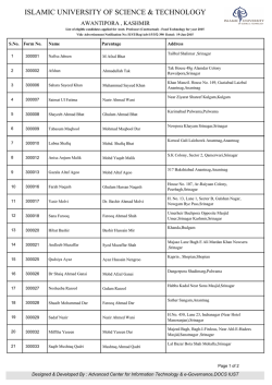
Studies on the m orphology of som e oxyurid nem atodes from com m on toad in K ashm ir. D. N, FOTEDAR & R. K. TIKOO Post-Graduate Department of Zoology, J & K University, Srinagar, Kashmir, ( India ). P A R T III ( A B ST R A C T S ) O F T H E PRCEEDINGS OF T H E 53rd SESSION OF T H E INDIAN SCIEN CE CONGRESS — CH ANDIGARH , 1965-66. Reprinted From : firoca's Press, Srinagar. 63A. Studies on the m orphology of som e oxyurid nem atodes from com m on toad In K ashm ir. D. N. F O T E D A R & R. K. TIKOO, Srinagar. Present studies pertain to two oxyurid nematodes, Oxysomatium srinagarensis F o t e d a r , 1960 and Cosmocerca kashmirensis F o t e d a r , 1959, recovered from the rectum of Bufo viridis from various areas in Kashmir. Heavy infection in the host was recorded in September. Many toads were found to harbour both the forms of oxyurids. Several variations were recorded in the two species. Their brief description and revised specific diagnosis is given in the paper. In 0 . srinagarensis, variations are recorded in the number of papillae and body measurements including those of spicules. The papillae in male post-cloacal region are found to vary from 8 to 10 pairs, those in cloacal region from 3 to 5 pairs. Paired papillae are also recorded in pre-cloacal region. In C. kashmirensis, variations are mainly recorded in plectanes and in the distribution of simple papillae in the male caudal region. While the typical number of 19 plectanes, (8 pairs in two irregular row’s and 3 additional plectanes-rosettes), some specimens have only 7 pairs of fully developed plectanes and 3 additional rosettes. The position of lateral rosette also varies. Three additional rosettes were not seen in specimens which were otherwise well developed. As regards the post-cloacal papillae, the number is variable, being 1 to 5 pairs ventral, 3 to 4 pairs dorsal and 1 pair caudal. O b a New Species of the Nem atode Genus Kalicephalus Molin, 1861 from a Snake in K ashm ir. D. N. FOTEDAR, M. K. RAINA AND RATNI TIKOO Reprinted from K A S H M IR SCIEN CE, Vol. Ill, Nos. 1-2, December 1966. On a New Species of the N em atode Genus Kalicepkalus Molin, 1861 from a Snake in K ashm ir. D. N. Fotedar, M . K, R ain a & Ratni T ikoo Post - Graduate Department o f Zoology, J & K University, Srinagar, K ashm ir. INDIA Several specimens of the genus KaVicephalus M o l i n , 1861 were collec ted from the oesophagus and stomach of two common snakes of Kashmir, Ptyas mucosus and Tropidonolus platyceps. From the former snake eight specimens, three females and five males, were found to constitute a new species. One of the female specimens was collected from the stomach of another host of the same species and the remaining specimens from the oesophagus. The specimens collected from the stomach of Tropidonotus platyceps, one male and two females, were found to belong to K. indicus O r t l e p p , 1923 which are briefly described herein. Strongylata Railliet & Henery, 1913. Strongyloidea Weinland, 1858. Diaphanocephalidae Travassos, 1919. Kalicepkalus Molin, 1861. Kalicepkalus kashmirensis n. sp. Host : Ptyas mucosus Location : Oesophagus and Stomach Locality : Srinagar, Kashmir. The worms are stout and cylindrical with anterior end dorso - ventrally broad and obliquely truncated. The maximum diameter is attained at about the middle of body, wherefrom it tapers slightly posteriorily. In both the sexes the cuticle shows longitudinal striations and bears minute to large papilla—like structures which are profusely developed in the anterior and pos terior regions of the body. The posterior region also shows transverse cuticular striations. The buccal capsule is compressed laterally and is without teeth. Mouth is a dorso-ventrally elongated slit, bounded by two lateral lips. Each lip bears three papillae which correspond to three parenchymatous bands in the buccal capsule. The buccal capsule is supported at its base by chitinous 94 D. N. FOTEDAR, M. K. RAINA AND RATNI TIKOO triangular plates and transverse bars. The buccal capsule is usually broader than long. The oesophagus is flask shaped and lined internally by chitin, and the billb has a diameter more than half the length of oesophagus. The oesophageal glands are well developed. The intestine runs straight to the posterior end and has roughly a uniform diameter throughout its length. The excretory pore is present at the level of the junction of oesophagus and intestine. Male Males are smaller than females. The bursa is broad and has two small ventral lobes, two large lateral lobes and a median dorsal lobe. The bursa is supported by rays. The ventro - ventral, and ventro - lateral arise from the same stem and are of equal size. The externo - lateral, medio - lateral and postero - lateral also arise from a common stem. The postero - lateral is the longest of the three and the externo lateral is the shortest. Externo - dorsal rays arise at the base of the dorsal and are long, directed outwards and backwards. The dorsal ray divides into two branches, each of which gives rise to three branches in turn. The first branch is given out on the outer side after which the main branch extends a short distance before dividing into two branches, the inner one being longer than the outer one. The two spicules are long and unequal or sub - equal. They are provided with alae, usually on the inner side with a rough margin. The spicules are broad at the base showing clear transverse striations and gradually tapering towards the tips where they form a long spear like expansion, The gubernaculum is not well represented but instead the whole wall of the cloaca seems to be chitinised forming a well de/eloped telamon at the tip. On either side of the so formed genital cone there are two extensions, the inner one smaller and papillae like and the outer one larger and flap like. Pre - bursal papillae are not seen. Female The females are broad anteriorly and tapering towards the posterior end. The tail, is short bearing a small acuminate spine - like tip. The ovaries are much coiled and long. The vulva is on a prominent protuberance situated in the posterior region of body. Behind the said valvar protuberance is a slight bulbous enlargement of the body. The vagina is short. The anterior uterus runs straight towards the anterior end, while the posterior one after taking a slight turn runs parallel to the first one. This is a constant feature in all the specimens. The eggs are ovoid and immature in all the specimens and measure 0.045 X 0.03 mm in size. Measurements ( in m m .) M ale A B C D Length. 5.7 5.925 5.925 7.35 Max. breadth at middip 0.24 0.255 0.255 0.27 Breadth oes. level 0.21 0.225 0.21 0.27 Breadth Pre-bursal level 0.15 0.18 0.195 0.21 Oes. length. 0.255 0.27 0.255 0.33 95 NEW SPECIES OF KALICEPHALUS 0.15 0.165 0.15 0.21 0.39 0.405 0.405 0.36 0.405 0.395 0.36 0.345 Buccal capsule length. 0.135 0.135 0.135 0.15 Buccal capsule breadth. 0.15 0.135 0.15 0.165 Oes. length, Oes. breadth. 1.7 1.63 1.7 1.57 Bursa length. 0.18 1.18 1.18 1.195 Bursa breadth. 0.48 0.57 0.54 0.63 Oes. breadth. Spicule length (a) (b ) A Length. 7.5 B 8.55 Max. breadth at middle. 0.345 0.375 0.375 Breadth at oes. 0.225 0.225 0.225 Breadth at preanal region. 0.135 0.12 0.135 Oes. length. 0.285 0.315 0.3 Oes. breadth. 0.165 0.18 0.18 Distance from ant. end to vulva 5.85 6.525 6.75 Length of vulvar projection. Tail length. 0.075 0.195 0.075 0.2 0.09 0.18 Buccal capsule length. 0.15 0.15 0.15 Buccal capsule breadth. Total length Distance from ant. end to vulva Tatal length Distance from post, end to vulva 0.18 0.18 0.18 1.28 1.31 1.33 4.54 4.2 4 C 9 96 D. N. FOTEDAR, M. K. RAINA AND RATNI TIKOO DISCUSSION At present the family Diaphanocephalidae T r a v a s s o s , 1920 includes three genera, namely Diaphanocephalus D i e s i n g , 1851; Kalicepkalus M o l i n , 1861 and Occipitodontus O r t l e p p , 1923. As regards Occipitodontus there is much controversy. The genus was raised by O r t l e p p (1923) for Kalicepkalus willeyi, L i n s t o w , 1904, on the basis of the structure of buccal capsule which is different from the other two genera. B a y 1 i s and D a u b n e y ( 1925 ) synonimised it again with Kalicephalus which was also recognised by M a p l e s t o n e (1931) and later followed by Y a m a g u t i (1961). Y e h ( 1956) is of the opinion that Occipitodontus is a valid genus as recognised earlier by S k r j a b i n ( 1952). The validity of the genus is quite evident, because the structure of the buccal capsule is taken as an important taxonomic character, the buccal capsule being distinct from the other two genera in Occipitodontus. As regards the fourth genus Kalicephaloides proposed by Y e h ( 1956 ) for Kalicephalus minutus ( B a y l i s and D a u b n e y , 1922 ), the authors are of the opinion that it cannot be separated from all other species of Kalicep halus and given the rank of a genus only on the basis of unequal spicules and the slight difference in the branching of the dorsal ray. These differences are of specific value and this species is considered here as a member of the genus Kalicephalus. The present form is assigned to Kalicephalus because its buccal capsule and other morphological characters are similar to the genus. It however resembles Diaphanocephalus D i e s i n g , 1851 in having unequal spicules, but differs from it in many other characters. The authors do not think it plausi ble to raise it to a new genus, although it differs from all species of Kalicep halus in having a 5 - lobed bursa and unequal spicules. ( Unequal spicules have been reported in K. naiae M a p l e s t o n e , 1931 but Yamaguti (1961) includes equal spicules as one of the diagnostic features of Kalicephalus). There are at present about 48 species of Kalicephalus as recognised by S k r j a b i n ( 1961 ) while Y a m a g u t i (1961) recognises 50 species, in which he includes species of Occipitodontus also. Out of all these species only two have been reported from the Oesophagus ( besides other parts of g u t) of snakes; these are K. willeyi L i n s t o w , 1904 and K. tennesseensis H a r d w o o d , 1934. There are four species which have also been reported from the same host from which the present specimens were collected. There a r e : K. bengalensis M a p l e s t o n e , 1929; K. indicus O r t l e p p , 1923, K. brachycephalus M a p l e s t o n e , 1923 and K. elongatus M a p l e s t o n e , 1931, but none of these reported pertain to the oesophagus of the snake. From, all the existing species of the genus the present form seems to be nearer to the above men tioned species as also to K. parvus M a p l e s t o n e , 1931. From the latter it differs in size ratios and in having uneqal or subequal spicules and 5 lobed bursa. The vulvar region which is prominent on a protuberance and has a post vulvar enlargement in the species under discussion is not so in K. indicus. NEW SPECIES OF KALICEPHALUS 97 In K. bengalensis the bursa ends in two sharp points ventrally and a pre-bursa! papilla is present. Both the features are absent in the present form. M oreover, the inner last branch of the dorsal ray is the longest in the present form which are equal in K. bengalensis. The telamon is very well developed in the present species and is U - shaped with one of its limbs slightly longer. In K. bengalensis only the tips of the spicules are alate, while in the present form the spicules are alate along their whole length. A gubernaclum is apparen tly absent in the present form. The females of the two species resemble in having a protuberant vagina, which is more prominent* in our specimens ( K. kashmirensis). In K. kashmirensis the vagina is very short and divides into two uteri, the posterior one taking a short turn before running parallel to the anterior uteru s; but in K. bengalensis the posterior uterus starts half way between the vulva and the anus. A comparsion of K. brachycephalus with K. kashmirensis suggests that the manner of division of the dorsal ray is vividly different in our species. They also differ in the structure of spicules. The present form resembles K. parvus M a p l e s t o n e , 1931 in having excretory pore at the same level and in the manner of branching of dorsal and other rays, but differs in the structure of bursa and spicules. In view of the above discussion based on characters different from those possessed by the existing species, the present form is regarded as a new species under the genus Kalicephalus M o l i n 1861 for which the name Kalicephalus kashmirensis is proposed. ( The specimens deposited in the Helminthological laboratory of P. G. Department of Zoology, J & K University, Srinagar. — Collection No. N 30A ) Kalicephalus indicus O r 1 1 e p p , 1923. H o st: Tropidonotus platyceps. Location : Stomach. Locality: Pahalgam (K ashm ir), The parasites are relatively small and the cuticle shows the usual longitudinal striations. T he papillae over the cuticle are sparse. The buccal capsule is like other species showing variations in size only. The duct of dorsal oesophageal gland projects into the mouth cavity. Male The male is smaller than the females and has a Companulate trilobed bursa. Ventral rays are long and closely approximated. Laterals arise from common trunk and are separated from each other. All the laterals are shorter than the ventrals, the externo lateral being the shortest. Externo - dorsals are short and stout arising from the dorsal ray. The dorsal ray further divides into two branches, each branch gives off two small branches one after the other and a third branch which is the longest. Spicules are equal, long and thin. They are broad at the base and tapering towards the apex and are twisted a little above apex. Gubernaculum is stout, rod-shaped and dorsal to the spicule. It is less than- half the length of the spicule. Telamon is not seen. There are no pre-bursal papillae. NEW SPECIES OF KALICEPHALUS 99 Female The tail is small and conical. It is tapering gradually and has transversely striated cuticle. Vulva is located in the posterior region. The uteri are diver gent continuing a little distance behind before turning anteriorly. Immature eggs are oval in outline. The body papillae are large and scanty. Measurements ( in m m .) Male Female 5.2-5.6 Length 4 0.18 0.24 Breadth Buccal capsule length 0.1 0.12 Buccal capsule breadth 0.1 0.1 Length of Oesophagus 0.21 0.23 Breadth of Oesophagus 0.09 0.1 Nerve ring ( from ant. end ) 0.16 — Length of bursa 0.33 — W idth of bursa 0.19 — Length of spicule 0.22 - — Length of gubernaculum 0.09 — Length of tail — 0.19 Distance between vagina and tail tip — 1.5 SUMMARY 1. Kalicephalus kashmirensis n. sp. is described from Ply as mucosus of Kashmir. It has unequal and alate spicules with distal spear-like expansions, well developed telamon and two extensions on either side of genital cone. The females have a prominent vulvar protuberance and a short acuminate caudal spine. 2. Kalicephaloides Y e h , 1956 is suppressed as synonym of Kalicephalus. 3. Kalicephalus indicus O r t l e p p , platyceps of Kashmir. 1923 is recorded from Topidonotus 100 D. N. FOTEDAR, M. K. RAINA AND RATNI TIKOO REFERENCES Maplestone, P. A., 1929.- Two new species of nematodes from Indian hosts. Ree. Indian. Mus., 31 s 87 - 92. 1932.- Parasitic Nematodes obtained from animals dying in the Calcutta Zoological Gardens. Part II. Rec. Indian. M us., 34 : 251-261. 1952.- Stongylata. In Skrjabin, K. I. Key to parasitic Skrjabin, K. I., nematodes ( in Russian tex t) vol. III. Acad. Schikhobalova, N. P Science Moscow 882 pp. English translation by Schulz, R. S., Birron, A., and Cole Z. S. (1961 ) Israel Programme Popova, T. I., Sci. Translation Jeruselem, 890 pp. Boev, S. N„ and Deylamure, S. L., 1961.- Systema Helminthum ; vol. III. The Nematodes of Yamaguti, S., Vertebrates. Parts I & II Interscience Pub. N. Y. 1261 pp. Yeh, Liang-Sheng., 1956.- O n Kalicephalus hongkongensis n. sp. from Elaphe moellendoiffi and the erection of a new genus Kalicephaloides. J . Helminth. 30 ( 4 ) : 203-210. 1926.- The nematode parasites of vertebrates. London, York, W ., & 536 pp. Maplestone, P. A., Printed at Br oca's Artistic Press, Srinagar, Kashmir. Ccsmocerca kashmirensis Fotedar, Bufoviridisin Kashmir- On the life cycle c f 1959 com m on nem atode parasite o f D. N. FO T E D A R A N D R A T N I TIK O O P o st-G rad u ate D epartm ent of Zoojogy, J . <& K . University, Srinagar. Kashmir-India R eprinted from : Proceeding of the 54th Session of the Indian Science Corgress, Hyderabad 1967, P art IV (Late A bstracts), p. 21 p. 21 8. On the life cycle of Cosmocerca kashmirensis Fotedar, 1959, common nematode parasite of Bufo viridis in Kashmir. D. N . F O T E D A R & R A T N I T IK O ‘ Srinagar F otedar (1959) described Cosmocerca kashmirensis from the rectum of Bufo viridis, common toad in Kashm ir. This is the first record of th e genus from Indian region. F otedar and Tikoo (1955) rep o rted several variations in this parasite and the o th er common oxyurid parasite of Bufo, Oxysomatium srinagwensis F otedar, 1960. P resen t studies p ertain to th e life cycle of Cosmocercj. kashmirensis. The egg and its early segm entation, first and second stage h rv a of the parasite have been studied by th e authors. G ravid fem ales, recovered from th e toads, w ere allow ed to oviposit in several media. R at faeces boiled in w ater and the solution filtered was used as satisfactory m edium for the developm ent of larvae H ost faeces gave equally satisfac tory results The em bryonated eggs after being released by th e female, hatched w ithin 2 —4 hours a t room tem p eratu re A t 24” C it takes 4—5 days for th e second stage larvae to develop The second stage or th e infective stage larvae are less active. They w ere fed orally to laboratory bred toads. Young adults were developed w ithin 8— 10 days and m ature ones w ithin 15—20 days. Further studies on the life cycle of Cosmocerca kashmirensis Bufo Fotedar, 1959, common oxyurid nematode parasite of viridis in Kashmir. D. N. FO T E D A R A N D R A T N I TIK OO P o st-G rad u ate D epartm ent of Zoology, J . c& K . University, Srinagar, Kashmir-India Reprinted from : Proceedings of the 55th Session of the Indian Science Congress, Vatana&i, January 1968, P art III (A bstracts), p. 460. p 460 studies on the life cy cle o f Cosmocerca k25. ashmiFurther rensis Fotedar, 1959, com m on oxyurid nem atode parasite o f Bufo v iridis in Kashmir D. N. FO T E D A R & R A T N I TIKOO Srinagar. CosmocercaKashmvensisis a common parasite of the rectum of Bufoviridis in Kashmir. F otedar and Tikoo (1966) studied its m orphology and th at of Oxysomatium srinagarensis Fotedar, I960 in detail and recorded several morphological variations. The present w ork on the life cycle of C- Kashmirensis includes the study of egg and its early segm entation, I and Il-stage free living juveniles and mode of infection to the host. R at faeces boiled in w ater and solution filtered was used as a satisfactory medium for the juveniles. M edium prepared from host faeces was equally satisfactory. The gravid females released eggs in tap w ater and norm al salt solution as easily as in the said media. The em bryonated eggs hatched w ithin 2-4 hours at n_om tem p eratu re (22-32°C) afcer th eir release from gravid females T he I-stage juvenile is 0.45-0.67 mm. long and has a short cylindical buccal capsule The oesophagus is rhabditiform type. It is highly mobile and continues to be so for 4-5 days after w hich lethargus sets in for a few hours and then it undergoes m oulting The Il-stage (infective stage) juvenile resem bles filiform larv a. It rem ains sheathed for some tim e, moving about w ithin old cuticle. Soon it sheds off the cuticle and shows active m ovem ents for a brief period of 24-36 hours. It is 0 5-0 7 mm. long. T he buccal capsule in reduced and oesophagus elongated The in testin al cells are in tw o prom inent rows and the genital prim ordia are along ventral side. E arlier presum ption of the authors th a t rhe juveniles rea ched d ire c t to the norm al site of the host for fu rth er grow th and sexual developm ent was found to be wrong by subsequent experim ents on the mode of infaction. Il-stage juveniles are capable of active penetration through host skin. Visceral m igration was found necessary for the juveniles. Juveniles w ith larger size w ere recovered 3 days after the toads were ex posed to infection by placing th eir skin in close contact w ith the infective stage juveniles in shallow containers. The toads previously tested for negative infection, w ere also successfully infected by subcutaneous injection of the juveniles. It takes 10-14 days for the parasites to reach th e norm al site for fu rth er grow th and sexual m aturity. T he longevity of the adults and the viability of eggs and juveniles in differen t m edia and at different tem peratures are also being studied. llnrr-n Iqbal T-ibr i ' "1 hesas Ac* No - ‘■1*


© Copyright 2026