
Desmopressin Induces Endothelial P-Selectin Expression
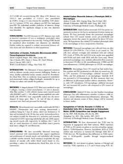
From www.bloodjournal.org by guest on February 6, 2015. For personal use only.
Desmopressin Induces Endothelial P-Selectin Expression and Leukocyte
Rolling in Postcapillary Venules
By Samina Kanwar, Richard C. Woodman, M.C. Poon, Toyoaki Murohara, Allan M. Lefer, Kelly L. Davenpeck,
and Paul Kubes
Desmopressin, (DDAVP; l-desamino-8-D-arginine vasopressin) increases the release and activity of von Willebrand factor (vWF); however, its effects on the other major constituent ofendothelial Weibel-Palade bodies, P-selectin, has
not
been investigated. DDAVP-induced P-selectin expression
may explain DDAVP's efficacy in bleeding disorders in which
vWF levels are normal. Therefore, the objective of this study
is t o assess the effect of DDAVP on P-selectin expression
on endothelial cells of postcapillary venules in vivo and on
human umbilicalvein endothelium in vitro, and t o determine
whether DDAVP has direct effects on leukocyte behavior in
postcapillary venules. DDAVP (0.1 and 1.0 pg/mL) induced
a significant but transient increase in P-selectin expression
on human umbilical vein endothelial cells as well as on rat
and human platelets. Immunohistochemical analysis of rat
postcapillary venules showed that in contrast t o saline,
DDAVP injection (1 pglkg, intravenous) induced significant
endothelial P-selectin expression.DDAVP
administration
also induced a rapid and significant increase in leukocyte
rolling in rat mesenteric venules in vivo. This response was
entirely dependent on P-selectin, as an anti+-selectin antibody rapidly reversed the DDAVP-inducedincrease in leukocyte rolling.DDAVP induced leukocyte rolling in medium (20
t o 40 pm) and large (>40 pm), but not small (c20pm),
postcapillary venules. In animals that were treated with
DDAVP, there wasa steady and significant increase in leukocyte adhesion. This study shows that DDAVP can directly
induce P-selectin expression on endothelium in vitro and in
vivo and that the latter response is capable of supporting
prolonged leukocyte rolling in rat postcapillary venules.
0 1995 by 7'he American Society of Hematology.
F
bodies of endothelial cells? Although increases in plasma
vWF and factor VI11 activity may explain the efficacy of
DDAVP in patients with type I vWD and hemophilia A,',',',"'
the mechanism(s) in patients who have normal or elevated
vWF remains unknown. Mechanisms independent of endothelial cell release of vWF have also been p r o p ~ s e d . ~ ~ ~ ~ ~ " ~ ~
Besides vWF and its related propolypeptide, P-selectin is
the only other constituent within WP bodies that is known
to be expressed on endothelial cells and to have an adhesive
f ~ n c t i o n . ' ~Recent
" ~ studies have shown that endothelial cell
expression of P-selectin is important for the very earlyleukocyte-endothelial cell interaction known as leukocyte rolling,17-20
an absolute prerequisite for leukocyte adhesion and
Although several agonists have been reported
to mediate vWF release and P-selectin upregulation on endothelial cell^,'^^'^^^^ the effect of DDAVP on endothelial cell
P-selectin expression and leukocyte-endothelia1 cell interactions has not been characterized. It is conceivable that
DDAVP's efficacy in disorders other than type 1 vWD and
hemophilia A may be partially caused by its ability to induce
P-selectin expression and thereby mediate leukocyte-endothelia1 cell interactions.
Therefore, in this study we systematically examined the
potential effect of DDAVP on P-selectin expression and
function on endothelial cells of postcapillary venules. This
was accomplished using immunohistochemical staining of
rat mesenteric venules exposed to DDAVP and intravital
microscopy to visualize leukocyte behavior in single postcapillary venules.
OR MORE THAN a decade desmopressin (DDAVP; 1desamino-8-D-arginine vasopressin), a synthetic derivative of the hormone L-arginine vasopressin, has been effectively used as a hemostatic agent in a variety of acquired
and inherited bleeding disorder^."^ DDAVP has been shown
to rapidly shorten bleeding time and reduce blood loss in
patients withmild and moderate hemophilia A, selected
types of von Willebrand disease (vWD) and several forms
of platelet dysf~nction.~-'
In certain circumstances the need
for blood products, and consequently the risk of transfusiontransmitted diseases, has been reduced because of DDAVP
administration.
Although a single unifying mechanism to explain the beneficial effects of DDAVP in the above heterogeneous group
of disorders is lacking, there is considerable evidence to
implicate an improvement, albeit transient, in hemostasis
after DDAVP administration."' It has generally beenassumed that the mechanism of action of DDAVP is to induce
release of preformed high- molecular-weight multimers of
von Willebrand factor (vWF) from the Weibel-Palade (WP)
From the Immunology Research Groupand the Division of Hematology,University of Calgary,Calgary,Alberta,Canada;
andthe
Department of Physiology, Jefferson Medical College, Thomas Jefferson University, Philadelphia, PA.
Submitted February 21, 1995; accepted May 31, 1995.
Supported by grants from the Alberta Heritage Foundation for
Medical Research (AHFMR),the Medical Research Council (MRC),
and the National Institute of General Medical Sciences of the National Institutes of Health. P.K. is an MRC and AHFMR scholar
and R. C.W. is a Clinical Investigator of the AHFMR.
Address reprint requests to Samina Kanwar, Departmentof Medical Physiology, Faculty of Medicine, Universityof Calgary, Calgary,
Alberta, T2N 4NI Canada.
The publication costsof this article weredefrayed in part by page
charge payment. This article must therefore be hereby marked
"advertisement" in accordance with 18 U.S.C. section 1734 solely to
indicate this fact.
0 1995 by The American Society of Hematology.
0006-4971/95/8607-0027$3.00/0
2760
MATERIALS AND METHODS
Intravital microscopypreparation.
Male Sprague-Dawley rats
(180 to 250 g) were maintained on a purified laboratory diet and
fasted for 24 hours before surgery. The animals were initially anesthetized with pentobarbitol sodium (65 mgkg body weight), and the
right carotid artery and jugular vein were cannulated for systemic
arterial blood pressure measurements (Statham F'23A pressure transducer and a Grass physiologic recorder, Scarborough, Canada) and
intravenous ( W ) drug administration, respectively. A midline abdominal incision was made and a segment of the mid-jejunal mesenBlood, Vol 86, No 7 (October l ) , 1995: pp 2760-2766
From www.bloodjournal.org by guest on February 6, 2015. For personal use only.
2761
DDAVP, P-SELECTIN, AND LEUKOCYTE ROLLING IN VIVO
minutes at room temperature and then washed once with 500 pL
tery was gently exteriorized and carefully placed over an optically
PBS. The wells were blocked with 1 mL of PBS containing 1%
clear viewing pedestal that permitted transillumination of a 2-cm2
bovine serum albumin (wt/vol) and 0.01% NaN3 for 30 minutes
segment of mesentery. Small, single unbranched postcapillary vefollowed by incubation with 100 pL of the primary antibody (S121
nules were visualized and the following parameters were measured:
solution (10 pg/mL) for 30 minutes. This antibody was kindly provenular diameter, leukocyte rolling and adhesion, leukocyte rolling
vided by Dr R. McEver (University of Oklahoma). After three
velocity, centerline red blood cell ( W C ) velocity, and shear rates.
washes with 0.05% ’TWEEN 20 (Sigma Chemical CO, St Louis,
This preparation has been extensively used by us25-27
and other^^^-^"
MO)/PBS, 100 pL of the secondary antibody (peroxidase-labeled
to examine leukocyte-endothelia1cell interactions in vivo.
goat-antimouse IgG; Kirkegaard and Perry Laboratories Inc, MaryExperimental protocol. Immediately upon finding a postcapilland) was added to the wells, incubated for 30 minutes, and washed
lary venule, the image was recorded for 5 minutes followed by three
three times. A substrate solution, ABTS peroxidase substrate (Kiradditional 5- minute recordings at 15, 30, and 60 minutes. For each
kegaard and Perry Laboratories Inc), was then added to the wells
set of experiments, all animals were pretreated before laparotomy
and the reaction allowed to develop for 30 minutes. Absorbance was
with sodium cromoglycate (5 mgkg IV) to minimize surgically
then measured at 405 nm.
induced leukocyte rolling as previously de~cribed.~’
Animals reFlow cytometric analysis of P-selectin expression on platelets.
ceived DDAVP in normal saline at 1 pgkg as an IV bolus. In other
Flow cytometric analysis of P-selectin expression on rat and human
animal models, investigators have used DDAVP ranging from 0.3
platelets was performed by a method previously de~cribed.~’
In brief,
to 4.0 pgkg body weight and have demonstrated enhanced hemoblood was collected from six male Sprague-Dawley rats and was
static and antidiuretic effe~ts.~’”~
The mesentery was continuously
anticoagulated with sodium citrate phosphate buffer (Sigma Chemisuperfused with warmed bicarbonate-buffered saline, and leukocyte
cal CO, St Louis, MO). Human blood was obtained from healthy
rolling and adhesion in the postcapillary venules were recorded for
adult volunteers. Platelet-rich plasma (PM) was obtained and centrithe remainder of the experiment.
To determine if the DDAVP-induced leukocyte rolling was mediated fuged to form a platelet-rich pellet. The pellet was washed and
resuspended in Dulbecco’s PBS. Aliquots of the platelet suspensions
by P-selectin, another series of experiments was conducted. In these
experiments, at 5 minutes
of the experimental protocol, animals received were either incubated with buffer alone (unstimulated) or stimulated
with hydrogen peroxide (HZOz;
100 pmoVL),aknown P-selectin
DDAVP followed15 minutes laterby 2 mgkg of an immunoneutralizinducer, or DDAVP (0.1 pg/mL) at 37°C for 15 minutes without
inganti-P-selectinantibody,PB1.3(P-selectinblocking
IgGI-clone
stirring. The platelet suspensions werethen treated withhuman
our
352;CytelCorp. San Diego,CA).Dose-responsestudiesfrom
blocking IgG (4.0 mg/mL; Sigma Chemical CO)and then the primary
laboratory have previously shown that PB1.3 at 2 mgkg completely
inhibitedP-selectin-dependentleukocyterollinginvivo.”Although
anti-P-selectin antibody, PB 1.3 (20 pg/mL; Cytel Corp) was added
and maintained at4°C for 60 minutes. The platelets were then
medium-sized (20 to 40 pm in diameter)postcapillaryvenules are
generally used to study leukocyte rolling in v ~ v o ~ ~ -in” a. ~separate
’
washed and F(ab’), fragments ofa goat-antimouse IgG-phycoerseries of experimentswealsoexaminedtheeffect
of DDAVPon
ythrin conjugate (Tago, Burlingame, CA) were used as secondary
leukocyte rolling in postcapillary venules of various diameters: small
antibody, and the cells analyzed by flow cytometry (FACScan; Bec(<20 pm), medium (20 to 40 pm), and large (>40pm).
ton Dickinson, San Jose, CA).
P-selectin expression in vivo. Thirty minutes after DDAVP adStatistical analysis. Data were analyzed using an analysis of
ministration the superior mesenteric artery and vein were both cannuvariance and the Student’s t-test witha bonferroni correction for
lated for perfusion of the mesenteric circulation. The ileum was then
multiple comparisons. All values are expressed as means 2 SE with
perfused free of blood with modified Krebs-Henseleit buffer warmed
significance at P < .05.
to37°C and bubbled with 95% O2 and 5% COz. The tissue was
subsequently fixed in4% paraformaldehyde and immunohistochemiRESULTS
cal localization of P-selectin accomplished using a modified avidinTable
1 summarizes the baseline hemodynamic paramebiotin immunoperoxidase technique as previously described.35Thirty
ters in animals before (0 minutes) and after (60 minutes)
venules were examined and the percentage of positive staining veDDAVP administration. There were no significant differnules determined. For control purposes similar surgical procedures
ences in RBC velocity, venular diameter, and shear rates
were also performed on a group of sham operated animals that
over time with DDAVP treatment. These data are consistent
received only saline. We also measured concentrations of vWF in
rat plasma before and after DDAVP administration using an enzymewith the observations that DDAVP acts through a V2 (vasolinked immunosorbent assay previously de~cribed.’~
pressin)-receptor on endothelium, rather than a VI-receptor,
P-selectin expression on human umbilical vein endothelial cells
which is found primarily on smooth muscle cells and causes
(HUVEC) in vitro. Endothelial cells were isolated from human
contra~tion.~~
Table 1 also shows that DDAVP did not induce
umbilical cords as previously described.26Briefly, umbilical cord
changes in leukocyte rolling velocity.
veins were rinsed of formed blood elements with phosphate-buffered
DDAVP administration at 5 minutes induced a significant
saline (PBS) containing penicillin (100 U/mL) and streptomycin
increase in the flux of rolling leukocytes that was sustained
(100 pg/mL). Collagenase (2.5 mg/mL; 149 U h g ) was instilled into
the vein and the cord incubated for 20 minutes at 37°C and gently
massaged to cause detachment of endothelial cells from the vessel
Table 1. Baseline Hemodynamic Parameters in Animals Before
wall. The digest was collected into centrifuge tubes, the collagenase
(time 0 minutes) and After (time 60 minutas)
inactivated with fetal calf serum, and centrifuged (400g for 10 miDDAVP Administration
nutes at 25‘C). The pellet was resuspended in M199, without cell
mitogens, containing 10% fetal calf serum and antibiotics plated
Time 0 rnin
Time 60 rnin
onto 48-well plates. Cultures were incubated in 5% COz, at 37°C
RBC
velocity
(rnm/s)
2.0
3- 0.3
2.2 3- 0.5
and 96% humidity, for 24 hours.
Venular
diameter
(pm)
32.7
2 0.8
3- 0.8
31.5
The monolayers were exposed to Hanks’ buffered salt solution or
306 2 56
Shear rates (S”)
346 ? 98
a solution of DDAVP (0.1 or 1.O yg/mL) for 2, 10,20, or 60 minutes.
Leukocyte rolling velocity (pmls)
40.8 3- 3.8
43.0 ? 2.2
The cells were then fixed with 500 p L of cold 1% formalin for 30
~
~
~~~~
From www.bloodjournal.org by guest on February 6, 2015. For personal use only.
2762
KANWARET
*
T
rn
Ev 5 0 t
0
:
5 .g
p2 20
40
Z-
0
b
30
D
D
j
P
Ez
111
F
5l
A
a
3
X
g&
B-
Fro
2E! 5 -
0
15
60
W
50 1
%
X-
y” !.
0 &
40
-
30 -
Z Q
zz
2
3:20l&-
0
10 -
l
x
S
0
-
0 vessels
0 vessels
> 40 urn
< 20 um
*
T
fi
DDAW
c/
0
0
15
TIME (min)
k
13 0- ,
for the entire duration of the experiment (Fig 1). The vessels
used in this experiment ranged in diameter from 20 to 40
pm (medium size). DDAVP also induced a significant and
sustained increase in leukocyte rolling in relatively large
(>40 pm) venules (Fig 2). The peak increase in leukocyte
rolling flux was similar in these vessels as in the mediumsized (20 to 40 pm) venules shown inFig 1. However,
DDAVP treatment did not support leukocyte rolling in postcapillary venules smaller than 20 pm (Fig 2 ) .
The number of adherent leukocytes per 100 pm venule
length in medium-sized vessels in control and DDAVPtreated animals is illustrated in Fig 3 . In contrast to controls,
animals that received DDAVP had a steady, time-dependent
U
0
25
1
Fig 1. DDAVPadministration at 5 minutes of the experimental protocol induced a significant increase in the flux of rolling leukocytes in
medium venules (20 to 40 pm). This increase in leukocyte rolling was
maintainedfor the entire durationof the experiment( 8 0 minutes), n=
6. * P < .05 relative to time 0 minutes.
rn
$10-
O Z
l0
o
1
0 CONTROL
0 DDAVP
Eg
V
TIME (min)
3
*+1
G2
0
ETl
AL
60
Fig 2. This figure shows the induction of leukocyte rolling in venules of various sizes. DDAVP induces
a significant increase in leukocyte rolling in large postcapillary venules(>40 p m in diameter, n =
8). but has no effect on leukocyterolling in small venules (<20 pm,
n = 4). * P < .05 relative to 0 minutes.
0
15
30
TIME (min)
60
Fig 3. This figure shows the number of adherentleukocytes
within a given segmentof medium-sized (20 to 40 p m in diameter)
postcapillary venules. DDAVP administered at 5 minutes induces a
subtle but significant increase in leukocyte adhesionat 60 minutes,
n = 6. * P < .05 relative to time 0 minutes value. t P < .05 relative to
respective control value.
increase in leukocyte adhesion that reached significance at
60 minutes. It should be noted that the increase in adhesion
in some venules was as high as 22 cells/100 pm length of
venules. Comparatively, leukocyte adhesion with pro-inflammatory agents such as platelet-activating factor and leukotriene B, is approximately 15 to 30 cells/100 pm length
venule,3’ suggesting a reasonably profound increase in leukocyte adhesion with DDAVP.
Figure 4 shows that DDAVP induced a rapid increase in
leukocyte rolling flux that could be rapidly and completely
anti-P-selectin
antibody
rn
W
Ex 5 ‘g
2:
0
40
.
30
0 %
20
27
r40
10
3
Er
X
0
3
0
15
30
60
TIME (min)
Fig 4. DDAVP increases leukocyte rolling via P-selectin. DDAVP
administration at 5 minutes induces a significant increase in leukocyte rolling in medium sized vessels (20 to 40 p m in diameter). An
anti-P-selectin antibody, PB1.3, administered at 20 minutes rapidly
reverses the DDAVP-induced increasein leukocyte rolling, n = 4. * P
< .05 relative to time 0 minutest P < .05 relative to time 15 minutes.
From www.bloodjournal.org by guest on February 6, 2015. For personal use only.
DDAVP,P-SELECTIN, AND LEUKOCYTE ROLLING IN VIVO
2763
B
Fig 5. Immunohistochemicalanalysis of P-selectinexpression in vivo in DDAVP-treated (A) and control (B)rats. DDAVP significantly
increased P-selectin expressionin medium-sized postcapillary venules compared with similar-sized venules from control rats.
reversedwithananti-P-selectin
antibody, PB1.3. This
PB 1.3-associated reduction inleukocyte rolling persisted for
the remainder of the experiment. To confirm that DDAVP
was inducing W P body mobilization and P-selectin expression, immunohistochemical analysis of P-selectin expression
and plasma levels of vWF were measured in untreated and
DDAVP-treated animals. In animals treated with DDAVP,
51.2% ? 7.3% of postcapillary venules were strongly positive for P-selectin staining. This is in contrast to tissue sections from animals treated with saline alone in which only
13.4% ? 2.3%of venules stained positive for P-selectin.
Virtually no P-selectin expression was observed in arterial
endothelium or in postcapillary venules smaller than 20 pm
in diameter (data not included) from either group of animals.
Figure 5 shows P-selectin expression in DDAVP-treated (A)
and control (B) mesenteric preparations. Plasma concentration of v W F was significantly increased after DDAVP administration from 0.39 t 0.03 to 0.62 ? 0.1 U/mL (n = 5).
To determine whether DDAVP induced P-selectin expression
on
human
endothelium,
we
exposed HUVEC
to
DDAVP.In contrast to untreated HUVEC,incubationof
HUVECwith DDAVP (0.1and1.0 pg/mL) increased P-
selectin expression within 2 minutes (Fig 6). This response
was maintained for at least 20 minutes. Interestingly, at 60
minutes the DDAVP (0.1 or 1.0 pg/mL) induced P-selectin
expression had returnedto baseline. This transient P-selectin
expression in vitro is consistent with results of endothelial
cell P-selectin expression induced by other agonists, includthrombin
ing
and
It should be noted
that
DDAVP did not induce P-selectin expression on passaged
endothelial cell cultures or primary cultures that received a
cocktail of growth mitogens (Endothelial Mitogen obtained
from Biomedical Technologies, Inc, Stoughton, MA) overnight (data not shown).
To confirm that DDAVPalso affected platelet^,'^ we measured P-selectin expression on rat and human platelets.Table
2 shows the percent positive staining for P-selectin on unstimulated platelets and platelets stimulated with H202or
DDAVP. The anti-P-selectin antibody bound to only asmall
percentage of rat and human platelets. However, after incubation with H202 and DDAVP the binding of the anti-Pselectin antibody to both the rat and human platelet surface
was significantly increased, suggesting thatboth
H202
and DDAVP significantly increase platelet P-selectinexpression.
From www.bloodjournal.org by guest on February 6, 2015. For personal use only.
2764
KANWAR ET AL
0
10
20
30
40
50
60
DURATION OF EXPOSURE TO STIMULUS
(min)
Fig 6. This figure shows a dosaresponse of DDAVP-inducadPselectin expression on monolayers of HUVEC in vitro. After 2, IO,
and 20 minuter of exposure, DDAVP inducad significant P-selectin
expressionat 0.1 (V)and 1.0 pg/mL (0).By 60 minutea,DDAVPinduced P-selectin expression returned
to control values.(V) Desmopressin (0 pg/mL). * P c .05 relative to respective control.
DISCUSSION
Commonly the beneficial hemostatic effects of DDAVP
have been attributed to the release of high-molecular-weight
v W F multimers from endothelial cells due to exocytosis of
WP bodies.lS9Increased plasma levels of vWF and factor
VI11 would certainly explain the efficacy of DDAVP in type
I vWD and hemophilia A; however, in the other DDAVPresponsive bleeding disorders, associated with normalor elevated plasma levels of vWF, the mechanism of action of
DDAVP remains unknown. It has been suggested that
DDAVP may have additional mechanism(s) of action irrespective of DDAVP's ability to increase plasma VWF.'.'."
One potential consideration is a direct and local effect of
DDAVP on endothelial cells independent of vWF release.
In this study, immunohistochemical staining of rat postcapillary venules showed that DDAVP induced significant
endothelial P-selectin expression. This may be a direct effect
of DDAVP on the endothelium inasmuch as DDAVP also
directly caused P-selectin expression on cultured endothelial
cells. The effects of DDAVP on endothelial P-selectin expression in vivo has, to our knowledge, not been previously
reported. It might be predicted that because P-selectin is
stored within WP bodies of endothelial cells, DDAVP would
influence P-selectin expression in a manner similar to other
endothelial cell secretagogues. Thrombin, calcium ionophore, complement components C5b-9, phorbol esters, and
histamine have all been previously reported to increase Pselectin expression simultaneously with the release of
vWF.'~,'~,"
Stimulation with these agonists leads to a rapid
exocytosis of WP bodies releasing preformed vWF
multimers and exposing P-selectin on the surface of the
plasma membrane.24P-selectin upregulation occurred within
5 minutes and persisted for nearly 30 minutes before under-
going re-endocytosis." Our results with DDAVP showed a
similar time course of endothelial P-selectin expression in
vitro, but a more prolonged time course in vivo. We have
also shown for the first time that DDAVP has a direct effect
on rat and human platelet P-selectin expression in vitro.
The functional significance of DDAVP-induced platelet Pselectin expression remains to be characterized.
In addition to the increased P-selectin expression in vivo,
there was a profound increase in leukocyte rolling after
DDAVP administration that persisted for the entire duration
of the experiment (60 minutes). These two events were causally related as ananti-P-selectin antibody completely reversed the DDAVP-induced increase in leukocyte rolling.
These observations suggest that DDAVP directly stimulates
vascular endothelial cells to express P-selectin, which is
functionally manifested as increased leukocyte rolling in
postcapillary venules. The increase in leukocyte rolling was
evident in medium and large postcapillary venules, but not
in small venules. Although it is tempting to conclude from
this functional data that P-selectin expression was invoked
only in vessels larger than 20 pm, it is also possible that in
these very small vessels wherein the diameter of the leukocyte approaches that of the vessel lumen, recruitment of
additional rolling cells may be less likely to occur.
Although this is the first documentation that DDAVP directly affects leukocyte rolling via P-selectin expression,
there is ample evidence both in vitro and in vivo to implicate
a role for P-selectin as an important adhesion molecule involved in leukocyte rolling. Reconstitution of P-selectin in
lipid bilayers or activation of the endothelium with histamine
has been shown to support leukocyte rolling under shear
conditions in flow ~harnbers.'~,*~
More recently, Kubes and
Kanwar3' and Asako et a1', showed that histamine administration can support prolonged (60 minutes), P-selectin-dependent leukocyte rolling in postcapillary venules. DDAVP
induced a very similar response in vivo to that reported for
histamine in that the P-selectin-dependent leukocyte rolling
persisted for 60 minutes despite the fact that in vitro,
DDAVP, as well as histamine, only induced transient Pselectin expression (<W minutes). A possible explanation
for this difference may be that in vitro endothelium isolated
from large veins responds differently than the postcapillary
venular endothelium, which is the normal site of leukocyte
rolling in vivo.
It is interesting that DDAVP has previously been reported
not to mobilize WP bodies to the surface of endothelial cells
in vitro?' However, in those studies, passaged endothelium
Table 2. Flow Cytometric Analysis of P-Selectin Expression
RatPlatelets
Platelets
Human
% Positive
Cells
n
14 rt 6
76 ? 5*
45 rt 2 t
4
4
3
% Positive
Cells
n
~~
Unstimulated
H20, (100 pmol/L)
DDAVP (0.1 pg/mL)
* P < .01 relative to unstimulated group.
t P < .05 relative to unstimulated group.
23 rt 9
63 -c 4*
55 rt 1 4 t
4
4
3
From www.bloodjournal.org by guest on February 6, 2015. For personal use only.
DDAVP, P-SELECTIN, AND LEUKOCYTE ROLLING IN VIVO
2765
9. Vischer UM, Wagner DD: von Willebrand factor proteolytic
was used whereas herein we used primary cultures of endoprocessing and multimerization precede the formation of Weibelthelium.Indeed, DDAVP hadno effect inour systemon
passaged endothelium or on primary cultures wherein growthPalade bodies. Blood 83:3536, 1994
10. Fressinaud E, Federici AB, Castaman G,Rothschild C, Rodefactop were added (data not shown). The need for primary
ghiero F, Baumgartner HR, Mannucci PM, Meyer D: The role of
culturestoinduceP-selectinexpression
is consistentwith
platelet von Willebrand factor in platelet adhesion and thrombus
observations by other investigators16 and highlights the very formation: A study of 34 patients with various subtypes of type I
specific conditions required to mimic the in vivo situation.
von Willebrand disease. Br J Haematol 86:327, 1994
Collins et a l l 6 have reported that trypsin (commonly used to
11. Lethagen S, Nilsson I M : DDAVP-induced enhancement of
platelet retention: Its dependence on platelet-von Willebrand factor
passage endothelium)affects WP body mobilization,perhaps
explaining the lackof effect of DDAVP on P-selectin expres- and the platelet receptor GP IIbAIIa. Eur J Haematol 49:7, 1992
12. Barnhart MI, Chen S, Lusher JM: DDAVP Does the drug
sion on passaged endothelial cells.
have
a direct effect on the vessel wall? Thromb Res 31:239, 1983
The results from our studies have several important impli13. Wun T, Paglieroni T, Lachant NA: Desmopressin stimulates
cations.First,theysupportthegrowingbody
of evidence
P-selectin expression on human platelets in vitro. Blood 84:159a,
for theassigned roleof P-selectin mobilizedfrom WP bodies
1994 (abstr, suppl 1)
in mediating leukocyterolling.’”20Second, DDAVP’s ability
14. Bonfanti R, Furie BC, Furie B, Wagner DD:PADGEM
to enhance leukocyte-endothelialcell interactions raises the
(GMP140) is a component of Weibel-Palade bodies of human endoquestion as to whether DDAVP’s efficacy in disorders other
thelial cells. Blood 73:1109, 1989
thantype 1 vWD and hemophilia A (inwhich DDAVP’s
15. Hattori R, Hamilton KK, Fugate RD. McEver RP, Sims PJ:
mechanism of action is throughthe release of endothelial
Stimulated secretion of endothelial von Willebrand factor is accompanied by rapid redistribution to the cell surface of the intracellular
vWF”~)is through adirect effect onendothelial cells to
granule membrane protein GMP-140. J Biol Chem 264:7768, 1989
enhanceP-selectinexpression.WhetherthisDDAVP-in16. Collins PW, Macey MG, Cahill MR, Newland AC: von Willeducedendothelial cell P-selectinexpression is associated
brand
factor release and P-selectin expression is stimulated by
withimprovedplatelet-endothelialinteractionsremainsto
be characterized. The clinical consequences and applications thrombin and trypsin but not L 1 in cultured human endothelial
cells. Thromb Haemost 70:346, 1993
of these novel observations are incompletely understood but
17. Dore M, Korthuis RJ, Granger DN, Entman ML, Smith CW:
merit further investigation.
P-selectin mediates spontaneous leukocyte rolling in vivo. Blood
ACKNOWLEDGMENT
We are grateful to Dr R. McEver for the generous gift of the antiP-selectin antibody, S12. We would also thank P. Reinhardt and Dr
Gary Sinclair for technical assistance.
REFERENCES
1. Mannucci PM: Desmopressin: A nontransfusional form of
treatment for congenital and acquired bleeding disorders. Blood
72:1449, 1988
2. Mannucci PM: Desmopressin (DDAVP) for treatment of disorders of hemostasis. Prog Hemost Thromb 72:19, 1986
3. Rose EH, Aledort LM: Nasal spray desmopressin (DDAVP)
for mild hemophilia A and von Willebrand disease. Ann Intern Med
114563, 1991
4. Mannucci PM, Vicente V, Vianello L, Cattaneo M, Alberca I,
Coccato MP, Faioni E, Mari D: Controlled trial of desmopressin
in liver cirrhosis and other conditions associated with a prolonged
bleeding time. Blood 67:1148, 1986
5 . Cattaneo M, Moia M, Della Valle P, Castellana P, Mannuci
PM: DDAVP shortens the prolonged bleeding times of patients with
severe von Willebrand disease treated with cryoprecipitate. Evidence
for a mechanism of action independent of released von Willebrand
factor. Blood 74:1972, 1989
6. Holmberg L, Nilsson IM, Borge L, Gunnarsson M, Sjorin E:
Platelet aggregation induced by 1-desamino-8-D-arginine vasopressin (DDAVP) in type IIb von Willebrand’s disease. N Engl J Med
309:816, 1983
7. Miller JL, Boselli BD, Kupinski JM: In vivo interaction of von
Willebrand factor with platelets following cryoprecipitate transfusion in platelet-type von Willebrand’s disease. Blood 63:226, 1984
8. Sloand EM, Alyono D, Klein HG, Chang P, Yu M, Lightfoot
FG, Kessler G: l-deamino-8-D-arginine vasopressin (DDAVP) increases platelet membrane expression of glycoprotein Ib in patients
with disorders of platelet function and after cardiopulmonary bypass.
Am J Hematol 46: 199, 1994
82:1308, 1993
18. Fiebig E, Ley K, Arfors KE: Rapid leukocyte accumulation
by “spontaneous” rolling and adhesion inthe exteriorized rabbit
mesentery. Int J Microcirc Clin Exp 10:127, 1991
19. Jones DA, Abbassi 0, McIntire LV, McEver RP, Smith CW:
P-selectin mediates neutrophil rolling on histamine-stimulated endothelial cells. Biophys J 65:1560, 1993
20. Mayadas TN, Johnson RC, Rayburn H, Hynes RO, Wagner
DD: Leukocyte rolling and extravasation are severely compromised
in P-selectin-deficient mice. Cell 74:541, 1993
21. Von Andrian UH, Hansel1 P,Chambers JD, Berger EM, Filho
IT, Butcher EC, Arfors KE: L-selectin function is required for p2integrin-mediated neutrophil adhesion at physiological shear rates
in vivo. Am J Physiol 263:H1034, 1992
22. Lindbom L, Xie X,Raud J, Hedqvist P: Chemoattractantinduced firm adhesion of leukocytes to vascular endothelium in vivo
is critically dependent on initial leukocyte rolling. Acta Physiol
Scand 146:415, 1992
23. Lawrence MB, Springer TA: Leukocytes roll on a selectin at
physiologic flow rates: Distinction from and prerequisite for adhesion through integrins. Cell 65859, 1991
24. Hattori R, Hamilton KK, McEver RP, Sims PJ: Complement
proteins C5b-9 induce secretion of high molecular weight multimers
of endothelial von Willebrand factor and translocation of granule
membrane protein GMP-I40 to the cell surface. J Biol Chem
264:9053, 1989
25. Wallace JL, Hogaboam CM, Kubes P Immunopathology of
NSAID-gastropathy: Inhibitory effects of interleukin-l and cyclosporin A. Ann NY Acad Sci 664:400, 1992
26. Woodman RC, Reinhardt PH, Kanwar S, Johnston FL, Kubes
P: The effects of human neutrophil elastase (HNE) on neutrophil
function in vitro and in inflamed microvessels. Blood 82:2188, 1993
27. Kubes P, Kanwar S, Niu X-F, Gaboury J: Nitric oxide synthesis inhibition induces leukocyte adhesion via superoxide and mast
cells. FASEB J 7:1293, 1993
28. Suematsu M, Kurose I, Asako H, Miura S, Tsuchiya M:In
From www.bloodjournal.org by guest on February 6, 2015. For personal use only.
2766
vivo visualization of oxyradical-dependent photoemission during endothelium-granulocyte interaction in microvascular beds treated with
platelet-activating factor. J Biochem 106:355, 1989
29. Asako H,Wolf RE, Granger DN, Korthuis RJ: Phalloidin
prevents leukocyte emigration induced by proinflammatory stimuli
in rat mesentery. Am J Physiol 263:H1, 1992
30. Arndt H, Smith CW, Granger DN: Leukocyte-endothelial cell
adhesion in spontaneously hypertensive and normotensive rats. Hypertension 21:667, 1993
31. Kubes P, Kanwar S: Histamine induces leukocyte rolling in
post-capillary venules: A P-selectin-mediated event. J lmmunol
152:3570, 1994
32. Vilhardt H, Barth T, Melin P, Aurell C-J: Antidiuretic activity
and release of factor VI11 byvasopressin analogues. Eur J Pharmacol
232:223, 1993
33. Paloma MJ, Paramo JA, Rocha E: Effect of DDAVP on endotoxin-induced intravascular coagulation in rabbits. Thromb Haemost
68:306, 1992
34. Kaufman S, Stewart M, Gordon PA: Evaluation of hemostatic
activity of desamino-D-arginine vasopressin (DDAVP) in uremic
rats. Thromb Res 73:53, 1994
35. Davenpeck K L , Gauthier TW, Lefer AM: Inhibition of endo-
KANWAR ET AL
thelial-derived nitric oxide promotes P-selectin expression and actions in the rat microcirculation. Gastroenterology 107:1050, 1994
36. Poon M-C, Hoar DI, Low S, Pon JK, Anand S, Sinclair GD:
Hemophilia A carrier detection by restriction fragment length polymorphism analysis and discriminant analysis basedonELISA of
factor VI11 and vWf. J Lab Clin Med 119:751, 1992
37. Weyrich AS, Ma X-L, Lefer DJ, Albertine KH, Lefer AM:
In vivo neutralization of P-selectin protects feline heart and endothelium in myocardial ischemia and reperfusion injury. J Clin Invest
9 1 :2620, 1993
38. Richardson DW, Robinson AG: Desmopressin. Ann lntern
Med 103:228, 1985
39. Gaboury J, Woodman RC, Granger DN, Reinhardt P, Kubes
P: Nitric oxide prevents leukocyte adherence: Role of superoxide.
Am J Physiol 265:H862, 1993
40. Asako H, Kurose I, Wolf R, DeFrees S, Zheng Z-L, Phillips
ML, Paulson JC, Granger DN: Role of H1 receptors and P-selectin
in histamine-induced leukocyte rolling and adhesion in postcapillary
venules. J Clin Invest 93:1508, 1994
41. Hashemi S, Palmer DS, Aye MT, Ganz PR: Platelet-activating
factor secreted by DDAVP-treated monocytes mediates von Willebrand factor release from endothelial cells. J Cell Physiol 154:496,
I993
From www.bloodjournal.org by guest on February 6, 2015. For personal use only.
1995 86: 2760-2766
Desmopressin induces endothelial P-selectin expression and
leukocyte rolling in postcapillary venules
S Kanwar, RC Woodman, MC Poon, T Murohara, AM Lefer, KL Davenpeck and P Kubes
Updated information and services can be found at:
http://www.bloodjournal.org/content/86/7/2760.full.html
Articles on similar topics can be found in the following Blood collections
Information about reproducing this article in parts or in its entirety may be found online at:
http://www.bloodjournal.org/site/misc/rights.xhtml#repub_requests
Information about ordering reprints may be found online at:
http://www.bloodjournal.org/site/misc/rights.xhtml#reprints
Information about subscriptions and ASH membership may be found online at:
http://www.bloodjournal.org/site/subscriptions/index.xhtml
Blood (print ISSN 0006-4971, online ISSN 1528-0020), is published weekly by the American
Society of Hematology, 2021 L St, NW, Suite 900, Washington DC 20036.
Copyright 2011 by The American Society of Hematology; all rights reserved.


© Copyright 2026