
Inhibition of Leukocyte Rolling in Venules by Protamine and
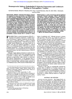
From www.bloodjournal.org by guest on February 6, 2015. For personal use only.
Inhibition of Leukocyte Rolling in Venules by Protamine
and Sulfated Polysaccharides
By Geert Jan Tangelder and Karl-E. Arfors
Intravital video microscopy was used to investigate leukocyte marginationin 80 mesenteric venules (19 to 54 bm) of 50
anesthetized rabbits. After intravenous (IV) bolus injection,
sulfated polysaccharides reduced in a reversible and dosedependent way the number of leukocytes rolling slowly
along the venular wall. The presence of sulfate groups is
essential because other negatively charged or neutralpolysaccharides had no effect. It was not caused by an increase in
RBC velocity or chelation of divalent cations. Inhibition by
sulfated dextrans (n = 7) was independent of molecular
weight (mol wt 13,000 to 500,000) but was influenced by the
average number of sulfate groups per monosaccharide. With
substitution 0.13, the gO%-inhibition dose was 104 mg/kg,
with 0.7 it was 56 mg/kg, and between substitution 1 and 2 it
ranged from 20 to 23 mg/kg. At 100 mg/kg, plasma concentra-
tion was 0.6 to 0.7 mg/mL. Xylan sulfate (mol wt 6,000,
substitution 1.8) gave 90% inhibition at 11 mg/kg, and
heparin gave 90% inhibition at 97 mg/kg. Duration of inhibition (0.5 to 2 hours) depended on mol wt and appeared to be
related to plasma clearance. Because protamine also inhibited rolling (12 mg/kg; <10 minutes), we propose that
repetitive formation and breakup of ionic bonds between
sulfate groups and positivelycharged amino acids is involved
in leukocyte rolling. During inhibition of rolling, systemic
lymphocyte/monocytelevels appeared to increase. Granulocyte counts did not change, indicating that rolling is not the
main mechanism responsible for the marginal granulocyte
pool.
o 1991 by The American Society of Hematology.
I
tized with 20% urethane (8 to 10 mLkg), injected in a marginal ear
vein. A catheter was placed in the left jugular vein for additional
small doses of urethane and injection of substances. A tracheal
tube was placed to facilitate breathing. Rectal temperature (36" to
39°C throughout the experiments) was used to control the heating
pad of the animal.
In 20 animals, a catheter was positioned in the right carotid
artery for collection of blood and measurement of blood pressure
(external transducer: Century Technology Company [CTC], Inglewood, CA; CP-01). Gas values and pH of blood samples were
measured with an acid-base analyzer (ABL 3, radiometer). Throughout these experiments, values of mean arterial pressure (MAP)
generally were within 70 to 90 mm Hg, arterial pH within 7.30 to
7.45, PCO, 30 to 50 mm Hg, and PO, 70 to 100 mm Hg.
Leukocyte levels in the systemic circulation were determined
with a Burker chamber. Blood was collected in Turks solution (0.2
mg Gentian violet in 1 mL glacial acetic acid, 6.25% volhol).
Shortly after induction of anesthesia, but before injection of any
substance, total leukocyte count ranged from 2.7 to 9.0 x 10³/pL
(median 5.85). Monomorphonuclear cell counts, ie, lymphocytes
and monocytes, ranged from 1.5 to 5.6 x 10³/pL(median 3.35); and
polymorphonuclear counts, ie, granulocytes, ranged from 0.8 to 5.7
x 103/pL (median 2.15). Granulocyte counts as a percentage of
total leukocyte counts ranged from 23% to 64% (median 39.5%).
Cell counts remained relatively constant during the first 1.5 to 2
hours of an experiment, but thereafter started to increase. At
approximately 3 hours, total leukocyte counts were on the average
2.5 to 3.0 times the initial value.
N TISSUES prepared for intravital microscopic observation, leukocytes are often observed rolling or sliding
along the wall of venules. Their velocity is distinctly less
than that of the other blood
Leukocyte rolling is
usually not observed in arterioles. It is probably the first
step of leukocyte margination in venules during inflammatory processes, ultimately leading to prolonged adhesion,
diapedesis, and extravascular migration.) In addition, rolling might be one of the mechanisms responsible for the
existence of the marginal leukocyte ~ 0 0 1 s .The
~ cells in
these pools can be rapidly mobilized into the circulation:
Much is known about the biophysical aspects of leukocyte rolling in venule^'^^^^^*; however, the cell surface structures mediating rolling have not been explored. Besides
glycoproteins: proteoglycans are also involved in cell adhesion phenomena." They contain long and negatively charged
polysaccharide chains such as chondroitin sulfate, dermatan sulfate, and heparan sulfate, all of which can be
synthesized by endothelial cells." Content and composition
of endothelial cell surface proteoglycans may differ depending on vessel type and location, even in microvessels." In
addition, sulfated macromolecules appear to be involved in
lymphocyte recirculation.' Therefore, sulfated polysaccharide chains may play a role in the rolling of leukocytes along
venular endothelium.
We wished to investigate in vivo whether sulfated polysaccharides injected intravenously (IV) could block binding
sites important for leukocyte rolling. When these substances proved to inhibit leukocyte rolling, additional experiments were performed to determine the possible mechanism, including studies examining the role of the negative
electrical charge. Whether rolling contributed to formation
of the marginal leukocyte pools was also evaluated. In
addition, experiments were performed with protamine, a
positively charged polypeptide that readily binds to sulfated
polysaccharides."
MATERIALS AND METHODS
Animals and intravital microscom. Fifty New Zealand White
rabbits of either sex (weight 1.7 to 3.2 kg) received diazepam
intramuscularly (IM 4 to 5 mgkg). Thereafter, they were anestheBlood, Vol 77, No 7 (April I),
1991: pp 1565-1571
From La Jolla Institute for Experimental Medicine, La Jolla, and
the Department of Physiology, University of Limburg, Maastricht, The
Netherlands.
Submitted August 9,1990; accepted November 27, 1990.
Suppried by Grant No. S91-156from the Netherlands organization
for the advancement ofpure research (ZWO).
Address reprint requests to G.J. Tangelder, MD, PhD, Depamnent
of Physiology, Biomedical Center, University of LimbuT, PO Box 616,
6200 MD Maastricht, The Netherlands.
The publication costs of this article were defrayed in pari by page
charge payment. This article must therefore be hereby marked
"advertisement" in accordance with 18 U.S.C. section I734 solely to
indicate this fact.
0 1991 by The American Society of Hematology.
0006-4971/91/7707-0022$3.00/0
1565
From www.bloodjournal.org by guest on February 6, 2015. For personal use only.
1566
TANGELDER AND ARFORS
For intravital microscopic observation, the mesentery was used.
Leukocyte rolling is invariably present in this thin and transparent
tissue after its exposure. Through a midline abdominal incision, the
distal part of the ileum was exteriorized. The mesenterywas spread
over a siliconized glass plate mounted in an electrically heated
microscope table (37°C). Care was taken not to touch or stretch the
mesentery itself. The preparation was continuously superfused
with a buffered (pH 7.4) Krebs-Henseleit or Tyrode’s solution kept
at 36“to 37°C. The bowels were kept moist with overlying wet gauze
and covered with Saran wrap to minimize evaporation and cooling.
Preparations were allowed to stabilize for at least 10 minutes. In 22
animals, a second fresh mesenteric loop was exposed after 1 to 2
hours. Most of the experiments were performed within 2.5 hours
after exposure of a mesenteric loop (median 80 minutes, maximum
4 hours).
Observations were made with a Leitz intravital microscope,
using water-immersion objectives (salt water [SW] 25 X or ultropak
objective [UO] 23 X ,numerical aperture 0.6 and 0.55, respectively).
Transillumination was performed with a tungsten lamp. In all
experiments, the level of sharp focus was in the median plane of a
venule. Images were recorded on videotape (Panasonic or MGA)
through a TV camera (Panasonic or SIT). A zoom lens (Leitz) was
used to obtain a final magnification at the front plane of the TV
camera of 50X. Vessel diameters were measured with vernier
calipers. In three animals, venular RBC velocity was measured with
the dual-slit method,I4using transillumination with a mercury arc.
In three animals, we performed fluorescence microscopy. The
intravascular leukocytes were labeled by IV injection of 2.8 to 3.0
mg/kg acridine orange (Chroma, FRG; 0.2% in physiologic saline).” They were visualized with flashes from a xenon arc
(Chadwick Helmuth power supply), using incident illumination
(Leitz Ploemopak, excitation filter BG12 and BG38; dichroic
mirror K490 nm; barrier filter 530 nm).
Substances injected to influence leukocyte rolling. The polysaccharides used are shown in Tables 1 and 2, except a sulfated dextran
with molecular weight (mol wt) 70,000 and degree of substitution
0.07, ie, average number of sulfate groups per monosaccharide.
The structure of a polysaccharide substituted with electrically
charged groups is shown in Fig 1. Dextrans were obtained from
Pharmacia Fine Chemicals (Uppsala, Sweden). The neutral dextrans and FITC-labeled dextran, which is slightly negative charged
(Table 2), as well as the dextran sulfate with mol wt 500,000 (Table
1), were obtained commercially. The other dextrans were synthesized and purified with salt precipitation by Dr T. de Belder
(Pharmacia AB, Uppsala).
Xylan sulfate was purchased as SP54 from Bene Chemie
(Munchen, FRG). Its xylose units (a pentose) have a ring structure
Table 1. Sulfated PolysaccharidesInhibiting Leukocyte Rolling
Type of Molecular Degree of
Substance Weight Substitution*
Xylan
Dextran
Dextran
Dextran
Heparin
6,000
13,100
13,500
59,400
70,000
500,000
70,000
70,000
13,000
1.8
2.0
1.22
1.12
1.55
1.9
0.7
0.13
1.3
90% Inhibition
(dose,mglkglt
11.1 (10.9-18.3)
22.5 (18.7-26.6)
22.3 (18.5-28.7)
22.2 (22.1-26.5)
22.0 (19.5-25.5)
20.2 (18.9-25.4)
56.1 (47.2-58.5)
103.7 (97.8-116.5)
97.1 (86.9-110.7)
Fined Polynomial
Order, r*
2
2
1
2
1
2
2
2
1
0.990
0.984
0.895
0.990
0.979
0.989
0.998
0.999
0.972
*Average number of sulfate groups added per monosaccharide;
heparin contained carboxyl groups as well (substitution0.5).
t95% Confidence limits in parentheses.
SP < ,001 in all cases.
Table 2. Substances With No Influence on Leukocyte Rolling
Substance
Dextran (neutral)
Dextran
Dextran carboxymethyl
Glucose-6-sulfate
Sodium sulfate
Molecular
Weight
Degree of
Substitution*
Dose
(mg/kg)t
70,000
500,000
-
150,000
70,000
150,000
298
142
0.1
0.85
0.11
1.o
220
80
150
415
330
32
108
-
-
‘Average number of FITC, carboxymethyl, or sulfate groups added
per monosaccharide.
tup to and including the doses used.
similar to that of glucose (a hexose, Fig l), but without the sixth
carbon group; they are linked 1 to 4.16 Heparin was obtained from
Lyphomed (Rosemont, IL) and Organon (Oss, Holland), with a
potency of USP 140 to 160lmg (10,000 USPlmL). Its hexose units
contain both carboxyl and sulfate groups. Commercial preparations range in mol wt from 5,000 to 30,000 with a mean value of
12,000 to 15,000.’’
Protamine (mol wt -4,000)13 was obtained as protamine sulfate
from Lilly (10 mg/mL). Glucose-6-sulfate was purchased from
Sigma, and sodium sulfate, calcium chloride, and magnesium
chloride were purchased from Mallinckrodt.
Substances were dissolved (1% to 10%) in physiologic saline or
water, yielding an isotonic solution. They were injected IV as a
bolus (1 to 8 mL, lasting 10 to 30 s). Often a series of injections was
given (maximum 6; interval 2 to 5 minutes). Cumulative doses were
then used to construct a dose-response curve. Because of short
duration of the effect, only the first injection was used with
protamine and xylan, and the first and second were used for the
substances with mol wt 13,000 (Table 1).
In two animals, plasma concentrations of the sulfated dextran
with substitution 0.13 were determined. After injection of 100
mg/kg, samples were taken at 1 and 5 and then every 10 minutes.
After acid hydrolysis in 1 N HCI (3 hours at 1Oo”C), the increase in
glucose concentration was determined with the o-toluidine method.’“
The method could not be used at higher degrees of sulfate
substitution.
Analysis of rolling. Collecting venules (diameter > 15 wm)
were selected with a sufficient number of rolling leukocytes. Video
recordings for off-line analysis were made before, during, and after
an injection (series). In vessels of this size, the number of
leukocytes rolling on the endothelium was not influenced by
differencesin blood flow velocity.’.’’ As observed in many rabbits,”
blood flow velocity in mesenteric venules (20 to 40 pm) ranged
from 1 to 6 mmls.
Figure 2 (top) shows leukocyte rolling. Leukocytes indicated as 1
and 2 move slowly in a rolling or sliding fashion along the vessel
wall, with a velocity of less than 50 ~ 4 s This
. is 1 to 2 orders of
I
SO@
Fig 1. Schematic of molecular structure of a sulfated polysaccharide (sulfated dextran). The average number of sulfate groups per
monosaccharide or degree of substitution is 1 in this example. The
distribution of sulfate groups may be inhomogeneous.
From www.bloodjournal.org by guest on February 6, 2015. For personal use only.
INHIBITION OF LEUKOCYTE ROLLING IN VENULES
1567
Fig 2 Example of a mesenteric venule (32 to 33
pm) before and after inhibitionof leukocyterolling by
a sulfated dextran (mol wt 59,400; 43 mg/kg). Top
two panels (before inhibition): in 1 second, leukocytes marked 1 and 2 moved less than 50 pm, the
distance indicated by the white bar in the bottom
right panel. Direction of flow is from left to right
(arrow). Top left panel: *Leukocytes that did not roll
but adhered firmly to the vessel wall. These cells
remained after inhibitionof rolling (bottom panels).
magnitude less than blood flow velocity. Individual cells moving at
flow velocity move too quickly to be detectable by the eye when
continuous illumination is used. Therefore, all moving WBC that
could be detected by eye were considered rolling cells. They either
moved steadily or alternated short periods of almost complete
standstill with a quicker move to another position at the wall.
Leukocytes rolling out of focus could also be detected easily.
Leukocyte rolling was quantified from video images, if necessary
at reduced speed. We counted in triplicate or duplicate the number
of all leukocytes rolling through a vessel segment (length one to
two diameters) during a period of 1to 7 minutes, depending on the
number of rollers. Cells present in the segment at the beginning of
the period of counting were not included. Rolling was expressed as
the average number of cells passing per minute. For construction of
dose-response curves, we expressed the number of rolling leukocytes remaining after an injection as a percentage of the number of
rolling cells in the control situation (Fig 3).
Stat3tics. Data were analyzed with SPSS/PC+ statistical package (SPSS, Chicago, IL). Stray values were defined according to
Tukey as outliers or extremes in a box-plot?' In all tests, a P-value
greater than 0.05 was considered nonsignificant. Correlations were
performed with the Spearman rank test, except for dose-response
relations. Paired data groups were compared (two-sided) with the
Wilcoxon signed-rank test.
ff
1
Multiple regression analysis was used to describe the doseresponse curves?' Starting with straight line regression, we fitted
successivehigher order polynomials. Significanceof a fit and of the
improvement by adding an extra term were tested with the F
statistic. In two curves, significant improvement by a higher (third)
order term was caused solely by the presence of one high dose
point lying far beyond the 90%-inhibition dose. These two points
were therefore excluded from the analysis. An example is the point
at 80 mgkg (open diamond) of the left curve shown in Fig 3.
The appropriate fit (first or second order) was used to calculate
the dose giving 90% inhibition. For comparison, 95% confidence
intervals were calculated using the 95% confidence bands" of the
best fitted straight lines (r 0.90 to 0.98, P < .001).In four curves,
the highest data point was excluded. An example is the point at 149
mgkg in the right curve of Fig 3. The 90%-inhibition doses based
on these straight lines differed from the former by less than 2.5 and
4 mgkg for the first six and last three substances in Table 1,
respectively.
RESULTS
In 80 venules, leukocyte rolling was observed before
(control) and during 93 injections or injection series.
Venular diameters ranged from 19 to 54 pm (median 32
logdose 2
Fig 3. Dose-responsecurves with best f ~ ofs two
sulfated dextrans. Concomitant log dose-affect plots
(inset) with effect shown as percentageof inhibition.
For the substance with dose-response curve shown
at left (open symbols), mol wt was 59,400 and degree
= 100.2 - 6 . 2 1 +
~ 0.096~';
of substitutionwas 1 . 1 2 1 ~
r = 991, P < .001). For the substance with doseresponse curve shown at right (closed symbols)
70,000 and 0.13, respectively ( y = 99.6
1 . 3 5 0.OOW; r = .999, P < .001). Different symbols in the dose-response curves represent experiments in different animals; level of 90% inhibkion
(dashed line).
+
Dose (mglkg)
-
From www.bloodjournal.org by guest on February 6, 2015. For personal use only.
1568
pm). Control values of leukocyte rolling ranged from 12 to
188/min (stray value 223). The distribution was skewed to
the right with a mode of 37/min (median 64/min). Skewness
was 1.03 (SE 0.25), which is significantly higher than zero,
the value expected in case of a normal distribution. No
correlations could be found between the number of rolling
leukocytes and vessel diameter or time elapsed after the
abdomen was opened.
Sulfated po&saccharides. Table 1 shows the sulfated
polysaccharides that were able to inhibit leukocyte rolling.
Within seconds after injection, the number of rolling
leukocytes decreased or even disappeared completely, depending on the dose. In Fig 2, leukocyte rolling, illustrated
at t = 0 and 1second, could no longer be observed anymore
after injection (t = 8.34 and 9.34 seconds). In contrast,
WBC that had stuck to the wall for several minutes were not
removed.
Inhibition of leukocyte rolling by sulfated polysaccharides was a reversible phenomenon. With the lower mol-wt
substances (mol wt < 15,000), rolling cells returned after 10
to 20 minutes and had usually reached levels of about 50%
to 100% of control within 20 to 40 minutes. With mol wt
60,000 to 70,000, inhibition lasted 40 to 70 minutes; with
mol wt 500,000 inhibition lasted for the remainder of the
experiment, ie, at least 90 to 105 minutes. Plasma concentrations of the sulfated dextran with mol wt 70,000 and
substitution 0.13 decreased rapidly. Within 45 to 65 minutes
after injection of 100 mgikg, 90% had been removed. One
minute after injection of this dose, the plasma concentration was 0.6 to 0.7 mg/mL. This is only 25% of the value
expected on the basis of the average plasma volume (39
mL/kg) of rabbits?'
Figure 3 shows the dose-response curves of two dextrans
with similar mol wt (60,000 to 70,000) but different degree
of substitution. The substance with substitution 0.13 (Fig 3,
right) was far less effective than that with substitution 1.12.
Each curve was best fitted with a second-order polynomial,
also shown in Fig 3. When the dose-axis of each curve was
normalized with respect to the intersection of its best fit
with the 90%-inhibition level (Fig 3), both curves coincided,
or, alternatively, a log dose-effect plot of both curves
indicated a similar slope (Fig 3, inset). This suggests that
the difference in potency of both substances is related to a
difference in affinity for the cell surface structures to which
they bind?3
The doses yielding 90% inhibition are shown in Table 1,
as well as the correlation coefficients ( I ) and polynomial
order of the appropriate fits. Comparison of the 95%confidence limits of these doses indicates that (1) the xylan
is significantly more effective than the dextrans, (2) dextrans with a degree of substitution exceeding unity are
equally effective despite the huge variation in mol wt, (3)
the effect decreases with substitution below unity, and (4)
heparin is far less effective than the other substances with a
high degree of substitution ( > 1).
Injection of sulfated dextrans at doses inhibiting leukocyte rolling did not cause a change in venular RBC velocity
(range 0.8 to 2.4 mm/s; four venules), except for the 500,000
mol-wt dextran. This substance could cause a transitory
TANGELDER AND ARFORS
decrease in velocity (maximum 50%) for 5 to 10 minutes.
Injection of sulfated polysaccharides did not change blood
pressure (BP).
Other substances and protamine. The sulfated dextran
with degree of substitution 0.07 did not reduce the number
of rolling leukocytes more than about 50%, up to and
including a dose of 600 mgikg. The substances that had no
influence on the number of rolling leukocytes are shown in
Table 2; the carboxymethyl-dextrans shown in Table 2 have
a degree of substitution similar to that of the last two
sulfated dextrans shown in Table 1. Comparison of these
four dextrans showed that negative charge alone is not
enough to influence WBC rolling. Inhibition of leukocyte
rolling specifically requires the presence of sulfate groups.
Free calcium or magnesium ions have been suggested to
be important for leukocyte rolling.' Therefore, we tested in
two animals whether inhibition by sulfated polysaccharides
could have been caused by a sequestration of these ions.
Injection of excess calcium or magnesium ions, however, up
to and including 10 times the dose needed to saturate all
chelating sites, did not cause leukocyte rolling to recur.
Injection of calcium or magnesium chloride alone did not
influence the number of rolling leukocytes.
Protamine was also able to- inhibit leukocyte rolling.
After a first injection, the effect was short. Rolling leukocytes started to return within 30 to 60 seconds and were
back at 50% to 100% of control after 5 to 10 minutes. The
dose-response curve was best fitted with a second-order
polynomial (y = 100.4 -ll.&c + 0.352, r = .98, P < .001).
The 90%-inhibition dose was 11.8 mgikg (10.1 to 13.8; 95%
confidence limits). Each successive injection within a series
caused a new decrease in leukocyte rolling, which then
lasted longer. After a cumulative dose of 20 to 25 mag,
return to control levels took approximately 20 minutes. At
these latter doses, protamine could induce a transient
decrease in blood flow velocity, probably caused by a
decrease in BP. Even at doses of 5 to 10 mg/kg, short
decreases (10% to 20%) in BP were noted.
Influence on systemic leukocyte counts. Table 3 shows
that disappearance of rolling leukocytes as caused by the
substances shown in Table 1 did not result from a removal
of WBCs from the circulation. The same was true of
protamine. The last two dextrans shown in Table 1 could at
their highest doses induce decreases in granulocyte counts
to 30% to 40% of control; however, these decreases usually
occurred 1 or more minutes after leukocyte rolling had
disappeared.
Experiments with fluorescent labeling confirmed that
circulating leukocytes were indeed delivered to and flowing
in the venules when leukocyte rolling had been inhibited.
Table 3. Systemic Leukocyte Counts During Inhibitionof Rolling by
Sulfated Polysaccharidesas Percentage of PieinjectionCount
Duration of
Experiment (h)
Granulocytes
Lymphocytes
and Monocytes
< 2 (n = 9)
2-4(n = 8)
90%' (65-190).
NS
100% (70-239)t.
NS
121% (75-133),P
< .10
144% (73-315l.P< .05
'Median values, with range in parentheses.
tone stray value of 632%.
From www.bloodjournal.org by guest on February 6, 2015. For personal use only.
1569
INHIBITION OF LEUKOCYTE ROLLING IN VENULES
All sulfated polysaccharides shown in Table 1 were used,
except two dextrans (substitution 1.55 and 0.7). The pattern
of leukocyte flux in venules became similar to that observed
in arterioles, with leukocytes rapidly passing by, often in the
center of the stream (Fig 4).
Table 3 also indicates that inhibition of leukocyte rolling
by sulfated polysaccharides did not increase the level of
circulating granulocytes, eg, owing to mobilization of the
marginal granulocyte pool. In contrast, the circulating
lymphocytelmonocyte levels appeared to increase. Leukocyte counts remained relatively constant during the first 2
hours of an experiment, but thereafter started to increase.
Because this might have influenced the comparison between postinjection and preinjection counts, Table 3 distinguishes between counts obtained before and after 2 hours.
In the first group, the change in lymphocytelmonocyte
counts approached statistical significance. The data in both
groups support each other, however, and in combination
suggest an increase in lymphocyte andlor monocyte levels
Fig 4. Fluorescentlylabeled leukocytes rolling in a venule (38 km;
top) and flowing in the midstream (bottom) after injection of the
sulfated xylan (15 mglkg). Direction of flow (arrow). The two cells at
the wall in the lower picture are adherent leukocytes. The tail of
after-images of leukocytes flowing in the midstream resulted from
one flash in each TV frame (60/s) and slow camera decay.
during inhibition of leukocyte rolling by a sulfated polysaccharide.
DISCUSSION
The present study showed in vivo that sulfated polysaccharides are able to inhibit the rolling or sliding of leukocytes
along the venular wall. Protamine, which is positively
charged, also inhibits rolling. Inhibition is reversible and
dose dependent. Leukocytes that had already adhered to
the wall for some time remained. Inhibition by sulfated
polysaccharides does not result solely from their negative
charge: the presence of sulfate groups is essential. The
effect increases with the number of sulfate groups up to
about one per monosaccharide. The dose needed was
independent of mol wt but was influenced by the type of
polysaccharide. The inhibition was not caused by an increase in RBC velocity or chelation of divalent cations. The
levels of circulating lymphocytes andlor monocytes appeared to increase, but granulocyte counts did not change
during inhibition of leukocyte rolling.
In the exposed rabbit mesenteries used in this study,
leukocyte rolling was always present without application of
chemotactic agents. This is also reported for mesentery of
other
No significant relation was found between
the number of rolling leukocytes and the duration of an
experiment, suggesting a relatively constant level of the
inflammatory factors involved. These as yet unknown factors differ in their action from the often-used chemotactic
agents formyl-methionyl-leucyl-phenyl-alanine(fMLP) and
phorbol myristate acetate (PMA). Addition of fMLP to the
superfusion solution reduces the number of rolling leukocytes in mesenteric venule^^^^^ As soon as fMLP reaches
the preparation, the rolling cells immediately stick more
firmly and permanently to the vessel wall. In contrast to
PMA-stimulated leukocytes,2srolling cells in vivo appear to
be round and do not show overt shape change with long
pseudopods. Therefore, in vitro adhesion assays using these
agents may represent firm, prolonged leukocyte sticking
rather than rolling.
The levels of wall shear stress in mesenteric venules are
29 dynes/cm2on the average?6 This is an order of magnitude
higher than the levels at which in vitro leukocyte adhesion
can be achieved. Adhesion of granulocytes to cultured
human umbilical vein endothelium or artificial surfaces
decreases with shear and is absent at shear stresses exceeding about 3 dynes/cm2,even after stimulation of one or both
cell types with fMLP, PMA, or interIe~kin-l.~*~’*~*
In contrast, in venules of the size used in our study the number of
leukocytes rolling on the endothelium is not influenced by
differences in blood flow velocity or wall shear rate.”” This
is another reason why current in vitro assays are not
representative of leukocyte rolling in venules. The source of
endothelium used might be critical, because in arterioles
that have the same flow conditions as venules leukocyte
rolling is absent.
The reduction in the number of leukocytes rolling on the
endothelium after injection of a sulfated polysaccharide or
protamine is not caused by a sudden increase in hydrodynamic dispersal forces acting on the rolling WBC. RBC
From www.bloodjournal.org by guest on February 6, 2015. For personal use only.
1570
TANGELDER AND ARFORS
velocity and BP did not increase. Moreover, if this had
occurred, the neutral and other dextrans (Table 2) might
have been expected to show an inhibitory effect as well.
This latter finding also indicates that the inhibition did not
result from an increase in plasma viscosity or osmolality.
Changes in osmolality can influence leukocyte deformability.29
Inhibition of leukocyte rolling did not increase the level
of circulating granulocytes. Approximately half to three
fourths of the granulocytes inside the vascular compartment do not c i r ~ u l a t e . They
~ . ~ are present mainly in lung
m i c r o v e ~ ~ e l ~and
, ~ " ~this
~ ' marginal pool can be rapidly
m~bilized."~~~'
The finding that a reduction in the number of
leukocytes rolling in mesenteric venules did not lead to an
increase in circulating granulocyte counts indicates that this
leukocyte-endothelial interaction is not the main mechanism responsible for the marginal granulocyte pool. In
addition, it suggests that in noninflamed tissue granulocyte
rolling is a rare phenomenon. Exposure of the mesentery
probably induces a mild inflammatory response in this thin
tissue.
The reversible nature of the inhibition of leukocyte
rolling by sulfated polysaccharides appears to be related to
their plasma clearance. The 90%-inhibition dose of a
sulfated dextran with mol wt 70,000 was cleared in about 1
hour. Rapid sequestration and desulfation mainly by the
liver have been reported in humans for heparin and the
xylan used in this
A relation between plasma
clearance and duration of inhibition might indicate that the
molecular structures involved are only temporarily affected.
This is supported by the short action of lower mol-wt
substances such as heparin. The immediate effect after
injection suggests that structures already present and involved in leukocyte rolling were blocked.
Dextrans charged negatively with carboxymethyl groups
had no effect on the number of rolling leukocytes; this
probably was not caused by a lack in degree of ionization;
eg, carboxy-methylcellulose,which like dextran consists of
glucose units, has a pKa of 5.5.3' Hence, at blood pH its
degree of ionization exceeds 98.5%. In vitro, binding of
lymphocytes to autologous erythrocytes could be blocked
effectively by sulfated polysaccharides but not by phosphated or carboxylated
To be effective, the sulfate groups must be present on a
sufficiently large molecule. Glucose-sulfate and sulfate ions
had no effect. On the other hand, inhibition of rolling was
independent of mol wt indicating that fewer molecules are
needed of a large sulfated polysaccharide than of a small
one as long as their total mass is the same. It suggests that a
large polysaccharide molecule can simultaneously block
more than one binding site important for leukocyte rolling.
Heparin was far less effective than dextran or xylan
sulfate. Xylan was more effective than the dextrans. These
results indicate that in addition to the influence of the
density of sulfate groups, the presence of other, nonfunctional moieties and type of sugar has an influence.
Protamine also inhibited leukocyte rolling in a reversible
way. It contains 80% to 90% arginine residues and binds
readily to sulfated polysaccharides." Therefore, an analogous interaction may be involved in leukocyte rolling along
venular endothelium. Negatively charged sulfate groups of
proteoglycans on one cell could interact with positively
charged arginine and/or lysine moieties of glycoproteins on
the other. Repetitive formation and breakup of ionic bonds
complies with the transitory nature of the interaction
between both cells during rolling. If endothelial proteoglycans are involved in leukocyte rolling, differences in sulfate
content, other substituted groups, and type of sugar might
explain the difference in leukocyte rolling between arterioles and venules.
In conclusion, sulfate groups but not other negatively
charged moieties on polysaccharides can reversibly bind to
and block in vivo molecular structures important for leukocyte rolling in venules. The positively charged arginine
residues of protamine can do the same. These results leave
intact the hypothesis that cell surface proteoglycans play a
role in leukocyte rolling. Reduction in the number of rolling
leukocytes in mesenteric venules did not lead to an increase
in circulating granulocyte counts, indicating that this type of
leukocyte-endothelial interaction is not the main mechanism responsible for the marginal granulocyte pool.
ACKNOWLEDGMENT
We are indebted to Sabrina Weijmer-van Velzen for technical
assistance.
REFERENCES
1. Atherton A, Born GVR: Quantitative investigations of the
adhesiveness of circulating polymorphonuclearleukocytes to blood
vessel walls. J Physiol222447,1972
2. Schmid-Schonbein GW, Usami S, Skalak R, Chien S: The
interaction of leukocytes and erythrocytes in capillary and postcapillary vessels. Microvasc Res 19:45,1980
3. Harlan JM: Leukocyte-endothelial interactions. Blood 65:
513,1985
4. Bierman HR, Kelly KH, Cordes FL, Byron RL, Polhemus BS,
Rappoport S: The release of leukocytes and platelets from the
pulmonary circulation by epinephrine. Blood 7:683,1952
5. House SD, Lipowsky HH: Leukocyte-endothelium adhesion:
Microhemodynamics in mesentery of the cat. Microvasc Res
34363,1987
6. House SD, Lipowsky HH: In vivo determination of the force
of leukocyte-endothelium adhesion in the mesenteric microvasculature of the cat. Circ Res 63:658,1988
7. Mayrovitz HN, Kang SJ, Herscovici B, Sampsell RN: Leukocyte adherence initiation in skeletal muscle capillaries and venules.
Microvasc Res 33:22,1987
8. Bagge U, Karlsson R: Maintenance of white blood cell
margination at the passage through small venular junctions. Microvasc Res 2092,1980
9. Hogg N: The leukocyte integrins. Immunol Today 10:111,
1989
10. Rapraeger A, Jalkanen M, Bernfield M: Integral membrane
proteoglycans as matrix receptors: role in cytoskeleton and matrix
assembly at the epithelial cell surface, in Wight TN, Mecham RP
From www.bloodjournal.org by guest on February 6, 2015. For personal use only.
INHIBITION OF LEUKOCYTE ROLLING IN VENULES
(eds): Biology of Proteoglycans. San Diego, CA, Academic, 1987,
p 129
11. Heifetz A, Allen D: Biosynthesis of cell surface sulfated
glycoproteins by cultured vascular endothelial cells. Biochemistry
21:171, 1982
12. Ausprunk DH, Boudreau CL, Nelson DA: Proteoglycans in
the microvasculature: I. Histochemical localization in microvessels
of the rabbit eye. Am J Pathol103:353,1981
13. Ando T, Yamasaki M, Suzuki K: Protamines: Isolation,
characterization, structure and function, in Molecular Biology,
Biochemistry and Biophysics 12. New York, Springer, 1973
14. Slaaf DW, Arts T, Jeurens TJM, Tangelder GJ, Reneman
RS: Electronic measurement of red blood cell velocity and volume
flow in microvessels, in Chayen J, Bitensky L (eds): Investigative
Microtechniques in Medicine and Biology. New York, Dekker,
1984, p 327
15. Tangelder GJ, Slaaf DW, Reneman RS: Fluorescent labeling of blood platelets in vivo. Thromb Res 282303, 1982
16. MacGregor IR, Dawes J, Paton L, Pepper DS, Prowse CV,
Smith M: Metabolism of sodium pentosan polysulphate in manCatabolism of iodinated derivatives. Thromb Haemost 51:321,
1984
17. Holmer E, Soderberg K, Bergqvist D, Lindahl U: Heparin
and its low molecular weight derivatives: Anticoagulant and
antithrombotic properties. Haemostasis 16:1, 1986 (suppl2)
18. Hultman E: Carbohydrates and related compounds. A
Glucose, galactose, fructose, inulin and ketone bodies in blood and
urine, in Curtius HCh, Roth M (eds): Clinical Biochemistry,
Principles and Methods. Berlin, de Gruyter, 1974, p 908
19. Oude Egbrink M G A Thromboembolic reaction following
vessel wall injury in arterioles and venules. Thesis, University of
Limburg, Maastricht, The Netherlands, 1989
20. Tukey JW: Exploratory Data Analysis. Reading, MA, Addison-Wesley, 1977
21. Kleinbaum DG, Kupper LL: Applied Regression Analysis
and Other Multivariable Methods. North Scituate, MA, Duxbury
Press, 1978
22. Kozma C, Macklin W, Cummins LM, Mauer R: The anatomy, physiology and the biochemistry of the rabbit, in Weisbroth
1571
SH, Flatt RE, Kraus AL (eds): The Biology of the Laboratory
Rabbit. San Diego, CA, Academic, 1974, p 50
23. Goodman Gilman A, Goodman LS, Rall TW,Murad F The
Pharmacological Basis of Therapeutics. New York, Macmillan,
1985
24. Tangelder GJ, Neumann C, Arfors K-E: Reduction of
leukocyte sticking by dextran-sulfate induced inhibition of leukocyte rolling. FASEB J 2:A 1881,1988 (abstr)
25. Ley K, Lundgren E, Berger E, Arfors K-E: Shear-dependent
inhibition of granulocyte adhesion to cultured endothelium by
dextran sulfate. Blood 73:1324,1989
26. Lipowsky HH, Kovalcheck S, Zweifach BW: The distribution of blood rheological parameters in the microvasculature of cat
mesentery. Circ Res 43:738,1978
27. Lawrence MB, McIntire LV, Eskin SG: Effect of flow on
polymorphonuclear leukocyte/endotheliaI cell adhesion. Blood
70:1284,1987
28. Lawrence MB, Smith C W ,Eskin SG, McIntire LV: Effect of
venous shear stress on CD-18 mediated neutrophil adhesion to
cultured endothelium. Blood 75227,1990
29. Chien S, Sung KLP, Skalak R, Schmid-Schonbein GW:
Viscoelastic properties of leukocytes in passive deformation, in
Bagge U, Born GVR, Gaehtgens P (eds): White Blood Cells:
Morphology and Rheology as Related to Function. The Hague,
Martinus Nijhoff, 1982, p 11
30. Doerschuk CM, Downey GP, Doherty DE, English D, Gie
RP, Ohgami M, Worthen GS, Henson PM, Hogg J C Leukocyte
and platelet margination within microvasculature of rabbit lungs. J
Appl Physiol68:1956,1990
31. Hogg JC: Neutrophil kinetics and lung injury. Physiol Rev
67:1249, 1987
32. Dawes J, Pepper DS: Catabolism of low-dose heparin in
man. Thromb Res 14845,1979
33. Windholz M, Budavari S, Blumetti RF, Otterbein ES (eds):
The Merck Index. Rahway NJ, Merck, 1983
34. Parish CR, Rylatt DB, Snowden JM: Demonstration of
lymphocyte surface lectins that recognize sulfated polysaccharides.
J Cell Sci 67:145,1984
From www.bloodjournal.org by guest on February 6, 2015. For personal use only.
1991 77: 1565-1571
Inhibition of leukocyte rolling in venules by protamine and sulfated
polysaccharides
GJ Tangelder and KE Arfors
Updated information and services can be found at:
http://www.bloodjournal.org/content/77/7/1565.full.html
Articles on similar topics can be found in the following Blood collections
Information about reproducing this article in parts or in its entirety may be found online at:
http://www.bloodjournal.org/site/misc/rights.xhtml#repub_requests
Information about ordering reprints may be found online at:
http://www.bloodjournal.org/site/misc/rights.xhtml#reprints
Information about subscriptions and ASH membership may be found online at:
http://www.bloodjournal.org/site/subscriptions/index.xhtml
Blood (print ISSN 0006-4971, online ISSN 1528-0020), is published weekly by the American
Society of Hematology, 2021 L St, NW, Suite 900, Washington DC 20036.
Copyright 2011 by The American Society of Hematology; all rights reserved.


© Copyright 2026