
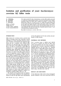
Characterization of the functional organization of yeast K2 killer
Characterization of the functional organization of yeast K2 killer preprotoxin gene Characterization of the functional organization of yeast K2 killer preprotoxin gene G. Gulbinienë Institute of Botany, Þaliøjø eþerø 49, LT-2021 Vilnius, Lithuania. E-mail: [email protected] Expression of a cDNA copy of the yeast K2 preprotoxin gene confers the complete K2 killer phenotype on sensitive cells. To determine the functional domains of killer preprotoxin, we analyzed the phenotypes of a set of mutations throughout regions encoding the δ-, α- and β-toxin subunits. Mutations within the β-subunit indicate it to be essential for the killing of sensitive cells and forming of immunity. Mutations within C-terminal region of α causing loss of toxicity also cause loss of immunity, implicating α in expression of both functions. Our results indicate that the leader sequence of preprotoxin is involved in ensuring the immunity. Key words: Saccharomyces cerevisiae, yeast, killer toxin, immunity INTRODUCTION Killer strains of Saccharomyces cerevisiae secrete a polypeptide toxin to which they are immune themselves. On the basis of killing profiles and missing cross-immunity, toxin-secreting strains have been classified into three major types (K1, K2, K28) [1]. The S. cerevisiae K1 killer system is one of the many described among the yeasts. The K1 precursor peptide has a δ-α-γ-β domain organization. Mature K1 toxin is secreted as an α/β heterodimer [2]. Mutational analysis confirmed that the hydrophilic βsubunit of K1 toxin is essential for binding to a cell wall receptor. The α-subunit, in contrast, is multifunctional, having regions necessary for killing, immunity, and cell wall receptor binding that appear to overlap in the polypeptide. The immunity-coding region of K1 extends through the C-terminal half of the α-subunit into the N-terminal part of the γ glycopeptide. β-subunit of K1 is not involved in formation of immunity [3]. The organization of K2 killer system is not so extensively studied. Meðkauskas and Èitavièius [4] have synthesized, cloned, sequenced and expressed in yeast the cDNA copy of M2-1 fragment of M2 dsRNA, encoding type K2 killer preprotoxin. Expression of the K2 killer precursor gene by the yeast ADH1 promoter in S. cerevisiae conferred both the K2 killer and immunity phenotypes on sensitive host yeast strains. The primary nucleic acids sequence of K2 gene shows no identity with gene coding for the sequence of the K1 toxin, but K2 precursor appears to have a similar overall structure to that of K1 [4, 5]. Here, we report results of a mutational analysis of K2 killer preprotoxin. We show that both the αand β-subunits appear to be required for toxicity and immunity, and the leader sequence of preprotoxin is necessary for ensuring immunity. MATERIALS AND METHODS The S. cerevisiae strain α1 (MATα leu2 [kil-0]) [6] was used as a recipient for investigation of phenotypes of K2 gene mutations and as a sensitive tester for killer toxin activity. The E. coli strain DH5α [7] was used for the routine growth and maintenance of plasmids. All media for the growth of DH5α and procedures of transformation were standard [8]. Media for the growth of S. cerevisiae have been described in [9]. Mutagenesis at restriction sites was accomplished with the K2 killer preprotoxin gene from plasmid pYEX12 [4]. General methods for DNA manipulations involving restriction digestion, Klenow fragment treatment, dephosphorylation, ligation, electrophoresis, extraction of DNA from agarose gel were performed essentially as described in [8] and according to the product manufacturers recommendations. Transformation into S. cerevisiae α1 was performed by the LiCl procedure [10]. Transformants were selected by complementation of LEU2 auxotrophy and tested for the killer phenotype as described in [11]. Immunity was assessed by spotting 107 cells of tester K1, K2 and K28 killer strains onto an agar plate seeded with 105 cells of transformant per ml [11]. All plasmid constructions were checked by restriction mapping and retransformation. ISSN 13920146. B i o l o g i j a . 2002. Nr. 3 !% G. Gulbinienë RESULTS AND DISCUSSION The toxin secretion and immunity exhibited by K2 killer yeast to their toxin are conferred by the precursor gene carried on the cDNA expression plasmid pYEX12. This gene defines 100% killer toxin production and wild-type immunity in this work. Our strategy to determine the functional domains of the K2 preprotoxin was to construct mutations Fig. 1. Localization of deletions introduced into the K2 preprotoxin gene. throughtout the δ-, α- and The restriction map at the top shows the preprotoxin gene as it is cloned into the SalI SalI sites of pYEX12; only sites used in this work are shown. Below the subunit β-encoding regions. By structure of the K2 killer preprotoxin is shown. The mutations are indicated here by analyzing the ability of the designations of plasmids containing them. The horizontal arrows indicate the extent mutant toxins to kill sen- of gene deletions sitive cells and confer immunity, the respective domains could be determined. The α-subunit of K1 preprotoxin contains two The actual mutations are summarized in Fig. 1. hydrophobic regions separated by a short hydrophiC-terminal deletions of preprotoxin were const- lic region [2]. Mutations altering the hydrophobic ructed to determine the role of K2 β-subunit in the regions of K1 α-subunit were found to be defective expression of immunity and killer functions. MluI both in killing and immunity [3]. The overall hydrestriction endonuclease was used to construct dele- rophobic/hydropathic organization is not preserved tion of 56 amino acids in the C-end of β. The ob- between K1 and K2 preprotoxins, but the α-subunit tained plasmid pMlu∆ was introduced into the sen- of K2 is also relatively hydrophobic [5]. In order to sitive S. cerevisiae strain α1. All the transformants investigate the influence of the hydrophobic C-tertested were incapable of killing the sensitive strain minal region of K2 α-subunit on the phenotype, we α1 and remained sensitive to killer toxins of K2 constructed plasmid pEco∆ by in-frame deletion of type as well as types K1 and K28. Thus, deletion of 21 C-terminal amino acids of α. Transformants con56 C-terminal amino acids of β-subunit totally abo- taining this construct were non-killers and sensitive lished the ability of the toxin to render immunity to K1, K2 and K28 toxins. Based on these findings, and kill sensitive cells. Therefore, we decided to we suppose that the C-terminal region (184205 amiconstruct a shorter deletion in the C-end of β-sub- no acids) of K2 α-subunit is necessary to ensure unit. We used the restriction endonuclease StuI, re- killing and immunity like that of K1. moved the 16 C-terminal amino acid region and Analysis of the amino acid sequence of K2 prepobtained the plasmid pStu∆. The α1-pStu∆ trans- rotoxin shows a hydrophobic sequence in δ, between formants had non-killer phenotypes similar to that residues 2743, postulated to act as a secretion of the α1-pMlu∆, suggesting that the β-subunit of leader sequence [5]. This sequence is preceded by a K2 preprotoxin was responsible for the formation of hydrophilic 126 amino acid segment whose funcimmunity. By contrast, all of the reported mutations tion remains unknown [4]. We investigated the role in the β-subunit of K1 preprotoxin retained the im- of this hydrophilic sequence in the expression of munity phenotype the transformants were fully re- killer phenotype. There are two potential in-frame sistant to toxins type K1 [3]. In addition, deletions initiation ATG codons at the start of the K2 preproin K2 β-subunit were also inactive in killing sensiti- toxin gene (codons 79 and 7678) and the most 5 ve cells, while mutations in the same region of K1 7-9 ATG is designated as the initiation codon [5]: allowed secretion of 30 1 10 BamHI 20 30 40 35% of toxin 5- GAA AAA ATG GGG ATC CGG GCC ACC AGC CTG GTG CAA GAC GAG compared with the wild type 5 60 CfrI 70 80 Pfl23II CTG ACA CTA GGT GAG CCG GCC ACC CGA GCA AGG ATG TGC GTA CGT -3 [3]. !& Characterization of the functional organization of yeast K2 killer preprotoxin gene We used the restriction endonuclease BamHI, deleted 11 bp form 5-end of K2 cDNA and obtained cDNA lacking the first in-phase start codon (79 ATG) carried on plasmid pBam∆-D [12]. Strain α1 containing this construct was able to kill sensitive cells, but the mutation led to a reduction in the size of the killing zone around the transformants to 66% (Fig. 2, I, line 4) in comparison with the wildtype plasmid pYEX12 (Fig. 2, I, line 3). However, α1-pBam∆-D transformants were still sensitive to K2 toxins produced by Rom-K100 (Fig. 2, II, line 1, C) and M437 (Fig. 2, II, line 1, D) killer strains, as well as to K1 (Fig.2, II, line 1, A, B) and K28 (Fig. 2, II, line 2, A, B, C) toxins, and thus exhibited a greatly reduced immunity. The α1-pBam∆-D transformants were immune only to their own toxin (Fig. 2, II, 4) and to that of α1-pYEX12 transformants (Fig. 2, II, line 3). We deleted 159 bp and 181 bp from 5-end of K2 cDNR by using CfrI and Pfl23II restriction endonucleases, respectively. Plate tests indicated that the deletions carried on both pCfr∆ and pPfl∆ plasmids clearly led to a defective killer phenotype transformants showed no killing zones on the lawn of the sensitive strain and were sensitive to the action of killer toxins of all types. Thus, elimination of the first initiation codon (plasmid pBam∆-D) had a dramatic effect on the formation of immunity, but the gene was still expressed. Deletion of 159 bp region preceding the second potential initiation codon (plasmid pCfr∆) resulted in a sensitive phenotype as did the deletion of the second initiation codon (plasmid pPfl∆). These findings enabled us to suppose that the second potential in-frame initiation codon 7678 ATG could be used as the initiation codon for protein synthesis, and the 1159 bp region was important as well. This resulted in an assembly of the mutant N-terminal region of the leader sequence, implicating the hydrophilic region (126 amino acids) of the leader sequence in the expression of immunity. Our results indicate some differences in the functional organization of K1 and K2 killer toxins. We have demonstrated that the β-subunit of K2 killer toxin is involved in forming the K2 immunity mutations in the β-subunit C-terminal-coding region resulted in a sensitive phenotype. The leader sequence of K2 preprotoxin was important in the expression of immunity as well mutations of the leader peptide-coding region partially or totally inactivated the immunity. We also demonstrated that the hydrophobic C-terminal region of α-subunit was necessary for immunity and toxic functions. References Fig. 2. K2 immunity mutant α1-pBam∆-D. I K2 activity. Test cultures were spotted on a tester α1 plate. Line 1, A, B wild type (wt) K1 killer strains, K7, DBY4975; line 1, C, D wt K2 killer strains, Rom-K100, M437; line 2, A, B, C wt K28 killer strains, 28, VKM2472, MS300; line 2, D recipient strain, α1; line 3 wt K2 cDNA strain, α1[pYEX12]; line 4 pBam∆D in a sensitive strain, α1[pBam∆-D]. II Immunity. Plasmid pBam∆-D was transformed into sensitive strain α1, and the transformed strain was embedded in agar. Then standard tester strains were spotted onto the plate. Line 1, A, B wt K1 killer strains, K7, DBY4975; line 1, C, D wt K2 killer strains, Rom-K100, M437; line 2, A, B, C wt K28 killer strains, 28, VKM2472, MS300; line 2, D recipient strain, α1; line 3 wt K2 cDNA strain, α1[pYEX12]; 4 pBam∆-D in a sensitive strain, α1[pBam∆-D] 1. Schmitt MJ, Eisfeld K. Recent Res Devel Virol 1999; 1: 52545. 2. Bostian KA, Elliot Q, Bussey H et al. Cell 1984; 36: 74151. 3. Zhu H, Bussey H. Mol Cell Biol 1991; 11 (1): 17581. 4. Meðkauskas A, Èitavièius D. Gene 1992; 111: 1359. 5. Dignard D, Whiteway M, Germain D et al. Mol Gen Genet 1991; 227: 12736. 6. ×èòàâè÷þñ Ä, Èíãå-Âå÷òîìîâ ÑÃ. Ãåíåòèêà 1972; 1 (1): 95102. 7. Woodcock DM, Crowther PJ, Doherty J et al. Nucleic Acids Res 1989; 17 (9): 346978. 8. Sambrook J, Fritsch EF, Maniatis T. Molecular Cloning: a Laboratory Manual. Cold Spring Harbor Laboratory, Cold Spring Harbor, New York, 1989. 9. Sherman JF, Fink GR, Hiks JB. Methods in Yeast Genetics. Cold Spring Harbor Laboratory, Cold Spring Harbor, New York, 1986. 10. Ito H, Fukuda Y, Murata K et al. J Bacteriol 1983; 153: 1638. 11. Íàóìîâà ÒÈ, Íàóìîâ ÃÈ. Ãåíåòèêà 1973; 9 (4): 8590. 12. Gulbinienë G, Jokantaitë T, Melvydas V. Biologija 2001; 4: 79. !' G. Gulbinienë G. Gulbinienë MIELIØ K2 KILERINIO PREPROTOKSINO GENO FUNKCINËS ORGANIZACIJOS CHARAKTERISTIKA Santrauka Siekdami nustatyti K2 kilerinio preprotoksino funkciðkai svarbias sritis, tyrëme lyderinæ sekà δ, taip pat α ir β toksino subvienetus koduojanèiø geno regionø mutacijø fenotipus. Nustatyta, kad Saccharomyces cerevisiae K2 tipo kilerinio preprotoksino funkcinë organizacija skiriasi " nuo K1 tipo preprotoksino. K2 preprotoksino β subvienetas yra svarbus uþtikrinant toksiðkumà ir imunitetà. Tuo tarpu K1 preprotoksino β subvieneto mutacijos tik sumaþina kilerinio toksino produkcijà ir neturi átakos imunitetui. K2 preprotoksino hidrofilinë 126 aminorûgðèiø seka, esanti prieð signaliná peptidà, dalyvauja imuniteto sudaryme: ðios sekos mutacijos lemia tik daliná atsparumà, arba visiðkà K2 imuniteto praradimà. K2 preprotoksino α subvieneto C-galinë hidrofobinë sritis (184205 aminorûgðtys) dalyvauja uþtikrinant ir imunitetà, ir kileriná aktyvumà.


© Copyright 2026