
Glycans in the immune system and The Altered Glycan Theory of
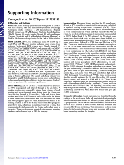
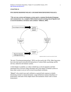
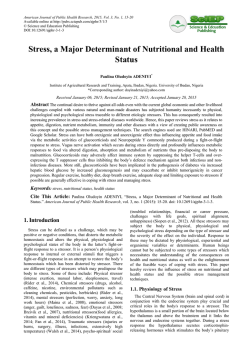
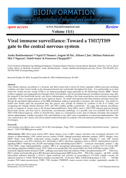
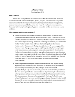
Journal of Autoimmunity xxx (2014) 1e13
Contents lists available at ScienceDirect
Journal of Autoimmunity
journal homepage: www.elsevier.com/locate/jautimm
Review
Glycans in the immune system and The Altered Glycan Theory
of Autoimmunity: A critical review
Emanual Maverakis a, *, Kyoungmi Kim b, Michiko Shimoda a, M. Eric Gershwin c,
Forum Patel a, Reason Wilken a, Siba Raychaudhuri c, L. Renee Ruhaak b,
Carlito B. Lebrilla d
a
Department of Dermatology, University of California, Davis School of Medicine, 3301 C Street, Suite 1400, Sacramento, CA 95816, USA
Department of Public Health Sciences, Division of Biostatistics, University of California, Davis Medical Center, Sacramento, CA 95816, USA
Department of Internal Medicine, Division of Rheumatology, University of California, Davis School of Medicine, Sacramento, CA 95817, USA
d
Department of Chemistry, University of California, Davis, One Shields Avenue, Davis, CA 95616, USA
b
c
a r t i c l e i n f o
a b s t r a c t
Article history:
Received 20 November 2014
Accepted 3 December 2014
Available online xxx
Herein we will review the role of glycans in the immune system. Specific topics covered include: the
glycosylation sites of IgE, IgM, IgD, IgE, IgA, and IgG; how glycans can encode “self” identity
by functioning as either danger associated molecular patterns (DAMPs) or self-associated molecular
patterns (SAMPs); the role of glycans as markers of protein integrity and age; how the glycocalyx can
dictate the migration pattern of immune cells; and how the combination of Fc N-glycans and Ig isotype
dictate the effector function of immunoglobulins. We speculate that the latter may be responsible for the
well-documented association between alterations of the serum glycome and autoimmunity. Due to
technological limitations, the extent of these autoimmune-associated glycan alterations and their role in
disease pathophysiology has not been fully elucidated. Thus, we also review the current technologies
available for glycan analysis, placing an emphasis on Multiple Reaction Monitoring (MRM), a rapid
high-throughput technology that has great potential for glycan biomarker research. Finally, we put forth
The Altered Glycan Theory of Autoimmunity, which states that each autoimmune disease will have a unique
glycan signature characterized by the site-specific relative abundances of individual glycan structures on
immune cells and extracellular proteins, especially the site-specific glycosylation patterns of the different
immunoglobulin(Ig) classes and subclasses.
© 2014 The Authors. Published by Elsevier Ltd. This is an open access article under the CC BY license
(http://creativecommons.org/licenses/by/4.0/).
Keywords:
Glycan
Glycome
Glycosylation
Immunoglobulin
Autoimmunity
1. Introduction and The Altered Glycan Theory of
Autoimmunity
Since the discovery of altered IgG glycosylation in patients with
rheumatoid arthritis [1], there has been mounting evidence favoring the role of glycans in the pathophysiology of autoimmunity. For
example, it is now well established that the type of glycan present at
residue Asn-180 of IgG1 helps dictate the effector function of the
antibody, with some glycans being pro-inflammatory while others
possessing anti-inflammatory properties [2,3]. As is the case for the
Asn-180 glycan, the antibody isotype also has a strong influence
over its functionality [4]. In fact, some autoimmune diseases are
strongly linked to a particular Ig class or subclass. Prototypic
examples include the IgG4-mediated diseases, pemphigus foliaceus
* Corresponding author. Tel.: þ1 (916) 734 1512; fax: þ1 (916) 442 5702.
E-mail address: [email protected] (E. Maverakis).
and autoimmune pancreatitis [5,6]. Thus, in antibody-mediated
autoimmunity, antigen specificity will determine the site of attack
whereas the glycan/Ig isotype combination will dictate the physical
nature of the attack. Based on these and other fundamentals
described herein we put forth The Altered Glycan Theory of Autoimmunity, which states that each autoimmune disease will have a
unique glycan signature characterized by the site-specific relative
abundances of individual glycan structures present on immune cells
and extracellular proteins, especially the site-specific glycosylation
patterns of the different Ig classes and subclasses. This review will
also discuss the role of glycans in the immune system and how
novel Mass Spectrometry (MS) technologies, specifically Multiple
Reaction Monitoring (MRM), can be used to rapidly identify the
glycan signatures of the different autoimmune diseases. [For
clarification Asn-180 of IgG1 corresponds to Asn-176 of IgG2, Asn227 of IgG3, and Asn-177 of IgG4. Using the International
Immunogenetics Information System (IMGT) numbering protocol
http://dx.doi.org/10.1016/j.jaut.2014.12.002
0896-8411/© 2014 The Authors. Published by Elsevier Ltd. This is an open access article under the CC BY license (http://creativecommons.org/licenses/by/4.0/).
Please cite this article in press as: Maverakis E, et al., Glycans in the immune system and The Altered Glycan Theory of Autoimmunity: A critical
review, Journal of Autoimmunity (2014), http://dx.doi.org/10.1016/j.jaut.2014.12.002
2
E. Maverakis et al. / Journal of Autoimmunity xxx (2014) 1e13
the position for each of these conserved glycosylations has the same
number, CH2-84.4, regardless of IgG subclass. An addition point of
potential confusion is that Asn-180 is also sometimes referred to as
Asn-297, if one attempts to sequentially number all amino acids
within the IgG molecule. Thus, for simplicity, this review will refer
to the conserved IgG glycosylation site as CH2-84.4].
2. A brief overview of antibody structure
Humans have five distinct classes of immunoglobulins (Igs): IgG,
IgM, IgA, IgE, and IgD (Fig. 1). IgA and IgG can be further divided into
two (IgA1-2) and four (IgG1-4) subclasses, respectively. All Igs
are comprised of two 50e77 kDa class-specific heavy chains (g, m, a,
ε and d) that are joined together by one or more disulfide bonds
(Fig. 1). Each heavy chain is also joined by a disulfide bond to a
25 kDa light chain, which can be one of two different isoforms
(k and l). For IgM and IgA, disulfide bonds can further connect
individual Igs (an their associated J chains) to form pentamer and
dimer structures, respectively (Fig. 1). The antigen-recognition
region of an Ig is referred to as its Fab fragment. In contrast, the
Fc fragment is comprised of the heavy chain region that interacts
with the Fc receptors on immune cells. In the IgA, IgD, and IgG
isoforms, a flexible linker, which can be decorated with glycans,
separates the Fab and Fc regions. IgM and IgE lack this hinge region
and are thus more rigid in structure. IgG1,2,4 have a single
conserved N glycosylation site at residue CH2-84.4 where large
(2 KDa) flexible glycans attach. The other Igs are more heavily
glycosylated (Fig. 1). As will be discussed in later sections, these
glycan modifications are critical for the appropriate function of all
Igs.
3. A vast amount of information is stored in a cell's glycocalyx
Along with nucleic acids, proteins, and lipids, glycans are one of
the four fundamental classes of molecules that make up all living
systems [7]. However, in comparison to the advances made in the
fields of genomics and proteomics, glycoscience remains relatively
understudied, a disturbing fact given that glycans play a major
role in the etiology of all human diseases [8]. Traditionally, the information stream of a cell is viewed as starting in the genome
Fig. 1. Immunoglobulin Isotypes and their sites of glycosylation. Depicted here are the antibody structures including their sites of glycosylation: IgM [(N-glycans at Asn-46, 209,
272, 279, and 439 (UNIPROT), or CH1-45, CH2-120, CH3-81, CH3-84.4, and CHS-7 (IMGT)]; IgA1 [(N-glycans at Asn-144 and 352 (UNIPROT) or CH2-20, CHS-7 (IMGT)]; IgA2
[(N-glycans at Asn-47, 92, 131, 205, 327 (UNIPROT) or CH1-45.2, CH1-114, CH2-20, CH2-120 and CHS-7 (IMGT)]; IgG [(N-glycans at Asn-180 (IgG1), Asn-176 (IgG2), Asn-227 (IgG3),
Asn-177 (IgG4), and Asn-322 (IgG3) (UNIPROT) or CH2-84.4 (IgG1-4) and CH3-79 (IgG3) (IMGT)]; IgD [(N-glycans at Asn-225, 316, and 367 (UNIPROT) or CH2-84.4, CH3-45.4, CH3116 (IMGT)], and IgE [(N-glycans at Asn-21, 49, 99, 146, 252, 264, 275 (UNIPROT) or CH1-15.2, CH1-45.2, CH1-118, CH2-38, CH3-38, CH3-77, and CH3-84.4 (IMGT)]. Each immunoglobulin is comprised of two heavy chains (blue) and two light chains (purple) that are linked together by disulfide bonds (black lines). IgA, IgD, and IgG have a flexible hinge
region that link the Igs' antigen-binding Fab region to their Fc receptor-binding region. O-glycosylation sites are depicted in yellow and N-glycosylation sites are depicted in brown.
The depicted glycans are important for the structural integrity of the antibodies and their effector function [2]. (For interpretation of the references to color in this figure legend, the
reader is referred to the web version of this article.)
Please cite this article in press as: Maverakis E, et al., Glycans in the immune system and The Altered Glycan Theory of Autoimmunity: A critical
review, Journal of Autoimmunity (2014), http://dx.doi.org/10.1016/j.jaut.2014.12.002
E. Maverakis et al. / Journal of Autoimmunity xxx (2014) 1e13
and ending with a set of expressed proteins, representing the
cell's phenotype. Once synthesized, proteins can interact with other
proteins to form networks. However in order for a protein to
function appropriately, it often requires a post-translational modification and glycans are one of the most commonly added modifiers
(Fig. 2). Thus, they can be considered the final step in the cell's
information stream. Following this logic, it has been suggested that
the proteome predicts the phenotype but the glycome actually is the
phenotype [8]. Supporting this view is the fact that glycans can
function as protein “on and off” switches or as “analog regulators” to
fine-tune protein function [8]. But how is information stored within
the glycome?
First, let us establish that glycan structures are sufficiently
complex for information storage. A cell is able to synthesize
thousands of unique glycan structures by linking together a finite
set of sugar monomers [9] (Fig. 2). Unlike DNA, RNA and proteins,
glycan synthesis is not a template driven linear process. The specific glycans found at a particular site along a protein can be very
heterogeneous, reflecting the cell's narration including its history
of expressed genes and its environmental encounters. Ultimately,
each glycan structure will contain some information about the
cell. This information is different from, but complementary to, the
genetic information stored in the cell's nucleus [8]. When one
considers the massive 3-dimensional structural diversity of
3
glycans combined with the variation in their attachment sites, the
potential for information to be stored within the glycocalyx parallels that of the genome. But in contrast to a cell's genetic information, we are just beginning to understand the information
stored within the glycome. In this regard, glycoscience is similar to
where the field of genetics was during the initial stages of the
genome project [8].
If the glycan code has yet to be deciphered, and for the most part
the exact structures and attachment sites of human glycans
are largely unknown, how can we be certain that information is
actually stored within the glycome? The answer is simple. Although
science has yet to develop the tools needed to understand the
glycome, nature has already done so. Lectins are carbohydratebinding proteins that are used by cells and microbial pathogens
to interpret the glycome [10]. They have complex specificities that
not only incorporate select sugar monomers such as galactose,
mannose, and fucose but also carbohydrate branching, spacing, and
multivalency. To highlight how lectins can harvest the information
stored within the glycome, we provide the following three
examples.
Example 1: Self Identity is displayed by the glycocalyx. The role
that glycans play in the pathophysiology of disease is not surprising considering every cell in the human body is decorated
Fig. 2. A limited number of sugar monomers can create thousands of complex glycans. Post-translational glycan modifications are generally thought to be important for protein
folding, steric protection from proteolytic degradation, and regulation of proteineprotein interactions. It is estimated that up to 70% of mammalian proteins are glycosylated.
The glycans are attached to proteins via “N” or “O” linkages, with N-glycosylations being more common. N-glycans are attached to asparagine (Asn) residues, whereas O-glycans are
attached to amino acids serine (Ser) or threonine (Thr). Depicted here is the process of N-glycosylation, which begins in the endoplasmic reticulum (ER) and ends in the Golgi.
N-glycans are attached to proteins at specific motifs; Asparagine-X-Serine or Asparagine-X-threonine, where X can be any amino acid except proline. During the process of
N-glycosylation, monosaccharides (often donated by UDP or GDP-sugars) are sequentially added to the glycan structure. Initially, two N-Acetylglucosamine residues are added
consecutively to Dol in the cytosol. This is followed by the addition of several mannose (Man) residues. After formation of the intermediate (Man5HexNAc2-PP-Dol), the complex is
flipped into the ER-lumen. Then, four additional Man residues are added. This is followed by the addition of 3 glucose (Glc) residues, donated by Glc-P-dolichol, to form the
Glc3Man9GlcNAc2-PP-dolichol precursor glycan, which is then transferred to an Asn residue on a newly synthesized protein. Glycosidases and glycosyltransferases then modify the
precursor glycan to potentially generate over 10 thousand unique structures, which can be separated into three very broad structural categories (High Mannose, Hybrid, and
Complex). Although not depicted here, N-glycans containing a “bisecting” N-acetylglucosamine residue can also be generated.
Please cite this article in press as: Maverakis E, et al., Glycans in the immune system and The Altered Glycan Theory of Autoimmunity: A critical
review, Journal of Autoimmunity (2014), http://dx.doi.org/10.1016/j.jaut.2014.12.002
4
E. Maverakis et al. / Journal of Autoimmunity xxx (2014) 1e13
with a thick layer of glycans, the glycocalyx. Because the immune system is tasked with surveying the body for “danger”,
the glycocalyx will be routinely engaged when an immune cell
contacts another cell or for that matter any component of its
environment [11]. In such interactions, glycans help dictate the
behavior of immune cells. Although the exact molecular structures and attachment sites of the glycan components of the
human immune system are poorly characterized, it is clear that
they play a major role in all of the fundamental functions of the
immune system, the most basic of which is “self/non-self”
discrimination, as described below.
In order for the immune system to respond to an invading
pathogen or other imminent threat, it needs to first identify the
threat as “non-self”. Since the late 1990's several seminal discoveries have demonstrated how the immune system can recognize
and respond to foreign patterns [12e14]. As it turns out many of the
“pathogen-associated molecular patterns” (PAMPs) and “danger
associated molecular patterns” (DAMPs) are glycoconjugates, and
their immune receptors are lectins. Examples include the soluble
mannose-binding lectins (MBLs), which recognize foreign glycan
patterns present on microbes and injured host cells. MBLs form
complexes with MBL-associated serine proteases (MASPs), which in
turn activate complement to destroy the microbial pathogen or
potentially dangerous host cell [15] (Later in this review we will
provide a specific example of how glycan alterations in the setting
of autoimmunity can result in MBL-mediated tissue injury).
Many other pattern recognition receptors, including most of the
a-defensins, have similar carbohydrate-binding properties [16].
Not as well understood, but of equal importance, is how lectins
within the immune system recognize “self-associated molecular
patterns” (SAMPs) to prevent robust responses to non-pathogenic
stimuli [17]. Sialic acid-containing glycans likely function as
SAMPs and Siglecs (e.g., CD33), Factor H, and CD24 have all been
identified as potential SAMP-recognizing receptors that can repress
immune responses [18]. There are at least 16 sialic acid-binding
Ig-like lectins (Siglecs) expressed by different leukocyte populations [19]. Some of these interpret non-pathogen glycans as
“self” and deliver inhibitory signals to immune cells to prevent
them from becoming over stimulated. Certain C-type lectin receptors (CLRs) on the surface of dendritic cells likely also function
as SAMP receptors. CLRs help instruct dendritic cells as to when it is
appropriate to induce immune tolerance rather than lymphocyte
activation [20,21]. Examples of CLRs expressed by immature
monocyte derived dendritic cells include CD206, DEC-205,
DC-SIGN, BCDA-2, Dectin-1, DCIR, DCAL-1, C-LEC, and DC-ASGPR.
As a testament to how important the above interactions are,
“self” glycans and their receptors are rapidly evolving to foil attempts by pathogenic microbes to mimic SAMPs in an effort to
avoid immune recognition [22,23]. Similarly, “self” glycans also
evolve to hide from viruses and other microbes that use host
glycans as microbial-binding sites to establish infections [17].
With respect to autoimmunity, a deficiency in SAMP-mediated
signaling might predispose an individual to develop an autoreactive immune response. As described later, depending on their
structure, the glycans present on IgG can function as either DAMPs
or SAMPs, with the ability to potentiate or suppress an autoreactive
immune response, respectively. Careful characterization of these
glycans will likely yield new biomarkers of autoimmunity.
Ultimately the identification of novel serum SAMPs will enable
investigators to design 'glycomimetics' as immunomodulatory
drugs for the treatment of autoimmune diseases. .
Example 2: Information on “Age” is stored within the glycocalyx. A
classic example of this is the clearance of old erythrocytes and
glycoproteins from the blood [24]. As an erythrocyte ages, it
becomes progressively de-sialylated, which in turn increases the
density of exposed galactose moieties on its surface. This allows
for asialoglycoprotein receptors (ASGP-R) in the liver to identify
the old erythrocytes and signal for their destruction [25].
Within the immune system sialic acid moieties sometimes
identify the maturation state on immune cells. For example, the
density of sialic acid on the T cell surface changes over time. Naïve T
cells express CD45 that is modified with a2,6-linked sialic acid. The
amount of a2,6-linked sialic acid is significantly reduced following
T cell activation. This decrease in a2,6-linked sialic acid renders the
activated T cells more susceptible to galectin-1 mediated apoptosis
[26]. Interestingly, CD4þ Th2 cells are resistant to galectin-1
mediated apoptosis because their expression of a2,6-linked sialic
acid is not decreased [27]. The exact structure and location of the
sialic acid containing glycans have not been well established but
since these moieties are differentially expressed on activated versus
naïve T cells, they might contribute to a unique autoimmune glycan
signature.
As is the case with old erythrocytes, the liver also uses desialylation to purge non-functional proteins from the circulation
(Fig. 3). Clearance of IgA is mediated at least in part by ASGP-Rs,
which recognizes galactose-terminating IgA N-glycans [28,29].
Differing from IgG, IgA1 and IgA2 have two conserved N-liked
glycosylation sites, one on the CH2 domain and the other on their
CHS tailpiece [2]. Apart from these, IgA2 has three additional
N-linked glycosylation sites and IgA1 has several additional
O-glycosylation sites. As a result of their increased accessibility to
glycosyltransferases, IgA glycans are more likely to be sialylated,
which allows IgA clearance to be regulated by ASGP-Rs. In the setting
of IgA-mediated nephropathy, alterations in IgA1 glycans have been
well documented [30]. Decreased sialylation and galactosylation
results in altered IgA1 aggregation and impaired ASGP-R-mediated
clearance. Together these contribute to the onset of nephropathy
[31]. Theoretically, these isotype-specific glycan alterations are part
of a unique glycan signature indicative of IgA-mediated autoimmunity. (Of note, the liver does not target de-sialylated IgG for
clearance and thus the CH2-84.4-attached glycans do not regulate
IgG half-life. This task seems to be carried out by the neonatal Fc
receptor, which keeps IgG levels constant [32,33]).
Fig. 3. Asialoglycoprotein receptors. Sialylated serum proteins and cells are not
recognized by asialoglycoprotein receptors in the liver and are thus protected from
uptake and degradation. However, as an initially sialylated molecule ages it progressively loses its sialic acid moieties making it a target for asialoglycoprotein receptors. In
the liver these receptors identify desialylated proteins, targeting them for uptake and
degradation.
Please cite this article in press as: Maverakis E, et al., Glycans in the immune system and The Altered Glycan Theory of Autoimmunity: A critical
review, Journal of Autoimmunity (2014), http://dx.doi.org/10.1016/j.jaut.2014.12.002
E. Maverakis et al. / Journal of Autoimmunity xxx (2014) 1e13
Example 3: The glycocalyx dictates lymphocyte migration. In order
for a leukocyte to extravasate into a tissue, it must first slow
down and roll along the endothelial surface. Leukocyte rolling is
initiated by selectins, which are highly regulated glycan-binding
glycoproteins. Selectins are exquisitely specific at binding to
certain glycan structures. For example, the high endothelial
venules of the secondary lymphoid tissues express the ligand for
L-selectin, the glycosylation-dependent cell adhesion molecule1 (GlyCAM-1). Since L-selectin (also known as CD62L) is
expressed by naïve and central memory T cells, binding of
L-selectin to GlyCAM-1 homes these T cells to the lymph nodes.
In contrast, effector memory T cells lack L-selectin and thus stay
in the periphery [34]. In the above example, the lack of a glycoconjugate prevents the effector memory T cells from homing
to the lymph nodes. However, sometimes the difference is even
more subtle. For example, skin-infiltrating T cells express the
cutaneous lymphocyte-associated antigen (CLA), which contains a carbohydrate epitope recognized by the monoclonal
antibody, HECA-452. CLA mediates lymphocyte migration to the
skin through its interaction with E-selectin. Remarkably, it was
discovered that CLA is actually an inducible carbohydrate
modification of P-selectin glycoprotein ligand-1 (PSGL-1), a
glycoprotein expressed by all human T cells [35]. This is an
excellent example of a glycan functioning as an “on-off” switch.
In the case of CLA, the CLA-specific glycans “turn on” PSGL-1's
skin-homing function (Fig. 4). Other lectins such as DC-SIGN
might also participate as rolling receptors. Thus, the process is
regulated by a repertoire of glycans present on both the effector
and target cells (e.g. lymphocytes and endothelial cells,
respectively) [36]. Given that autoimmunity is often organspecific it is likely that the autoreactive cells will have unique
tissue-homing glycan signatures, which could potentially serve
as novel glycan biomarkers of autoimmunity. With a better
understanding of the glycan signatures that home
lymphocytes to different anatomical sites, exquisitely specific
5
therapeutics can be developed to inhibit inflammation in a
tissue-specific manner.
4. The impact of Ig isotype on immunoglobulin effector
function
Although an immunoglobulin's Fab region dictates its antigenspecificity (i.e. the site of attack for autoreactive antibodies),
effector function is determined by the Fc region [4]. For example,
FcR-bearing innate immune cells can initiate pro-inflammatory or
cytotoxic pathways following engagement of their FcRs with
antigen-bound multimeric Ig complexes [37e39]; the particular
type of response induced by this interaction will depend on the
FcRs that are engaged. Fc receptors are a family of glycoproteins
that are comprised of an IgG-binding subunit that, depending on
the receptor, may pair with accessory g, z, or b subunits, which
are important for receptor signaling [40]. In humans there are three
classes of FcRs: FcgRI, FcgRII, and FcgRIII (Fig. 5). These can be
broadly categorized as either inhibitory FcRs (FcgRIIB) or activating
FcRs (FcgRI, FcgRIIA, and FcgRIIIA) [4]. With the exception of FcgRI,
which has a high affinity for monomeric IgG, all other FcRs bind to
multimeric antigeneantibody complexes [4]. The affinity of the FcR
for these complexes depends on the subclass of the complexed IgGs
(Fig. 5).
With respect to the different activating FcRs, the consequences
of their engagement will depend on the type of cell that expresses
them (Fig. 5), as different cell types have markedly different
effector responses. Additionally, the same FcR can be linked to
different signaling molecules when expressed by different cell
types [4]. Common effector functions initiated by activating
FcRs include: phagocytosis, antigen processing, cytokine release,
degranulation and antibody-dependent cellular cytotoxicity
(ADCC) [41,42]. However, individual activating FcRs can initiate
different cellular responses even when expressed by the same
Fig. 4. Cutaneous Leukocyte Antigen (CLA) is a glycovariant of P-selectin glycoprotein ligand-1 (PSGL-1). A) PSGL-1, present on the surface of T cells, binds to P-selectin, which is
upregulated on endothelial cells in the setting of inflammation. PSGL-1 binding to P-selectin helps initiate leukocyte rolling. B) In contrast, PSGL-1 does not bind to E-selectin, which
is present on endothelial cells within the skin. C) Skin-homing T cells up-regulate the glycosylation enzyme FucT-VII, which leads to an increase in the number of sialyl-Lewis X
moieties on PSGL-1, bestowing it with the capacity to bind to E-selectin. The sialyl-Lewis X-decorated E-selectin-binding glycoform of PSGL-1 is called CLA, which also differs from
PSGL-1 in that it occurs as a monomer.
Please cite this article in press as: Maverakis E, et al., Glycans in the immune system and The Altered Glycan Theory of Autoimmunity: A critical
review, Journal of Autoimmunity (2014), http://dx.doi.org/10.1016/j.jaut.2014.12.002
6
E. Maverakis et al. / Journal of Autoimmunity xxx (2014) 1e13
Fig. 5. Fc receptors can have unique Ig specificities. FcgRI, FcgRIIA, FcgRIIIA are activating Fc receptors that differ in their affinities for the individual IgG subclasses; the only
commonality being that each of them binds best to IgG1. Of the activating receptors, FcgRI is unique in that it can bind with high affinity to monomeric IgG antibodies. In contrast,
FcgRIIA and FcgRIIIA are low affinity Fc receptors that bind only to antigeneantibody complexes. Activating signals originating from FcReIg interactions can be initiated by
intracellular immunoreceptor tyrosine-based activation motifs (ITAMs), either within the Fc receptor or as part of the Fc receptor complex. In contrast, FcgRIIB is a low affinity
inhibitory Fc receptor, which has an immunoreceptor tyrosine-based inhibition motif (ITIM) as part of its cytoplasmic domain. FcgRIIIB, found exclusively on neutrophils, is an
activating glycosylphosphatidylinositol (GPI)-linked receptor.
cell. For example, human neutrophils express two activating FcRs,
FcgRIIA and FcgRIIIB. When engaged, FcgRIIA increases L-selectin
expression [43] and promotes phagocytosis [44]. In contrast,
engagement of FcgRIIIB results in robust phosphorylation of
ERK and the transcription factor ELK-1 and increased b1 integrin
activation [45,46].
Signals originating from the activating FcRs are balanced by
those from the inhibitory FcR, FcgRIIB, which helps to regulate the
antigen-IgG-induced activation threshold of FcR-expressing
cells, maintain peripheral tolerance, and ultimately terminate IgG
mediated inflammatory effector responses. FcgRIIB is expressed by
a variety of innate immune effector cells (except for NK cells) but
unlike other FcRs, FcgRIIB is unique in that it is also expressed by B
cells. The expression pattern of the different FcRs is regulated by a
variety of environmental factors including the presence of: LPS,
interleukins, TNF, complement proteins, and TGF-b [38]. Aberrant
increases of a particular FcR will result in an imbalanced immune
response that can lead to either a pro- or anti-inflammatory state
depending on the integration of FcR-originating activating and
inhibitory signals. For example, without FcgRIIB, mice develop
autoimmunity in a B cell-autonomous manner, demonstrating
FcgRIIB's direct role in maintaining self-tolerance [47]. Clearly,
maintaining an appropriate balance between activating and inhibitory FcRs can help prevent the development of autoimmunity.
IgGs of different subclasses preferentially interact with different
FcRs. It is therefore not surprising that several antibody-mediated
autoimmune diseases have been associated with a particular Ig
isotype (Table 1). IgG4-mediated diseases include pemphigus
vulgaris and autoimmune pancreatitis. In such cases the pathophysiology of the autoimmune disease is likely linked either to the
particular Fc-binding profile of the autoreactive Igs, or to some
other immunologically relevant lectin receptor that can interact
with the Igs' Fc glycan(s) (Fig. 1). As described later, detection of
biologically relevant perturbations of serum IgG subclass concentrations can be problematic given the large variation in normal IgG
concentrations. In the future, the relationship between antibodymediated autoimmunity and Ig isotype will become increasingly
apparent as investigators focus more on the relative abundance of
the different isotypes rather than their absolute concentrations. By
determining the relative abundance of the different Ig class and
subclasses and integrating this with the site-specific glycosylation
profiles we predict that disease-specific glycan signatures will
emerge for most autoimmune diseases.
4.1. Serum glycopeptides as biomarkers of autoimmunity?
With a glycoprotein concentration of 40 g/L, serum is an
excellent source of glycans to search for novel biomarkers of human disease. Unlike RNA and protein, there is no template for
glycan synthesis. Glycosylation is a process influenced by a variety
of factors including: the type of cell and its activation state;
environmental factors, such as the presence of available metabolites; the age of the cell, as glycan moieties can be lost over time;
and inflammatory mediators, such as cytokines and chemokines.
All of these factors may be altered in the setting of autoimmunity.
For example, some autoimmune diseases have a predominant
Table 1
Disease specific antigens and immunoglobulin subclasses.
Autoimmune disease
Known antigens
Ig
Reference
Autoimmune Pancreatitis
Chronic Inflammatory
Demyelinating
Polyradiculoneuropathy
(CIDP)
Hashimoto's
Thyroiditis (HT)
Pemphigus Foliaceus
Unspecified
Neurofascin 155
IgG4
IgG4
[6]
[101]
Thyroid Peroxidase
Thyroglobulin
Desmosome-associated
glycoprotein
Desmoglein 3
Unspecified
Calcium binding protein
(calsequestrin)
Thyroid Stimulating
Hormone Receptor
Antimitochondrial
antibodies (AMAs)
Antinuclear
antibodies (ANA)
Rim-like/membrane
(RL/M)
Multiple nuclear
dot (MND)
IgG4
IgG4
IgG4
[102]
IgG1, IgG4
IgA1/IgA
IgG1, IgG3
[103]
[104]
[105]
IgG3/IgG
[106]
IgM
[107,108]
Pemphigus Vulgaris
IgA Nephropathy
Thyroid Eye
Disease (TED)
Grave's Disease
Primary Biliary
Cirrhosis (PBC)
[5]
IgG3
IgG1
IgG3
Please cite this article in press as: Maverakis E, et al., Glycans in the immune system and The Altered Glycan Theory of Autoimmunity: A critical
review, Journal of Autoimmunity (2014), http://dx.doi.org/10.1016/j.jaut.2014.12.002
E. Maverakis et al. / Journal of Autoimmunity xxx (2014) 1e13
7
Table 2
Variances in antibody glycan composition.
Rheumatoid Arthritis
IgA Nephropathy
Gastric Cancer
Henoch-Schonlein Purpura
HIV
Influenza and Tetanus Vaccination
Lambert-Eaton Syndrome
ANCA Vasculitis
Myasthenia Gravis
Ovarian Cancer
PR3-ANCA Vasculitis
Sjogren's Syndrome
Glycan Modification
Reference
Decreased N-linked galactosylation and sialylation of IgG
Decreased N-linked galactosylation and terminal sialylation of ACPA IgG1
Increased fucosylation of ACPA-IgG1
Decreased terminal O-linked galactosylation on the hinge region of IgA1 heavy chains
Decreased galactosylation and sialylation of IgG
Decreased galactosylation and sialylation of IgA1
Decreased galactosylation, sialylation, and fucosylation in HIV-specific antibodies associated with
enhanced Fc-mediated reduction of viral replication and enhanced Fc receptor binding
Increased galactosylation and sialylation of anti-vaccine IgG
Increased number of sialic acid per glucose during vaccination
Decreased IgG1 bisecting N-acetylglucosamine
Decreased galactosylation of IgG1 and IgG2
Increased levels of bisecting N-acetylglucosamine on IgG1 and IgG2 in patients less than 50 years old
Decreased galactosylation of IgG
Decreased sialylation of IgG Fab’2
Decreased galactosylation of IgG2
Increased agalactosylated biantennary glycanson IgG heavy chains
Decreased N-glycan 2,6-linked sialylation
Decreased galactosylation of IgG
Decreased sialylation of IgG
[109,110]
[111e114]
[60]
[111]
[51]
[61]
[59]
[56,115]
[59]
[58]
[56,116]
[117]
ACPA: anti-citrullinated protein antibodies; Ig: immunoglobulin; MPO: myeloperoxidase; PR3: anti-proteinase 3.
cytokine signature. These cytokines will profoundly influence the
expression of glycosidases, sialidases and glycosyltransferases,
which are known to impact glycan structure [48,49]. Thus, a
particular cytokine signature should theoretically be reflected in
the serum glycome. In some cases, cells will only synthesize a
particular glycoprotein under extreme conditions, such as within
an inflamed joint of a rheumatoid arthritis (RA) patient. Indeed,
during episodes of inflammation there are large fluctuations in
serum glycoproteins, including the acute phase proteins [50].
Another factor to consider is that Ig glycosylation can be antigendependent. When the glycosylation patterns of anti-HIV antibodies were characterized in HIV-infected patients, researchers
discovered that anti-gp120-specific antibodies tended to be less
sialylated, and more likely to be of the G0 type when compared to
bulk anti-HIV antibodies from the same patients [51]. Since
different autoantigens are dominant in patients with various
autoimmune diseases, it is possible that this will drive unique
disease-specific glycosylation profiles. In summary, the resulting
serum glycome is an expression of the overall state of the individual, making it possible for unique autoimmune signatures to be
detectable in the serum. Our specific hypothesis is that each
particular autoimmune disease will have a unique glycan profile.
To identify these disease-specific signatures, we have begun to
characterize the composition and relative abundance of the
different glycan structures at specific glycosylation sites in patients with autoimmunity.
Fig. 6. IgG Glycoforms and their inflammatory properties. Ig heavy chain residue CH2-84.4 is post-translationally modified with the addition of an N-glycan, depicted here as a
blue, green, and yellow Y-shaped structure between the two IgG heavy chains. This glycosylation site is conserved in all IgG subclasses (IgG1-4). To accommodate its CH2-84.4linked glycan, IgG has a hydrophobic patch (not depicted). The CH2-84.4-linked glycan can be classified broadly as being either G0, G1, or G2. G0 glycans have a higher affinity
for FcgRIII and are associated with a variety of autoimmune diseases. G0 glycans terminate with GlcNac residues and thus have zero galactose residues, hence their name. In
contrast, G2 glycans terminate with two galactose residues. CH2-84.4 glycans can also be sialylated or fucosylated, which can bestow the antibody with anti-inflammatory
properties because these modifications decrease Ig affinity for FcgRIII and also allow the antibody to interact with endogenous lectins on antigen presenting cells, e.g. sialylated antibodies likely bind to DC-SIGN. (For interpretation of the references to color in this figure legend, the reader is referred to the web version of this article.)
Please cite this article in press as: Maverakis E, et al., Glycans in the immune system and The Altered Glycan Theory of Autoimmunity: A critical
review, Journal of Autoimmunity (2014), http://dx.doi.org/10.1016/j.jaut.2014.12.002
8
E. Maverakis et al. / Journal of Autoimmunity xxx (2014) 1e13
Please cite this article in press as: Maverakis E, et al., Glycans in the immune system and The Altered Glycan Theory of Autoimmunity: A critical
review, Journal of Autoimmunity (2014), http://dx.doi.org/10.1016/j.jaut.2014.12.002
E. Maverakis et al. / Journal of Autoimmunity xxx (2014) 1e13
5. The impact of glycosylation on immunoglobulin effector
function
The idea that glycans will be differentially expressed in the
setting of autoimmunity is supported by decades-old research.
Early insightful but technologically limited studies revealed alterations of haptoglobin glycosylation in diseases such as rheumatoid
arthritis and Crohn's disease [52,53]. Other studies focused on
characterizing the IgG and IgA-linked glycans. Patients with rheumatoid arthritis, Lambert-Eaton myasthenic syndrome, myasthenia
gravis, Crohn's disease, juvenile arthritis, systemic lupus erythematosus, IgA nephropathy and systemic vasculitis have all been
shown to have altered Ig glycosylation [1,54e64] (Table 2). The
observed changes in Ig glycosylation are of immense biological
significance because the N-glycans present within the Fc region of
Ig help to dictate the antibody's effector function. The Fc region
CH2-84.4 glycosylation site is conserved in all IgG subclasses
(IgG1-4) and over 30 different glycans have been shown to attach
there [54]. Other classes of immunoglobulins also have N-glycans
within their Fc regions. These occur at asparagine residues 144 and
352 for IgA1; 47, 92, 131, 205 and 327 for IgA2; 21, 49, 99, 146, 252,
264, and 275 for IgE; and 46, 209, 272, 279, and 439 for IgM (Fig. 1).
However, CH2-84.4 of IgG remains the most well-characterized
glycosylation site.
To accommodate its CH2-84.4-linked glycan, IgG has a hydrophobic patch that utilizes more than 70 non-covalent bonds to
configure the glycan within the interstitial space between its CH2
domains. These non-covalent interactions help maintain the quaternary structure and thermodynamic stability of the Ig Fc region
[65]. Thus, the CH2-84.4 glycan is critical for normal Fc function
[66]. Because of their location, CH2-84.4 glycans are difficult substrates for glycosyltransferases. This is of importance because the
type of glycan present at CH2-84.4 will directly impact Ig effector
function. For example, fucose containing CH2-84.4 glycans reduce
Ig affinity for FcgRIIIa [67]. Specifically, fucose containing glycans at
IgG CH2-84.4 create steric hindrance and thereby prevent FcgRIIIa's
Asn-162 glycan from interacting with the Ig Fc region. Evidently,
the FcgRIIIa Asn-162 glycan is required for high avidity interactions
with the Fc region of IgG and fucose containing glycans at IgG
CH2-84.4 prevent these interactions from occurring [68].
Irrespective of their fucosylation status, IgG CH2-84.4 glycans
can be rudimentarily classified as belonging to one of three glycoforms: G0, G1, or G2 (Fig. 6), each having different FcR affinities. G0
glycans lack galactose and terminate instead with GlcNAc moieties.
In contrast, G1 and G2 glycans contain one or two galactose residues, respectively. In patients with RA, juvenile RA, Crohn's disease,
and some forms of lupus, the glycans at CH2-84.4 were found to
often lack galactose, terminating instead with GlcNAc moieties, the
so-called G0 glycans [1,63] (Fig. 6). Increased levels of G0 glycans
also correlate with RA disease activity [69], but the cause for their
increase is not well understood. Some reports have demonstrated
an association between increases in G0 glycans and decreased
9
galactosyltransferase (GTase) activity [70,71], which may be one
mechanism. Regardless of how they are created, the proinflammatory nature of G0 glycans is becoming increasingly evident.
Investigators have demonstrated that IgGs bearing G0 glycans
at CH2-84.4 have increased affinity for FcgRIII, an activating FcR.
They also serve as epitopes for lectin binding, including the
complement-activating mannose-binding lectins (MBLs) [72].
MBLs interact most efficiently with IgG-G0 clusters, an interaction
that has been linked to rapidly progressive RA [73]. In addition to
activation of the lectin pathway of complement, G0-decorated IgGs
can also activate both the classical and alternative complement
pathways making them especially problematic in the setting of
autoimmunity [74]. Another interesting finding is that elevations
in G0 glycans correlate with the onset of autoimmunity, as they
appear with increasing frequency after the age of 25 [75]. Lastly,
matching the epidemiology of autoimmunity, is the finding that
glycosylation profiles are altered in males and pregnant females.
The latter produce antibodies with increased galactose and sialic
acid moieties, which would fit an anti-inflammatory profile [69].
Similar to the effect observed with fucose, sialic acid containing
glycans at CH2-84.4 reduce IgG affinity for FcgRIII, bestowing
it with anti-inflammatory properties [76]. Correlating with this
finding is the fact that sialylated IgGs are often decreased in the
setting of autoimmunity, promoting a pro-inflammatory state.
There is also a link between the therapeutic effect of intravenous
immunoglobulin (IVIG) and sialylation, which epitomizes the
importance of glycans containing sialic acid at CH2-84.4 [77]. IVIG
is a pooled preparation of immunoglobulin made from thousands
of donors and its anti-inflammatory properties have been known
for more than three decades [78,79]. IVIG's mechanism of action is
multifactorial but there is good evidence that it can function as a
SAMP. By binding to SAMP receptors on antigen presenting cells
IVIG can increase the expression of the inhibitory FcR, FcgRIIB, and
shorten the half-life of auto-reactive antibodies [80e82]. With
respect to sialylation, the anti-inflammatory properties of IVIG have
been mapped to the CH2-84.4 glycan within the Fc region of
IgG. Specifically, de-sialylated preparations of IVIG lose their therapeutic activity and the anti-inflammatory effects of IVIG can be
recapitulated with administration of recombinant sialylated
IgG1 Fc [76]. From these findings, it is likely that in healthy individuals sialylated antibodies may function as endogenous
SAMPs, providing immune-modulatory effects by binding to SAMP
receptors. To date, SAMP receptors for sialylated IgG have not been
fully characterized but some evidence points to known immunologically relevant lectins such as DC-SIGN, which are thought to be
required for the anti-inflammatory properties of IVIG [77].
The importance of CH2-84.4 glycans has also been extensively
demonstrated for commercial monoclonal antibodies. For example,
the efficacy of rituximab, an anti-CD20 monoclonal used to treat
lymphoma, appears to be linked to its ability to bind FcgRIIIa
[83,84]. By producing anti-cancer monoclonal antibodies in a variety of cell lines that differ in their glycosylation machinery it was
Fig. 7. Multiple Reaction Monitoring (MRM) to identify glycopeptide biomarkers of autoimmunity. A) Without the need for additional purification, serum or plasma from
peripheral blood is digested with trypsin to yield peptide fragments, including glycopeptides. A C18 Nano-LC Chip is then employed to separate the peptides and glycopeptides form
one another utilizing Ultra High-Pressure Liquid Chromatography. The separated sample is then ionized using electron spray ionization and analyzed using a triple quadrupole time
of flight mass spectrometry (QqQ-MS) using multiple reaction monitoring (MRM). B) MRM requires prior knowledge of the collision-induced dissociation (CID) behavior of the
peptides and glycopeptides of interest. This knowledge allows for the appropriate MRM transitions to be developed for QqQ-MS detection. The process also requires a great deal of
instrument optimization and knowledge of the peptide and glycopeptide retention times, but once established MRM can rapidly identify peptide and glycopeptides from serum
samples with great sensitivity. Depicted here, a tryptic peptide common to the Fc region of all four IgG subclasses (red arrow) is used for absolute quantitation of total IgG. IgG
subclass-specific peptides (light green, purple, black, and dark green arrows) are then used for comparison to the common Fc region peptide to determine the relative abundance of
the individual IgG subclasses. C) Using a data set of theoretical IgA values, no significant difference between total IgA or IgA subclass-specific titers is seen between healthy controls
and patients with two different autoimmune diseases (AutoD1, AutoD2). However, when the data is graphed as the relative abundance of the different IgA subclasses, it becomes
clear that patients with AutoD1 have an increase in IgA1 that is highly significant. (For interpretation of the references to color in this figure legend, the reader is referred to the web
version of this article.)
Please cite this article in press as: Maverakis E, et al., Glycans in the immune system and The Altered Glycan Theory of Autoimmunity: A critical
review, Journal of Autoimmunity (2014), http://dx.doi.org/10.1016/j.jaut.2014.12.002
10
E. Maverakis et al. / Journal of Autoimmunity xxx (2014) 1e13
discovered that the presence of a bisecting GlcNAc correlated with
superior anti-cancer properties [85]. Subsequently, it was demonstrated that ADCC activity could be increased by producing
monoclonal antibodies in CHO cells engineered to over express
GnTIII, an enzyme that adds bisecting GlcNac residues [86,87]. An
alternative explanatory model for these results is that bisecting
glycans often lack fucose, the absence of which would increase IgG
affinity for FcgRIIIa and subsequently promote ADCC [88e90].
Indeed the activity of rituximab is also increased when it is
produced in CHO cells that lack a(1e6) fucosyl transferase [88e90].
Although glycosylation of IgG has been extensively studied in
the setting of autoimmunity, less is known about the glycosylation
patterns of other immunoglobulins such as IgA and IgM in patients
with autoimmune diseases. This is due in part to our current limitations in technology and the complexity of these glycosylated
proteins. IgA has two subclasses: IgA1 and IgA2, where IgA1 is
decorated with 5 O-glycans and 2 N-glycans and IgA2 is decorated
with 5 N-glycans. IgM, which typically exists as a homopentamer in
blood, also has 5 N-glycosylation sites on each of its heavy chains
(Fig. 1). It is likely that glycan alterations at these sites will have an
impact on how the immunoglobulins interact with other components of the immune system and the relative abundances of these
glycans may also be indicative of specific autoimmune disease
states, which we are currently investigating.
6. Peptide and glycopeptide-specific technological
advancements in mass spectrometery
The majority of the early glycan profiling research used matrixassisted laser desorption/ionization (MALDI) mass spectrometry
(MS) [91e95] and generally focused on enzymatically released
glycans. Analyzing released N-glycans by MALDI MS yields
composition profiles that can be converted to putative structures
based on known biology, which is highly conserved. (N-Glycans
contain a common core composed of a chitobiose and a trimannosyl moiety) (Fig. 2). Perhaps for this reason, the majority of
glycan profiling studies have characterized N-glycosylations.
However, MALDI MS profiling is limited because it does not provide
information on isomers and focusing on released glycans provides
no information about the originating sites of glycosylation. Such
information is important in order to develop a full understanding of
how glycans are related to the pathophysiology of autoimmunity.
Details on glycosylation sites are also important for biomarker
discovery research. We believe that accurately measuring the
relative abundance of individual site-specific glycan structures
within the immune system is the key to identifying glycan
biomarkers of autoimmunity.
One of the major goals set forth by the National Research Council
of the National Academies is for the development of novel technology for the characterization of glycan structures [11]. To rapidly
identify autoimmune glycan signatures a technology that provides
absolute quantitation of all major serum proteins and their individual glycoforms would be ideal. This would require, for example,
determination of the relative and absolute abundance of each IgG
subclass including their site-specific glycosylations. With this goal
in mind, our laboratory (CL) has used multiple reaction monitoring
(MRM) to reliably quantify the absolute and relative Ig glycoforms
directly from serum or plasma without the need for additional
enrichment procedures [96]. Although MRM has been used
extensively in metabolomics and proteomics [97e100], its high
sensitivity and linear response over a wide dynamic range make it
especially suited for glycan biomarker research and discovery. MRM
is performed on a triple quadrupole (QqQ) instrument, which is set
to detect a predetermined precursor ion in the first quadrupole, a
fragmented in the collision quadrupole, and a predetermined
fragment ion in the third quadrupole. It is a non-scanning technique, wherein each transition is detected individually and the
detection of multiple transitions occurs concurrently in duty cycles.
Altering the cycle time (time spent monitoring all transitions in one
duty cycle) affects sampling efficiency and therefore data quality
while changes in dwell time (time spent acquiring a specific transition) affects the signal-to-noise ratio. To reduce the number of
concurrent transitions the number of monitored transitions per
glycopeptide can be decreased to a single transition. Single transition monitoring is possible because the typical fragment ions of a
glycan, the so-called “oxonium ions”, can easily identify the compound as a glycopeptide and provide good quantitation.
The main advantage of MRM is that it allows the site-specific
glycosylation profile to be normalized to the absolute protein
concentration. For IgG quantitation, a tryptic peptide common to the
Fc region of all four subclasses of IgG is used for total IgG quantification (Fig. 7). Simultaneously, subclass-specific peptides are used
to determine the absolute and relative quantities of all four IgG
subclasses (Fig. 7). Since IgM occurs in a class all by itself, relative IgM
can be calculated using values for the high affinity IgGs. By focusing
on relative abundance instead of total Ig concentration, significant
disease-associated elevations of an Ig subclass become easy to
identify (Fig. 7). Importantly, this method does not require special
immunoglobulin enrichment procedures that may create bias for
specific structures and specific glycoforms. It is also rapid, allowing
for high throughput site-specific analyses, ideal for autoimmune
biomarker discovery. Combining MRM with ultrahigh pressure
liquid chromatography (UHPLC), utilizing a C18 nano-LC column,
provides excellent separation of glycopeptides and non-glycosylated
peptides at great speed (10e15 min versus 50e90 min in HPLC) prior
to mass spectrometry (Fig. 7). UHPLC also reduces charge competition during electrospray ionization, making the technique more
sensitive for glycoconjugate detection. Together this approach will
allow us to accurately determine an individual's serum glycome,
which will ultimately lead to the identification of novel glycan
signatures of autoimmunity.
7. Summary and future directions
Although the role of glycans in the immune system is too broad
of a topic to successfully cover in one review, we have attempted to
highlight some of their important functions, especially with respect
to immune homeostasis. We predict that as we continue to develop
the appropriate analysis tools, it will become increasingly apparent
that a full understanding of one's immune glycome will provide the
greatest insight into their overall immune health, including their
likelihood of developing autoimmunity or other immune abnormality. As described above, MRM, is one innovative method capable
of quickly characterizing the relative abundance of different glycoconjugates within the serum of an individual. This technology
will be the key to identifying novel glycan biomarkers of autoimmunity as well as other immunopathologies. The same technique
can also be applied to other human tissues and to experimental
systems, including animal models. Ultimately such research will
provide additional measures of disease phenotype, help predict
patients' responsiveness to treatment, and provide new insight into
the pathogenic immune response responsible for their disease.
We anticipate that in the near future glycan analysis will become
integral to the diagnosis and management of human disease.
Funding sources
EM is an early career awardee of the Howard Hughes Medical
Institute and the Burroughs Wellcome Fund. This work was
supported by NIH DP2OD008752 and NIH DK 39588.
Please cite this article in press as: Maverakis E, et al., Glycans in the immune system and The Altered Glycan Theory of Autoimmunity: A critical
review, Journal of Autoimmunity (2014), http://dx.doi.org/10.1016/j.jaut.2014.12.002
E. Maverakis et al. / Journal of Autoimmunity xxx (2014) 1e13
Financial disclosures
The authors have no conflict of interest to declare
References
[1] Parekh RB, Dwek RA, Sutton BJ, Fernandes DL, Leung A, Stanworth D, et al.
Association of rheumatoid arthritis and primary osteoarthritis with changes
in the glycosylation pattern of total serum IgG. Nature 1985;316(6027):
452e7.
[2] Arnold JN, Wormald MR, Sim RB, Rudd PM, Dwek RA. The impact of glycosylation on the biological function and structure of human immunoglobulins.
Annu Rev Immunol 2007;25:21e50.
[3] Jefferis R. Glycosylation as a strategy to improve antibody-based therapeutics. Nat Rev Drug Discov 2009;8(3):226e34.
[4] Ravetch JV, Bolland S. IgG Fc receptors. Annu Rev Immunol 2001;19:275e90.
[5] Rock B, Martins CR, Theofilopoulos AN, Balderas RS, Anhalt GJ, Labib RS, et al.
The pathogenic effect of IgG4 autoantibodies in endemic pemphigus foliaceus (fogo selvagem). N Engl J Med 1989;320(22):1463e9.
[6] Kamisawa T, Funata N, Hayashi Y, Eishi Y, Koike M, Tsuruta K, et al.
A new clinicopathological entity of IgG4-related autoimmune disease.
J Gastroenterol 2003;38(10):982e4.
[7] Apweiler R, Hermjakob H, Sharon N. On the frequency of protein glycosylation, as deduced from analysis of the SWISS-PROT database. Biochim
Biophys Acta 1999;1473(1):4e8.
[8] Transforming glycoscience: a roadmap for the future. 2012. Washington
(DC).
[9] Cummings RD. The repertoire of glycan determinants in the human glycome.
Mol Biosyst 2009;5(10):1087e104.
[10] Smith DF, Cummings RD. Application of microarrays for deciphering
the structure and function of the human glycome. Mol Cell Proteomics
2013;12(4):902e12.
[11] Matzinger P. Tolerance, danger, and the extended family. Annu Rev Immunol
1994;12:991e1045.
[12] Medzhitov R, Preston-Hurlburt P, Janeway Jr CA. A human homologue of the
Drosophila Toll protein signals activation of adaptive immunity. Nature
1997;388(6640):394e7.
[13] Medzhitov R, Janeway Jr CA. Innate immunity: the virtues of a nonclonal
system of recognition. Cell 1997;91(3):295e8.
[14] Gallucci S, Matzinger P. Danger signals: SOS to the immune system. Curr
Opin Immunol 2001;13(1):114e9.
[15] Banda NK, Takahashi M, Takahashi K, Stahl GL, Hyatt S, Glogowska M, et al.
Mechanisms of mannose-binding lectin-associated serine proteases-1/3
activation of the alternative pathway of complement. Mol Immunol
2011;49(1e2):281e9.
[16] Lehrer RI, Jung G, Ruchala P, Andre S, Gabius HJ, Lu W. Multivalent binding of
carbohydrates by the human alpha-defensin, HD5. J Immunol 2009;183(1):
480e90.
[17] Varki A, Gagneux P. Human-specific evolution of sialic acid targets:
explaining the malignant malaria mystery? Proc Natl Acad Sci U S A
2009;106(35):14739e40.
[18] Chen HY, Fermin A, Vardhana S, Weng IC, Lo KF, Chang EY, et al. Galectin-3
negatively regulates TCR-mediated CD4þ T-cell activation at the immunological synapse. Proc Natl Acad Sci U S A 2009;106(34):14496e501.
[19] Pillai S, Netravali IA, Cariappa A, Mattoo H. Siglecs and immune regulation.
Annu Rev Immunol 2012;30:357e92.
[20] Geijtenbeek TB, van Vliet SJ, Engering A, t Hart BA, van Kooyk Y. Self- and
nonself-recognition by C-type lectins on dendritic cells. Annu Rev Immunol
2004;22:33e54.
[21] Steinman RM, Hawiger D, Nussenzweig MC. Tolerogenic dendritic cells.
Annu Rev Immunol 2003;21:685e711.
[22] Carlin AF, Uchiyama S, Chang YC, Lewis AL, Nizet V, Varki A. Molecular
mimicry of host sialylated glycans allows a bacterial pathogen to engage
neutrophil Siglec-9 and dampen the innate immune response. Blood
2009;113(14):3333e6.
[23] Khatua B, Ghoshal A, Bhattacharya K, Mandal C, Saha B, Crocker PR, et al.
Sialic acids acquired by Pseudomonas aeruginosa are involved in reduced
complement deposition and siglec mediated host-cell recognition. FEBS Lett
2010;584(3):555e61.
[24] Ashwell G, Harford J. Carbohydrate-specific receptors of the liver. Annu Rev
Biochem 1982;51:531e54.
[25] Aminoff D, Bruegge WF, Bell WC, Sarpolis K, Williams R. Role of sialic acid in
survival of erythrocytes in the circulation: interaction of neuraminidasetreated and untreated erythrocytes with spleen and liver at the cellular
level. Proc Natl Acad Sci U S A 1977;74(4):1521e4.
[26] Earl LA, Bi S, Baum LG. N- and O-glycans modulate galectin-1 binding, CD45
signaling, and T cell death. J Biol Chem 2010;285(4):2232e44.
[27] Toscano MA, Bianco GA, Ilarregui JM, Croci DO, Correale J, Hernandez JD,
et al. Differential glycosylation of TH1, TH2 and TH-17 effector cells selectively regulates susceptibility to cell death. Nat Immunol 2007;8(8):825e34.
[28] Basset C, Devauchelle V, Durand V, Jamin C, Pennec YL, Youinou P, et al.
Glycosylation of immunoglobulin A influences its receptor binding. Scand J
Immunol 1999;50(6):572e9.
11
[29] Stockert RJ. The asialoglycoprotein receptor: relationships between structure, function, and expression. Physiol Rev 1995;75(3):591e609.
[30] Hiki Y, Odani H, Takahashi M, Yasuda Y, Nishimoto A, Iwase H, et al. Mass
spectrometry proves under-O-glycosylation of glomerular IgA1 in IgA nephropathy. Kidney Int 2001;59(3):1077e85.
[31] Kokubo T, Hiki Y, Iwase H, Tanaka A, Toma K, Hotta K, et al. Protective role of
IgA1 glycans against IgA1 self-aggregation and adhesion to extracellular
matrix proteins. J Am Soc Nephrol 1998;9(11):2048e54.
[32] Ghetie V, Ward ES. Multiple roles for the major histocompatibility complex
class I- related receptor FcRn. Annu Rev Immunol 2000;18:739e66.
[33] Roopenian DC, Akilesh S. FcRn: the neonatal Fc receptor comes of age. Nat
Rev Immunol 2007;7(9):715e25.
[34] Sallusto F, Lenig D, Forster R, Lipp M, Lanzavecchia A. Two subsets of memory
T lymphocytes with distinct homing potentials and effector functions. Nature
1999;401(6754):708e12.
[35] Fuhlbrigge RC, Kieffer JD, Armerding D, Kupper TS. Cutaneous lymphocyte
antigen is a specialized form of PSGL-1 expressed on skin-homing T cells.
Nature 1997;389(6654):978e81.
[36] Vestweber D, Blanks JE. Mechanisms that regulate the function of the
selectins and their ligands. Physiol Rev 1999;79(1):181e213.
[37] Carroll MC. The complement system in regulation of adaptive immunity. Nat
Immunol 2004;5(10):981e6.
[38] Nimmerjahn F, Ravetch JV. Fcgamma receptors: old friends and new family
members. Immunity 2006;24(1):19e28.
[39] Nimmerjahn F, Ravetch JV. Anti-inflammatory actions of intravenous
immunoglobulin. Annu Rev Immunol 2008;26:513e33.
[40] Rosales C, Uribe-Querol E. Fc receptors: cell activators of antibody functions.
Adv Biosci Biotechnol 2013;4:21e33.
[41] Daeron M. Fc receptor biology. Annu Rev Immunol 1997;15:203e34.
[42] Hulett MD, Hogarth PM. Molecular basis of Fc receptor function. Adv
Immunol 1994;57:1e127.
[43] Kocher M, Siegel ME, Edberg JC, Kimberly RP. Cross-linking of Fc gamma
receptor IIa and Fc gamma receptor IIIb induces different proadhesive phenotypes on human neutrophils. J Immunol 1997;159(8):3940e8.
[44] Rivas-Fuentes S, Garcia-Garcia E, Nieto-Castaneda G, Rosales C. Fcgamma
receptors exhibit different phagocytosis potential in human neutrophils. Cell
Immunol 2010;263(1):114e21.
[45] Garcia-Garcia E, Nieto-Castaneda G, Ruiz-Saldana M, Mora N, Rosales C.
FcgammaRIIA and FcgammaRIIIB mediate nuclear factor activation through
separate signaling pathways in human neutrophils. J Immunol 2009;182(8):
4547e56.
[46] Ortiz-Stern A, Rosales C. Fc gammaRIIIB stimulation promotes beta1 integrin
activation in human neutrophils. J Leukoc Biol 2005;77(5):787e99.
[47] Bolland S, Ravetch JV. Spontaneous autoimmune disease in Fc(gamma)RIIBdeficient mice results from strain-specific epistasis. Immunity 2000;13(2):
277e85.
[48] De Graaf TW, Van der Stelt ME, Anbergen MG, van Dijk W. Inflammationinduced expression of sialyl Lewis X-containing glycan structures on alpha
1-acid glycoprotein (orosomucoid) in human sera. J Exp Med 1993;177(3):
657e66.
[49] Azuma Y, Murata M, Matsumoto K. Alteration of sugar chains on alpha(1)acid glycoprotein secreted following cytokine stimulation of HuH-7 cells
in vitro. Clin Chim Acta 2000;294(1e2):93e103.
[50] Gabay C, Kushner I. Acute-phase proteins and other systemic responses to
inflammation. N Engl J Med 1999;340(6):448e54.
[51] Ackerman ME, Crispin M, Yu X, Baruah K, Boesch AW, Harvey DJ, et al.
Natural variation in Fc glycosylation of HIV-specific antibodies impacts
antiviral activity. J Clin Invest 2013;123(5):2183e92.
[52] Thompson S, Dargan E, Griffiths ID, Kelly CA, Turner GA. The glycosylation
of haptoglobin in rheumatoid arthritis. Clin Chim Acta 1993;220(1):
107e14.
[53] Goodarzi MT, Turner GA. Reproducible and sensitive determination of
charged oligosaccharides from haptoglobin by PNGase F digestion and
HPAEC/PAD analysis: glycan composition varies with disease. Glycoconj J
1998;15(5):469e75.
[54] Parekh RB, Roitt IM, Isenberg DA, Dwek RA, Ansell BM, Rademacher TW.
Galactosylation of IgG associated oligosaccharides: reduction in patients
with adult and juvenile onset rheumatoid arthritis and relation to disease
activity. Lancet 1988;1(8592):966e9.
[55] Moore JS, Wu X, Kulhavy R, Tomana M, Novak J, Moldoveanu Z, et al.
Increased levels of galactose-deficient IgG in sera of HIV-1-infected individuals. AIDS 2005;19(4):381e9.
[56] Holland M, Yagi H, Takahashi N, Kato K, Savage CO, Goodall DM, et al. Differential glycosylation of polyclonal IgG, IgG-Fc and IgG-Fab isolated from
the sera of patients with ANCA-associated systemic vasculitis. Biochim Biophys Acta 2006;1760(4):669e77.
[57] Homma H, Tozawa K, Yasui T, Itoh Y, Hayashi Y, Kohri K. Abnormal glycosylation of serum IgG in patients with IgA nephropathy. Clin Exp Nephrol
2006;10(3):180e5.
[58] Saldova R, Royle L, Radcliffe CM, Abd Hamid UM, Evans R, Arnold JN, et al.
Ovarian cancer is associated with changes in glycosylation in both acutephase proteins and IgG. Glycobiology 2007;17(12):1344e56.
[59] Selman MH, Niks EH, Titulaer MJ, Verschuuren JJ, Wuhrer M, Deelder AM.
IgG fc N-glycosylation changes in Lambert-Eaton myasthenic syndrome and
myasthenia gravis. J Proteome Res 2011;10(1):143e52.
Please cite this article in press as: Maverakis E, et al., Glycans in the immune system and The Altered Glycan Theory of Autoimmunity: A critical
review, Journal of Autoimmunity (2014), http://dx.doi.org/10.1016/j.jaut.2014.12.002
12
E. Maverakis et al. / Journal of Autoimmunity xxx (2014) 1e13
[60] Kodar K, Stadlmann J, Klaamas K, Sergeyev B, Kurtenkov O. Immunoglobulin
G Fc N-glycan profiling in patients with gastric cancer by LC-ESI-MS: relation
to tumor progression and survival. Glycoconj J 2012;29(1):57e66.
[61] Selman MH, de Jong SE, Soonawala D, Kroon FP, Adegnika AA, Deelder AM,
et al. Changes in antigen-specific IgG1 Fc N-glycosylation upon influenza and
tetanus vaccination. Mol Cell Proteomics 2012;11(4). p. M111 014563.
[62] Ruhaak LR, Nguyen UT, Stroble C, Taylor SL, Taguchi A, Hanash SM, et al.
Enrichment strategies in glycomics-based lung cancer biomarker development. Proteomics Clin Appl 2013. http://dx.doi.org/10.1002/prca.201200131
[Epub ahead of print].
[63] Parekh R, Isenberg D, Rook G, Roitt I, Dwek R, Rademacher T. A comparative
analysis of disease-associated changes in the galactosylation of serum IgG.
J Autoimmun 1989;2(2):101e14.
[64] Bond A, Alavi A, Axford JS, Bourke BE, Bruckner FE, Kerr MA, et al. A detailed
lectin analysis of IgG glycosylation, demonstrating disease specific changes
in terminal galactose and N-acetylglucosamine. J Autoimmun 1997;10(1):
77e85.
[65] Mimura Y, Sondermann P, Ghirlando R, Lund J, Young SP, Goodall M, et al.
Role of oligosaccharide residues of IgG1-Fc in Fc gamma RIIb binding. J Biol
Chem 2001;276(49):45539e47.
[66] Lund J, Takahashi N, Pound JD, Goodall M, Jefferis R. Multiple interactions of
IgG with its core oligosaccharide can modulate recognition by complement
and human Fc gamma receptor I and influence the synthesis of its oligosaccharide chains. J Immunol 1996;157(11):4963e9.
[67] Okazaki A, Shoji-Hosaka E, Nakamura K, Wakitani M, Uchida K, Kakita S, et al.
Fucose depletion from human IgG1 oligosaccharide enhances binding
enthalpy and association rate between IgG1 and FcgammaRIIIa. J Mol Biol
2004;336(5):1239e49.
[68] Ferrara C, Stuart F, Sondermann P, Brunker P, Umana P. The carbohydrate at
FcgammaRIIIa Asn-162. An element required for high affinity binding to nonfucosylated IgG glycoforms. J Biol Chem 2006;281(8):5032e6.
[69] Rook GA, Steele J, Brealey R, Whyte A, Isenberg D, Sumar N, et al. Changes in
IgG glycoform levels are associated with remission of arthritis during pregnancy. J Autoimmun 1991;4(5):779e94.
[70] Axford JS, Mackenzie L, Lydyard PM, Hay FC, Isenberg DA, Roitt IM. Reduced
B-cell galactosyltransferase activity in rheumatoid arthritis. Lancet
1987;2(8574):1486e8.
[71] Axford JS, Sumar N, Alavi A, Isenberg DA, Young A, Bodman KB, et al. Changes
in normal glycosylation mechanisms in autoimmune rheumatic disease.
J Clin Invest 1992;89(3):1021e31.
[72] Malhotra R, Wormald MR, Rudd PM, Fischer PB, Dwek RA, Sim RB. Glycosylation changes of IgG associated with rheumatoid arthritis can activate
complement via the mannose-binding protein. Nat Med 1995;1(3):237e43.
[73] Garred P, Madsen HO, Marquart H, Hansen TM, Sorensen SF, Petersen J, et al.
Two edged role of mannose binding lectin in rheumatoid arthritis: a cross
sectional study. J Rheumatol 2000;27(1):26e34.
[74] Banda NK, Wood AK, Takahashi K, Levitt B, Rudd PM, Royle L, et al. Initiation
of the alternative pathway of murine complement by immune complexes is
dependent on N-glycans in IgG antibodies. Arthritis Rheum 2008;58(10):
3081e9.
[75] Parekh R, Roitt I, Isenberg D, Dwek R, Rademacher T. Age-related galactosylation of the N-linked oligosaccharides of human serum IgG. J Exp Med
1988;167(5):1731e6.
[76] Kaneko Y, Nimmerjahn F, Ravetch JV. Anti-inflammatory activity of immunoglobulin G resulting from Fc sialylation. Science 2006;313(5787):670e3.
[77] Anthony RM, Wermeling F, Karlsson MC, Ravetch JV. Identification of a receptor
required for the anti-inflammatory activity of IVIG. Proc Natl Acad Sci U S A
2008;105(50):19571e8.
[78] Fehr J, Hofmann V, Kappeler U. Transient reversal of thrombocytopenia in
idiopathic thrombocytopenic purpura by high-dose intravenous gamma
globulin. N Engl J Med 1982;306(21):1254e8.
[79] Imbach P, Barandun S, Baumgartner C, Hirt A, Hofer F, Wagner HP. High-dose
intravenous gammaglobulin therapy of refractory, in particular idiopathic
thrombocytopenia in childhood. Helv Paediatr Acta 1981;36(1):81e6.
[80] Bruhns P, Samuelsson A, Pollard JW, Ravetch JV. Colony-stimulating factor-1dependent macrophages are responsible for IVIG protection in antibodyinduced autoimmune disease. Immunity 2003;18(4):573e81.
[81] Tackenberg B, Jelcic I, Baerenwaldt A, Oertel WH, Sommer N, Nimmerjahn F,
et al. Impaired inhibitory Fcgamma receptor IIB expression on B cells in
chronic inflammatory demyelinating polyneuropathy. Proc Natl Acad Sci U.
S. A 2009;106(12):4788e92.
[82] Hansen RJ, Balthasar JP. Effects of intravenous immunoglobulin on platelet
count and antiplatelet antibody disposition in a rat model of immune
thrombocytopenia. Blood 2002;100(6):2087e93.
[83] Watier H. Variability factors in the clinical response to recombinant
antibodies and IgG Fc-containing fusion proteins. Expert Opin Biol Ther
2005;5(Suppl. 1):S29e36.
[84] Marcus R, Hagenbeek A. The therapeutic use of rituximab in non-Hodgkin's
lymphoma. Eur J Haematol Suppl 2007;67:5e14.
[85] Lifely MR, Hale C, Boyce S, Keen MJ, Phillips J. Glycosylation and biological
activity of CAMPATH-1H expressed in different cell lines and grown under
different culture conditions. Glycobiology 1995;5(8):813e22.
[86] Umana P, Jean-Mairet J, Moudry R, Amstutz H, Bailey JE. Engineered glycoforms of an antineuroblastoma IgG1 with optimized antibody-dependent
cellular cytotoxic activity. Nat Biotechnol 1999;17(2):176e80.
[87] Davies J, Jiang L, Pan LZ, LaBarre MJ, Anderson D, Reff M. Expression of
GnTIII in a recombinant anti-CD20 CHO production cell line: Expression
of antibodies with altered glycoforms leads to an increase in ADCC
through higher affinity for FC gamma RIII. Biotechnol Bioeng 2001;74(4):
288e94.
[88] Niwa R, Natsume A, Uehara A, Wakitani M, Iida S, Uchida K, et al. IgG
subclass-independent improvement of antibody-dependent cellular cytotoxicity by fucose removal from Asn297-linked oligosaccharides. J Immunol
Methods 2005;306(1e2):151e60.
[89] Imai-Nishiya H, Mori K, Inoue M, Wakitani M, Iida S, Shitara K, et al. Double
knockdown of alpha1,6-fucosyltransferase (FUT8) and GDP-mannose 4,6dehydratase (GMD) in antibody-producing cells: a new strategy for generating fully non-fucosylated therapeutic antibodies with enhanced ADCC.
BMC Biotechnol 2007;7:84.
[90] Yamane-Ohnuki N, Kinoshita S, Inoue-Urakubo M, Kusunoki M, Iida S,
Nakano R, et al. Establishment of FUT8 knockout Chinese hamster ovary
cells: an ideal host cell line for producing completely defucosylated antibodies with enhanced antibody-dependent cellular cytotoxicity. Biotechnol
Bioeng 2004;87(5):614e22.
[91] An HJ, Ninonuevo M, Aguilan J, Liu H, Lebrilla CB, Alvarenga LS, et al. Glycomics analyses of tear fluid for the diagnostic detection of ocular rosacea.
J Proteome Res 2005;4(6):1981e7.
[92] An HJ, Miyamoto S, Lancaster KS, Kirmiz C, Li B, Lam KS, et al. Profiling of
glycans in serum for the discovery of potential biomarkers for ovarian cancer. J Proteome Res 2006;5(7):1626e35.
[93] An HJ, Kronewitter SR, de Leoz ML, Lebrilla CB. Glycomics and disease
markers. Curr Opin Chem Biol 2009;13(5e6):601e7.
[94] Kronewitter SR, de Leoz ML, Peacock KS, McBride KR, An HJ, Miyamoto S,
et al. Human serum processing and analysis methods for rapid and reproducible N-glycan mass profiling. J Proteome Res 2010;9(10):4952e9.
[95] Kronewitter SR, De Leoz ML, Strum JS, An HJ, Dimapasoc LM, Guerrero A,
et al. The glycolyzer: automated glycan annotation software for high performance mass spectrometry and its application to ovarian cancer glycan
biomarker discovery. Proteomics 2012;12(15e16):2523e38.
[96] Hong Q, Lebrilla CB, Miyamoto S, Ruhaak LR. Absolute quantitation of
immunoglobulin G and its glycoforms using multiple reaction monitoring.
Anal Chem 2013;85(18):8585e93.
[97] Li AC, Alton D, Bryant MS, Shou WZ. Simultaneously quantifying parent
drugs and screening for metabolites in plasma pharmacokinetic samples
using selected reaction monitoring information-dependent acquisition on
a QTrap instrument. Rapid Commun Mass Spectrom 2005;19(14):
1943e50.
[98] Xiao JF, Zhou B, Ressom HW. Metabolite identification and quantitation in
LC-MS/MS-based metabolomics. Trends Anal Chem 2012;32:1e14.
[99] Kitteringham NR, Jenkins RE, Lane CS, Elliott VL, Park BK. Multiple reaction
monitoring for quantitative biomarker analysis in proteomics and metabolomics. J Chromatogr B Analyt Technol Biomed Life Sci 2009;877(13):
1229e39.
[100] Gallien S, Duriez E, Domon B. Selected reaction monitoring applied to
proteomics. J Mass Spectrom 2011;46(3):298e312.
[101] Querol L, Nogales-Gadea G, Rojas-Garcia R, Diaz-Manera J, Pardo J, OrtegaMoreno A, et al. Neurofascin IgG4 antibodies in CIDP associate with disabling
tremor and poor response to IVIg. Neurology 2014;82(10):879e86.
[102] Zhang J, Zhao L, Gao Y, Liu M, Li T, Huang Y, et al. A classification of Hashimoto's thyroiditis based on immunohistochemistry for IgG4 and IgG. Thyroid 2014;24(2):364e70.
[103] Dhandha MM, Seiffert-Sinha K, Sinha AA. Specific immunoglobulin isotypes
correlate with disease activity, morphology, duration and HLA association in
Pemphigus vulgaris. Autoimmunity 2012;45(7):516e26.
[104] Liu H, Peng Y, Liu F, Xiao W, Zhang Y, Li W. Expression of IgA class switching
gene in tonsillar mononuclear cells in patients with IgA nephropathy.
Inflamm Res 2011;60(9):869e78.
[105] de Haan S, Lahooti H, Morris O, Wall JR. Epitopes, immunoglobulin classes
and immunoglobulin G subclasses of calsequestrin antibodies in patients
with thyroid eye disease. Autoimmunity 2010;43(8):698e703.
[106] Santoh T, Watanabe M, Kuroda S, Ueda D, Iwatani Y. Ratio of serum IgG3 to
total IgG concentration and goiter size are independent factors in intractability of Graves' disease. Endocr J 2007;54(6):887e94.
[107] Lleo A, Liao J, Invernizzi P, Zhao M, Bernuzzi F, Ma L, et al. Immunoglobulin M
levels inversely correlate with CD40 ligand promoter methylation in patients
with primary biliary cirrhosis. Hepatology 2012;55(1):153e60.
[108] Rigopoulou EI, Davies ET, Pares A, Zachou K, Liaskos C, Bogdanos DP, et al.
Prevalence and clinical significance of isotype specific antinuclear antibodies
in primary biliary cirrhosis. Gut 2005;54(4):528e32.
[109] Scherer HU, van der Woude D, Ioan-Facsinay A, el Bannoudi H, Trouw LA,
Wang J, et al. Glycan profiling of anti-citrullinated protein antibodies isolated from human serum and synovial fluid. Arthritis Rheum 2010;62(6):
1620e9.
[110] Matsumoto A, Shikata K, Takeuchi F, Kojima N, Mizuochi T. Autoantibody
activity of IgG rheumatoid factor increases with decreasing levels of galactosylation and sialylation. J Biochem 2000;128(4):621e8.
[111] Novak J, Moldoveanu Z, Renfrow MB, Yanagihara T, Suzuki H, Raska M, et al.
IgA nephropathy and Henoch-Schoenlein purpura nephritis: aberrant
glycosylation of IgA1, formation of IgA1-containing immune complexes, and
activation of mesangial cells. Contrib Nephrol 2007;157:134e8.
Please cite this article in press as: Maverakis E, et al., Glycans in the immune system and The Altered Glycan Theory of Autoimmunity: A critical
review, Journal of Autoimmunity (2014), http://dx.doi.org/10.1016/j.jaut.2014.12.002
E. Maverakis et al. / Journal of Autoimmunity xxx (2014) 1e13
[112] Mestecky J, Tomana M, Crowley-Nowick PA, Moldoveanu Z, Julian BA,
Jackson S. Defective galactosylation and clearance of IgA1 molecules as a
possible etiopathogenic factor in IgA nephropathy. Contrib Nephrol
1993;104:172e82.
[113] Mestecky J, Tomana M, Moldoveanu Z, Julian BA, Suzuki H, Matousovic K,
et al. Role of aberrant glycosylation of IgA1 molecules in the pathogenesis of
IgA nephropathy. Kidney Blood Press Res 2008;31(1):29e37.
[114] Allen AC, Harper SJ, Feehally J. Galactosylation of N- and O-linked carbohydrate moieties of IgA1 and IgG in IgA nephropathy. Clin Exp Immunol
1995;100(3):470e4.
13
[115] Holland M, Takada K, Okumoto T, Takahashi N, Kato K, Adu D, et al. Hypogalactosylation of serum IgG in patients with ANCA-associated systemic
vasculitis. Clin Exp Immunol 2002;129(1):183e90.
[116] Espy C, Morelle W, Kavian N, Grange P, Goulvestre C, Viallon V, et al. Sialylation levels of anti-proteinase 3 antibodies are associated with the activity
of granulomatosis with polyangiitis (Wegener's). Arthritis Rheum
2011;63(7):2105e15.
[117] Youinou P, Pennec YL, Casburn-Budd R, Dueymes M, Letoux G, Lamour A.
Galactose terminating oligosaccharides of IgG in patients with primary
Sjogren's syndrome. J Autoimmun 1992;5(3):393e400.
Please cite this article in press as: Maverakis E, et al., Glycans in the immune system and The Altered Glycan Theory of Autoimmunity: A critical
review, Journal of Autoimmunity (2014), http://dx.doi.org/10.1016/j.jaut.2014.12.002

© Copyright 2026