
Flexible Bronchoscopy and its Role in the Staging of Non
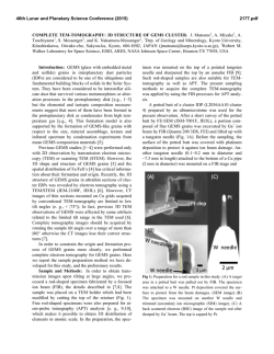
F l e x i b l e Bro n c h o s c o p y
and its Role in the
S t a g i n g of N o n – S m a l l
Cell Lung Cancer
Felix J.F. Herth, MD, DSc, FCCP*, Ralf Eberhardt, MD
KEYWORDS
The first ever bronchoscopy was performed in
1887 by Gustav Killian of Freiburg, Germany.1
During the early years of the development of bronchoscopy, the indications for the procedure were
primarily therapeutic: removal of foreign bodies
and dilation of strictures from tuberculosis and
diphtheria. In the early part of the twentieth
century, Chevalier Jackson, the father of American
bronchoesophagology, further advanced bronchoscopic techniques and designed modern rigid
bronchoscopes.2 Again, the primary indication
was often therapeutic.
The flexible bronchoscope was developed in the
late 1960s by Ikeda3 and has become the mainstay investigation in the evaluation of patients suspected of lung cancer. It is used mainly as
a diagnostic tool providing tissue to determine
the histologic type of tumor. Bronchoscopy also
has a role in disease staging and an extended
role in delivering therapeutic modalities. Flexible
bronchoscopy (FB) is easier to perform and is
safe and well tolerated by patients. The requirement of only a moderate sedation makes it an
acceptable outpatient procedure. It has almost
completely replaced rigid bronchoscopy in the
initial assessment. The development of video
bronchoscopes has the added advantage of facilitating teaching and rendering the procedure more
interesting for the observers in the bronchoscopy
suite.
The flexibility of the bronchoscope allows the
operator to inspect most of fourth order and often
up to sixth order bronchi. In addition, the operator
may directly assess mucosal details, such as color
and vascularity. Relative contraindications to the
procedure are few and include hypoxemia refractory to supplemental oxygen, intractable bleeding
diathesis, severe pulmonary hypertension, cardiovascular instability, and acute hypercapnia.4
FB is safe with a complication rate of 0.12% and
a mortality rate of 0.04%.5 The dangers of hemorrhage and pneumothorax relate to the biopsy
procedure used and are discussed later. In all
patients, the bronchoscope causes a temporary
increase in airflow obstruction, which may result
in hypercapnia.6 Inappropriate sedation with
benzodiazepines or opiates increases the likelihood of respiratory complications and high-risk
patients could be identified by prior measurement
of arterial blood gases.5–7 Supplemental oxygen
should be provided and patients monitored
throughout the procedure with pulse oximetry.
Cardiac monitoring should be used for those
patients with a history of ischemic heart disease
and
resuscitation
equipment
immediately
available.
Although FB has largely replaced rigid bronchoscopy in the initial assessment of the patient,
the rigid scope has advantages in certain situations.8 It may provide more accurate information
regarding the endobronchial location of a tumor
before resection. Additionally, manipulation of
the scope allows assessment of the mobility of
the proximal airways providing an indirect
Department of Pneumology and Critical Care Medicine, Thoraxklinik, University of Heidelberg, Amalienstrasse
5, D-69126 Heidelberg, Germany
* Corresponding author.
E-mail address: [email protected] (F.J.F. Herth).
Clin Chest Med 31 (2010) 87–100
doi:10.1016/j.ccm.2009.08.006
0272-5231/10/$ – see front matter ª 2010 Elsevier Inc. All rights reserved.
chestmed.theclinics.com
Flexible bronchoscopy Transbronchial needle aspiration
Endobronchial ultrasound Electromagnetic navigation
88
Herth & Eberhardt
evaluation of mediastinal nodal involvement.
Airway obstruction is less and the rigid scope
may be preferable in exploring patients with
tracheal narrowing in whom the flexible scope
may produce critical airway narrowing. It provides
superior suction, facilitating the assessment and
biopsy of potentially hemorrhagic lesions and the
debulking of large tumors.8–10 In addition, many
physicians are now acquiring skills in this technique to facilitate endobronchial laser therapy
and stent placement.11
THE DIAGNOSTIC YIELD OF FB
The expected diagnostic yield from FB depends
on the location and the size of the lesion. Central
endobronchial lesions yield the highest diagnostic
return (>90%), whereas small peripheral lesions
often prove more elusive unless more demanding
and time-consuming techniques are used. The
question of which combination of cytologic and
histologic procedures provides the optimum diagnostic yield has not been conclusively answered
but probably depends on the expertise available
in any individual center. The routine techniques
include bronchial washings, brushings, and biopsies but these may be augmented by the use of
transbronchial needle aspiration (TBNA) and bronchoalveolar lavage (BAL).12
More than 70% of lung carcinomas can be approached with FB and although the yield is dependent on operator’s experience, a high level of
diagnostic accuracy can be achieved by taking
between three and five biopsy specimens and
a combination of brushing, biopsy, and bronchial
washes can expect to establish a diagnosis in
more than 60% of cases.6,7,13,14 When the tumor
is visible but is intramural rather than endobronchial in distribution the diagnostic yield falls to
55% and is reduced further when the tumor lies
beyond the bronchoscopist’s vision.6,7,12
The main role of BAL in patients with lung cancer
is the diagnosis of opportunistic infections, especially in patients undergoing chemotherapy. BAL
may have an extended role, however, in the diagnosis of malignancy itself. A high diagnostic yield
has been shown in the detection of pulmonary
hematologic malignancies, primary bronchoalveolar cell carcinoma, and metastatic adenocarcinoma of the breast.15–17
Information on the role of BAL in the diagnosis of
primary lung cancer remains sparse. Examination
of BAL from 55 patients with a peripheral lung
lesion demonstrated a diagnostic yield of around
30% with no false-positive results and only one
instance of incorrect cell typing. Additionally,
in combination with bronchial washings and
postbronchoscopy
sputum
analysis
BAL
increased the yield to 56%.18 Examination of
BAL in 162 patients with malignant lung infiltrates
revealed improved sensitivity in cases of bronchoalveolar cell carcinoma (93%) and lymphangitic carcinomatosis (83%). Forty-five percent of
non-Hodgkin lymphoma could be detected and
immunocytochemistry is of value in identification
and classification.19
BAL is safe; bleeding and pneumothorax are
uncommon and the fever and transient loss of
lung function reported are rarely serious and there
is no need for fluoroscopy. Furthermore, the diagnostic yield is high in diseases other than cancer,
such as pulmonary tuberculosis. Advances in cell
and molecular biology may complement the
technique of BAL to improve the rate of tumor
diagnosis in peripheral lesions (particularly adenocarcinoma) and may also provide a useful tool to
explore the molecular mechanisms governing the
genesis of lung cancer.20–22
VISIBLE ENDOBRONCHIAL LESIONS
Central tumors can present as exophytic mass
lesions, with partial or total occlusion of the bronchial lumen, as peribronchial tumors with extrinsic
compression of the airway, or with submucosal
infiltration of tumor. The changes with peribronchial tumors or with submucosal infiltration are
often subtle. The airways should be examined
closely for characteristic changes, such as
erythema, loss of bronchial markings, and nodularity of the mucosal surface. Central lesions are
usually sampled with a combination of bronchial
washes, bronchial brushings, and endobronchial
biopsies. The yield of endobronchial biopsies is
highest for exophytic lesions, with a diagnostic
yield of approximately 90%.23–25 Three to four
biopsies are likely adequate in this situation.
Attempts should be made to obtain the biopsies
from areas of the lesion that seem viable (Fig. 1).
For submucosal lesions, TBNA can be performed by inserting the needle into the submucosal plane at an oblique angle, and in patients
with peribronchial disease and extrinsic compression, the needle should be passed through the
bronchial wall into the lesion.26,27 It is particularly
frustrating when apparently adequate biopsy
specimens from visible endobronchial disease
fail to achieve a diagnosis (Fig. 2). Reasons for
this include the presence of surface necrosis or
the presence of crush artifact (particularly
common with samples from small cell carcinoma).
In these circumstances, TBNA may improve diagnostic yield.28,29
Bronchoscopy
Fig. 1. Endobronchial biopsy from a lesion in the right
main bronchus.
Regarding the T (T, N, M classification) staging,
FB may allow the operator to determine that the
tumor is beyond resection. Pointers to inoperability
include paralysis of a vocal cord, tumor to the level
of the right tracheobronchial junction or to within
2 cm of the left tracheobronchial junction, and definite carinal or tracheal involvement (Fig. 3).
PERIPHERAL LUNG LESIONS
Peripheral lesions are usually sampled with
a combination of bronchial wash, brushes,
Fig. 2. TBNA needle before passing the bronchial wall
in case of an endobronchial compression of the distal
trachea.
Fig. 3. Tumor in right main bronchus, reaching the
level of the main carina.
transbronchial biopsy (TBBx), and TBNA. The
diagnostic yield of bronchoscopy for peripheral
lesions depends on a number of factors, including
lesion size, the distance of the lesion from the
hilum, and the relationship between the lesion
and bronchus. The yield of bronchoscopy for
lesions smaller than 3 cm varies from 14% to
50% compared with a diagnostic yield of 46% to
80% when the lesion is larger than 3 cm.13,30,31
The presence of a bronchus sign on chest CT
predicts a much higher yield of bronchoscopy for
peripheral lung lesions. In these cases, fluoroscopic guidance should be used to ensure proper
positioning of the diagnostic accessory (Fig. 4).
Fluoroscopy increases the diagnostic yield from
TBBx in focal lung lesions but it is time-consuming,
requires experience, and is not universally available. If the disease process is diffuse, however,
such as in lymphangitic carcinomatosis, yields
are similar whether or not fluoroscopy is used.32
Indeed, TBBx may be regarded as the procedure
of choice for lymphangitic carcinomatosis.
Complications from TBBx include pneumothorax
and hemorrhage but these are generally low and
rarely serious.
In situations where bronchial biopsies cannot be
obtained examination of bronchial washings may
still yield useful information and often provide
complementary information.25,33 It is often prudent
to perform all types of sampling procedure to
maximize the yield.12
Several studies have demonstrated that TBNA
may be used to obtain diagnostic tissue from
peripheral lesions. Typical results report an
89
90
Herth & Eberhardt
Fig. 4. Positive bronchus sign in a peripheral lung
lesion in the left upper lobe.
increase in the diagnostic yield from percentage
figures in the mid thirties up to the high
sixties.12,34–36 As with TBBx, the size of the
peripheral lesion seems to be important, although
this is not a feature of all studies. Optimum yields
are provided by using a combination of diagnostic
techniques. TBNA may represent an alternative to
TBBx when the airway externally compressed to
such a degree that it is not possible to negotiate
the biopsy forceps.
The need to work-up and manage pulmonary
nodules and masses is encountered with
increasing frequency in chest medicine. In patients
with such nodules, the diagnostic procedure is
usually performed as a TBBx under fluoroscopic
guidance. This commonly performed procedure
is associated with a low yield in coin lesions smaller
than 3 cm or fluoroscopically invisible lesions.25,37
Nodules that are too small to be visualized by
conventional fluoroscopy during the procedure
pose a particular problem and usually require
further, often surgical, biopsy procedures. Promising new technologies, such as electromagnetic
navigation
and
endobronchial
ultrasound
(EBUS), may help overcome the limitations.
ELECTROMAGNETIC NAVIGATION
The electromagnetic navigation system is
a device that assists in localizing and placing
endobronchial accessories (eg, forceps, brush,
and needle) in the desired areas of the lung.
The system uses low-frequency electromagnetic
waves, which are emitted from an electromagnetic board placed under the bronchoscopy
table mattress. A 1-mm diameter, 8-mm long
sensor probe mounted on the tip of a flexible
metal cable constitutes the main assembly of
the device (locatable guide). Once the probe is
placed within the electromagnetic field, its position in the X, Y, and Z planes, and its orientation
(roll, pitch, and yaw movements), are captured
by the electromagnetic navigation system. This
information is then displayed on a monitor in
real time (Fig. 5). The locatable guide also has
an added feature that allows its distal section
to be steered 360 degrees. The fully retractable
probe is incorporated into a flexible catheter
(serving as an extended working channel), which
once placed in the desired location creates an
easy access for bronchoscopic accessories.
The computer software and monitor allow the
bronchoscopist to view the reconstructed
three-dimensional CT scans of the object’s
anatomy in coronal, sagittal, and axial views
together with superimposed graphic information
depicting the position of the sensor probe.
There are still some major limitations to the technique. For planning, a CT scan is necessary with
a special protocol (1-mm cuts and tight overlay).
For the planning of the procedure, use of the electromagnetic navigation bronchoscopy (ENB) software is required. The planning can be done even
on the system or on a special dedicated laptop
before the procedure; the planning needs some
time, up to 10 minutes even in trained hands.
The whole procedure time is prolonged compared
with a traditional diagnostic bronchoscopy with
fluoroscopy; but equal to that required by the
CT-guided percutaneous needle aspiration. The
locatable guide is a single-use device and costs
between $500 and $1000.
Schwarz and colleagues38 performed a trial to
determine the practicality, accuracy, and safety
of real-time electromagnetic navigation in locating
artificially created peripheral lung lesions in a swine
model. No adverse effects, such as pneumothorax
or internal bleeding, were encountered in any
animal in this study. Schwarz and colleagues38
concluded that real-time electromagnetic positioning technology, coupled with previously
acquired CT scans, is an accurate technology
that can augment standard bronchoscopy to
assist in reaching peripheral lung lesions and performing biopsies. Based on the results of Schwarz
and colleagues,38 Becker and colleagues39 performed a pilot study in humans. They examined
the use of the system in 30 consecutive patients
presenting for endoscopic evaluation of lung
nodules and masses. The lesion size in this population varied from 12 to 106 mm but was specifically not controlled for in this early trial.
Evaluation was possible in 29 patients, and in 20
Bronchoscopy
Fig. 5. Navigation screen during an electromagnetic navigation procedure. In the right lower lobe the relation
from the sensor is seen.
patients a definitive diagnosis was established,
with no complications related to the navigation
device. In an uncontrolled study, again Schwarz
and colleagues40 confirmed that the procedure
was safe and added only an average of 15 minutes
to the time of a conventional bronchoscopy.
Successful diagnostic biopsies were obtained in
69% of patients. A follow-up study of 60
patients,41,42 published in 2006, successfully
reached the target lesion in 100% of cases. Bronchoscopy with electromagnetic navigation diagnosed 80.3% of the lesions, 74% of the
peripheral lesions, and 100% of the lymph nodes.
Of the lesions, 57% were less than 2 cm in size.
Diagnostic yield did not differ significantly based
on the size of the lesion. The accuracy of ENB
navigation has been proved in animal studies
and against fluoroscopically40,41 verified reference
points in humans. Nevertheless, all preceding
diagnostic studies using ENB also used fluoroscopy to guide biopsies. The role of ENB as
a stand-alone technology is still unproved and
concerns remain that biopsy instruments may
dislodge an accurately positioned extended
working channel when replacing the sensor probe.
Eberhardt and colleagues42 examined the yield
of ENB without fluoroscopy in the diagnosis of
peripheral lung lesions and solitary pulmonary
nodules. Ninety-two peripheral lung lesions were
biopsied in the 89 subjects. The diagnostic yield
of ENB was 67%, which was independent of lesion
size. The mean navigation error was 9 Æ 6 mm;
range was 1 to 31. When analyzed by lobar distribution, there was a trend toward a higher ENB yield
in diagnosing lesions in the right middle lobe (88%).
Eberhardt and colleagues42 concluded that ENB
could be used as a stand-alone bronchoscopic
technique without compromising diagnostic yield
or increasing pneumothorax risk. This may result
in sizable time saving and avoids radiation exposure. Makris and colleagues43 confirmed these
results. In 40 patients all target lesions but one
was reached and the overall diagnostic yield was
62.5% (25 of 40). Also, the French group summarized that electromagnetic navigation–guided
bronchoscopy has the potential to improve the
diagnostic yield of transbronchial biopsies without
further fluoroscopic guidance and may be useful in
early diagnosis of lung cancer, particularly in nonoperable patients.
91
92
Herth & Eberhardt
EBUS
Two different types of EBUS systems are available
in the market. The linear EBUS bronchoscope,
which incorporates the ultrasound transducer at
its distal end, uses a fixed array of transducers
aligned in a curvilinear pattern. Because of the
size of the scope, the system is usable only in
the central airways. For the peripheral lung the
radial EBUS must be used. The radial EBUS
system consists of a mechanical radial miniprobe
(Fig. 6). Two types of miniprobe are available,
one with a notch at the tip for a water-fillable
balloon catheter, one without a notch. Particularly
with the notch type, ultrasonography can be performed using the balloon method; the balloon is inflated at the distal end of the probe after the probe
has been inserted into the working channel. The
balloon method makes possible easy delineation
of ultrasound images even at sites where it is difficult to retain defecated water. The limitation is the
size; with the balloon sheath an endoscope with
2.8 mm or larger diameter channel has to be
used. The notchless probe is smaller (1.7 mm)
and can also be used in smaller bronchoscopes.
The 20-MHz frequency is commonly used,
although 12- and 30-MHz probes are also
available.
For use in the peripheral lung, most commonly
the probe is placed through a guide sheath in the
working channel of the bronchoscope. After localization of the lesion (Fig. 7), the bronchoscope is
kept in place at the nearest visible subsegmental
carina, and the miniprobe removed. Through the
guide sheath, the forceps are guided to the lesion
(Fig. 8). By using the guide sheath, EBUS-guided
TBBx can be performed without losing the position
of the nodule. The initial studies were performed
without the guide sheath but most recently the
use of the guide sheath is considered as the
technique of choice. The feasibility trial of EBUSguided TBBx44 without the use of fluoroscopy
showed that EBUS can provide an alternative to
Fig. 6. The endobronchial miniprobe for peripheral
lung biopsy.
Fig. 7. EBUS image of a coin lesion.
fluoroscopy for image guidance in biopsies for
peripheral lesions. In the study, a trend toward
superior results with EBUS was particularly strong
in lesions less than 3 cm in diameter. The same
results were shown by Shirakawa and
colleagues.45 After the feasibility trial, investigators
began to examine the use of EBUS as an adjunct
for the diagnosis of peripheral lung lesion and solitary pulmonary nodules. A large prospective study
by Paone and colleagues46 compared traditional
TBBx with EBUS for peripheral lesions. They found
that EBUS-guided bronchoscopy had a sensitivity
of 0.83 for lesions greater than 3 cm in size and
0.75 for lesions less than 3 cm in size; compared
with the traditional TBBx EBUS also showed
promise when used for nodules less than 3 cm in
size. These types of lesion are often difficult to
Fig. 8. The theoretical background of an EBUS-guided
TBBx.
Bronchoscopy
visualize fluoroscopically for TBBx, and conventional bronchoscopy has a low diagnostic yield in
such settings.
Most of the published studies used radiographic fluoroscopy, with radiation exposure for
both the patient and medical staff. This result
is in line with the study by Herth and
colleagues47 including only peripheral pulmonary
lesions less than 30 mm and reporting a diagnostic yield of 87% for EBUS-guided TBBx
without the need for radiographic equipment or
radiation exposure.
For lesions less than 20 mm, the yield of EBUSguided detection and pathologic diagnosis
decreased fewer than 30% yield.48 By contrast,
Japanese groups have reported diagnostic yields
of 53% and 72%, respectively, for lesions less
than 20 mm using EBUS-guided TBBX with a catheter sheath and under radiographic fluoroscopy.49,50 More recently, a Japanese group
performed EBUS-guided TBBX using virtual bronchoscopic navigation and detected 67% of lesions
less than 20 mm on EBUS, resulting in a diagnostic
yield of 44%.51 The limitation to the systems is
a significant learning curve, and methods of physician training and education still need to be established. EBUS lacks a navigational system,
however, and requires the operator to maneuver
the bronchoscope blindly to the lesion with the
knowledge of prior radiologic investigations, such
as CT scans.
Biopsies using ENB have not always resulted in
a diagnosis despite accurate navigation in most
cases to within 10 mm of the target center. Respiratory variations causing larger than anticipated
navigation errors and dislodgement of the
extended working channel when biopsy instruments were introduced may account for this lower
than expected diagnostic yield. ENB lacks
a means to directly visualize lesions before
biopsy. The role of combining EBUS with ENB
to gain the benefits and minimize the limitations
of either technique has never been reported.
Eberhardt and colleagues52 performed a prospective randomized controlled trial comprising three
arms with EBUS only, ENB only, and combined
EBUS-ENB to test this hypothesis. Of the 120
patients recruited, 118 had a definitive histologic
diagnosis and were included in the final analysis.
The diagnostic yield of the combined procedure
(88%) was greater than either EBUS (69%) or
electromagnetic navigation alone (59%; P 5
.02). The group concluded that combined EBUS
and electromagnetic navigation improves the
diagnostic yield of flexible bronchoscopy in
peripheral lung lesions without compromising
safety.
MEDIASTINAL STAGING
TBNA
The first description of sampling mediastinal
lymph nodes through the tracheal carina using
a rigid bronchoscope was by Schieppati,53,54 an
Argentinean physician who presented the technique at the Argentine Meeting of Bronchoesophagology in 1949. In 1978, Wang and colleagues55
demonstrated that with this technique it was also
possible to sample paratracheal nodes. In 1979,
Oho and colleagues56 introduced a flexible needle
that could be used through a bronchofiberscope
and in 1983, Wang and coworkers57,58 pointed
out the diagnostic possibility of the method in
staging of lung cancer and developed new types
of needles. Subsequent publications highlighted
its use in the diagnosis of endobronchial and
peripheral lesions and the ability of TBNA to
provide a diagnosis even in the absence of endobronchial disease, in a nonsurgical fashion,
confirmed its usefulness to bronchoscopists
(Fig. 9).59–61
Operators have reported the use of 21- and 22gauge cytology needles and 19-gauge histology
needles.62 Samples are provided by rinsing the
needle with a small volume of normal saline and
collecting the ‘‘flush solution’’ for analysis.
Although TBNA is not widely used, it seems to
improve the diagnostic yield when sampling from
visible endobronchial, submucosal, and peripheral
lesions. Additionally, the technique may detect
mediastinal disease potentially allowing the operator to diagnose and stage a lung tumor in one
procedure performed under local anesthetic.
TBNA may be used to sample lymph nodes that
lie immediately adjacent to the trachea and major
bronchi. Care must be taken to perform TBNA
before inspection of the distal airways and other
sampling procedures because contamination
with exfoliated malignant cells is a recognized
Fig. 9. Fluoroscopy of a TBNA in the left distal
trachea.
93
94
Herth & Eberhardt
cause of false-positive results. Studies have reported sensitivity rates of between 43% and
83% and positive predictive values of 89% to
100%.63–68 Use of a 19-gauge needle provides
greater sensitivity than a 22-gauge needle but
a combination of samples provides the best
yields.62,69 Although the positive predictive value
is high (often 100%) the negative predictive value
is low and does not obviate the requirement for
further surgical staging.59–69 A potential limitation
of mediastinal lymph node staging with TBNA is
that it is a blind procedure. The technique may
be combined with that of EBUS by the miniprobes.70,71 The numerous papers on TBNA performed in the last few years confirm the safety of
the procedure. No cases of mortality related to
TBNA have been described. The rare complications reported are pneumothorax,72 pneumomediastinum,73 hemomediastinum,74 bacteremia,75
and pericarditis.76 None of these complications
determined clinical major consequences. One of
the major complications of TBNA is the possible
severe damage to the working channel of the
scope.61
Endoesophageal Ultrasound
with Fine-needle Aspiration
EUS fine-needle aspiration (FNA) is a relatively
new method first described in 1991.77 Since then
several studies have been published and it has
been demonstrated that generally all lesions outlined by EUS may be punctured, and even small
lesions down to the size of 5 mm may be
diagnosed.78
EUS-FNA is performed with the aid of esophagoscopy and a biopsy needle is passed through
the working channel of the endoscope, through
the esophageal wall and guided ultrasonographically toward the lesion of interest in the mediastinum (Fig. 10). The procedure is performed
under local anesthesia and moderate sedation.
This method gives an excellent overview of mediastinal structures, including a good access to the
paraesophageal space, the aorticopulmonary
window, the subcarinal region, and the region
around the left atrium (level 4, 5, and 7).79–81 EUS
has the advantage of being noninvasive, safe,
and cost effective.82 An area anterior to the airfilled trachea, however, cannot be visualized. The
echoendoscope is initially introduced up to the
level of celiac axis and gradually withdrawn
upward for a detailed mediastinal imaging.
Because the ultrasound waves are emitted parallel
to the long axis of the endoscope, the entire needle could be visualized approaching a target in
the sector-shaped sound field. Pulse-wave
Fig. 10. EUS procedure of an enlarged lymph node
(4 left). The vessel is seen by the color Doppler flow.
Doppler ultrasonography imaging is performed,
whenever vascular structures are suspected in
the pathway of the needle or adjacent to it, to
correct the target line if necessary.81 The needle
is advanced through the wall of the esophagus
and guided into the target lesion. The central stylet
is removed, and a special 10-mL syringe attached
to the hub of the needle to apply suction as the
needle is moved back and forth within the mass.
The suction is released slowly, and the needle
assembly removed out of the biopsy channel.
One to two needle passes are made to obtain
adequate tissue.83,84
Visual assessment of mediastinal lymph nodes
by EUS gave various observers sensitivity of 0.54
to 0.75, specificity of 0.71 to 0.98, positive predictive values of 0.46 to 0.77, and negative predictive
values of 0.85 to 0.93, in a total number of patients
studied being more than 1000.85
These studies varied widely with regard to the
number of examined mediastinal lymph node
levels, visual criteria for malignancy, and patient
population characteristics. Compared with CT,
the detection rate of malignant lymph nodes was
higher with EUS, with less false-positive results.86
EUS can assess mediastinal lymph nodes at most
levels, particularly at levels 4 left, 5, 7, 8, and 9, and
metastasis in the left adrenal gland. Levels 1, 2, 3,
and 4 right are not always assessable, because of
interference by air in the larger airways.79,81 When
enlarged, however, detection is easier.78,85 Properties of lymph nodes indicating possible malignancy are a hypoechoic core, sharp edges,
round shape, and a long axis diameter exceeding
10 mm.85 Signs of benignancy are a hyperechoic
core (fat); central calcification (old granulomatous
disease); ill-defined edges; and a long and narrow
shape.86,87 False-negative results may have been
introduced by an occasional poor lymph node
Bronchoscopy
sampling during EUS-FNA (sampling only the most
suspicious nodes). Many outcomes have been
supported by not only clinical but also surgical
follow-up. Despite these drawbacks, the clinical
impact of EUS-FNA is illustrated by a change in
the management of non–small cell lung cancer
patients after EUS-FNA in 66% of the patients, or
cancellation of 68% and 49% of the scheduled
mediastinoscopies and thoracotomies, respectively. According to Hunerbein and colleagues,83
EUS-FNA made an unexpected diagnosis of
malignancy in 30% of the procedures. In two
studies with decision-analysis models, EUS-FNA
was shown to be less expensive compared with
mediastinoscopy for the assessment of the entire
mediastinum or only for subcarinal lymph nodes.79,88,89 Barawi and colleagues90 prospectively
studied the incidence of complications associated
with EUS-FNA. In 842 mediastinal EUS-FNA
procedures, one infection, two hemorrhages, and
one inexplicable transient hypotension were
reported.
EUS-FNA is contraindicated in patients with
a Zenker diverticulum or bleeding tendency.91
FNA of a cystic mediastinal lesion should be
avoided, or when necessary be preceded by
prophylactic antibiotics.92
Real-time EBUS with TBNA
Lymph node staging is also the main indication for
use of the new EBUS-TBNA scope. An ultrasound
transducer integrated into a bronchoscope with
a separate working channel potentially increases
the yield of TBNA by allowing direct visualization
of needle placement within the area of interest. A
special ultrasonic puncture bronchoscope by integrating a convex probe at the tip of the FB has
been developed. With this bronchoscope direct
TBNA under real-time convex probe EBUS
(EBUS-TBNA-bronchoscopy) guidance is now
possible (Fig. 11).
EBUS-TBNA is performed by direct transducer
contact with the wall of the trachea or bronchus.
When a lesion is identified, a 22-gauge full-length
steel needle is introduced through the biopsy
channel of the endoscope. Power Doppler examination may be performed before the biopsy to
avoid inadvertent puncture of mediastinal vessels.
Under real-time ultrasonic guidance the needle is
placed in the lesion (Fig. 12). Suction is applied
with a syringe, and the needle is moved back
and forth inside the lesion.93
Endobronchial,
ultrasound-guided,
TBNA
(EBUS-TBNA) has been available for more than 5
years. A growing body of research supports its
usefulness in airway assessment and procedure
Fig. 11. The tip of the EBUS-TBNA scope with the
ultrasound system. The TBNA needle is inserted.
guidance, especially since the availability of positron emission tomography scanning.94–97 EBUSTBNA has access to all of the mediastinal lymph
node stations accessible by mediastinoscopy and
N1 nodes. The largest trial reported the results of
using the method in 502 patients98; 572 lymph nodes were punctured, and 535 (94%) resulted in
a diagnosis. Biopsies were taken from all reachable
lymph node stations (2l, 2r, 3, 4r, 4l, 7, 10r, 10l, 11r,
and 11l). Mean (SD) diameter of the nodes was 1.6
cm (0.36 cm) and the range was 0.8 to 3.2 cm.
Sensitivity was 92%, specificity was 100%, and
the positive predictive value was 93%. Like in all
other trials no complications occurred. The
Danish-German group99 examined in addition the
accuracy of EBUS-TBNA in sampling nodes less
than 1 cm in diameter. Among 100 patients, 119
lymph nodes with a size between 4 and 10 mm
were detected and sampled. Malignancy was detected in 19 patients but missed in 2 others; all diagnoses were confirmed by surgical findings. The
mean (SD) diameter of the punctured lymph nodes
was 8.1 mm. The sensitivity of EBUS-TBNA for detecting malignancy was 92.3%, the specificity was
Fig. 12. EBUS-TBNA of an enlarged mediastinal lymph
node. The needle is clearly visible within the node.
95
96
Herth & Eberhardt
100%, and the negative predictive value was
96.3%. No complications occurred. They summarized that EBUS-TBNA can sample even small
mediastinal nodes, avoiding unnecessary surgical
exploration in one of five patients who have no CT
evidence of mediastinal disease. Potentially operable patients with clinically nonmetastatic non–
small cell lung cancer may benefit from presurgical
EBUS-TBNA biopsies and staging.
A study comparing EBUS-TBNA, CT, and positron emission tomography for lymph node staging
of lung cancer showed a high yield for EBUSTBNA.100 Altogether, 102 potentially operable
patients with proved (N 5 96) or radiologically suspected (N 5 6) lung cancer were included in the
study. CT, positron emission tomography, and
EBUS-TBNA were performed before surgery for
the evaluation of mediastinal and hilar lymph
node metastasis. The sensitivities of CT, positron
emission tomography, and EBUS-TBNA for the
correct diagnosis of mediastinal and hilar lymph
node staging were 76.9%, 80%, and 92.3%; the
specificities were 55.3%, 70.1%, and 100%; and
the diagnostic accuracies were 60.8%, 72.5%,
and 98%, respectively. EBUS-TBNA was proved
to have high sensitivity and specificity, compared
with CT or positron emission tomography, for
mediastinal staging in patients with potentially
resectable lung cancer.
Restaging of the mediastinum is another area of
growing interest for the treatment strategy of lung
cancer. In cases of advanced lymph node stage
lung cancer, induction chemotherapy before
surgical resection is an option. Mediastinoscopy
is considered the gold standard for staging the
mediastinum. Remediastinoscopy can be technically difficult, however, and is not commonly performed. The ability to perform multiple, repeat
biopsies using EBUS-TBNA allows restaging of
the mediastinum after the introduction of chemotherapy. A group of 124 consecutive patients
with tissue-proved IIIA-N2 disease who were
treated with induction chemotherapy underwent
mediastinal restaging by EBUS-TBNA. The sensitivity, specificity, positive predictive value, negative predictive value, and diagnostic accuracy of
EBUS-TBNA for mediastinal restaging following
induction chemotherapy were 76%, 100%,
100%, 20%, and 77%, respectively.
EBUS–TBNA is an accurate, minimally invasive
test for mediastinal restaging of patients with
non–small cell lung cancer. Because of the low
negative predictive value, however, tumor-negative findings should be confirmed by surgical
staging.101
EBUS-TBNA also can be used for the diagnosis
of intrapulmonary nodules and mediastinal and
hilar lymph nodes. The limitation is the reach of
EBUS-TBNA, which depends on the size of the
bronchus. Usually the EBUS-TBNA can be inserted as far as the lobar bronchus. Lung tumors
located adjacent to the airway within reach of
EBUS-TBNA can be diagnosed with EBUSTBNA. Tournoy and colleagues102 have reported
their experience for this indication. In 60 patients
who had an initial nondiagnostic bronchoscopy,
they were able to establish the definitive diagnosis
in 77% without any complication.
Complications related to the procedure are rare
and similar to those of conventional TBNA
including bleeding from major vessels, pneumomediastinum, mediastinitis, pneumothorax, bronchospasm, and laryngospasm. Authors have not
encountered any major complications related to
EBUS-TBNA. Although EBUS has enabled the
bronchoscopist to see beyond the airway, one
must be aware of the possible complications
related to the procedure.103,104
Rapid On-site Evaluation
Rapid on-site evaluation (ROSE) is comparable
with the intraoperative frozen-section examination.
The technique requires the cytopathologist
and the pathology technician to process and interpret the stained wet film of the aspirate immediately and report the result to the bronchoscopist.
Several studies have shown that ROSE reduces
the incidence of inadequate specimens, an important cause of nondiagnostic TBNA aspirates.105–108
Davenport109 studied the value of ROSE in 73
aspirates and compared the results with 134 specimens processed routinely. The aspirates were obtained from the mediastinal lymph nodes and the
peripheral lung nodules. With ROSE, the proportion of aspirates showing malignant cells
increased from 31% to 56%. The proportion of
the inadequate negative specimens dropped
from 56% to 18%. The negative aspirate with
ROSE had a higher negative predictive value
than that of routinely prepared specimens. In
a recent prospective study, Diette and
colleagues110 evaluated TBNA aspirates with
ROSE in 81 of 204 cases. The overall diagnostic
yield was 81% when ROSE was used compared
with a 50% yield when specimens were processed
in the usual manner. Multivariate analysis showed
that ROSE was an independent predictor of a positive aspirate for malignant cells with an odds ratio
of 4.5. The mean number of needle attempts was
slightly greater with ROSE. The concordance
between the preliminary diagnosis made in the
bronchoscopy suite and the final diagnosis was
reached after subsequent review of material in
Bronchoscopy
the cytopathology laboratory was 87%, indicating
that the on-site evaluation of needle aspirate is
fairly accurate but not perfect.
Although ROSE seems to improve the diagnostic yield of TBNA, its cost-effectiveness
remains unclear. Successful use of ROSE requires
services of an expert cytopathologist. Many
pathologists do not favor ROSE because of the
extra time and effort involved. The reimbursement
from a third party payor for these services is highly
variable. Presently, the decision to use ROSE
remains institution-specific.
10.
11.
12.
13.
SUMMARY
Technologic advances in bronchoscopy continue
to improve the ability to perform minimally invasive, accurate evaluations of the tracheobronchial
tree and to perform an ever-increasing array of
diagnostic, staging, therapeutic, and palliative
interventions. The role of both old and new diagnostic bronchoscopy will continue to evolve as
further improvements are made in bronchoscopes, accessory equipment, and imaging technologies. The major challenge is the adoption of
the many new bronchoscopic techniques into
routine clinical practice. There is a need for welldesigned studies to delineate the appropriate
use of these interventions and to better define their
limitations and cost effectiveness.
REFERENCES
1. KilIian G. Ueber directe bronchoscopie. MMW
1898;27:844–7.
2. Jackson CH. The life of Chevalier Jackson: an autobiography. New York: Macmillan; 1938.
3. Ohata M. History and progress of bronchology in
Japan. JJSB 1998;20:539–46.
4. Barle´si F, Doddoli C, Greillier L, et al. Bronchoscopy in the diagnosis of lung cancer: an evaluation
of current practice. Rev Mal Respir 2006;23:17–26.
5. Becker HD, Shirakawa T, Tanaka F, et al. Transbronchial lung biopsy in the immunocompromised
patient. Eur Respir Mon 1998;9:193–208.
6. Mazzone P, Jain P, Arroliga AC, et al. Bronchoscopy and needle biopsy techniques for diagnosing
and staging of lung cancer. Clin Chest Med 2002;
23(1):137–58.
7. El-Bayoumi E, Silvestri GA. Bronchoscopy for the
diagnosis and staging of lung cancer. Semin Respir Crit Care Med 2008;29(3):261–70.
8. Wahidi MM, Herth FJ, Ernst A. State of the art: interventional pulmonology. Chest 2007;131(1):261–74.
9. Herth FJF, Ernst A. Innovative bronchoscopic diagnostic techniques: endobronchial ultrasound and
14.
15.
16.
17.
18.
19.
20.
21.
22.
23.
24.
25.
electromagnetic navigation. Curr Opin Pulm Med
2005;11(4):278–81.
Folch E, Mehta AC. Airway interventions in the
tracheobronchial tree. Semin Respir Crit Care
Med 2008;29(4):441–52.
Herth FJF, Eberhardt R. Interventional bronchoscopy. Minvera Pneumol 2004;43:189–201.
Gasparini S, Ferrety M, Such E, et al. Integration of
transbronchial and percutaneous approach in the
diagnosis of peripheral pulmonary nodules or
masses: experience with 1027 consecutive cases.
Chest 1995;108:131–7.
Govert JA, Dodd LG, Kussin PS, et al. A prospective comparison of fiberoptic transbronchial needle
aspiration and bronchial biopsy for bronchoscopically visible lung carcinoma. Cancer 1999;87:
129–34.
Govert JA, Kopita JM, Matehar D, et al. Cost-effectiveness of collecting cytologic specimens during
fiberoptic bronchoscopy for endoscopically visible
lung tumors. Chest 1996;109:451–6.
De Gracia J, Bravo C, Miravitalles M, et al. Diagnostic value of bronchoalveolar lavage in peripheral
lung cancer. Am Rev Respir Dis 1993;147:649–52.
Fabin E, Nagy M, Meszaros G. Experiences with
bronchial brushing method. Acta Cytol 1975;19:
320–1.
Fedullo AJ, Ettensohn DB. Bronchoalveolar lavage
in the lymphangitic spread of adenocarcinoma to
the lung. Chest 1985;87:129–31.
Semenzato G, Spatafora M, Feruglio C, et al. Bronchoalveolar lavage and the immunology of lung
cancer. Lung 1990;168:1041–9.
Rennard SI. Bronchoalveolar lavage in the diagnosis of cancer. Lung 1990;168:1035–40.
Garg S, Handa U, Mohan H, et al. Comparative
analysis of various cytohistological techniques in
diagnosis of lung diseases. Diagn Cytopathol
2007;35(1):26–31.
Emad A, Emad V. The value of BAL fluid LDH level
in differentiating benign from malignant solitary
pulmonary nodules. J Cancer Res Clin Oncol
2008;134(4):489–93.
Azoulay E, Schlemmer B. Diagnostic strategy in
cancer patients with acute respiratory failure. Intensive Care Med 2006;32(6):808–22.
Wilson RW, Frazier AA. Pathological-radiological
correlations: pathological and radiological correlation of endobronchial neoplasms: part II, malignant
tumors. Ann Diagn Pathol 1998;2(1):31–4.
Simoff MJ. Endobronchial management of
advanced lung cancer. Cancer Control 2001;8(4):
337–43.
Schreiber G, McCrory DC. Performance characteristics of different modalities for diagnosis of suspected lung cancer: summary of published
evidence. Chest 2003;123(Suppl 1):115S–28S.
97
98
Herth & Eberhardt
26. Dasgupta A, Jain P, Minai OA, et al. Utility of transbronchial needle aspiration in the diagnosis of
endobronchial lesions. Chest 1999;115:1237–41.
27. Dasgupta A, Mehta AC. Transbronchial needle
aspiration: an underused diagnostic technique.
Clin Chest Med 1999;20:39–51.
28. Gasparini S. Evolving role of interventional pulmonology in the interdisciplinary approach to the
staging and management of lung cancer: bronchoscopic mediastinal staging of lung cancer. Clin
Lung Cancer 2006;8(2):110–5.
29. Horsley JR, Miller RE, Amy RWM, et al. Bronchial
submucosal needle aspiration performed through
the fiberoptic bronchoscope. Acta Cytol 1984;28:
211–7.
30. Gasparini S. Bronchoscopic biopsy techniques in
the diagnosis and staging of lung cancer. Monaldi
Arch Chest Dis 1997;4:392–8.
31. Hanson RR, Zavala DC, Rhodes ML, et al. Transbronchial biopsy via flexible fiberoptic bronchoscope: result in 164 patients. Am Rev Respir Dis
1976;114:67–72.
32. Pisani RJ, Wright AJ. Clinical utility of bronchoalveolar lavage in immunocompromised hosts.
Mayo Clin Proc 1992;67(3):221–7.
33. Cortese DA, McDougaIl JC. Biopsy and brushing
of peripheral lung cancers with fluoroscopic guidance. Chest 1979;75:141–5.
34. Yung RC. Tissue diagnosis of suspected lung
cancer: selecting between bronchoscopy, transthoracic needle aspiration, and resectional biopsy.
Respir Care Clin N Am 2003;9(1):51–76.
35. Liam CK, Pang YK, Poosparajah S. Diagnostic
yield of flexible bronchoscopic procedures in lung
cancer patients according to tumour location.
Singapore Med J 2007;48(7):625–31.
36. Ellis JH Jr. Transbronchial biopsy via the fiberoptic
bronchoscope: experience with 107 consecutive
cases and comparison with bronchial brushing.
Chest 1975;68:524–32.
37. Baaklini WA, Reinoso MA, Gorin AB, et al. Diagnostic yield of fiberoptic bronchoscopy in evaluating solitary pulmonary nodules. Chest 2000;
117(4):1049–54.
38. Schwarz Y, Mehta AC, Ernst A, et al. Electromagnetic navigation during flexible bronchoscopy.
Respiration 2003;70:516–22.
39. Becker HD, Herth F, Ernst A, et al. Bronchoscopic
biopsy of peripheral lung lesions under electromagnetic guidance. J Bronchol 2005;12:9–13.
40. Schwarz Y, Greif J, Becker HD, et al. Real-time
electromagnetic navigation bronchoscopy to
peripheral lung lesions using overlaid CT
images: the first human study. Chest 2006;129:
988–94.
41. Gildea TR, Mazzone PJ, Karnak D, et al. Electromagnetic navigation diagnostic bronchoscopy: a
42.
43.
44.
45.
46.
47.
48.
49.
50.
51.
52.
53.
54.
55.
prospective study. Am J Respir Crit Care Med
2006;174:982–9.
Eberhardt R, Anantham D, Herth FJF, et al. Electromagnetic navigation diagnostic bronchoscopy in
peripheral lung lesions. Chest 2007;131:1800–5.
Makris D, Scherpereel A, Leroy S, et al. Electromagnetic navigation diagnostic bronchoscopy for
small peripheral lung lesions. Eur Respir J 2007;
29(6):1187–92.
Herth F, Ernst A, Becker H. Endobronchial ultrasound-guided transbronchial lung biopsy in solitary pulmonary nodules and peripheral lesions.
Eur Respir J 2002;20:972–4.
Shirakawa T, Imamura F, Hamamoto J, et al. Usefulness of endobronchial ultrasonography for transbronchial lung biopsies of peripheral lung lesions.
Respiration 2004;71:260–8.
Paone G, Nicastri E, Lucantoni G, et al. Endobronchial ultrasound-driven biopsy in the diagnosis of peripheral lung lesions. Chest 2005;
128:3551–7.
Herth FJ, Becker HD, Ernst A, et al. Endobronchial
ultrasound-guided transbronchial lung biopsy in
fluoroscopically invisible solitary pulmonary
nodules: a prospective trial. Chest 2006;129:
147–50.
Dooms CA, Verbeken EK, Becker HD, et al. Endobronchial ultrasonography in bronchoscopic
occult pulmonary lesions. J Thorac Oncol 2007;
2:121–4.
Kurimoto N, Miyazawa T, Okimasa S, et al. Endobronchial ultrasonography using a guide sheath
increases the ability to diagnose peripheral pulmonary lesions endoscopically. Chest 2004;126:
959–65.
Kikuchi E, Yamazaki K, Sukoh N, et al. Endobronchial ultrasonography with guide-sheath for peripheral pulmonary lesions. Eur Respir J 2004;24:
533–7.
Asahina H, Yamazaki K, Onodera Y, et al. Transbronchial biopsy using endobronchial ultrasonography with a guide sheath and virtual
bronchoscopic navigation. Chest 2005;128:
1761–5.
Eberhardt R, Anantham D, Ernst A, et al. Multimodality bronchoscopic diagnosis of peripheral lung
lesions: a randomized controlled trial. Am J Respir
Crit Care Med 2007;176:36–41.
Schieppati E. La puncion mediastinal a traves del
espolon traqueal. Rev Asoc Med Argent 1949;
663:497–9.
Schieppati E. Mediastinal lymph nodes puncture
through the tracheal carina. Surg Gynecol Obstet
1958;107:243–6.
Wang KP, Terry PB, Marsh B. Bronchoscopic
needle aspiration biopsy of paratracheal tumors.
Am Rev Respir Dis 1978;118:17–21.
Bronchoscopy
56. Oho K, Kato H, Ogawa I, et al. A new needle for
transfiberoptic bronchoscope use. Chest 1979;76:
492.
57. Wang KP, Marsh BR, Summer WR, et al. Transbronchial needle aspiration for diagnosis of lung
cancer. Chest 1981;80:48–50.
58. Wang KP, Terry PB. Transbronchial needle aspiration in the diagnosis and staging of bronchogenic
carcinoma. Am Rev Respir Dis 1983;127:344–7.
59. Wang KP, Britt EJ, Haponik EF, et al. Rigid transbronchial needle aspiration biopsy for histological
specimens. Ann Otol Rhinol Laryngol 1985;94:
382–5.
60. Schenk DA, Bower JH, Bryan CL, et al. Transbronchial needle aspiration staging of bronchogenic
carcinoma. Am Rev Respir Dis 1986;134:146–8.
61. Dasgupta A, Mehta AC, Wang KP. Transbronchial
needle aspiration. Semin Respir Crit Care Med
1997;18:571–81.
62. Schenk DA, Chambers SL, Derdak S, et al.
Comparison of the Wang 19 gauge and 22 gauge
needles in the mediastinal staging of lung cancer.
Am Rev Respir Dis 1993;147:1251–8.
63. Salazar AM, Westcott JL. The role of transthoracic
needle biopsy for the diagnosis and staging of
lung cancer. Clin Chest Med 1993;14:99–110.
64. Jain P, Arroliga A, Mehta AC. Cost-effectiveness of
transbronchial needle aspiration in the staging of
lung cancer. Chest 1996;110:24s.
65. Wang KP, Haponik EF, Gupta PK, et al. Flexible
transbronchial needle aspiration: technical considerations. Ann Otol Rhinol Laryngol 1984;93:233–6.
66. Shure D, Fedullo PF. The role of transcarinal needle
aspiration in the staging of bronchogenic carcinoma. Chest 1984;86:693–6.
67. Utz JP, Ashok MP, Edell ES. The role of transcarinal
needle aspiration in the staging of bronchogenic
carcinoma. Chest 1993;104:1012–6.
68. Chin R Jr, McCain TW, Lucia MA, et al. Transbronchial needle aspiration in diagnosing and staging
lung cancer. How many aspirates are needed?
Am J Respir Crit Care Med 2002;166:377–81.
69. Gasparini S, Zuccatosta L, De Nictolis M. Transbronchial needle aspiration of mediastinal lesions.
Monaldi Arch Chest Dis 2000;1:29–32.
70. Herth FJ, Becker HD, Ernst A. Ultrasound-guided
transbronchial needle aspiration: an experience in
242 patients. Chest 2003;123:604–7.
71. Herth F, Becker HD, Ernst A. Conventional vs endobronchial ultrasound-guided transbronchial needle
aspiration: a randomized trial. Chest 2004;125(1):
322–5.
72. Wang KP, Brower R, Haponik EF, et al. Flexible
transbronchial needle aspiration for staging of
bronchogenic carcinoma. Chest 1983;84:571–6.
73. Harrow EM, Abi-Saleh W, Blum J, et al. The utility of
transbronchial needle aspiration in the staging of
74.
75.
76.
77.
78.
79.
80.
81.
82.
83.
84.
85.
86.
87.
bronchogenic carcinoma. Am J Respir Crit Care
Med 2000;161:601–7.
Talebian M, Recanatini A, Zuccatosta L, et al. Hemomediastinum as a consequence of transbronchial needle aspiration. J Bronchol 2004;11:
178–80.
Witte MC, Opal SM, Gilbert JG, et al. Incidence of
fever and bacteraemia following transbronchial
needle aspiration. Chest 1986;89:85–7.
Sterling BE. Complication with a transbronchial
histology needle. Chest 1990;98:783–4.
Schuder G, Isringhaus H, Kubale B, et al. Endoscopic ultrasonography of the mediastinum in the
diagnosis of bronchial carcinoma. Thorac Cardiovasc Surg 1991;39:299–303.
Vilmann P. Endoscopic ultrasonography-guided
fine-needle aspiration biopsy of lymph nodes. Gastrointest Endosc 1996;43:S24–9.
Vilmann P. Endoscopic ultrasound-guided fineneedle biopsy in Europe. Endoscopy 1998;30:
161–2.
Wiersema MJ, Vilmann P, Giovannini M, et al. Endosonography-guided fine-needle aspiration biopsy:
diagnostic accuracy and complication assessment. Gastroenterology 1997;112:1087–95.
Vilmann P, Hancke S, Henriksen FW, et al. Endosonographically-guided fine needle aspiration biopsy
of malignant lesions in the upper gastrointestinal
tract. Endoscopy 1993;25(8):523–7.
Aabakken L, Silvestri GA, Hawes RH, et al. Costefficacy of endoscopic ultrasonography with fineneedle aspiration vs. mediastinotomy in patients
with lung cancer and suspected mediastinal adenopathy. Endoscopy 1999;31:707–11.
Hunerbein M, Ghadimi BM, Haensch W, et al.
Transesophageal biopsy of mediastinal and pulmonary tumors by means of endoscopic ultrasound
guidance. J Thorac Cardiovasc Surg 1998;116:
554–9.
Claussen M, Annema JT, Welker L, et al. Endoscopic ultrasound-guided fine-needle aspiration
in pulmonary medicine. Pneumologie 2004;58(6):
435–42.
Micames CG, McCrory DC, Pavey DA, et al. Endoscopic ultrasound-guided fine-needle aspiration
for non-small cell lung cancer staging: a systematic
review and metaanalysis. Chest 2007;131(2):
539–48.
Fritscher-Ravens A, Sriram PV, Bobrowski C, et al.
Mediastinal lymphadenopathy in patients with or
without previous malignancy: EUS-FNA-based
differential cytodiagnosis in 153 patients. Am J
Gastroenterol 2000;95:2278–84.
Chang KJ, Erickson RA, Nguyen P. Endoscopic
ultrasound (EUS) and EUS-guided fine-needle
aspiration of the left adrenal gland. Gastrointest
Endosc 1996;44:568–72.
99
100
Herth & Eberhardt
88. Annema JT, Hoekstra OS, Smit EF, et al. Towards
a minimally invasive staging strategy in NSCLC:
analysis of PET positive mediastinal lesions by
EUS-FNA. Lung Cancer 2004;44(1):53–60.
89. Hawes RH, Gress FG, Kesler KA, et al. Endoscopic
ultrasound versus computed tomography in the
evaluation of the mediastinum in patients with nonsmall-cell lung cancer. Endoscopy 1994;26:784–7.
90. Barawi M, Gottlieb K, Cunha B, et al. A prospective
evaluation of the incidence of bacteremia associated with EUS-guided fine-needle aspiration. Gastrointest Endosc 2001;53:189–92.
91. Rabe KF, Welker L, Magnussen H. Endoscopic
ultrasonography (EUS) of the mediastinum: safety,
specificity, and results of cytology. Eur Respir J
1998;12:974.
92. Annema JT, Veselic M, Versteegh MI, et al. Mediastinitis caused by EUS-FNA of a bronchogenic
cyst. Endoscopy 2003;35(9):791–3.
93. Herth FJF, Krasnik M, Yasufuku K, et al. Endobronchial ultrasound-guided transbronchial needle
aspiration: how I do it. J Bronchol 2006;13(2):
84–91.
94. Krasnik M, Vilmann P, Larsen SS, et al. Preliminary
experience with a new method of endoscopic
transbronchial real time ultrasound guided biopsy
for diagnosis of mediastinal and hilar lesions.
Thorax 2003;58(12):1083–6.
95. Yasufuku K, Chhajed PN, Sekine Y, et al. Endobronchial ultrasound using a new convex probe:
a preliminary study on surgically resected specimens. Oncol Rep 2004;11(2):293–6.
96. Yasufuku K, Chiyo M, Sekine Y, et al. Real-time
endobronchial ultrasound-guided transbronchial
needle aspiration of mediastinal and hilar lymph
nodes. Chest 2004;126:122–8.
97. Rintoul RC, Skwarski KM, Murchison JT, et al. Endobronchial and endoscopic ultrasound-guided
real-time fine-needle aspiration for mediastinal
staging. Eur Respir J 2005;25:416–21.
98. Herth FJ, Eberhardt R, Vilmann P, et al. Real-time
endobronchial ultrasound guided transbronchial
needle aspiration for sampling mediastinal lymph
nodes. Thorax 2006;61(9):795–8.
99. Herth FJ, Ernst A, Eberhardt R, et al. Endobronchial
ultrasound-guided transbronchial needle aspiration of lymph nodes in the radiologically normal
mediastinum. Eur Respir J 2006;28:910–4.
100. Yasufuku K, Nakajima T, Motoori K, et al. Comparison of endobronchial ultrasound, positron emission tomography, and computed tomography for
lymph node staging of lung cancer. Chest 2006;
130:710–8.
101. Herth FJ, Annema JT, Eberhardt R, et al. Endobronchial ultrasound with transbronchial needle aspiration for restaging the mediastinum in lung cancer.
J Clin Oncol 2008;26:3346–50.
102. Tournoy KG, Rintoul RC, van Meerbeeck JP, et al.
EBUS-TBNA for the diagnosis of central parenchymal lung lesions not visible at routine bronchoscopy. Lung Cancer 2009;63:45–9.
103. Herth FJ, Rabe KF, Gasparini S, et al. Transbronchial and transoesophageal (ultrasound-guided)
needle aspirations for the analysis of mediastinal
lesions. Eur Respir J 2006;28:1264–75.
104. Herth FJ, Eberhardt R. Actual role of endobronchial
ultrasound (EBUS). Eur Radiol 2007;17(7):
1806–12.
105. Uchida J, Imamura F, Takenaka A, et al. Improved
diagnostic efficacy by rapid cytology test in fluoroscopy-guided bronchoscopy. J Thorac Oncol
2006;1(4):314–8.
106. Baram D, Garcia RB, Richman PS. Impact of
rapid on-site cytologic evaluation during transbronchial needle aspiration. Chest 2005;128(2):
869–75.
107. Gasparini S. It is time for this ‘ROSE’ to flower.
Respiration 2005;72(2):129–31.
108. Omiya H, Nagatomo I, Yamamoto S, et al. Rapid
staining with the modified Gill-Shorr method for reliable, rapid bronchoscopic diagnosis. Acta Cytol
2006;50(4):444–6.
109. Davenport RD. Rapid on-site evaluation of transbronchial aspirates. Chest 1990;98:59–61.
110. Diette GB, White P Jr, Terry P, et al. Utility of onsite
cytopathology assessment for bronchoscopic evaluation of lung masses and adenopathy. Chest
2000;117:1186–90.
© Copyright 2026