
Analysis of the Mechanism of Anagrelide-Induced
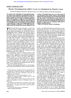
From www.bloodjournal.org by guest on February 6, 2015. For personal use only.
Analysis of the Mechanism of Anagrelide-Induced Thrombocytopenia in Humans
By Eric M. Mazur, Alan G. Rosmarin, Patricia A. Sohl, Julie L. Newton, and Amirthini Narendran
Anagrelide is a new therapeutic compound recently demonstrated to have a rapid and selective thrombocytopenic
effect in humans. The effects of anagrelidewere evaluated in
plasma clot and liquid suspension cultures of optimally
stimulated normal human peripheral blood megakaryocyte
progenitors in order to determine the mechanism of its
thrombocytopenic activity. In plasma clot cultures, at clinically relevant, therapeutic concentrations (5 t o 50 ng/mL),
anagrelide exerted no significant inhibitory effect on
megakaryocyte colony numbers or colony size. Only at
anagrelide concentrations of 10 t o 500 times therapeutic
doses did anagrelide inhibit megakaryocyte colony development: an anagrelide concentration of 5 pg/mL reduced
colony numbers by 57% and colony size by 31%. In contrast,
lower, therapeutic anagrelide concentrations exerted profound effects in liquid culture on megakaryocytecytoplasmic
maturation, size, and DNA content. When present for the
entire 12-day culture duration, anagrelide induced Ieftshifted megakaryocyte maturation and reduced both
megakaryocyte ploidy and megakaryocyte diameter. Anagrelide, at concentrations of 5 to 50 ng/mL, shifted the modal
cultured megakaryocyte morphologic stage from 111 t o II,
reduced the modal ploidy value from 16N t o EN, and de-
creased the mean megakaryocyte diameter by up to 22%.
from 27.6 pm to 21.6 pm. Megakaryocyte diameter was
significantly reduced in most instances, even when analyzed
as a function of morphologic stage. When anagrelide was
added to the cultures after 6- and 9-day delays (during the
final 6 and 3 days, respectively, of culture), similar inhibitory
effects on megakaryocytematuration stage and ploidy distribution were observed. However, the magnitude of the leftshift in ploidy appeared to be less as the duration of
anagrelide exposure was reduced. Conversely, megakaryocyte diameter was not significantly affected by the shorter 3and &day anagrelide exposures. These data indicate that
therapeutic concentrations of anagrelide influence primarily
the postmitotic phase of megakaryocyte development, decreasing platelet production by reducing megakaryocytesize
and ploidy, as well as by disrupting full megakaryocyte
maturation. Inhibition of megakaryocytediameter appears to
require more prolonged anagrelide exposure than inhibition
of maturation stage and ploidy. The molecular mechanisms
responsiblefor the inhibitory effects of anagrelide on megakaryocytopoiesisremain to be defined.
0 1992by The American Society of Hematology.
A
forming progenitors were observed in the three anagrelideresponsive patients evaluated.‘ Thus, anagrelide does not
appear to cause thrombocytopenia either by direct stem cell
toxicity or by inhibiting the production of recognizable
megakaryocytes. The thrombocytopenia induced by anagrelide also does not appear to result from a shortened
platelet circulation time. Preclinical testing in 10 healthy
volunteers demonstrated no significant effect of anagrelide
on platelet survival time as measured by ”Cr labeli~~g.~’
In vitro, anagrelide has been shown to have a lineagespecific inhibitory effect on human CFU-Meg-derived colony development? However, inhibition was observed only
at supratherapeutic anagrelide concentrations in culture.
Half-maximal megakaryocyte colony inhibition occurred at
anagrelide concentrations of 0.1 to 0.3 pg/mL, whereas the
peak anagrelide plasma concentration following a standard
oral dose of 1mg is either 5 ng/mL of unchanged drug or 50
ng/mL of drug plus metabolites.’~~
A subsequent evaluation of patients receiving anagrelide
demonstrated that their bone marrow megakaryocytes,
NAGRELIDE is a recently developed imidazo(2,l-b)
quinazolin-2-1 compound that has been demonstrated to have a potent thrombocytopenic effect in human
recipients.I4 Originally developed as a platelet antiaggregating drug, anagrelide exhibited no effect on circulating
platelet concentrations in preclinical animal toxicology
studies involving 11animal species, including five species of
It was not until the initial multiple-dose safety
studies in humans that the potent thrombocytopenic effect
of anagrelide was recognized. Thrombocytopenia developed almost universally in test subjects receiving anagrelide
for 7 days and was observed at doses of anagrelide significantly below those required to produce optimal platelet
aggregation antagonism?> Because hematologic toxicity
prohibited the chronic administration of anagrelide as a
clinical antithrombotic agent, the developmental approach
for anagrelide as a pharmaceutical was refocused to exploit
its capacity to induce isolated thrombocytopenia. A recent
phase I1 study involving 577 patients with thrombocytosis
and myeloproliferate disorders demonstrated that anagrelide at doses of 2 to 4 mg/d will reduce the platelet count
by 50% or to less than 6OO,OOO/~Lin 93% of evaluable
patients:
Despite its potency in vivo in humans, the thrombocytopenic effect of anagrelide has not been reproduced to date in
any experimental animal model
Therefore, data
evaluating the mechanism by which anagrelide induces
thrombocytopenia are limited. In the preliminary clinical
studies of anagrelide, there were no reported changes in
bone marrow morphology. Despite substantial decreases in
patients’ circulating platelet counts during the course of
anagrelide therapy, both the number and appearance of
their bone marrow megakaryocytes remained unchanged.”’
In addition, no changes in the frequencies of bone marrow
erythroid (BFU-E) and megakaryocytic(CFU-Meg) colonyBlood, Vol79, No 8 (April 15). 1992: pp 1931-1937
From the Department of Medicine, Miliam Hospital and Brown
Universe, Providence, RI.
Submitted November 26,1990; accepted November 1, 1991.
Supported in part by a grant from the American Heart Association,
M e Island Afiliate, and finds provided by a Biomedical Research
Support Grant from the National Institutes of Health to the Miriam
Hospital (SO7 RR05818).
Address reprint requests to Eric M. Mazur, MD, Department of
Medicine, Miriam Hospital, 164 Summit Ave, Providence, RI 02906.
The publication costs of this article were defrayed in part by page
charge payment. This article must therefore be hereby marked
“advertisement”in accordance with 18 U.S.C. section 1734 solely to
indicate thb fact.
0 1992 by The A m e r i c aSociety
n
of Hematology.
0006-497119217908-0123$3.OO/O
1931
From www.bloodjournal.org by guest on February 6, 2015. For personal use only.
1932
MAZUR ET AL
while normal in number, were significantly reduced in size
when quantitated by computerized image analysis.6 This
effect on megakaryocyte size could be reproduced in vitro,
but only at the supratherapeutic anagrelide concentration
of 5 Fg/mL6 (Dr L. Solberg, personal communication).
Our investigation was undertaken to further elucidate
the mechanism by which anagrelide inhibits platelet production in man. Particular attention was directed at defining
those changes in megakaryocyte development that could be
induced by anagrelide at concentrations which were clinically therapeutic (ie, 2 5 0 ng/mL). The effects of anagrelide on human megakaryocytopoiesis were analyzed in
vitro using two different target assays: plasma clot cultures
for CFU-Meg7-9and a liquid culture system that supports
the development of mature, polypoid megakaryocytes from
peripheral blood stem cells.'" These two assays permit the
examination of complementary developmental components
of megakaryocytopoiesis. Colony-forming assays require
stem cell mobilization and proliferation, and the acquisition
of megakaryocyte phenotypic markers by the derivative
cells. Liquid cultures permit accurate analyses of megakaryocyte size, ploidy, and extent of cytoplasmic maturation. Our
data indicate that therapeutic concentrations of anagrelide
reduce platelet production in man by affecting the later
phases of megakaryocyte development, inhibiting polyploidization, cytoplasmic size, and maturation.
MATERIALS AND METHODS
Human subjects. Peripheral blood for CFU-Meg cultures and
normal AB plasma and serum were obtained by routine venipuncture of consenting healthy adult donors, all of whom provided
written informed consent. Frequent donors were chosen as the
source of peripheral blood progenitor cells because higher concentrations of circulating CFU-Meg are typically observed in such
individuals.
Megakaryocyte progenitor cell cultures. Mononuclear cells were
prepared from the peripheral blood of normal volunteer donors
using Ficoll 400-sodium diatrizoate (Pharmacia, Piscataway, NJ)
density centrifugation. Cells were further depleted of monocytes by
two cycles of plastic adherence and plated at 5 X 10⁵ cells/mL in
plasma clot cultures' modified as we have reported."' For this
investigation, the plasma clot cultures were further improved by
substituting citrated, platelet-poor human AB plasma at 15%
(vol/vol) for both the human AB serum and citrated beef plasma,
which had been previous constituents of the cultures. Growth
stimulation was provided by maximally stimulating concentrations
of the active ammonium sulfate fraction of aplastic canine serum."
Liquid suspension cultures of adherent-depleted, normal peripheral blood mononuclear cells were prepared as we have described." Briefly, mononuclear cells were suspended at 5 x l@to
lo6 cells/mL in 10- to 30"
volumes in tissue culture flasks
(Corning Glass Works, Corning, NY).The culture medium consisted of 20% heparinized, platelet-poor human AB plasma in
supplemented Iscove's modified Dulbecco's medium (IMDM;
GIBCO, Grand Island, NY).Supplemented IMDM was prepared
by adding 1.0 mL each of the following to 100 mL of IMDM: lOOX
minimal essential medium (MEM) nonessential amino acids, l O O X
L-glutamine, 1 0 0 ~MEM vitamins ( 1 0 0 ~supplements obtained
from GIBCO), 10% (wt/vol) deionized bovine serum albumin
(BSA), and 1 mmol/L a-thioglycerol (final concentration, 10
pmol/L). In all cultures, megakaryocyte colony-stimulating activity
was provided by the 0% to 60% ammonium sulfate fraction of
aplastic canine serum" at 1 to 2 mg/mL. Suspension cultures were
incubated for 12 to 13 days at 37°C in a 100% humidified
atmosphere of 5% CO, in air.
Megakaryocytes were enriched from the liquid cultures by
counterflow centrifugal elutriation." Cultured cells were suspended and eluted in Mega-buffer," a medium specifically designed to preserve megakaryocyte integrity. Elutriation was performed using a Beckman 52-21 centrifuge (Beckman Instruments,
Palo Alto, CA) equipped with a J E d B elutriation rotor and a
Sanderson separation chamber. After introduction of the cultured
cells into the elutriation chamber at a rotor speed of 2,500 rpm,
eluent flow rates were increased incrementally from 31 mL/min to
45 mL/min. Megakaryocytes were concentrated in the cellular
fraction remaining in the elutriation chamber. Megakaryocytes
constituted 58% of cells in this fraction and exhibited an average
viability of 67%.1° For cytologic analyses, megakaryocytes were
spun onto poly-L-lysine-coated glass slides by cytocentrifugation
(Shandon Cytospin 2, Sewickley, PA).
Anagrelide. Anagrelide was provided in powdered form (Batch
1708-1) by the Pharmaceutical Research and Development Division, Bristol-Myers Company, Wallingford, CT.A stock solution of
anagrelide (5 mg/mL) in dimethylsulfoxide (DMSO) was prepared
and stored frozen at -20°C. For use in culture, anagrelide was
diluted in a-medium with 1% BSA (wtlvol) and filter-sterilized. At
the maximum concentration of anagrelide evaluated (5 pg/mL),
DMSO constituted only 0.1% of the culture volume. At this
concentration, DMSO alone exhibited no effects on megakaryocyte
colony number, colony size, ploidy, and maturation (data not
shown).
Cultured megakaryocyte analyses. Plasma clot megakaryocyte
colony cultures were fixed and labeled fluorescently using a rabbit
polyclonal antiplatelet glycoprotein antiserum as we have reported.' In each plasma clot experiment, megakaryocyte colony
numbers were averaged from duplicate culture plates. Colony size
was determined by averaging the numbers of megakaryocytes in all
colonies within individual culture plates.
Analyses of megakaryocyte size, maturation stage, and ploidy
were performed using liquid culture-derived megakaryocytes
mounted on glass slides. Compared with megakaryocytes grown in
plasma clot, liquid culture-derived megakaryocytes exhibit much
better preserved cytologic detail (Fig 1) and tighter coefficients of
variation on ploidy analyses.
Megakaryocyte size and maturation stage were determined after
Wright-Giemsa staining. Megakaryocytes were identified by their
characteristic light microscopic morphology. Diameters were calculated as the average of perpendicular measurements obtained
using a calibrated eyepiece micrometer." Megakaryocytes were
assigned to maturation stages I through IV using the criteria of
Levine et a1.l3.l4
For determination of megakaryocyte ploidy, unstained cytocentrifuge preparations were labeled using a modified Feulgen tech1'])-1,3,4 oxidiazole (I) (Fluka
n i q ~ e . ' ~2,5-bis-(4'-aminophenyl-[
.'~
Chemical, Happauge, NY) was chosen as the quantitative DNA
fluorochrome because its relative stability to ultraviolet irradiation
minimizes the influence of light exposure duration on measurements of DNA flu~rescence.~~
Cellular DNA content was quantitated by microfluorometry as
we have described.1s*'6In every experiment, ploidy distributions
were derived from at least 200 megakaryocytes, which were
evaluated sequentially on cytocentrifuge preparations. Megakaryocytes were identified morphologically based on their characteristically large nuclear size and/or nuclear multilobation. Diploid cell
DNA fluorescence was determined on each slide in a ratio of
approximately 1:2 with the megakaryocytes.16Diploid cells consisted of the lymphocytes and macrophages, which contaminated
From www.bloodjournal.org by guest on February 6, 2015. For personal use only.
1933
ANAGRELIDE AND HUMAN MEGAKARYOCYTOPOIESIS
E
7
1
I
500
5Mx)
0
cc
a
W
5
50
CONCENTRATION OF ANALGRELIDE hid)
Fig 1. Representative megakaryocyte obtained from liquid culture
of normal human peripheral blood mononuclear cells. This stage 111
megakaryocyte exhibits characteristic nuclear lobulation and cytoplasmic granulation. (Wright-Giemsa, stain; original magnification x313.)
the megakaryocyte-rich elutriation fraction. In these investigations,
the coefficients of variation for the 2N fluorescent signals averaged
16.2% (54.4% [SD]). Because of the uncertainty in identifying the
low-ploidy megakaryocytes, megakaryocyte ploidy distributions
were limited to include only cells with DNA contents greater than
or equal to 8N.
Srurisricul unulysis. Megakaryocyte diameters, colony numbers,
and colony sizes were compared using two-tailed Student’s r tests.
Megakaryocyte stage and ploidy distributions were evaluated
statistically using Pearson’s xztest of independence.”
Fig 2. Effect of anagrelide on megakaryocyte colony formation in
plasma clot culture. Varying concentrations of anagrelide (0.5 ng/mL
t o 5 pg/mL) were added at time 0 t o cultures optimally stimulated
with the active ammonium sulfate fraction of aplastic canine serum.”
Stimulated megakaryocyte colony numbers in the absence of anagrelide (control) averaged 62 ? 17 (SD) (range, 36.5 t o 74.0)colonies
per 5 x lo5 mononuclear cells plated. Data are normalized in each
experiment by the number of colonies in the optimally stimulated,
anagrelide-free cultures. Data are derived from four separate experiments, each performed in duplicate, and are analyzed statistically by
the two-tailed Student‘s t test. *P = .058, **P < .001.
10-fold higher than the peak therapeutic concentrations
achieved in vivo (ie, 50 ng/mL).
In contrast, lower, therapeutic concentrations of anagrelide were found to exert profound effects on maturation
stage, size, and ploidy of developing megakaryocytes grown
in liquid culture. Three concentrations of anagrelide, 5
ng/mL, 15 ng/mL, and 50 ng/mL, were evaluated and
exhibited similar effects. Compared with control cultures,
anagrelide-containing cultures exhibited a significant leftshift in morphologic stage distribution in all experiments, at
RESULTS
Figure 2 illustrates the effects of varying concentrations
of anagrelide on human megakaryocyte colony development in vitro. Colony growth was inhibited significantly only
at anagrelide concentrations greater than 500 ng/mL.
Half-maximal reduction of colony numbers was observed at
an anagrelide concentration of approximately 1.1 pg/mL.
Anagrelide also inhibited megakaryocyte colony size in
culture, although the magnitude of this effect was smaller
and the concentrations of anagrelide required for inhibition
were higher (Fig 3). A significant reduction in megakaryocyte numbers per colony was obtained only at the relatively
high anagrelide concentration of 5 pg/mL. The halfmaximal inhibitory concentration of anagrelide on colony
size was not reached at the highest anagrelide concentration tested. Similar inhibitory effects of anagrelide on
megakaryocyte colony number and size were observed
when optimal stimulatory concentrations of interleukin-3
were substituted for the aplastic canine serum fraction as a
source of colony-stimulating activity in the plasma clot
cultures (data not shown). For both megakaryocyte colony
number and size, these experiments demonstrated significant inhibition by anagrelide only at concentrations at least
120,
O‘
T
7
d5
5
5’0
560
sdoo
CONCENTRATION OF ANAORELIDE (nglml)
Fig 3. Effect of anagrelide on megakaryocyte colony size in plasma
clot culture. Varying concentrations of anagrelide (0.5 ng/mL t o 5
pg/mL) were added at time 0 t o cultures optimally stimulated with
the actlve ammonium sulfate fraction of aplastic canine serum.”
Stimulated colony size in control cultures averaged 7 ? 3 (SD) (range,
5.2 t o 10.5) cells per colony. Data are normalized in each experiment
by the colony size in optimally stimulated control cultures. Data are
consolidated from three separate experiments and are analyzed
statistically by a two-tailed Student’s t-test. *P < .01.
From www.bloodjournal.org by guest on February 6, 2015. For personal use only.
1934
MAZUR ET AL
all anagrelide concentrations tested (Table 1). Whereas an
average of 43.7% f 3.4% (SD) of megakaryocytes were
stages I and I1 in the control cultures, 64.4% f 4.8% were
stages I and I1 when grown in the presence of anagrelide
(P < .001 by Student's t test).
Dose-response effects of anagrelide on cultured
megakaryocyte stage were not clearly evident across different experiments (Table 1).However, within experiment
4 (Table l),the megakaryocyte morphologic stage distribution was significantly further left-shifted by anagrelide at 15
ng/mL compared with 5 ng/mL (P < .OS by Pearson's xz
test. In that same experiment, the stage distributions at
anagrelide concentrations of 15 ng/mL and 50 ng/mL did
not differ significantly.
Therapeutic concentrations of anagrelide in culture also
exhibited a substantial inhibitory effect on megakaryocyte
size (Table 2). Overall, anagrelide reduced mean megakaryocyte diameter by 17% to 22%. Since cell volume is a
function of diameter cubed, these changes represent a 43%
to 53% reduction in megakaryocyte cytoplasmic volume
and thus platelet production capacity.
Because megakaryocyte size is in part dependent on the
extent of cytoplasmic mat~ration,'~
mean megakaryocyte
diameters were also compared within each maturation
stage (Table 3). Even when segregated by stage, in most
instances exposure of the developing megakaryocytes to
anagrelide resulted in significant reductions in diameter
within each stage category.
Table 1. Effect of Anagrelide on Liquid Culture-Derived
Megakaryocyte Stage Distribution
Table 2. Effect of Anagrelide on Cultured Megakaryocyte Diameter
Anagrelide
Concentration
(nglmL)
No.of
ExperimentsITotal
Megakaryocytes
Analyzed
4/704
2/400
1/200
3/500
0
5
15
50
Experiment 1 (400)*
Control
Anagrelide
5 ng/mL
Experiment 2 (300)
Control
Anagrelide
50 ng/mL
Experiment 3 (304)
Control
Anagrelide
50 ng/mL
Experiment 4 (800)
Control
Anagrelide
5 ng/mL
15 ng/mL
50 ng/mL
II
Ill
IV
PValuet
2.5
41.0
54.5
2.0
-
11.0
52.5
33.0
3.5
<.001
4.0
44.5
49.0
2.5
18.0
53.0
27.0
2.0
6.7
34.6
52.9
5.8
11.0
51.0
35.5
2.5
7.0
34.5
55.0
3.5
8.5
10.5
9.5
48.5
55.5
57.5
42.0
34.0
33.0
1.0
0
0
< .001
< ,001
<.001
< .001
< ,001
*Numbers represent the mean percentageof total megakaryocytes in
each morphologic stage category. The modal stage for each experimental condition is set in boldface.
tstatistical significance is determined in each experiment by comparing megakaryocyte morphologic stage distributions in the anagrelidecontaining cultures to the relevant controls. Distributions are compared
by Pearson's x2 test."
SNumbers in parentheses reflect the total number of megakaryocytes
analyzed in each experiment.
* SD lwn)
27.6 2 1.4
21.6 5 2.5
22.9
21.6 5 2.8
P Value*
.032
-
.013
*Statistical significance is determined by two-tailed Student's t tests.
For the anagrelideconcentration of 15 ng/mL, only a single experimental value was available and, therefore, significance testing was not
performed.
Across experiments, a dose-response effect of anagrelide
on stage-segregated megakaryocyte diameter was not evident (Table 3). However, in experiment 4 (Table 3), stage I
and I11 megakaryocytes exhibited decreasing megakaryocyte diameter with increasing anagrelide concentration. For
stage I megakaryocytes (experiment 4), this dose-response
effect was statistically significant (P= .027 by linear regression analysis).
Megakaryocyte size is also a function of DNA ~ 0 n t e n t . I ~
Therefore, we determined cultured megakaryocyte ploidy
distributions in control and anagrelide-containing liquid
suspension cultures. One experiment was performed with
anagrelide at 5 ng/mL, and two at 50 ng/mL. Since the
results did not differ significantly, data were combined for
Table 3. Effect on Anagrelide on Cultured Megakaryocyte Diameter
Segregated by Stage
Morphologic Stage*
Morphologic Stage*
I
Mean Diameter
Experiment 1 t
Control
Anagrelide
5 ng/mL
Experiment 2
Control
Anagrelide
50ng/mL
Experiment 3
Control
Anagrelide
50ng/mL
Experiment4
Control
Anagrelide
5ng/mL
15 ng/mL
50ng/mL
I
II
111
IV
16.1 f 2.6
20.8f 4.7
27.2 2 6.7
37.4 f 5.4
9.9 2 1.4'
15.5 f 4.6" 21.8 f 5.5" 30.4 rt 6.1d
13.6 2 1.9
20.6 f 3.5
10.7 rt 1.4'
15.0 f 3.5" 22.5 f 8.6" 39.8 rt 5.3
14.4
* 1.4
22.8 2 5.0
28.0 f 7.4
29.6 f 7.3
44.2 rt 6.8
44.3 f 3.9
10.6 f 2.8b 19.2 f 4.0" 27.1 -c 8.2d 35.0 rt 5.5b
I
14.6 f 2.7
20.8 f 4.0
14.4 f 1.7 20.9 rt 4.0
13.1 f 2.0d 21.3 f 4.2
11.6 f 2.1' 21.8 ? 3.5
31.0 -t 6.9
42.8 2 6.7
29.2 5 5.7d 48.1 f 2.7
28.3 f 6.9'
N/A
27.3 6.7.
N/A
Abbreviation: N/A, no cells in this category availablefor analysis.
'Numbers represent the mean f SD megakaryocyte diameter in
micrometers segregated by morphologic stage.
tExperimental designations and numbers of megakaryocytes analyzed are identical to those presented in Table l .
a-dStatistical
analysis is performed within each experiment and stage
category using a two-tailed Student's t test. Megakaryocyte diameter in
the presence of anagrelide is compared with control diameter. Significance levels are designated as follows: 'P I .001; bP 5 .01; 'P 9 .05;
d.05 < P < .lo.
From www.bloodjournal.org by guest on February 6, 2015. For personal use only.
ANAGRELIDE AND HUMAN MEGAKARYOCYTOPOIESIS
1935
Table 4. Effect of Anagrelide on Megakaryocyte Ploidy
in Liquid Culture
Table 6. Megakaryocyte Ploidy in Liquid Culture;
Anagrelide Exposure After Varying Culture Intervals
Ploidy Class’
Cont roI
Anagrelide
Ploidy Class+
8N
16N
32N
64N
29.4 k 16.4
50.8 2 14.4
56.1 ? 16.6
34.6 f 6.1
11.8 ~f:2.4
14.2 f 9.1
2.8 f 1.2
0.4 2 0.6
Data are derived from three separate, paired experiments (experiments 1 to 3, see Table 1) and incorporate ploidy determinations on a
total of 483 megakaryocytes 2 8N. Ploidy distributions differ significantly (P < ,001) by Pearson’s xz test.”
*Numbers represent the mean f SD percentage of total megakaryocytes in each ploidy class.
presentation. Table 4 illustrates that anagrelide exposure
resulted in a leftward shift in megakaryocyte ploidy. Anagrelide caused a small but statistically insignificant reduction in geometric mean megakaryocyte ploidy (14.8N & 1.8
to 12.6N ? 2.0). However, this was associated with a shift in
the modal ploidy value from 16N to 8N and a statistically
significant difference in the ploidy distribution histograms
(P < .001).
Our data indicate that anagrelide, at clinically relevant
concentrations, has significant inhibitory effects on postmitotic megakaryocyte development. Anagrelide appears to
exert its thrombocytopenic effect by reducing megakaryocyte size and ploidy, as well as by disrupting or preventing
full megakaryocyte maturation. To further evaluate this
hypothesis, liquid megakaryocyte cultures were exposed to
anagrelide only after 6 and 9 days of incubation (during the
final 6 and 3 days, respectively, of in vitro culture). One
experiment was performed using anagrelide at 50 ng/mL,
and three using anagrelide at 15 ng/mL. Data from these
four experiments were consolidated for the purposes of
presentation, because they did not differ significantly.Since
stem cell-derived megakaryocyte development exhibits partial synchrony in the liquid culture system,” this approach
permitted us to isolate the effects of anagrelide on the later,
nonmitotic components of megakaryocyte development. By
day 6 of culture, most of the mitotic expansion of megakaryocyte precursors from the progenitor cell has been completed, but only a few morphologically identifiable megakaryTable 5. Megakaryocyte Stage in Liquid Culture;
Anagrelide Exposure After Varying Culture Intervals
Morphologic Stage*
Controlt
Anagrelide
Day6
Day9
I
II
111
IV
4.4 -c 0.6
34.4 f 2.5
56.9 2 1.7
4.4% 2.1
6.8 f 1.9
6 . 0 2 0.9
50.7 f 4.3
53.1 f 3.1
39.9 2 4.7
38.5 ~f:3.1
2.6 f 1.0
2.5 2 1.3
Data are consolidated from four separate experiments and incorporate the analyses of 2,210 megakaryocytes. Anagrelide day 6 and day 9
morphologic stage distributions each differ significantly from control
(P < ,001) by Pearson‘s x2 test.”
*Numbers represent the mean f SD percentage of total megakaryocytes in each morphologic stage category. The modal stage for each
experimental condition is set in boldface.
tMegakaryocytes derived from control cultures were grown for 12
days in the absence of anagrelide.
8N
Control
Anagrelide
Day6
Day9
16N
32N
24.4 2 20.1
52.7 f 8.8
19.9
~f:11.5
42.3 ? 11.8
36.8 f 20.0
42.9 f 8.9
45.3 2 10.0
13.7% 6.6
15.3 f 8.8
64N
3.1 2 1.9
1.2 2 0.9
2.7 2 4.2
Data are derived from four separate experiments and include the
analyses of 1,835 megakaryocytes. Anagrelide day 6 and day 9 ploidy
distributions each differ significantly from control (P < ,001) by Pearson’s x2 test.”
‘Numbers represent the mean f SD percentage of total megakaryocytes in each ploidy class.
ocytes are present (data not shown). By day 9, of the
morphologically identifiable megakaryocytes, approximately 75% are stages I and 11.”
Table 5 demonstrates that anagrelide exposure limited to
the final 3 and 6 days of liquid culture also inhibited
megakaryocyte maturation. The extent of inhibition was
comparable to that observed when anagrelide was present
throughout the entire 12-day culture interval (Table 1).
Similarly, the inhibitory effect of anagrelide on megakaryocyte polyploidization was also observed with anagrelide
exposure limited to the final 3 and 6 days of culture (Table
6). However, the magnitude of the left-shift in ploidy
appeared to decrease as the duration of anagrelide exposure was reduced from 12 to 6 to 3 days (Tables 4 and 6).
In contrast, megakaryocyte size (segregated by morphologic stage) was not significantly reduced by the 3- and
6-day anagrelide exposures (Table 7). Reduction of
megakaryocyte diameter was observed only when developing megakaryocytes were exposed to anagrelide for the
entire 12-day culture duration (Tables 2 and 3). Thus, the
inhibitory effects of anagrelide on megakaryocyte diameter
appear to be exerted at an earlier megakaryocyte developmental stage, before day 6 of culture.
DISCUSSION
Anagrelide has been observed clinically to have a potent
and specific inhibitory effect on circulating platelet numbers in man. Initial in vitro studies were unsuccessful in
defining the mechanism of anagrelide-induced thrombocyt ~ p e n i aWhile
. ~ these studies did demonstrate an inhibitory
effect of anagrelide on human megakaryocyte colony develTable 7. Megakaryocyte Diameter Ploidy in Liquid Culture;
Anagrelide Exposure After Varying Culture Intervals
Diameter by Morphologic Stage*
Cont roI
Anagrelide
Day6
Day9
I
II
Ill
14.4 f 1.5
22.9 f 1.3
31.1 f 6.3
41.3
IV
13.52 2.6
13.6 f 2.4
23.1 ? 0.9
23.5 2 0.9
27.3 f 2.1
30.31f: 3.6
41.3 2 1.0
34.4 2 8.6
2
5.7
Data are consolidated from four separate experiments and incorporate measurements on 2,210 megakaryocytes.
‘Numbers represent the mean 2SD megakaryocyte diameter in
micrometers.
From www.bloodjournal.org by guest on February 6, 2015. For personal use only.
1936
opment, the effects were demonstrable only at anagrelide
plasma concentrations well above therapeutic levels. Our
data confirm these earlier observations. Significant inhibition of colony growth was observed at anagrelide concentrations greater than 0.5 Fg/mL. Since therapeutic levels of
anagrelide are in the range of 5 to 50 ng/mL, it is unlikely
that anagrelide reduces platelet production in the clinical
setting by inhibition at the level of the megakaryocyte
progenitor cell.
Our observations indicate that anagrelide-induced thrombocytopenia also is not due to inhibition of the mitotic
expansion of developing early megakaryocyte precursors.
Mitotic expansion can be quantitated in vitro by measuring
megakaryocyte colony size.” In our investigation,
megakaryocyte colony size was not reduced except at
anagrelide concentrations approximately 100-fold above
therapeutic levels.
These in vitro data are consistent with the in vivo
morphologic evaluations that have been performed on bone
marrow specimens obtained from patients receiving anagrelide. Such specimens consistently exhibit no changes in
megakaryocyte
suggesting no significant effect
of anagrelide on megakaryocyte development from the
committed stem cell (CFU-Meg) through the early, morphologically identifiable megakaryocyte.
Our data indicate that therapeutic levels of anagrelide
influence primarily the postmitotic phases of megakaryocyte development. We observed inhibitory effects in vitro
on megakaryocyte ploidy, size, and cytoplasmic maturation.
Any or all of these inhibitory effects may account for the
thrombocytopenic activity of anagrelide in vivo. Inhibition
of megakaryocyte endoreduplication would result in a
left-shifted megakaryocyte ploidy distribution. Since there
is believed to be a direct, constant relationship between
megakaryocyte DNA content and quantitative platelet
production per cell,’s a primary effect shifting megakaryocyte ploidy leftward would result secondarily in reductions
in megakaryocyte size and platelet output. A decrease in
cytoplasmic volume independent of ploidy could equally
well account for diminished platelet-producing capacity per
megakaryocyte. If this latter hypothesis is valid, then the
focus of anagrelide’s action would be the reduction of the
number of platelets produced per unit of megakaryocyte
DNA. Regardless, our data indicating that anagrelide
reduces megakaryocyte diameter by an average of 20% in
vitro translate into a 50% in vivo reduction in platelet
production capacity.
The mechanism whereby an anagrelide-induced leftward
shift in megakaryocyte cytoplasmic maturation could lead
to diminished platelet production is less clear. A simple
delay in the platelet output from a cohort of developing
megakaryocytes would not affect the net quantity of platelets produced by that cohort; at steady-state, platelet
production would remain unchanged. More likely is that
the observed cytoplasmic immaturity of megakaryocytes
exposed to anagrelide results from the induction of a
component of maturation arrest or ineffective megakaryocytopoiesis. If this postulate is true, fewer megakaryocytes
MAZUR ET AL
would actually mature through stage IV and thus reach the
stage at which platelets are shed. The potential quantitative
impact of this effect of anagrelide on platelet production in
vivo cannot be determined from our data.
It must also be emphasized that ploidy, size, and maturation stage are not independent parameters in megakaryocyte development. Higher ploidy megakaryocytes are as a
group larger and exhibit a greater degree of cytoplasmic
maturity.I3 Thus, one need not postulate that anagrelide
independently affects multiple separate components of
megakaryocyte development. A primary inhibitory activity
on a single, critical determinant of nonmitotic megakaryocyte maturation would satisfactorily explain the majority of
our in vitro observations.
Our data confirm the previous in vivo and in vitro
observations reported by Solberg et a1.6 That group demonstrated that bone marrow megakaryocytes obtained directly
from anagrelide-treated patients were present in normal
numbers, but were significantly reduced in size. In
megakaryocyte liquid suspension cultures containing high,
supratherapeutic concentrations of anagrelide (5 Fg/mL,
personal communication), they also detected a comparable
reduction of megakaryocyte size, as well as an inhibitory
effect on cultured megakaryocyte cytoplasmic maturation.
In Solberg’s investigation6 megakaryocyte maturation was
assessed by cultured cellular glycoprotein IIb/IIIa antibody
binding and measurements of cell contour regularity or
roundness, rather than morphologic stage assignation.
The proposition that anagrelide induces thrombocytopenia by disrupting megakaryocyte maturation is also supported by clinical observations in treated patients. In phase
I1 studies of more than 420 evaluable patients with thrombocythemia? the onset of action of anagrelide in vivo was
found to be relatively rapid. The median time to achieve a
50% reduction in the platelet count was 11 days4 In
addition, following the discontinuation of anagrelide, platelet counts have been observed to increase rapidly within 4
days4 This rapid onset and offset of the clinical activity of
anagrelide argues for a locus of action late in megakaryocyte development. If it were affecting megakaryocyte development at the stem cell level, one would expect changes in
circulating platelet counts to occur more slowly, comparable to the changes observed with conventional cytotoxic
drugs. Thrombocytopenia resulting from cytotoxic drugs
typically develops over 2 or more weeks and requires at
least 1week for resolution.
Further evidence that anagrelide acts late in megakaryocyte development can be derived from those experiments in
which the addition of anagrelide to liquid suspension
culture was delayed by 6 and 9 days. We have previously
demonstrated that by day 6 in suspension culture,
megakaryocyte precursors have undergone several rounds
of mitosis and have acquired surface membrane glycoprotein IIb/IIIa (unpublished data). By day 9, mitotic
megakaryocyte precursors disappear and both immature
megakaryocytes and earlier, nonmitotic IIb/IIIa-positive
precursors are present’ (unpublished data). Our current
data demonstrate that exposure of these day 6 and 9
From www.bloodjournal.org by guest on February 6, 2015. For personal use only.
1937
ANAGRELIDE AND HUMAN MEGAKARYOCYTOPOIESIS
megakaryocyte precursors to therapeutic concentrations of
anagrelide results in inhibitory effects on maturity and
ploidy comparable to those induced by anagrelide exposure
for the entire 12-day culture duration. Thus, the developmental target of the inhibitory activity of anagrelide can be
to a large extent isolated to the nonmitotic, late stage of
megakaryocyte development.
Somewhat confounding this interpretation are our data
indicating that the inhibition of megakaryocyte diameter
requires anagrelide exposure for more than 6 days. Despite
decreases in ploidy and maturation stage, no consistent
effect on megakaryocyte diameter could be demonstrated
in those cultures to which anagrelide addition was delayed
(Table 6). Thus, the effect of anagrelide on megakaryocyte
size appears to be exerted at a developmental time point
proximal to that at which ploidy and maturation stage are
affected. This latter observation suggests that more than a
single molecular mechanism may be involved in anagrelideinduced thrombocytopenia. However, since our data are
primarily descriptive in character, any discussion of potential molecular mechanisms is entirely speculative.
The apparent in vitro lineage specificity of anagrelide
demonstrated by Silverstein et a13 has been substantially
reproduced in large-scale, phase I1 clinical
Despite
continuous therapy with anagrelide for up to 2 years, no
significant decrease in circulating white blood cells has
been noted among more than 420 evaluable patients and
there have been only small (1 g/dL) changes in hemoglobin
c o n c e n t r a t i ~ nThe
. ~ ~ ~hematopoietic lineage and developmental stage specificities of anagrelide suggest that it will
have great future utility, not only in the clinical setting, but
also as a valuable research tool for studying the regulation
of human megakaryocytopoiesis and platelet production.
ACKNOWLEDGMENT
The authors gratefully acknowledge Drs J. Stewart Fleming and
Lee P. Schacter (Pharmaceutical Research and Development
Division, Bristol-Myers Company, Wallingford, CT)for providing
data on preclinical and phase 1/11 clinical testing of anagrelide.
The word processing assistance of Charlotte Carter-Edwards,
Anne B. Garvey, and Maureen A. Barlow is also greatly appreciated.
REFERENCES
1. Fleming JS, Buyniski JP: Anagrelide, in Scriabine A (ed):
New Drugs Annual: Cardiovascular Drugs. New York, NY,Raven,
1983, p 277
2. Schacter LP, Doyle G: Investigator’s Brochure: Anagrelide.
Wallingford, CT, Bristol-Myers Pharmaceutical Research and
Development Division, December 1,1989
3. Silverstein MN, Petitt RM, Solberg LA, Fleming JS, Knight
RC, Schacter LP: Anagrelide: A new drug for treating thrombocytosis. N Engl J Med 318:1292,1988
4. Silverstein MN, Solberg LA, Silver RT, Fitch T, Mazur E,
Allen S, Cuttner J, Frenkel G, Cherng NC, Yovan N, Grechko J,
Pettit RM, Rosenbloom B, Knospe W, Colon-Otero G, Doyle G,
Gilbert HS, Weick J, Miller W, Burroughs J, Khoory M, Schacter
LP: Anagrelide: A therapy for thrombocythemic states; experience
in 577 patients. Am J Med (in press)
5. Andes WA, Noveck RJ, Fleming JS, Buyniski JP: Selective
thrombocytopenia produced by the antiplatelet drug anagrelide.
Clin Res 30309A, 1982 (abstr)
6. Solberg LA, Oles KJ, Tarach JT, Petitt RM, Fleming JS,
Schacter LP, Silverstein MN: The effects of anagrelide on human
megakaryocytopoiesis. Blood 74:20a, 1989 (abstr, suppl 1)
7. Mazur EM, Hoffman R, Chasis J, Marchesi S, Bruno E
Immunofluorescent identification of human megakaryocyte colonies using an antiplatelet glycoprotein antiserum. Blood 57:277,
1981
8. Mazur EM, Cohen JL, Wong GG, Clark S C Modest stimulatory effect of recombinant human GM-CSF on colony growth from
peripheral blood human megakaryocyte progenitor cells. Exp
Hematol15:1128,1987
9. Mazur EM, Cohen JL, Bogart L, Mufson RA,Gesner TG,
Yang YC, Clark SC: Recombinant gibbon interleukin-3 stimulates
megakaryocyte colony growth in vitro from human peripheral
blood progenitor cells. J Cell Physiol136:439,1988
10. Mazur EM, Basilico D, Newton JL, Cohen JL, Charland C,
Soh1 PA, Narendran A. Isolation of large numbers of enriched
human megakaryocytes from liquid cultures of normal peripheral
blood progenitor cells. Blood 76:1771,1990
11. Mazur EM, South K Human megakaryocyte colonystimulating factor in sera from aplastic dogs: Partial purification,
characterization, and determination of hematopoietic cell lineage
specificity.Exp Hematol13:1164,1985
12. Shoff PK, Levine R F Elutriation for isolation of megakaryocytes. Blood Cells 15:285,1989
13. Levine RF, Hazzard KC, Lamberg JD: The significance of
megakaryocyte size. Blood 601122,1982
14. Williams N, Levine R F The origin, development, and
regulation of megakaryocytes. Br J Haematol52173,1982
15. Arriaga M, South K, Cohen JL, Mazur EM: Interrelationship between mitosis and endomitosis in cultures of human
megakaryocyte progenitor cells. Blood 69:486,1987
16. Mazur EM, Lindquist DL, de Alarcon PA, &hen J L
Evaluation of bone marrow megakaryocyte ploidy distributions in
persons with normal and abnormal platelet counts. J Lab Clin Med
111:194,1988
17. Hays W L Statistics for Psychologists. New York, NY,Holt,
Rinehart & Winston, 1963, p 578
18. Harker LA, Finch CA: Thrombokinetics in man. J Clin
Invest 48:963.1969
From www.bloodjournal.org by guest on February 6, 2015. For personal use only.
1992 79: 1931-1937
Analysis of the mechanism of anagrelide-induced thrombocytopenia
in humans
EM Mazur, AG Rosmarin, PA Sohl, JL Newton and A Narendran
Updated information and services can be found at:
http://www.bloodjournal.org/content/79/8/1931.full.html
Articles on similar topics can be found in the following Blood collections
Information about reproducing this article in parts or in its entirety may be found online at:
http://www.bloodjournal.org/site/misc/rights.xhtml#repub_requests
Information about ordering reprints may be found online at:
http://www.bloodjournal.org/site/misc/rights.xhtml#reprints
Information about subscriptions and ASH membership may be found online at:
http://www.bloodjournal.org/site/subscriptions/index.xhtml
Blood (print ISSN 0006-4971, online ISSN 1528-0020), is published weekly by the American
Society of Hematology, 2021 L St, NW, Suite 900, Washington DC 20036.
Copyright 2011 by The American Society of Hematology; all rights reserved.


© Copyright 2026