
Provisional PDF - Chemistry Central Journal
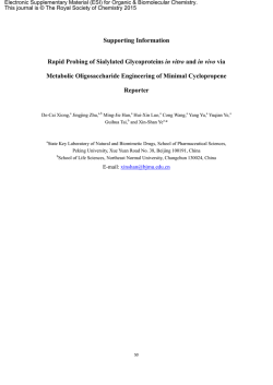
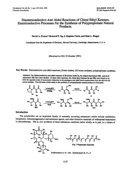
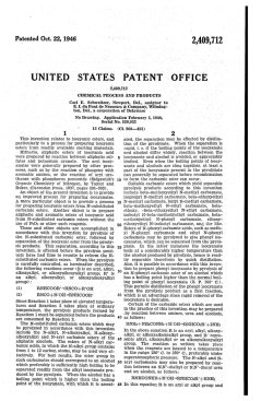
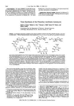
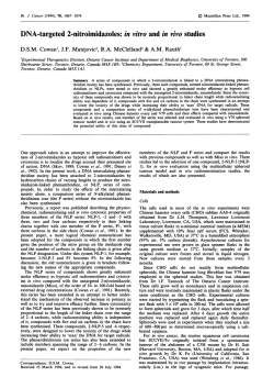
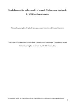
(2015)9:3
!
"#$##%&'#($&)*$#+*$$,-*#
./0
1
-02
3
4*56-0
!
.5
7
7
07
8 9
7
0
27
5
:3220
*:
*;*0
#,)-*#)(<
5
!
-/ -$#+
"#$
-&1 -$#+
"''2'#$##%&'#($&)*$#+*$$,-*#
*
=
2
3
>
2
>>
:
=2
"'';
''
'
>>
2
"''
?-$#)/
@
>>
>>
3 "''
2'
' '+$=
= =
=2
7
Cytotoxic activity of triazole-containing alkyl β-Dglucopyranosides on a human T-cell leukemia cell
line
E Davis Oldham1,†
Email: [email protected]
Larissa M Nunes2,†
Email: [email protected]
Armando Varela-Ramirez2
Email: [email protected]
Stephen E Rankin3
Email: [email protected]
Barbara L Knutson3
Email: [email protected]
Renato J Aguilera2*
Corresponding author
Email: [email protected]
*
Hans-Joachim Lehmler4*
*
Corresponding author
Email: [email protected]
1
Department of Chemistry, University of Mary Washington, 1300 College
Avenue, Fredericksburg, VA 22401, USA
2
Cytometry, Screening and Imaging Core Facility, Border Biomedical Research
Center, Department of Biological Sciences, Bioscience Research Building,
University of Texas at El Paso, 500 West University Ave., El Paso, TX 79968,
USA
3
Department of Chemical and Materials Engineering, University of Kentucky,
Lexington, KY 40506, USA
4
Department of Occupational and Environmental Health, The University of Iowa,
UI Research Park, Iowa City, IA 52242, USA
†
Equal contributors.
Abstract
Background
Simple glycoside surfactants represent a class of chemicals that are produced from renewable
raw materials. They are considered to be environmentally safe and, therefore, are increasingly
used as pharmaceuticals, detergents, and personal care products. Although they display low
to moderate toxicity in cells in culture, the underlying mechanisms of surfactant-mediated
cytotoxicity are poorly investigated.
Results
We synthesized a series of triazole-linked (fluoro)alkyl β-glucopyranosides using the coppercatalyzed azide-alkyne reaction, one of many popular “click” reactions that enable efficient
preparation of structurally diverse compounds, and investigate the toxicity of this novel class
of surfactant in the Jurkat cell line. Similar to other carbohydrate surfactants, the cytotoxicity
of the triazole-linked alkyl β-glucopyranosides was low, with IC50 values decreasing from
1198 to 24 µM as the hydrophobic tail length increased from 8 to 16 carbons. The two alkyl
β-glucopyranosides with the longest hydrophobic tails caused apoptosis by mechanisms
involving mitochondrial depolarization and caspase-3 activation.
Conclusions
Triazole-linked, glucose-based surfactants 4a-g and other carbohydrate surfactants may cause
apoptosis, and not necrosis, at low micromolar concentrations via induction of the intrinsic
apoptotic cascade; however, additional studies are needed to fully explore the molecular
mechanisms of their toxicity.
Graphical Abstract
Triazole-linked, glucose-based surfactants cause apoptosis, and not necrosis, at low
micromolar concentrations via induction of the intrinsic apoptotic cascade.
Background
Carbohydrate surfactants are an important class of surfactants that can be produced from
renewable raw materials (e.g., starch, cellulose, hemicellulose, etc.) and are considered to be
environmentally safe. Because of their interesting interfacial properties, carbohydrate
surfactants with hydrocarbon tails are useful for a broad range of applications, such as
pharmaceuticals, detergents, agrochemicals, food and personal care products [1-3].
Carbohydrate surfactants with partially fluorinated tails are of particular interest for
biomedical applications, including blood substitutes and pulmonary drug delivery [4-7]. One
feature of carbohydrate surfactants is the availability of an incredible number of structural
motifs, including varied head groups, hydrophobic tails and linkers [2]. For example, the
polar head group of carbohydrate surfactants can contain one or more mono- to
polysaccharide moieties; be cyclic or linear; or differ in the stereochemistry of the hydroxyl
groups. Furthermore, the carbohydrate head group can be linked by a variety of approaches,
for example glycosylation, esterification and etherification, and using different linkers to one
or more hydrophobic tails.
The copper-catalyzed azide-alkyne cycloaddition (CuAAC) [8] between an azide and an
alkyne represents an attractive and straightforward approach the link a polar carbohydrate
headgroup and a hydrophobic tail. Indeed, a considerable number of carbohydrate surfactants
containing a 1,2,3-triazole linker have been described, including simple alkyl xylopyranoside
[9] and glucopyranoside surfactants [10], structurally more complex glucose and maltosebased conjugates [11-14], alkyl and aryl O-xylosides and O-xylobiosides [15], 6-triazolelinked galacto- or glucolipids [16], branched fluorinated amphiphiles [17], bolaform
surfactants with glucose, galactose and lactose head groups [18], mannitol-based gemini
surfactants [19], and “star-like” carbohydrate surfactants [20]. Many triazole-linked
carbohydrate surfactants can be synthesized by the reaction of a carbohydrate group
containing an azide group with a suitable alkyne derivative, such as alkynes [18] or propargyl
derivatives of alcohols [12,14] and fatty acids [11,13,16]. Alternatively, a carbohydrate group
with a propargyl group can be reacted with alkyl azides to yield the desired triazole-linked
carbohydrate surfactants [9,10,15,18,19].
The synthesis and physicochemical properties of carbohydrate surfactants have been
investigated in considerable depths [3,21,22]. However, only limited structure-toxicity
relationships of carbohydrate surfactants in general and triazole-liked carbohydrate
surfactants in particular have been reported in mammalian systems. Typically, carbohydrate
surfactants display low toxicity in cells in culture, with IC50 values in the micro- to millimolar
concentration range [4-7,9,13,23-29]. For example, we observed IC50 values ranging from 26
to 890 µM for a series of triazole-linked alkyl β-D-xylopyranosides in several mammalian
cell lines, with the Jurkat cell line being the most sensitive cell line [9]. Despite the potential
use of carbohydrate surfactants in food and personal care products and biomedical
applications, the mechanisms underlying the cytotoxicity of carbohydrate surfactants have
not been explored systematically to date. Because of the potentially broad application of the
triazole-linker in the synthesis of structurally diverse carbohydrate surfactants, we prepared a
series of triazole-linked alkyl β-D-glucopyranosides with hydrocarbon and partially
fluorinated hydrophobic tails, and performed a preliminary investigation of possible
mechanisms of their toxicity in comparison to other carbohydrate surfactants in the Jurkat
cell line.
Results and discussion
Synthesis of triazole-linked alkyl glucopyranosides
The synthesis of the desired glucose-based surfactants was analogous to our previously
published synthesis of triazole-containing alkyl β-D-xylopyranosides [9]. These alkyl β-Dxylopyranosides contained a triazole ring incorporated through the CuAAC reaction [8], and
possessed surface-active properties. This approach utilizes the ability of this so-called “click”
reaction to quickly prepare a series of related molecules. Briefly, the synthesis began with a
β-selective glycosylation of commercially-available β-D-glucose pentaacetate (Scheme 1A).
This strategy was chosen to yield anomerically pure surfactants, as previous work has
suggested the β-alkyl anomers may be more biocompatible [29]. Glycosylation with
propargyl alcohol under Lewis-acid-promoted conditions afforded 2 as the β-anomer [30].
The CuAAC reaction between 2 and alkyl azides, which can easily be prepared from the
corresponding alkyl bromides or iodides [31], was carried out using 0.1 equiv CuSO4 and 0.2
equiv sodium ascorbate in aqueous tert-butanol to generate 3a–g [8]. In the last step the
acetate protecting groups were removed with sodium methoxide, followed by neutralization
with Dowex 50 W × 8–100 ion exchange resin to yield the triazole-linked surfactants 4a-g.
The final products were purified by recrystallization and provided satisfactory elemental
analysis. 1H NMR spectroscopy confirmed the anomeric stereochemistry; the only previously
reported synthesis of 4a-e used a Fisher glycosylation which resulted in a mixture of α and β
anomers [10]. Overall, the synthetic approach outlined in Scheme 1 offers a facile approach
to a large range of novel carbohydrate surfactants with well-defined stereochemistry at the
anomeric carbon.
Scheme 1 (A) Synthesis of triazole-containing alkyl β-D-glucopyranosides using the
CuAAC reaction; (B) Chemical structure and abbreviations of reference surfactants
used in the cell culture studies. 5, (1-octyl-1H-1,2,3-triazol-4-yl)methyl β-Dxylopyranoside; C7G1, heptyl-β-D-glucopyranoside; β-OTG, 1-S-octyl-β-Dthioglucopyranoside; C14G1, tetradecyl-β-D-glucopyranoside.
In vitro cytotoxicity of triazole-containing alkyl xyloside surfactants
Biocompatibility studies using mammalian cells in culture suggests that many carbohydrate
surfactants with hydrocarbon and partially fluorinated hydrophobic tails are relatively nontoxic in vitro, with no cytotoxicity observable even at millimolar concentrations for some
surfactants [4-7,9,13,16,23-29]. Growing experimental evidence suggests that many of these
surfactants cause cell death by a mechanism involving apoptosis, not necrosis [9,23,25].
Here, we initially investigated the cytotoxicity of a series of seven triazole-containing alkyl
glucopyranosides surfactants 4a-g (Scheme 1) with the MTS ([3-(4,5-dimethylthiazol-2-yl)5-(3-carboxymethoxyphenyl)-2-(4-sulfophenyl)-2H-tetrazolium]) assay in the Jurkat cell line
and, subsequently, explored possible mechanisms by which they cause cytotoxicity. Four
structurally related carbohydrate surfactants (Scheme 1B) were also included in our initial
cytotoxicity screening to facilitate the comparison with earlier studies [9,23-25].
The IC50 values of the triazole-containing alkyl β-D-glucopyranoside surfactants 4a-e
decreased with increasing alkyl chain length (Table 1), i.e., their cytotoxicity increased with
increasing chain-length. Similarly, the cytotoxicity of structurally related, hydrocarbon based
carbohydrate surfactants, such as triazole-containing alkyl β-D-xylopyranosides, alkyl β-Dxylopyranosides, alkyl α- and β-D-glactopyranosides, alkyl α- and β-D-glucopyranoside
surfactants, and 6-triazole-linked galacto- or glucolipids, increases from short to medium
hydrophobic tails [9,16,23-25,29]. One likely explanation for this effect is an increased
partitioning of the carbohydrate surfactants into the cell with increasing length of the
hydrophobic tail. As a result, the intracellular concentration of homologous carbohydrate
surfactants and, thus, their cytotoxicity increases as a function of hydrophobic tail length.
Consistent with this observation, we have shown that the apparent membrane partitioning
coefficient of carbohydrate surfactants is proportional to the hydrophobic tail length [24].
Unlike hydrocarbon surfactants 4a-e, many other carbohydrate surfactants investigated to
date display a “cut-off” effect [32], i.e., carbohydrate surfactants with a long hydrophobic tail
show a decrease in the cytotoxicity relative to medium length surfactant [9,23-25,29]. For
example, triazole-containing alkyl β-D-xylopyranosides displayed large IC50 values for short
chain (hexyl) and long chain surfactants (tetradecyl and hexadecyl), whereas the medium
chain alkyl β-D-xylopyranosides (decyl and dodecyl) was the most toxic compounds in this
series of surfactants [9]. Although we did not investigate longer hydrophobic tails due to the
poor aqueous solubility of the corresponding surfactant (i.e., > hexadecyl), we propose that
triazole-containing alkyl β-D-glucopyranoside surfactants 4 would display a “cut-off” effect
for surfactants with long hydrophobic tails. The structure-dependent factors likely involved
the “cut-off” effect, such as lipid and water solubility, critical aggregate concentration,
binding to proteins in the cell and cell culture medium, and diffusion through the cell
membrane, are poorly understood and warrant further investigation [32,33].
Table 1 IC50 values of hydrocarbon and fluorocarbon triazole-linked alkyl β-Dglucopyranosides 4a-g tested on Jurkat cellsa
Compound
Alkyl β-D-glucopyranosidesb
4a
4b
4c
4d
4e
4f
4g
Control surfactantsb
5
C14G1 β-OTG
C7G1
Hydrophobic tail
C8H17 C10H21 C12H25 C14H29 C16H33 C2H4C6F13
C2H4C8F17
C8H17 C14H29 C8H17
C7H15
1198 171
89
53
24
*
*
663
67
163
*
IC50 value [µM]
a
The inhibitory concentration 50% (IC50) in µM is defined as the concentration of experimental compound required to inhibited 50% of the
conversion of MTS to formazan, as compared with the absorbance produced by untreated cells after 16 h of incubation.
b
Please see Scheme 1 for the chemical structures and corresponding abbreviations for the alkyl β-D-glucopyranosides and the control
surfactants.
* Cytotoxicity was <50% at the highest compound concentration tested (2000 µM) and therefore their IC50 values could not be determined.
Comparison of the IC50 values of the triazole-containing alkyl β-D-glucopyranosides
surfactants 4 with structurally related surfactants reveals interesting structure-toxicity
relationships (Table 1). For example, the IC50 values of the alkyl β-D-glucopyranosides
C14G1 is comparable to the IC50 values of the structurally related triazole-containing alkyl
glucoside 4d. This observations is consistent with our earlier findings that the triazole-linker
does not markedly affect the cytotoxicity of triazole-containing alkyl xyloside [9] and the
more general expectation that the introduction of a carbohydrate group renders drug
molecules containing a triazole group less cytotoxic [34]. In contrast, the triazole-containing
octyl glucoside compound 4a appeared to be more toxic compared to its structural analogue,
C7G1, with IC50 values of 1198 µM and > 2,000 µM, respectively. It is also interesting to
note that the IC50 value of the triazole-containing octyl xyloside 5 was significantly lower
compared to its structural glucoside analog 4a, indicating that xyloside-based surfactants are
more cytotoxic compared to glucose-based surfactants. This observation is remarkable
because xylose and glucose differ only by a single hydroxymethyl group, but otherwise have
the same stereochemistry in the pyranose ring system.
Consistent with this observation, we have previously reported that small changes in the
structure of the carbohydrate head group of a surfactant can influence its toxicity [24]. Simple
hexadecyl and octadecyl glucopyranoside surfactants, but not structurally related galactoside
surfactants caused cytotoxicity at low millimolar concentrations in the B16F10 mouse
melanoma cell line. A similar observation has been reported for partially fluorinated glucovs. galactopyranoside in the B16 melanoma cell line [28] and for 6-triazole-linked galacto- or
glucolipids in A549 human lung adenocarcinoma epithelial cell line [16]. Moreover, there is
some evidence that the configuration at the anomeric center may play a role in the
cytotoxicity of carbohydrate surfactants in different cancer cell lines [29]; however this effect
of the stereochemistry on the anomeric center has not been observed in all studies [24],
possibly due to differences in the carbohydrate surfactants, cell lines and/or experimental
conditions employed. Since the stereochemistry of hydroxyl groups of some surfactants, such
as uronic acid-based surfactants, is known to drastically alter their physicochemical
properties [35,36], it is possible that small changes in the stereochemistry of the polar headgroup result in differences in the cytotoxicity by either indirectly by altering physicochemical
properties of macromolecular structures, such as the cell membrane, and/or direct interaction
with cellular targets.
The two partially fluorinated surfactants 4f (F-octyl) and 4g (F-decyl) displayed no
cytotoxicity in the Jurkat cell line over the entire concentration range investigated (8 µM to 2
mM). The hydrocarbon surfactant 4a, which is the structural analog of partially fluorinated
surfactant 4g, displayed low toxicity in the Jurkat cell line, with an IC50 value of 1,198 µM.
Similarly, many other studies have reported that the introduction of a perfluoroalkyl group in
a hydrocarbon surfactant is typically protective and significantly decreases its cytotoxicity in
mammalian cells in culture [5-7,9,24,25,27,28]. However, a perfluoroalkyl group in the
hydrophobic tail is not always protective, as we have shown for octyl versus F-octyl β-Dxylopyranosides [23]. These differences in the cytotoxicity of hydrocarbon and partially
fluorinated carbohydrate surfactants likely reflect differences in the physicochemical
properties of the respective carbohydrate surfactant caused by the introduction of varying
degrees of fluorination in the hydrophobic tail.
Annexin V/PI apoptosis/necrosis assay
Our previous studies demonstrate that structurally diverse carbohydrate surfactants, including
triazole-linked alkyl β-D-xylopyranosides cause cytotoxicity by apoptosis and not necrosis
[9,23,25]. We therefore assessed whether triazole-linked alkyl β-D-glucopyranosides 4d
(tetradecyl) and 4e (hexadecyl) cause cytotoxicity by apoptosis or necrosis. Because
phosphatidylserine translocation from the inner leaflet to the outer membrane is an early
event in apoptotic cell death [37], Annexin V-FITC, which has a high affinity for
phosphatidylserine, was used to detect phosphatidylserine as a marker of apoptosis in livecells by flow cytometry. As can be seen in Figure 1, a significant amount of
phosphatidylserine is externalized when Jurkat cells were treated with the tetradecyl
glycopyranoside C14G1 (~65%), while treatment with 4d and 4e resulted in lower (<20%)
but significant Annexin-V staining after a 16 h incubation. These findings demonstrate that,
similar to other carbohydrate surfactants, triazole-linked alkyl glucopyranosides 4d and 4e
cause apoptosis in the Jurkat cell line.
Figure 1 Triazole-containing alkyl β-D-glucopyranosides 4d and 4e and alkyl β-Dglucopyranosides C14G1 induced significant phosphatidylserine externalization on
Jurkat cell line. The mode of cell death induction, apoptosis or necrosis, was monitored via
flow cytometric assay after co-staining of cells with Annexin V-FITC and PI. Cells were
exposed to the ~ IC50 concentration of each compound as determined by MTS assay (see
Table 1). The total percentage of apoptotic cells is expressed as the sum of percentages of
both early and late stages of apoptosis (Annexin V-FITC positive; white bars), with green
fluorescence signal. Cells that were stained only with PI due to the loss of plasma membrane
integrity, but without FITC signal, are considered necrotic cells (gray bars). Analysis of the
apoptotic populations using the two-tailed Student's paired t-test of 4d, 4e and C14G1-treated
Jurkat cells against DMSO and untreated controls was P < 0.001 (*). Each bar represents the
average of three independent measurements values, and error bars represent the standard
deviation of the mean. Unt refers to untreated cells.
Surfactant-mediated cytotoxicity involves a mitochondria-dependent
apoptosis pathway
Apoptosis can be caused by the activation of cysteine-aspartic acid proteases (caspases)
through an intrinsic, mitochondria-mediated pathway or an extrinsic pathway involving
cellular death receptors, such as FAS/CD95 or tumor necrosis factor receptor 1 (TNFR1). To
gain further insights into the mechanisms involved in carbohydrate surfactant-mediated
apoptosis, we investigated the dissipation of mitochondrial membrane potential (∆Ψm), an
early facet in apoptosis that has been implicated in initiating the intrinsic pathway [38].
Briefly, Jurkat cells were treated for 6 h with carbohydrate surfactants 4d, 4e or C14G1,
stained
with
the
fluorophore
5,5',6,6'-tetrachloro-1,1',3,3'tetraethylbenzimidazolylcarbocyanine iodide (JC-1) and analyzed via flow cytometry [39,40].
Results indicated that 4d and C14G1 compounds provoked preferential accumulation of JC-1
monomers (green), an indicator of mitochondrial depolarization, and interfered with the
formation of JC-1 aggregates (red) (Figure 2). A similar distribution pattern was observed in
cells treated with the positive control, H2O2. The most potent compound causing
mitochondrial depolarization was C14G1 followed by 4d. These outcomes suggest that 4dand C14G1-mediated cytotoxicity appeared to be initiated via ∆Ψm depolarization, involving
the intrinsic apoptotic pathway in the initiation of cell death. In contrast, 4e toxicity appeared
to circumvent ∆Ψm dissipation to induce cell death.
Figure 2 Mitochondrial depolarization mediated by triazole-containing alkyl β-Dglucopyranosides 4d and 4e and alkyl β-D-glucopyranosides C14G1. Jurkat cells were
treated with triazole-containing alkyl β-D-glucopyranosides 4d and 4e and alkyl β-Dglucopyranosides C14G1 at their respective IC50 concentration and incubated for 6 h.
Changes in the mitochondrial ∆Ψm was determined by staining with 2 µM of JC-1. The IC50
concentration values that were used were the following: 4d = 53 µM; 4e = 24 µM; and
C14G1 = 66.5 µM. After dissipation of ∆Ψm, the JC-1 reagent emits a green fluorescence
signal, whereas cells with polarized mitochondrial membrane it emits a red signal.
Percentages of cells emitting green fluorescence signal (y-axis) are depicted. Each bar
represents the mean ± SD of four independent replicates. The following controls were
included: untreated cells as a negative control; cells treated with 0.1% v/v DMSO as a control
for solvent effects; and cells exposed to 1 mM H2O2 as a positive control. Approximately
1x10⁴ flow cytometry events were acquired and analyzed per sample using CXP software.
Surfactants inflict cytotoxicity via caspase-3 activation
Caspase-3 is activated by both the intrinsic and extrinsic apoptotic pathways. To examine
whether caspase-3 activation was involved in the cytotoxicity provoked by the selected
experimental compounds, a cell permeable fluorogenic reagent, NucView 488 Caspase-3
substrate, and Jurkat cells were utilized. This substrate allows the detection of caspase-3
activity in live cells via flow cytometry. Jurkat cells with active caspase-3 were significantly
detected after 6 h of incubation with 4d, 4e and alkyl β-D-glucopyranoside C14G1, as
compared with untreated and solvent controls (DMSO; P < 0.001; Figure 3) [39,40]. The
most efficient carbohydrate surfactant eliciting caspase-3 activation was C14G1 (Figure 3).
These observations suggest that the cytotoxicity induced by 4d, 4e and C14G1was indeed
mediated via apoptosis as initially detected by phosphatidylserine externalization and
corroborated by caspase-3 activation; both hallmarks of apoptosis.
Figure 3 Treatment of Jurkat with triazole-containing alkyl β-D-glucopyranosides 4d
and 4e and alkyl β-D-glucopyranosides C14G1 resulted in caspase-3 activation. Jurkat
cells were treated with compounds at their respective IC50 concentration. The percentage of
cells with activated caspase-3 as determined by emission of green fluorescence signal is
indicated on y axis. Each bar represents average of three independent measurements values,
and error bars their corresponding SD values. Data was analyzed using the two-tailed
Student's paired t-test of compound treated cells vs. DMSO treated cells with P-values within
statistically significant range of P < 0.0001 (*) and P < 0.008 (†).
Experimental
General procedures
The 1H and 13C NMR spectra were recorded on a Bruker DRX 400 Digital NMR
spectrometer. 19F spectra were recorded using a Bruker Avance 300. NMR assignments were
determined from a COSY spectrum of 3b. Representative 1H and 13C NMR spectra are
included in Additional file 1. High resolution mass spectra were obtained at the University of
California, Riverside Mass Spectrometry facility. Elemental analyses were obtained from
Atlantic Micro Lab Microanalysis Service (Atlanta, Georgia, USA). All reactions were
monitored by thin layer chromatography, followed by visualization with UV and
anisaldehyde-H2SO4. Azides were prepared using a known method [31] and used without
further purification. β-Propargyl 2,3,4,6-tetra-O-acetylglucopyranoisde was prepared from
commercially available β-D-glucopyranoside pentaacetate as previously described.
Compounds 3a-e and 4a-e have been reported in the literature [10]. Tetradecyl β-Dglucopyranoside (C14G1) was prepared as previously described [24]. 1-S-Octyl-β-Dthioglucopyranoside (β-OTG), propargyl alcohol and Dowex® 50W × 8-100 ion exchange
resin were obtained from Acros Organics/Fisher Scientific (Pittsburgh, PA). Boron trifluoride
diethyl ethereate and sodium methoxide were obtained from Alfa Aesar (Ward Hill, MA).
Sodium azide and sodium ascorbate were obtained from Aldrich (St. Louis, MO). Cupric
sulfate pentahydrate was obtained from Mallinckrodt (St. Louis, MO). All organic solvents
were reagent grade or higher and were used without further purification. Flash
chromatography was performed using 60 Å (40-63 µm, 230x400 mesh) silica gel.
General procedure for the CuAAC reaction
Triacetyl propargyl glucose (2) and n-alkyl azide (1.0 – 1.1 eq.) were combined with 2:1 tertbutanol : water (0.33 M) at room temperature. Sodium ascorbate (0.2 eq., 1.0 M in water) was
added, followed by CuSO4 pentahydrate (0.1 eq., 75 mg/mL in water), and the mixture stirred
at room temperature for 90 minutes. At this time the reactions often became homogeneous
and faint blue. The reaction mixture was diluted with water and extracted three times with
ethyl acetate. The combined extracts were washed with brine, dried over MgSO4 and
concentrated. The crude residue was purified by silica gel column chromatography
(hexanes/EtOAc), or used without further purification.
(1-Octyl-1H-1,2,3-triazol-4-yl)methyl 2,3,4-tri-O-acetyl-β-glucopyranoside (3a)
The general procedure was used with propargyl glucose (512 mg, 1.32 mmol) and 1azidooctane (208 mg, 1.32 mmol); after column chromatography using EtOAc:hexanes (2:1,
v/v), 636 mg (89%) of 3a were obtained as a clear oil which solidified upon standing. 1H
NMR (CDCl3, 400MHz): δ 7.49 (s, 1H, triazole-CH), 5.18 (app t, J = 9.4 Hz, 1H, H-3), 5.08
(app t, J = 9.9 Hz, 1H, H-4), 4.99 (dd, J = 9.5, 8.0 Hz, 1H, H-2), 4.92 (d, J = 12.6 Hz, 1H, H1’a), 4.80 (d, J = 12.6 Hz, 1H, H-1’b), 4.66 (d, J = 7.9 Hz, 1H, H-1), 4.32 (t, J = 7.3 Hz, 2H,
H-α), 4.26 (dd, J = 12.3, 4.8 Hz, 1H, H-6a), 4.14 (dd, J = 12.3, 2.3 Hz, 1H, H-6b), 3.72 (ddd,
J = 9.9, 4.6, 2.3 Hz, 1H, H-5), 2.07 (s, 3H, OAc), 2.00 (s, 3H, OAc), 1.98 (s, 3H, OAc), 1.96
(s, 3H, OAc), 1.84 - 1.91 (m, 2H, H-β), 1.15 - 1.37 (m, 10H, 5 x CH2), 0.85 (t, J = 6.5 Hz,
3H, H-ω); 13C NMR (CDCl3, 100 MHz): δ 170.6, 170.2, 169.4, 169.3, 144.0, 122.5, 99.7,
72.7, 71.8, 71.1, 68.1, 63.0, 61.7, 50.4, 31.6, 30.2, 29.0, 28.8, 26.4, 22.5, 20.7, 20.61, 20.56 (2
x C), 14.0; HRESIMS calcd for C25H40N3O10 (M + H)+: 542.2708; found: 542.2710.
(1-Decyl-1H-1,2,3-triazol-4-yl)methyl 2,3,4-tri-O-acetyl-β-glucopyranoside
(3b)
The general procedure was used with propargyl glucose (507 mg, 1.31 mmol) and 1azidodecane (240 mg, 1.31 mmol); after column chromatography using EtOAc:hexanes (2:1,
v/v), 669 mg (90%) of 3b were obtained as a waxy solid. 1H NMR (CDCl3, 400MHz): δ 7.50
(s, 1H, triazole-CH), 5.19 (app t, J = 9.4 Hz, 1H, H-3), 5.09 (app t, J = 9.8 Hz, 1H, H-4), 5.01
(dd, J = 9.5, 8.0 Hz, 1H, H-2), 4.93 (d, J = 12.5 Hz, 1H, H-1’a), 4.82 (d, J = 12.5 Hz, 1H, H1’b), 4.68 (d, J = 8.0 Hz, 1H, H-1), 4.33 (t, J = 7.3 Hz, 2H, H-α), 4.27 (dd, J = 12.3, 4.8 Hz,
1H, H-6a), 4.15 (dd, J = 12.3, 2.3 Hz, 1H, H-6b), 3.73 (ddd, J = 9.9, 4.7, 2.3 Hz, 1H, H-5),
2.09 (s, 3H, OAc), 2.02 (s, 3H, OAc), 1.99 (s, 3H, OAc), 1.98 (s, 2H, OAc), 1.84 - 1.94 (m,
2H, H-β), 1.18 - 1.37 (m, 14H, 7 x CH2), 0.87 (t, J = 6.5 Hz, 3H, H-ω); 13C NMR (CDCl3,
100 MHz): δ 170.5, 170.0, 169.3, 169.2, 143.3, 122.4, 99.5, 72.7, 71.8, 71.1, 68.2, 62.9, 61.7,
50.3, 31.7, 30.2, 29.35, 29.25, 29.1, 28.9, 26.4, 22.5, 20.6, 20.53, 20.47 (2 x C), 14.8;
HRESIMS calcd for C27H44N3O10: 570.3021; found: 570.3034.
(1-Dodecyl-1H-1,2,3-triazol-4-yl)methyl 2,3,4-tri-O-acetyl-β-glucopyranoside
(3c)
The general procedure was used with propargyl glucose (526 mg, 1.36 mmol) and 1azidododecane (286 mg, 1.36 mmol); after column chromatography using EtOAc:hexanes
(2:1, v/v), 666 mg (82%) of 3c were obtained as a waxy solid. 1H NMR (CDCl3, 400MHz): δ
7.50 (s, 1H, triazole-CH), 5.20 (app t, J = 9.5 Hz, 1H, H-3), 5.09 (app t, J = 9.9 Hz, 1H, H-4),
5.01 (dd, J = 9.5, 7.9 Hz, 1H, H-2), 4.94 (d, J = 12.5 Hz, 1H, H-1’a), 4.82 (d, J = 12.5 Hz,
1H, H-1’b), 4.69 (d, J = 8.0 Hz, 1H, H-1), 4.33 (t, J = 7.2 Hz, 2H, H-α), 4.27 (dd, J = 12.3,
4.8 Hz, 1H, H-6a), 4.15 (dd, J = 12.3, 2.4 Hz, 1H, H-6b), 3.73 (ddd, J = 10.0, 4.8, 2.4 Hz, 1H,
H-5), 2.09 (s, 3H, OAc), 2.03 (s, 3H, OAc), 2.00 (s, 3H, OAc), 1.98 (s, 3H, OAc), 1.85-1.95
(m, 2H, H-β), 1.19 - 1.36 (m, 18H, 9 x CH2), 0.88 (t, J = 6.7 Hz, 3H, H-ω); 13C NMR
(CDCl3, 100 MHz) δ 171.7, 171.2, 170.5, 170.4, 145.1, 123.5, 101.0, 73.9, 73.0, 72.3, 69.4,
64.1, 62.9, 51.5, 32.9, 31.4, 30.63 (2 x C), 30.56, 30.43, 30.36, 30.0, 27.6, 23.7, 21.8, 21.7,
21.6 (2 x C), 15.2; HRESIMS calcd for C29H48N3O10: 598.3334; found: 598.3339.
(1-Tetradecyl-1H-1,2,3-triazol-4-yl)methyl 2,3,4-tri-O-acetyl-βglucopyranoside (3d)
The general procedure was used with propargyl glucose (526 mg, 1.36 mmol) and 1azidotetradecane (371 mg, 1.55 mmol); after column chromatography using EtOAc:hexanes
(3:2, v/v), 636 mg (75%) of 3d were obtained as a waxy solid. 1H NMR (CDCl3, 400MHz): δ
7.49 (s, 1H, triazole-CH), 5.19 (app t, J = 9.4 Hz, 1H, H-3), 5.09 (app t, J = 9.9 Hz, 1H, H-4),
5.01 (dd, J = 9.5, 8.0 Hz, 1H, H-2), 4.93 (d, J = 12.5 Hz, 1H, H-1’a), 4.81 (d, J = 12.1 Hz,
1H, H-1’b), 4.68 (d, J = 8.1 Hz, 1H, H-1), 4.33 (t, J = 7.3 Hz, 2H, H-α), 4.27 (dd, J = 12.3,
4.8 Hz, 1H, H-6a), 4.14 (dd, J = 12.3, 2.3 Hz, 1H, H-6b), 3.69 - 3.78 (m, 1H, H-5), 2.08 (s,
3H, OAc), 2.02 (s, 3H, OAc), 1.99 (s, 3H, OAc), 1.97 (s, 3H, OAc), 1.83 - 1.94 (m, 2H, H-β),
1.17 - 1.40 (m, 22H, 11 x CH2), 0.87 (t, J = 6.8 Hz, 3H, H-ω); 13C NMR (CDCl3, 100 MHz):
δ 170.6, 170.1, 169.4, 169.3, 144.0, 122.4, 99.8, 72.7, 71.9, 71.2, 68.3, 63.0, 61.8, 50.4, 31.9,
30.3, 29.6, 29.6, 29.58 (2 x C), 29.54, 29.33, 29.29, 28.9, 26.5, 22.6, 20.7, 20.6, 20.5 (2 x C),
14.1; HRESIMS calcd for C31H52N3O10: 626.3647; found: 626.3670.
(1-Hexadecyl-1H-1,2,3-triazol-4-yl)methyl 2,3,4-tri-O-acetyl-βglucopyranoside (3e)
The general procedure was used with propargyl glucose (528 mg, 1.36 mmol) and 1azidohexadecane (373 mg, 1.38 mmol); after column chromatography using EtOAc:hexanes
(3:2, v/v), 692 mg (78%) of 3e were obtained as a white solid. 1H NMR (CDCl3, 400MHz): δ
7.49 (s, 1H, triazole-CH), 5.19 (app t, J = 9.5 Hz, 1H, H-3), 5.08 (app t, J = 9.8 Hz, 1H, H-4),
5.00 (dd, J = 9.5, 8.0 Hz, 1H, H-2), 4.93 (d, J = 12.6 Hz, 1H, H-1’a), 4.81 (d, J = 12.5 Hz,
1H, H-1’b), 4.68 (d, J = 7.9 Hz, 1H, H-1), 4.32 (t, J = 7.2 Hz, 2H, H-α), 4.27 (dd, J = 12.3,
4.8 Hz, 1H, H-6a), 4.14 (dd, J = 12.3, 2.3 Hz, 1H, H-6b), 3.73 (ddd, J = 9.9, 4.7, 2.4 Hz, 1H,
H-5), 2.08 (s, 3H, OAc), 2.01 (s, 3H, OAc), 1.98 (s, 3H, OAc), 1.97 (s, 3H, OAc), 1.81 - 1.94
(m, 2H, H-β), 1.16 - 1.36 (m, 26H, 13 x CH2), 0.87 (t, J = 6.6 Hz, 3H, H-ω); 13C NMR
(CDCl3, 100 MHz): δ 170.6, 170.1, 169.4, 169.3, 143.9, 122.5, 99.7, 72.7, 71.8, 71.1, 68.2,
62.9, 61.7, 50.3, 31.8, 30.9, 30.2, 29.59 (2 x C), 29.56 (2 x C), 29.52, 29.45, 29.3, 29.3, 28.9,
26.4, 22.6, 20.7, 20.6, 20.5, 14.1; HRESIMS calcd for C33H56N3O10: 654.3960; found:
654.3980.
(1-(3,3,4,4,5,5,6,6,7,7,8,8,8-Tridecafluorooctyl)-1H-1,2,3-triazol-4-yl)methyl
2,3,4-tri-O-acetyl-β-glucopyranoside (3f)
The general procedure was used with propargyl glucose (702 mg, 1.81 mmol) and 1-azido3,3,4,4,5,5,6,6,7,7,8,8,8-tridecafluorooctane (707 mg, 1.81 mmol); after column
chromatography using EtOAc:hexanes (3:2, v/v), 846 mg (60%) of 3f were obtained as a
white solid. 1H NMR 1H NMR (CDCl3, 400 MHz): δ 7.60 (s, 1H, triazole-CH), 5.20 (app t, J
= 9.3 Hz, 1H, H-3), 5.10 (app t, J = 9.8 Hz, 1H, H-4), 5.01 (dd, J = 9.4, 8.1 Hz, 1H, H-2),
4.94 (d, J = 12.4 Hz, 1H, H-1’a), 4.83 (d, J = 12.7 Hz, 1H, H-1’b), 4.66-4.69 (m, 3H, H-1, Hα), 4.25 (dd, J = 12.4, 4.6 Hz, 1H, H-6a), 4.15 (dd, J = 12.4, 2.1 Hz, 1H, H-6b), 3.73 (ddd, J
= 10.0, 4.4, 2.3 Hz, 1H, H-5), 2.83 (tt, J = 18.0, 7.5 Hz, 1H, H-β); 13C NMR (CDCl3, 100
MHz): δ 170.6, 170.2, 169.4, 169.4, 144.6, 123.4, 100.0, 72.6, 71.9, 71.1, 68.2, 63.0, 61.6,
42.3, 31.6, 20.7, 20.6 (3 x C); 19F NMR (282 MHz, CDCl3) δ -81.23, -114.70, -122.35, 123.37, -123.96, -126.64; HRESIMS calcd for C25H27N3O10F13: 776.1483; found: 776.1470.
(1-(3,3,4,4,5,5,6,6,7,7,8,8,9,9,10,10,10-Heptadecafluorodecyl)-1H-1,2,3-triazol4-yl)methyl 2,3,4-tri-O-acetyl-β-glucopyranoside (3g)
The general procedure was used with propargyl glucose (702 mg, 1.81 mmol) and 1-azido3,3,4,4,5,5,6,6,7,7,8,8,9,9,10,10,10-heptadecafluorooctane (707 mg, 1.81 mmol); after
column chromatography using EtOAc:hexanes (3:2, v/v), 846 mg (60%) of 3g were obtained
as a white solid. 1H NMR (CDCl3, 400 MHz): δ 7.59 (s, 1H, triazole-CH), 5.20 (app t, J = 9.3
Hz, 1H, H-3), 5.09 (app t, J = 9.8 Hz, 1H, H-4), 5.01 (dd, J = 9.4, 8.1 Hz, 1H, H-2), 4.93 (d, J
= 12.4 Hz, 1H, H-1’a), 4.83 (d, J = 12.7 Hz, 1H, H-1’b), 4.66-4.70 (m, 3H, H-1, H-α), 4.25
(dd, J = 12.4, 4.6 Hz, 1H, H-6a), 4.18 (dd, J = 12.4, 2.1 Hz, 1H, H-6b), 3.73 (ddd, J = 10.0,
4.4, 2.3 Hz, 1H, H-5), 2.83 (tt, J = 18.0, 7.5 Hz, 1H, H-β); 13C NMR (CDCl3, 100 MHz): δ
170.6, 170.2, 169.4, 169.4, 144.6, 123.4, 100.0, 72.6, 71.9, 71.1, 68.2, 63.0, 61.6, 42.3, 31.6,
20.7, 20.6 (3 x C); 19F NMR (282 MHz, CDCl3) δ -81.23, -114.68, -122.13, -122.44 (2 x
CF2), -123.22, -123.94, -126.62; HRESIMS calcd for C27H27N3O10F17: 867.1420; found:
876.1421.
General procedure for acetate deprotection
Triazole peracetates 3 were stirred in dry methanol. NaOMe (1 eq.) was added and the
solution stirred at room temperature for 2-4 hr. Dowex® 50W × 8-100 ion exchange resin
was added and the reaction mixture stirred for another 30 min. The resin was filtered and the
solvent concentrated. The crude residue was purified by recrystallization or column
chromatography to yield pure 1-alkyl-1H-1,2,3-triazol-4-ylmethyl β-D-glucopyranosides 4.
(1-Octyl-1H-1,2,3-triazol-4-yl)methyl β-D-glucopyranoside (4a)
Following the general procedure for acetate deprotection, 902 mg (1.66 mmol) of 3a and 90
mg (1.66 mmol) NaOMe were stirred in 6 mL MeOH. The crude product purified by column
chromatography, yielding 434 mg (70%) of 4a as a white solid. 1H NMR (MeOD, 400 MHz):
δ 8.03 (s, 1H, triazole-CH), 4.98 (d, J = 12.3 Hz, 1H, H-1’a), 4.79 (d, J = 12.4 Hz, 1H, H1’b), 4.37 - 4.43 (m, 3H, H-1, H-α), 3.91 (dd, J = 11.9, 1.7 Hz, 1H, H-6a), 3.69 (dd, J = 11.9,
5.6 Hz, 1H, H-6b), 3.18 - 3.41 (m, 4H, H-2, H-3, H-4, H-5), 1.85 - 1.98 (m, 2H, H-β), 1.22 1.42 (m, 10H, 5 x CH2), 0.90 (t, J = 6.7 Hz, 3H, H-ω); 13C NMR (MeOD, 100 MHz): δ 145.8,
125.4, 103.7, 78.2, 78.1, 75.1, 71.7, 63.1, 62.9, 51.5, 33.1, 31.5, 30.4, 30.2, 27.6, 23.8, 14.6;
HRESIMS calcd for C17H32N3O6 (M + H)+: 374.2286; found: 374.2289; Anal calcd for
C17H31N3O6: C 54.68, H 8.37, N 11.25; found: C 54.43, H 8.20, N 10.99.
(1-Decyl-1H-1,2,3-triazol-4-yl)methyl β-D-glucopyranoside (4b)
Following the general procedure for acetate deprotection, 597 mg (1.05 mmol) of 3b and 56
mg (1.05 mmol) NaOMe were stirred in 4 mL MeOH for 4 hours. The crude product was
purified by column chromatography (5:4:1 CH2Cl2:acetone:MeOH), yielding 266 mg (62%)
of 4b as a white solid. 1H NMR (MeOD, 400 MHz): δ 8.03 (s, 1H, triazole-CH), 4.78 (d, J =
12.4 Hz, 1H, H-1’a), 4.78 (d, J = 12.4 Hz, 1H, H-1’b), 4.36-4.44 (m, 3H, H-1, H-α), 3.90 (dd,
J = 11.9, 1.6 Hz, 1H, H-6a), 3.68 (dd, J = 11.9, 5.6 Hz, 1H, H-6b), 3.11 - 3.44 (m, 4H, H-2,
H-3, H-4, H-5), 1.81 - 2.00 (m, 2H, H-β), 1.17 - 1.44 (m, 14H, 7 x CH2), 0.90 (t, J = 6.7 Hz,
3H, H-ω); 13C NMR (MeOD, 100 MHz) δ 145.8, 125.4, 103.7, 78.2, 78.1, 75.1, 71.7, 63.1,
62.9, 51.5, 33.2, 31.4, 30.8, 30.7, 30.6, 30.3, 27.6, 23.9, 14.6; HRESIMS calcd for
C19H36N3O6 (M + H)+: 402.2599; found: 402.2599; Anal cald for C19H35N3O6 (H2O)0.4: C
55.84, H 8.83, N 10.28; found: C 55.90, H 8.77, N 10.09.
(1-Dodecyl-1H-1,2,3-triazol-4-yl)methyl β-D-glucopyranoside (4c)
Following the general procedure for acetate deprotection, 540 mg (0.904 mmol) of 3c and 48
mg (0.904 mmol) NaOMe were stirred in 4 mL MeOH for 4 hours. The crude product was
purified by recrystallization from acetone/hexane, yielding 266 mg (62%) of 4c as a white
solid. 1H NMR (MeOD, 400 MHz): δ 8.01 (s, 1H, triazole-CH), 4.97 (d, J = 12.3 Hz, 1H, H1’a), 4.78 (d, J = 12.4 Hz, 1H, H-1’b), 4.38-4.1 (m, 3H, H-1, H-α), 3.90 (dd, J = 11.7, 1.5 Hz,
1H, H-6a), 3.66 (dd, J = 11.9, 4.9 Hz, 1H, H-6b), 3.17 - 3.37 (m, 4H, H-2, H-3, H-4, H-5),
1.78 - 2.04 (m, 2H, H-β), 1.19 - 1.42 (m, 18H, 9 x CH2), 0.90 (t, J = 6.7 Hz, 3H, H-ω); 13C
NMR (MeOD, 100 MHz): δ 145.8, 125.4, 103.8, 78.2, 78.1, 75.2, 71.8, 63.2, 62.9, 51.5, 33.2,
31.4, 30.9 (2 x C), 30.8, 30.7, 30.6, 30.2, 27.6, 23.9, 14.6; HRESIMS calcd for C21H40N3O6
(M + H)+: 430.2912; found: 430.2917; Anal calcd for C21H39N3O6(H2O)0.25: C 58.11, H 9.17,
N 9.68: Found: C 58.33, H 9.14, N 9.37.
(1-Tetradecyl-1H-1,2,3-triazol-4-yl)methyl β-D-glucopyranoside (4d)
Following the general procedure for acetate deprotection, 394 mg (0.630 mmol) of 3d and 34
mg (0.63 mmol) NaOMe were stirred in 2.4 mL MeOH for 2 hours. The crude product was
purified by column chromatography (5:4:1 CH2Cl2:acetone:MeOH), yielding 177 mg (61%)
of 4d as a white solid. 1H NMR (MeOD, 400MHz): δ 8.01 (s, 1H, triazole-CH), 4.97 (d, J =
12.4 Hz, 1H, H-1’a), 4.78 (d, J = 12.4 Hz, 1H, H-1’b), 4.35 - 4.44 (m, 3H, H-1, H-α), 3.90
(dd, J = 11.8, 1.6 Hz, 1H, H-6a), 3.65 - 3.72 (m, 1H, H-6b), 3.19 - 3.34 (m, 4H, H-2, H-3, H4, H-5), 1.83 - 1.99 (m, 2H, H-β), 1.18 - 1.43 (m, 22H, 11 x CH2), 0.90 (t, J = 6.6 Hz, 3H, Hω); 13C NMR (MeOD, 100 MHz): δ 145.8, 125.4, 103.8, 78.2, 78.1, 75.2, 71.8, 63.2, 62.9,
51.5, 33.2, 31.4, 30.92, 30.90, 30.89, 30.87, 30.8, 30.7, 30.6, 30.2, 27.6, 23.9, 14.6;
HRESIMS calcd for C24H44N3O6 (M + H)+: 458.3225; found: 458.3246; Anal calcd for
C24H43N3O6: 60.37, H 9.47, N 9.18; found: C 59.97, H 9.29, N 8.91.
(1-Hexadecyl-1H-1,2,3-triazol-4-yl)methyl β-D-glucopyranoside (4e)
Following the general procedure for acetate deprotection, 800 mg (1.22 mmol) of 3e and 66
mg NaOMe (1.22 mmol) were stirred in 5 mL MeOH for 3.5 hours. The crude product was
purified by recrystallization from methanol, yielding 176 mg (30%) of 4e as a white solid. 1H
NMR (DMSO-d6, 400 MHz): δ 8.10 (s, 1H, triazole-CH), 4.83 (d, J = 12.1 Hz, 1H, H-1’a),
4.62 (d, J = 12.4 Hz, 1H, H-1’b), 4.32 (t, J = 7.1 Hz, 2H, H-α), 4.25 (d, J = 7.7 Hz), 3.71 (dd,
J = 11.7, 1.8 Hz, 1H, H-6a), 3.46 (dd, J = 11.8, 6.4 Hz, 1H, H-6b), 3.08 - 3.18 (m, 3H, H-3,
H-4, H-5), 2.98 (app t, J = 7.8 Hz, H-2), 1.73 - 1 .86 (m, 2H, H-β), 1.15 - 1.39 (m, 26H, 13 x
CH2), 0.85 (t, J = 7.3 Hz, H-ω); 13C NMR (DMSO-d6, 100 MHz): δ 143.7, 124.0, 102.1, 76.9,
76.7, 73.4, 70.1, 61.5, 61.2, 49.2, 31.3, 29.7, 29.01 (4 x C), 28.98 (2 x C), 28.93, 28.85, 28.7,
28.4, 25.8, 22.0, 13.9; HRESIMS calcd for C25H48N3O6 (M + H)+: 486.3538; found:
486.3530; Anal calcd for C25H47N3O6 : C 61.83, H 9.75, N 8.65; found: C 61.66, H 9.61, N
8.44.
(1-(3,3,4,4,5,5,6,6,7,7,8,8,8-Tridecafluorooctyl)-1H-1,2,3-triazol-4-yl)methyl βD-glucopyranoside (4f)
Following the general procedure for acetate deprotection, 397 mg of 3f and 28 mg NaOMe
were stirred in 2 mL MeOH for 3.5 hours. The crude product was purified by recrystallization
from acetone/hexane, yielding 128 mg (38%) of 4f as a white solid. 1H NMR (MeOD, 400
MHz): δ 8.10 (s, 1H, triazole-CH), 4.97 (d, J = 12.5 Hz, 1H, H-1’a), 4.75 - 4.83 (m, 3H, H1’a, H-α), 4.38 (d, J = 7.7 Hz, 1H, H-1), 3.89 (dd, J = 11.9, 2.0 Hz, 1H, H-6a), 3.67 (dd, J =
11.8, 5.3 Hz, 1H, H-6b), 3.26 - 3.41 (m, 3H (1H buried under solvent signal), H-3, H-4, H-5),
3.21 (dd, J = 8.9, 7.8 Hz, 1H, H-2), 2.95 (tt, J = 19.0, 7.1 Hz, 2H, H-β); 13C NMR (MeOD
100 MHz): δ 146.2, 126.0, 103.8, 78.2, 78.1, 75.2, 71.8, 63.1, 62.9, 43.6, 32.3; 19F NMR
(MeOD, 282 MHz) δ -80.7, -113.68, -121.17, -122.16, -122.85, -125.61; HRESI MS calcd for
C17H19N3O6 (M + H)+: 608.1061; found: 608.1064; Anal calcd for C17H18N3O6: C 33.62, H
2.99, N 6.92; found: C 33.62, H 2.91, N 6.85.
(1-(3,3,4,4,5,5,6,6,7,7,8,8,9,9,10,10,10-Heptadecafluorodecyl)-1H-1,2,3-triazol4-yl)methyl β-D-glucopyranoside (4g)
Following the general procedure for acetate deprotection, 425 mg (0.48 mmol) of 3g and 28
mg (0.48 mmol) NaOMe were stirred in 2 mL MeOH for 3.5 hours. The crude product was
purified by recrystallization from methanol, yielding 138 mg (41%) of 4g as a white solid. 1H
NMR (MeOD, 400MHz): δ (ppm) 8.11 (s, 1H), 4.98 (d, J = 12.5 Hz, 1H), 4.75 - 4.83 (m, 3H,
H-1’a, H-α), 4.39 (d, J = 7.7 Hz, 1H), 3.90 (dd, J = 11.9, 1.8 Hz, 3H), 3.68 (dd, J = 11.9, 5.6
Hz, 4H), 3.25 - 3.39 (m, 3H (1H buried under solvent signal), H-3, H-4, H-5), 3.21 (dd, J =
9.0, 7.8 Hz, 1H), 2.95 (tt, J = 18.7, 7.1 Hz, 8H); 13C NMR (MeOD, 100 MHz): δ (ppm)
146.2, 126.0, 103.8, 78.2, 78.1, 75.2, 71.8, 63.1, 62.9, 43.6, 32.3; 19F NMR (282 MHz,
MeOD) δ ppm -80.64, -113.67, -120.97, -121.14 (2 x CF2), -122.00, -122.79, -125.56 (br. s.);
HRESIMS calcd for C19H19N3O6F17: 708.0997; found: 708.1006; Anal calcd for C19H18N3O6:
C 32.26, H 2.56, N 5.94; found: 31.92, H 2.57, N 5.66.
Cell culture experiments
Dilutions of experimental chemical compounds
Chemical compounds stock solutions and their dilutions were prepared in dimethyl sulfoxide
(DMSO; Sigma-Aldrich, St Louis, MO) and as necessary aliquots were added directly to 24and 96-wells plates containing cells in complete media.
Cell line & culture conditions
The human acute leukemia T-lymphocytes Jurkat cell line (Jurkat; ATCC, Manassas, VA)
was used for the cytotoxicity assay [41]. Jurkat cells were derived from a 14 years old male
donor afflicted with non-Hodgkin T-lymphoma. The culture medium for Jurkat cells was
Roswell Park Memorial Institute medium (RPMI; HyClone, Logan, UT) with 10% heat
inactivated fetal bovine serum (FBS; HyClone). The medium was supplemented with 100
U/mL penicillin and 100 µg/mL streptomycin (Lonza, Walkersville, MD). Cells growing
exponentially around 60 – 75% confluence were counted and seeded into 96-well plate
format (Greiner Bio-One, Monroe, NC) at density of 25,000 cells in 100 µL culture media per
well. All the incubation conditions were 37°C in a humidified 5% CO2 atmosphere. To
guarantee high viability, cells were prepared as previously detailed [23]. All tests were
assessed in quadruplicate.
MTS colorimetric assay for cell viability
Jurkat cells were incubated with a gradient of the experimental compounds from 8 µM to
2000 µM. After 12 h of incubation, 20 µL of the MTS [3-(4,5-dimethylthiazol-2-yl)-5-(3carboxymethoxyphenyl)-2-(4-sulfophenyl)-2H-tetrazolium]
reagent
(CellTiter
96
AQueousOne Solution Cell Proliferation Assay; Promega, Madison, WI) were added to each
well and subsequently incubated for an additional 4 h for a total incubation period of 16 h.
The colored formazan product was measured by absorbance at 490 nm with a reference
wavelength of 650 nm using a microplate reader (SpectraMax 190 Absorbance Microplate
Reader, Molecular Devices, Sunnyvale, CA). Control wells, containing the same volumes of
culture medium and MTS reagent, were utilized to subtract background absorbance [9]. In
addition, 1 mM of hydrogen peroxide (H2O2; Sigma-Aldrich, St Louis, MO) was used as a
positive control for cytotoxicity. DMSO treated cells as solvent control and untreated (Unt)
cells were also included in each experimental plate. Data are expressed as the cell viability
percentage relative to DMSO treated control cells. Each experimental point was performed in
quadruplicate to obtain the mean and standard deviation values.
Inhibitory concentration 50% (IC50) in µM is defined as the concentration of experimental
compound required to inhibit 50% of the conversion of MTS to formazan, as compared with
the absorbance produced by untreated cells after 16 h of incubation. Data derived from the
MTS assay was used to determine the IC50. The two absorbance values closest to the 50%
point were plotted with its corresponded chemical compound concentration and the equation
of the regression line was utilized to calculate the IC50 as described previously [42].
Annexin V/PI apoptosis/necrosis assay
The triazole-containing alkyl β-D-glucopyranosides 4d and 4e were selected because of their
comparatively high toxicity (Table 1) to gain further insights into the mode and mechanism
of cell death caused by this class of surfactants. The structural analog of 4d, alkyl β-Dglucopyranosides C14G1, was selected as a control surfactant because its IC50 value is
comparable to 4d and 4e. Briefly, Jurkat cells were seeded in a 24-well flat bottom tissue
culture plate (Becton Dickinson, Franklin Lakes, NJ) at a cell density of 100,000 cells per
well in 1 mL of culture media as described above. Triazole-containing alkyl β-Dglucopyranosides 4d and 4e and C14G1 were added to the cells at their respective IC50
followed by additional incubation of 16 h. The following controls were included in each
experimental plate: (1) H2O2 (1 mM) was used as a positive control for apoptosis; (2) DMSO
(1% v/v) was used as a solvent control; and (3) untreated (Unt) cells that were not exposed to
DMSO or compound. All treatments including controls were run in quadruplicates. Cells
from each individual well were collected in a pre-chilled ice-water cytometric tube, washed
and processed essentially as detailed previously [40]. Briefly, cells were stained with a
solution containing a mix of Annexin V-FITC and PI in 100 µL of binding buffer (Beckman
Coulter, Miami, FL). After 15 min of incubation on ice in the dark, 300 µL of ice-cold
binding buffer was added to the cell suspensions and immediately examined via flow
cytometry (Cytomics FC 500; Beckman Coulter, Miami, FL). The total percentage of
apoptotic cells was interpreted as the sum of both early and late stages of apoptosis (Annexin
V-FITC positive), bottom and top right quadrants in a flow cytometric dot plots, respectively.
Cells undergoing necrosis only stain with PI and not with Annexin V-FITC. For each sample,
approximately 5,000 individual events were acquired per sample and analyzed with CXP
software (Beckman Coulter, Miami, FL). Prior to data acquisition, the flow cytometer was set
up and calibrated utilizing unstained, single- (PI or Annexin V-FITC) and double- (PI and
Annexin V-FITC) stained cells. FL1 and FL2 detectors were plotted at x-axis versus y-axis,
respectively.
Mitochondrial membrane potential (∆Ψm) polychromatic analysis
Jurkat cells, plated in a 24 well format, were treated for 6 h [3939] with IC50 concentration
values of compounds and stained with 2 µM of JC-1 (5,5',6,6'-tetrachloro-1,1',3,3'tetraethylbenzimidazolylcarbocyanine iodide) fluorophore following the manufacturer’s
instructions (MitoProbe; Life Technologies, Grand Island, NY). Cells with intact polarized
mitochondria allow JC-1 aggregation that emits a red fluorescence signal; whereas cells with
depolarized mitochondria result in the formation of JC-1 monomers that emit a green
fluorescence signal. The same controls described in the previous section were also included
in these analyses. Data acquisition and analysis was accomplished by using CXP software
(Beckman Coulter). Each data point was obtained from four independent replicates.
Live-cell detection of intracellular caspase-3 activation
Cysteine-aspartic protease-3 (caspase-3) activation was verified by using a fluorogenic
NucView 488 Caspase-3/7 substrate for live cells, following the vendor’s protocol (Biotium,
Hayward, CA). This substrate diffuses easily into cells with intact plasma membrane and
permits the detection of caspase-3 activation in live cells. Jurkat cells were seeded on a 24well plate format and treated with the IC50 concentration of experimental compounds for 6 h.
Cells exhibiting a green fluorescence signal, revealing of caspase-3 activation, were
monitored via flow cytometry (Cytomics FC500, Beckman Coulter). The same three controls
were also analyzed in parallel as described in previous sections. Each data point was obtained
from three replicates. Approximately 5,000 events were collected and analyzed per sample
using CXP software as described above.
Statistical analysis
Every experimental test was accomplished in quadruplicate. To denote experimental
variability, all data are plotted with the standard deviation of the mean. The statistical
importance of differences between two experimental samples was achieved via two-tailed
paired Student's t-tests. To define whether comparisons of two independent samples have
statistical significance, P < 0.01 value was considered significant.
Conclusions
The synthetic approach employed allows the rapid synthesis of novel triazole-linked, glucosebased surfactants 4a-g with well-defined stereochemistry at the anomeric carbon and
hydrocarbon or fluorocarbon hydrophobic tails. An initial toxicity assessment revealed that
selected triazole-containing alkyl β-D-glucopyranosides (4c-e) and the structurally related
tetradecyl β-D-glucopyranoside (i.e., C14G1) cause cytotoxic effects on Jurkat cells at low
micromolar concentrations. Jurkat cells treated with triazole-containing alkyl β-Dglucopyranosides 4d and 4e and alkyl β-D-glucopyranoside C14G1 exhibited
phosphatidylserine externalization, an early biochemical event of apoptosis. Furthermore,
selected compounds induced mitochondria depolarization and caspase-3 activation that are
features of induction of the intrinsic apoptotic cascade. Additional studies are needed to
explore the impact of triazole-containing alkyl β-D-glucopyranosides 4 and other
carbohydrate surfactants to better understand the molecular mechanisms of their toxicity.
Competing interests
The authors declare that they have no competing interests.
Authors’ contributions
EDO carried out the synthesis, purification and characterization of the compounds; LMN and
AVR performed the cell culture experiments and analyzed the results; AVR, SER, BLK, RJA
and HJL conceived the study, participated in its design and contributed to the writing of the
manuscript. All authors read and approved the final manuscript.
Acknowledgements
We thank Gladys Almodovar for critical review of the manuscript and cell culture expertise.
We also thank the Cytometry, Screening and Imaging Core Facility at the University of
Texas at El Paso (UTEP), supported by RCMI program Grant No. 2G12MD007592, to the
Border Biomedical Research Center (BBRC) at UTEP, from the National Center on Minority
Health and Health Disparities, a component of National Institutes of Health. The synthesis of
the triazole-containing alkyl β-D-glucoyranosides 4a-g was supported by grants from the
National Science Foundation (CBET-0967381/0967390) and the U.S. Department of
Agriculture Biomass Research and Development Initiative (Grant Agreement 68-3A75-7608) to HJL. The cell culture work was supported by NIGMS SCORE Grant
1SC3GM103713-01 to RJA.
References
1. Hill K, LeHen-Ferrenbach C. Sugar-Based Surfactants for Consumer Products and
Technical Applications. In: Ruiz CC, editor. Surfactant Sci Ser. Boca Raton, FL: CRC Press;
2008. p. 1.
2. Dembitsky VM. Astonishing diversity of natural surfactants: 1. Glycosides of fatty acids
and alcohols. Lipids. 2004;39(10):933–53.
3. Queneau Y, Chambert S, Besset C, Cheaib R. Recent progress in the synthesis of
carbohydrate-based amphiphilic materials: the examples of sucrose and isomaltulose.
Carbohydr Res. 2008;343(12):1999–2009.
4. Riess JG, Greiner J. Carbohydrate- and related polyol-derived fluorosurfactants: an update.
Carbohydr Res. 2000;327(1–2):147–68.
5. Milius A, Greiner J, Riess JG. Synthesis of F-alkylated glycosides as surfactants for in vivo
uses. Effects of the length of the hydrocarbonated spacer in the aglycone on Königs-Knorr
reaction, surface activity and biological properties. New J Chem. 1991;15:337–44.
6. Zarif L, Greiner J, Pace S, Riess JG. Synthesis of perfluoroalkylated xylitol ethers and
esters: new surfactants for biomedical uses. J Med Chem. 1990;33(4):1262–9.
7. Zarif L, Greiner J, Riess JG. Perfluoroalkylated monoesters of 1,4-D-sorbitan, isosorbide,
and isomannide: new surfactants for biomedical applications. J Fluorine Chem. 1989;44:73–
85.
8. Rostovtsev VV, Green LG, Fokin VV, Sharpless KB. A stepwise huisgen cycloaddition
process: copper(I)-catalyzed regioselective "ligation" of azides and terminal alkynes. Angew
Chem Int Ed Engl. 2002;41(14):2596–9.
9. Oldham ED, Seelam S, Lema C, Aguilera RJ, Fiegel J, Rankin SE, et al. Synthesis, surface
properties, and biocompatibility of 1,2,3-triazole-containing alkyl beta-D-xylopyranoside
surfactants. Carbohydr Res. 2013;379:68–77.
10. Sani FA, Heidelberg T, Hashim R. Farhanullah: Alkyl triazole glycosides (ATGs)–a new
class of bio-related surfactants. Colloids Surf, B. 2012;97:196–200.
11. Clemente MJ, Fitremann J, Mauzac M, Serrano JL, Oriol L. Synthesis and
characterization of maltose-based amphiphiles as supramolecular hydrogelators. Langmuir.
2011;27(24):15236–47.
12. Paul KJV, Loganathan D. Synthesis of novel glycolipids derived from glycopyranosyl
azides and N-(β-glycopyranosyl)azidoacetamides. Tetrahedron Lett. 2008;49(44):6356–9.
13. Song S-X, Zhang H-L, Kim C-G, Sheng L, He X-P, Long Y-T, et al. Expeditious
preparation of triazole-linked glycolipids via microwave accelerated click chemistry and their
electrochemical and biological assessments. Tetrahedron. 2010;66(52):9974–80.
14. Zhang H-L, He X-P, Deng Q, Long Y-T, Chen G-R, Chen K. Research on the structure–
surface adsorptive activity relationships of triazolyl glycolipid derivatives for mild steel in
HCl. Carbohydr Res. 2012;354:32–9.
15. Brusa C, Ochs M, Remond C, Muzard M, Plantier-Royon R. Chemoenzymatic synthesis
of "click" xylosides and xylobiosides from lignocellulosic biomass. RSC Advances.
2014;4(18):9330–8.
16. Zhang H-L, He X-P, Sheng L, Yao Y, Zhang W, Shi X-X, et al. Synthesis of novel 6triazologlycolipids via click chemistry and their preliminary cytotoxicity assessments. Mol
Divers. 2011;15(4):889–900.
17. Schuster T, Schellenberger S, Friedrich R, Klapper M, Müllen K. Branched fluorinated
amphiphiles based on carbohydrates. J Fluorine Chem. 2013;154:30–6.
18. Neto V, Granet R, Krausz P. Novel class of non-ionic monocatenary and bolaform
alkylglycoside surfactants. Synthesis by microwave-assisted glycosylation and olefin crossmetathesis or by ‘click-chemistry’: physicochemical studies. Tetrahedron. 2010;66(25):4633–
46.
19. Mohammed AI, Abboud ZH, Alghanimi AHO. Synthesis of D-mannitol substituted etherlinked bis-1,2,3-triazoles as models of gemini surfactants. Tetrahedron Lett.
2012;53(38):5081–3.
20. Neto V, Granet R, Mackenzie G, Krausz P. Efficient synthesis of “star‐like” surfactants
via “click chemistry” [3 + 2] copper (I)‐catalyzed cycloaddition. J Carbohydr Chem.
2008;27(4):231–7.
21. Stubenrauch C. Sugar surfactants - aggregation, interfacial, and adsorption phenomena.
Curr Opin Colloid Interface Sci. 2001;6(2):160–70.
22. Goodby JW, Goertz V, Cowling SJ, Mackenzie G, Martin P, Plusquellec D, et al.
Thermotropic liquid crystalline glycolipids. Chem Soc Rev. 2007;36(12):1971–2032.
23. Xu W, Osei-Prempeh G, Lema C, Davis Oldham E, Aguilera RJ, Parkin S, et al.
Synthesis, thermal properties, and cytotoxicity evaluation of hydrocarbon and fluorocarbon
alkyl β-D-xylopyranoside surfactants. Carbohydr Res. 2012;349:12–23.
24. Li X, Turanek J, Knoetigova P, Kudlackova H, Masek J, Parkin S, et al. Hydrophobic tail
length, degree of fluorination and headgroup stereochemistry are determinants of the
biocompatibility of (fluorinated) carbohydrate surfactants. Colloids Surf, B. 2009;73(1):65–
74.
25. Li X, Turánek J, Knötigová P, Kudlácková H, Mašek J, Pennington DB, et al. Synthesis
and biocompatibility evaluation of fluorinated, single-tailed glucopyranoside surfactants.
New J Chem. 2008;32:2169–79.
26. Söderlind E, Wollbratt M, von Corswant C. The usefulness of sugar surfactants as
solubilizing agents in parenteral formulations. Int J Pharm. 2003;252(1–2):61–71.
27. Kasuya MCZ, Cusi R, Ishihara O, Miyagawa A, Hashimoto K, Sato T, et al. Fluoroustagged compound: a viable scaffold to prime oligosaccharide synthesis by cellular enzymes.
Biochem Biophys Res Commun. 2004;316(3):599–604.
28. Kasuya MCZ, Ito A, Cusi R, Sato T, Hatanaka K. Cellular uptake and saccharide chain
elongation of "fluoro-amphiphilic" glycosides. Chem Lett. 2005;34(6):856–7.
29. Song S-X, Wu M-L, He X-P, Zhou Y-B, Sheng L, Li J, et al. The anomeric mixture of
some O-galactolipid derivatives is more toxic against cancer cells than either anomer alone.
Bioorg Med Chem Lett. 2012;22(5):2030–2.
30. Rowan AS, Nicely NI, Cochrane N, Wlassoff WA, Claiborne A, Hamilton CJ.
Nucleoside triphosphate mimicry: a sugar triazolyl nucleoside as an ATP-competitive
inhibitor of B. anthracis pantothenate kinase. Org Biomol Chem. 2009;7(19):4029–36.
31. Alvarez SG, Alvarez MT. A practical procedure for the synthesis of alkyl azides at
ambient temperature in dimethyl sulfoxide in high purity and yield. Synthesis. 1997;4:413–4.
32. Balgavy P, Devinsky F. Cut-off effects in biological activities of surfactants. Adv Colloid
Interf Sci. 1996;66:23–63.
33. Kamp F, Hamilton JA. How fatty acids of different chain length enter and leave cells by
free diffusion. Prostaglandins Leukot Essent Fatty Acids. 2006;75(3):149–59.
34. Zhou CH, Wang Y. Recent researches in triazole compounds as medicinal drugs. Curr
Med Chem. 2012;19(2):239–80.
35. Razafindralambo H, Richel A, Wathelet B, Blecker C, Wathelet JP, Brasseur R, et al.
Monolayer properties of uronic acid bicatenary derivatives at the air-water interface: effect of
hydroxyl group stereochemistry evidenced by experimental and computational approaches.
Phys Chem Chem Phys. 2011;13(33):15291–8.
36. Razafindralambo HL, Richel A, Paquot M, Lins L, Blecker C. Liquid crystalline phases
induced by the hydroxyl group stereochemistry of amphiphilic carbohydrate bicatenary
derivatives. J Phys Chem B. 2012;116(13):3998–4005.
37. Leventis PA, Grinstein S. The distribution and function of phosphatidylserine in cellular
membranes. Annu Rev Biophys. 2010;39:407–27.
38. Li H, Zhu H, Xu CJ, Yuan J. Cleavage of BID by caspase 8 mediates the mitochondrial
damage in the Fas pathway of apoptosis. Cell. 1998;94(4):491–501.
39. Robles-Escajeda E, Lerma D, Nyakeriga AM, Ross JA, Kirken RA, Aguilera RJ, et al.
Searching in mother nature for anti-cancer activity: anti-proliferative and pro-apoptotic effect
elicited by green barley on leukemia/lymphoma cells. PLoS One. 2013;8(9):e73508.
40. Robles-Escajeda E, Martinez A, Varela-Ramirez A, Sanchez-Delgado RA, Aguilera RJ.
Analysis of the cytotoxic effects of ruthenium-ketoconazole and ruthenium-clotrimazole
complexes on cancer cells. Cell Biol Toxicol. 2013;29(6):431–43.
41. Schneider U, Schwenk HU, Bornkamm G. Characterization of EBV-genome negative
"null" and "T" cell lines derived from children with acute lymphoblastic leukemia and
leukemic transformed non-Hodgkin lymphoma. Int J Cancer. 1977;19(5):621–6.
42. Varela-Ramirez A, Costanzo M, Carrasco YP, Pannell KH, Aguilera RJ. Cytotoxic
effects of two organotin compounds and their mode of inflicting cell death on four
mammalian cancer cells. Cell Biol Toxicol. 2011;27(3):159–68.
Additional files provided with this submission:
Additional file 1. The following additional data are available with the online version of this paper. Additional data file 1
contains copies of 1H and 13C NMR spectra (1859k)
http://journal.chemistrycentral.com/content/supplementary/s13065-014-0072-1-s1.pdf
© Copyright 2026