
Fig. 1 - ResearchGate
Biochimica et Biophysica Acta 1741 (2005) 85 – 94
http://www.elsevier.com/locate/bba
Cloning, characterization and DNA immunization of an Onchocerca
volvulus glyceraldehyde-3-phosphate dehydrogenase (Ov-GAPDH)B
Klaus D. Erttmanna,*, Andre´ Kleensanga, Erik Schneidera, Sven Hammerschmidtb,
Dietrich W. Bqttnera, Michaela Gallina
b
a
Bernhard Nocht Institute for Tropical Medecine, Bernhard-Nocht-Str.74, D-20359 Hamburg, Germany
Research Center for Infectious Diseases, University of Wu¨rzburg, Ro¨ntgenring 11, D-97070 Wu¨rzburg, Germany
Received 13 August 2004; received in revised form 5 December 2004; accepted 14 December 2004
Available online 5 January 2005
Abstract
In the search for Onchocerca volvulus antigens possibly involved in protection against human onchocerciasis, partial amino acid sequence
analysis of one of the O. volvulus antigens of the serologically identified proteins showed a close relationship to the glyceraldehyde-3phosphate dehydrogenase (GAPDH) protein family. Subsequent adult worm cDNA library screening and cloning produced a clone of 1650
bp. An open reading frame spans over 1020 bp encoding for a protein of 340 amino acids with an apparent molecular weight of 38 000.
Comparison of the complete amino acid sequence identified this protein as a member of the GAPDH protein family. The recombinantly
expressed protein shows GAPDH enzymatic activity as well as plasminogen-binding capacity. DNA sequence analysis of the corresponding
gene revealed the presence of two introns. Using immunohistology Ov-GAPDH was observed in microfilariae, infective larvae, and adult
male and female worms. Most striking was the labelling of the musculature of the body wall. Labelling was also observed in the
pseudocoeloma cavity and in a subset of cell nuclei, suggesting additional, non-glycolytic functions of the Ov-GAPDH. Gene gun
immunization with the DNA-construct in cattle led to specific humoral immune responses. Thus, the protective potential of the DNAconstruct of Ov-GAPDH can be evaluated in vaccination trials using animal models such as the cattle/Onchocerca ochengi model.
D 2004 Elsevier B.V. All rights reserved.
Keywords: Glyceraldehyde-3-phosphate dehydrogenase; Filaria; Onchocerca volvulus; Plasminogen-binding; Protective antigen; DNA immunization
1. Introduction
The parasitic nematode, Onchocerca volvulus, is a major
cause of blindness and dermal pathology in the tropics.
Chemotherapy with the microfilaricidal drug ivermectin,
which is the backbone of the present African Programme for
Onchocerciasis Control, even in combination with vector
control by larvicides, cannot eradicate the parasite reservoir
from hyperendemic areas in West Africa [1]. Since bovine
onchocerciasis is a vaccine-preventable disease [2], it has
B
The DNA sequence has been submitted to GenBank under the
accession number: Y09455.
* Corresponding author. Tel.: +49 40 42818 470; fax: +49 40 42818 377.
E-mail address: [email protected] (K.D. Erttmann).
0925-4439/$ - see front matter D 2004 Elsevier B.V. All rights reserved.
doi:10.1016/j.bbadis.2004.12.010
been proposed that in combination with these control
measures, a vaccine might fully stop transmission as well
as disease. Using DNA for vaccination greatly simplifies
vaccine development and production, as DNA vaccines
remain stable under local conditions, presumably without a
cold chain [3]. Several antigens of O. volvulus, for example
the DNA-construct of the O. volvulus chitinase [4], have
been shown to induce significant protection in animal
models. One of the major vaccine candidates against the
human pathogenic trematode Schistosoma mansoni was
identified as glyceraldehyde-3-phosphate dehydrogenase
(GAPDH) [5,6]. GAPDH has been suggested as major
therapeutical target in several parasitic diseases, as a vaccine
candidate or as a target for chemotherapeutic treatment. This
has been primarily attributed to the role of GAPDH as a key
enzyme in glycolysis and gluconeogenesis, thus being
86
K.D. Erttmann et al. / Biochimica et Biophysica Acta 1741 (2005) 85–94
crucial in energy production. Here we report the characterization of the O. volvulus GAPDH as well as the immune
response of cattle against the DNA-construct of the coding
sequence of the GAPDH of O. volvulus.
[12]. The further purification of the recombinant protein by
Ni2+ chelate chromatography and pH-shift under denaturing
conditions was performed according to the pET manual
(Novagen, Madison, WI, USA).
2.4. DNA sequence analysis
2. Materials and methods
2.1. Study population
After informed consent residents of West African areas
endemic for onchocerciasis in Liberia, Benin and Guinea
underwent physical and parasitological examinations, essentially as described [7]. The study procedures were in
accordance with the Declaration of Helsinki (1975 and its
revisions in 1983 and 2000).
2.2. Parasite preparation
Adult O. volvulus worms were obtained by collagenase
digestion from nodules surgically removed from Liberian
patients as previously described [8]. Onchocercomas
embedded in paraffin were available from several studies
in Liberia, Ghana, and Uganda [9–11]. The extirpation of
onchocercomas for research had been approved by the
Medical Board, Hamburg, Germany, and by authorities in
the African countries. Nodules with adult Onchocerca
ochengi had been collected from cattle in Ngaoundere in
Cameroon (supplied by PD. Dr. A. Renz, University of
Tqbingen, Germany). For the examination of infective
larvae, we used Simulium yahense that had been reared
from pupae and had been experimentally infected with O.
volvulus in Guinea (supplied by Dr. T. Kruppa, BNI
Hamburg) or S. soubrense from Liberia (supplied by PD
Dr. G. Strote, formerly BNI Hamburg).
2.3. Identification of the Ov-GAPDH cDNA and protein
expression
In order to identify the genomic structure of Ov-GAPDH,
an O. volvulus E FiXII-gDNA library was screened using
the same screening method as described above. Manual
sequencing was carried out employing the dideoxynucleotide chain termination method of Sanger et al. [13] using the
appropriate vector primer and synthetic internal primers
deduced from the partial sequence of the clone. Sequence
analysis was performed in both orientations. Automated
sequencing was performed on an Applied Biosystems
automated DNA sequencer. The derived sequence was
compared with the public protein and nucleotide database
(Genbank) by using the BLASTn and BLASTx algorithms.
The DNA sequence of Ov-GAPDH was deposited in
GenBank (accession no.Y09455).
2.5. Southern blot analysis
Human DNA was prepared from HL60 cells and O.
volvulus DNA from adult female worms. The isolation and
preparation of the DNA was done as described [14]. The
Southern blot analysis was carried out by standard methods
using the entire cDNA of Ov-GAPDH as a probe. The
Southern blot was prepared by separation of approximately
10 Ag human and adult O. volvulus EcoRI and HindIII
restricted genomic DNA on an 1% agarose gel. After
depurination, denaturation and neutralization, separated
DNA was transferred to nitrocellulose membrane
(Schleicher and Schuell, Dassel, Germany). The filter was
hybridized overnight at 55 8C using a radioactively labelled
probe.
2.6. Western blotting
A E ZapII expression cDNA library, prepared from
adult O. volvulus mRNA, was screened with a 32P-labelled
970 bp probe coding for Ov-GAPDH. The probe was
obtained by PCR amplification of O. volvulus cDNA using
primers derived from conserved regions of known GAPDH
nucleotide sequences. After plaque purification inserts
were subcloned into phagemids (pBluescript II SK,
Stratagene, Heidelberg, Germany) for sequencing using
the in vivo excision protocols as supplied by the
manufacturer (Stratagene).
PCR was carried out using synthetic oligonucleotides
spanning the entire coding region of the Ov-GAPDH
cDNA. The obtained fragment was digested with the
appropriate restriction enzymes and cloned into the
pJC45Flag expression vector modified according to Clos
and Brandau [12]. Expression was performed in Escherichia
coli strain PAPlaclQ (DE3) according to Clos and Brandau
The purified recombinant antigen was separated by
SDS-PAGE in a 10% acrylamide gel based according to
standard methods. Nitrocellulose strips were incubated
with sera at different dilutions and a 1:1000 dilution of
goat anti-rabbit IgG (H+L) horseradish peroxidase conjugate (HRP) (Biorad, Munich, Germany) and developed
using 4-chloro-naphtol/H2O2.
2.7. Enzyme-linked enzyme immunoassy (ELISA)
For antibody analysis, wells of Maxisorb plates (Nunc,
Wiesbaden, Germany) were coated at 37 8C with purified
Ov-GAPDH in carbonate-bicarbonate buffer, pH 9.6, 2 Ag/
well. After incubation with the primary antibody as
secondary reagent, horseradish peroxidase-conjugated
goat-anti-rabbit IgG (Sigma, Deisenhof, Germany) was
K.D. Erttmann et al. / Biochimica et Biophysica Acta 1741 (2005) 85–94
applied. The substrate was tetramethyl-benzidine (Sigma).
The absorbance was read at 450 nm.
2.8. Synthesis of cDNA
Total O. ochengi RNA was isolated with an RNA
extraction reagent (TRIzol Reagent, Gibco, Karlsruhe,
Germany) and cDNA was obtained by reverse transcription
using oligo-dT primers. cDNA from O. volvulus L3 was
derived from a ZAPII O. volvulus cDNA library. PCR
amplification was primed with a deduced GAPDH primer
set, obtained by sequence comparison of highly conserved
regions, such as the ATP binding site and NAD+ binding site
of members of the GAPDH protein family.
87
and haematoxylin (Merck, Darmstadt, Germany) as counterstaining. As negative controls the preimmune serum
from the immunized rabbit and human AB-serum were
used. To examine the specificity, the Ov-GAPDH antibodies were absorbed from the rabbit serum or from human
O. volvulus sera using the purified rOv-GAPDH antigen
transferred to nitrocellulose membrane and subsequently
tested by immunoblotting. The eluted antibodies showed
recognition of the rOvGAPDH as well as of a corresponding
single band of 36 kDa in O. volvulus worm extract. For
immunohistology they were used as primary antibodies
diluted 1:5.
2.12. Cloning and purification of the Ov-GAPDH DNA
vaccination construct
2.9. Enzymatic analysis of the recombinant protein
The activity of the GAPDH enzyme was determined by
measuring the decrease in absorbance of NADH at a
wavelength of 340 nm. Tests were performed on the
recombinantly expressed Ov-GAPDH. Rabbit GAPDH
(Sigma) was used as positive and nonrecombinant pJC40
vector as negative control.
2.10. Plasminogen-binding assay
Binding of human plasminogen (Sigma) was performed
using human plasminogen followed by anti-plasminogen
antibodies and secondary antibodies conjugated with
peroxidase. In blot-overlay assays plasminogen-binding
activity was detected by incubation of the membrane with
a substrate solution containing 1 mg/ml 4-chloro-1-naphthol
(Sigma) and 0.1% H2O2 in PBS. Control experiments were
performed as described using proteins from Streptococcus
pneumoniae serotype 2 strain and deletion mutants as
described [15].
2.11. Immunohistology
Onchocercomas had been fixed in 80% ethanol, 4%
buffered formaldehyde or Karnovsky solution (2% paraformaldehyde and 0.025% glutaraldehyde) and embedded in
paraffin. Blackflies with infective larvae of O. volvulus had
been fixed in 80% ethanol or Bouin solution. For
immunohistology the alkaline phosphatase anti-alkaline
phosphatase (APAAP) method was applied according to
the recommendations given by the manufacturer (DakoCytomation, Hamburg, Germany). Polyclonal antibodies
against rOV-GAPDH were raised by three consecutive
immunizations of a rabbit with 100 Ag recombinant OvGAPDH. The immunization was performed in the facilities
of Eurogentec (Ougree, Belgium). The rabbit polyclonal
antibody against rOv-GAPDH was used as primary antibody
diluted 1:200. As secondary antibody anti-rabbit mouse
immunoglobulins (clone MR12/53, DakoCytomation) were
applied. Fast Red TR salt (Sigma) was used as chromogen,
The entire coding region of the Ov-GAPDH was cloned
into the BamH1 and Not1 restriction sites of the vaccination
vector pcDNA3.1(+) (Invitrogen). The Ov-GAPDHpcDNA3.1(+) was then transformed into DH5a cells,
followed by bidirectional sequence analysis for proof
reading purposes. Plasmid-DNA for immunization was
obtained by fermentation. For purification of Ov-GAPDHpcDNA3.1(+) ion-exchange chromatography was performed using the Nucleobond PC Prep-100 kit (MachereyNagel, Dqren, Germany), following the protocol of the
manufacturer.
2.13. Immunization of rabbits and cattle
For gene gun immunization, plasmid DNA was precipitated onto gold beads (1.6 Am diameter) at a ratio of 1
Ag/mg gold. In all experiments rabbits and cattle were bled
before DNA immunization to collect preimmune samples
for antibody assays. Rabbits were DNA immunized on day
1 and boosted on days 33 and 69. Twenty-four shots of 0.2
Ag Ov-GAPDH-pcDNA3.1(+) were applied into the
shaved back using a Helios gene-gun (Biorad). Following
the protocol of the manufacturer, a pressure of 300 psi was
used. A similar protocol was applied for gene gun
immunization of two calves using for each immunization
on days 1, 27, and 66, ten shots of 1.25 Ag of OvGAPDH-pcDNA3.1(+) and a pressure of 300 psi. The
shots were applied intradermally into the shaved skin of
the neck.
3. Results
3.1. Cloning and sequence analysis of Ov-GAPDH
To identify O. volvulus antigens possibly involved in
protection, partial amino acid (AA) sequence analysis of an
O. volvulus antigen showed a close relationship to the
glyceraldehyde-3-phosphate dehydrogenase (GAPDH) protein family. Subsequent screening of an O. volvulus adult
88
K.D. Erttmann et al. / Biochimica et Biophysica Acta 1741 (2005) 85–94
worm cDNA library using the GAPDH-specific PCR
fragment resulted in a clone of 1650 bp. An open reading
frame spans over 1020 bp coding for a protein of 340 AA
with an apparent molecular weight of 38 000.
Comparison of the deduced AA sequence indicated a
high level of identity to members of the GAPDH protein
family (74% to human GAPDH) [16]. Furthermore,
Genbank comparison as well as the presumptive AA
-391
-361
T GTTCTTCAAA NGGNCNATCA TTGGTCTCAC
-301
CTTCAACACA TATTACCCAA AGCATACAGN ATTATTTTTC
-241
TTTGTAGCTT GTCGGGTGGG GACGNAATAA GTTATTTTTT
-181
AGGAAAAAAA AAAAAAAGGA AGTGTTTTAA ATGTTACGTA
-121
AAATCTTTGT CTACATTACA TTATTTTCTC ATGTTATTTA
-61
ATCGGCTGAT ATTTAATTTC AAGAAATCAA CAAATTAAGC
-1
TGTTGTGTAC TGTTTACGAA GGCATTTCCA CGTGTTCATC
-360
GNGATCACTT CCTGTTTNAC
-300
CGGTATTTTA AATCANAAGT
-240
TTTTTGCTTT TTCNCAAAAG
-180
TTTCTTGCTG GGACGGAGGC
-120
CGCAGTGTGT TTATGTTGTT
-60
TGTTATTACC ATATCAGCCC
1
ATG AGC AAA CCG AAG ATT GGA ATT AAT GGG TGA
61
TCA TAT TAA ACT AAT TGT TTG AAA AAT TTT CAG
121
TTA TTA AAG ATT CTG AAA TTT TTG TCT GAT GTC
181
AAA AAA ATT TCC TCA AAT TCA GAT TTG GTC GTA
241
TTG AAA AGG ACA CCG TTG AAG TAG TGG CTG TCA
301
TGG TAT ACA TGT TCA AAT ACG ACT CAA CAC ATG
361
AGG GTG GAA AGC TTA TTG TAA CAA ACG GCA AAA
421
GCA AAG ATC CTG CCG AAA TTC CAT GGG GAG TAG
481
CTG GTG TTT TAC AAC AAC GGA GAA AGC AAG CGC
541
CAT CAT TTC GGC TCC ATC TGC TGA TGC ACC GAT
601
GTA TGA TAA AGC AAA CAA TCA CAT CAT CTC TAA
661
GCC ATT GGC TAA GGT TAT CCA TGA TAA ATT TGG
721
ACA TGC AAC AAC GGC CAC TCA GAA GAC TGT TGA
781
TGG TCG AGG TGC TGG TCA GAA CAT CAT CCC AGC
841
AAA AGT CAT TCC GGA TCT GAA TGG AAA GCT AAC
901
GGA CGT ATC AGT TGT TGA TCT CAC TTG CCG ACT
961
TAA GGC CGC TGT GAA AGA GGC AGC TGC TGG ACC
1021
GGA CCA GGT ATG AGT TCC GAT TTT TTG TTC AAA
1081
TAA ACA TTG AAG ATA ATT TGT TCT TTC TCT GAA
1141
GCT TGT TCG TGC AAC AAA CAG TTG CAT AAA AAG
1201
CTG TTC CAA ATG ACT TAC CAT AAT TTC AGG TTG
1261
ATT CAT CAA TCT TTG ATG CTC TGG CAT GCA TTT
1321
TTG CTT GGT ACG ATA ATG AAT ATG GCT ACA GCA
1381
1394
TCG CGA GCA AAT AA
1395
1431
GCAGCAGTGG TACAACACTG TCATCATGGG CTTGATC
60
GTT GAT TTT TTC TTG AAA TTC ACC CAT
120
AAT CAG TGT CGG AAT TGA GAC ATA TTT
180
GTT ACG GTC CTT TGA GTT TTG GTA TCT
240
TCG GCC GAC TTG TTT TGA GAG CGG CAG
300
ATG ACC CCT TCA TCA ACA TCG ATT ACA
360
GAC GCT TTA AGG GTC ATG TTT CTG CTG
420
CGA CTC ATC AAA TCG CTG TAC ACA ACA
480
AAG GTG CAG AAT ATG TTG TCG AAT CTA
540
ACA TCT GAA GGG TGG CGC TAA GAA AGT
600
GTT CGT AAT GGG TGT TAA CAA CGA CAA
660
TGC TTC ATG CAC CAC CAA TTG TCT GGC
720
TAT CAT CGA GGG TTT GAT GAC CAC CGT
780
TGG ACC ATC TGG AAA GTT GTG GCG AGA
840
AAG TAC TGG TGC AGC AAA GGC TGT AGG
900
TGG AAT GGC TTT CCG TGT GCC AAC TCC
960
GCA GAA AGG TGC AAG TAT GGA TGA AAT
1020
AAT GAA GGG AAT TCT GGA ATA TAC TGA
1080
GCA AGT TTA ATT TCA AAA TCA TAA TGA
1140
GAT TCA GAT TGA GTT GCT TAT TTC TTT
1200
TTA TTT CCA AGA TTA ATT TTA TGA ATG
1260
TAT CAT CTG ATT TCG TTG GTG ATC CAC
1320
CAC TGA ATC CAA ACT TCG TTA AAT TGA
1380
ACC GTG TTG TTG ACC TTA TCT CTT ACA
Fig. 1. Nucleotide sequence of the Ov-GAPDH gene with the 5V and 3V flanking regions. The coding region starts at nt 1 and ends at nt 1394 and contains two
introns (underlined). A putative promotor sequence TGTTG at position 40 to 36 is also underlined.
K.D. Erttmann et al. / Biochimica et Biophysica Acta 1741 (2005) 85–94
89
3.3. Analysis of enzymatic activity of Ov-GAPDH and
binding analysis of human plasminogen
kb
6.5-
-5.0
4.3-3.5
1
2
3
4
Fig. 2. Southern hybridization of O. volvulus genomic DNA (lanes 1 and 3)
and human genomic DNA (lanes 2 and 4) with 32P-labelled Ov-GAPDH as
a probe. Restriction enzymes used are HindIII (lanes 1 and 2) and EcoRI
(lanes 3 and 4). The size in kilobases is indicated at the sides.
sequences for an ATP and NAD+ binding site and Sloop confirmed the relationship of the deduced AA
sequence to the GAPDH family. Finally, the proof of the
enzymatic activity and plasminogen-binding capacity of
the recombinant protein allowed designation of cDNA
clone Ov-GAPDH.
To analyze whether the purified recombinant OvGAPDH is biologically active, assays for GAPDH enzymatic activity were performed. The NADH-enzyme activity
was determined at 740 AM/min/mg GAPDH, representing a
moderate activity level.
Since GAPDH has been localized on the surface of
bacteria and was shown to bind plasminogen [15,17], the
plasminogen-binding activity of the purified recombinant
Ov-GAPDH was also examined. The plasminogen-binding
property of group A streptococci and pneumococci and
subsequent activation facilitate the penetration of the
pathogen during the invasive infection process [18]. The
plasminogen blot-overlay assay revealed binding activity of
Ov-GAPDH as demonstrated in Fig. 3.
kDa
47
38
3.2. Genomic structure
Following a 5V-noncoding region of bps 391, the coding
region of Ov-GAPDH is interrupted by 2 introns, with the
length of bps 173 of intron I and bps 202 of intron II,
followed by a 3V-noncoding region of bps 36 (Fig. 1). The
untranslated region contains a CAATT sequence at position
87 to 83, an AT-rich region at position 40 to 21 and a
TGTTG promoter sequence at position 40 to 36,
indicating the presence of the complete gene. The introns
indicate the eukaryotic origin of the Ov-GAPDH sequence.
Characterization of the genomic structure of Ov-GAPDH
by Southern blot analysis revealed a hybridization pattern of
two bands (Fig. 2, lanes 1 and 3). Control experiments
showed no hybridization of an Ov-GAPDH cDNA probe to
human genomic DNA (Fig. 2, lanes 2 and 4), confirming the
parasite origin of the GAPDH. Since the cDNA contains
single sites for the used restriction enzymes EcoRI as well
as HindIII, it suggests the existence of a single copy gene.
This is supported by analysis of the gene structure obtained
from nine clones of a FiXII-gDNA library of O. volvulus
which shows also no isoforms of the enzyme.
Since the Ov-GAPDH will be used as a vaccine
candidate antigen in the O. ochengi/cattle model, a
corresponding GAPDH cDNA clone of O. ochengi was
isolated and characterized for DNA and AA sequence
comparison. The deduced O. ochengi AA sequence shows a
99.1% identity to the AA sequence of Ov-GAPDH.
1
2
Fig. 3. Plasminogen-binding of the recombinant Ov-GAPDH protein.
Binding of human plasminogen to purified recombinant Ov-GAPDH
fragment (lane 1) and to purified a-enolase from S. pneumoniae as positive
control (lane 2) analysed with human plasminogen and anti-plasminogen
antibodies. The position of the 38-kDa recombinant Ov-GAPDH at 38 kDa
and of pneumococcal enolase at 47 kDa is marked by arrows.
90
K.D. Erttmann et al. / Biochimica et Biophysica Acta 1741 (2005) 85–94
3.4. Immunolocalization
Immunohistological expression of Ov-GAPDH was
demonstrated in adult male (Fig. 4A) and female O.
volvulus (Fig. 4B–F), in microfilariae in nodule tissues
(Fig. 4G) and in infective larvae in blackflies (Fig. 4H–J).
Both the serum from the immunized rabbit as well as the
affinity purified antibodies produced a distinct labelling
pattern, while no labelling was observed after application of
the preimmune serum or the human AB-serum (not shown).
In all sections of adult filariae, most striking was the
labelling of the afibrillary compartment of the muscles of
the body wall, where the mitochondria are (Fig. 4A–B), and
of the uterus (Fig. 4D). The hypodermis was usually
labelled to a lesser extent whereby often only the outer
zone was labelled where the folding of the cell membrane
forms a labyrinth in the vicinity of the cuticle (Fig. 4A).
Occasionally, the outer as well as the inner labyrinths of the
hypodermis were labelled (Fig. 4C). Labelling of some
nuclei was also observed in the uterus epithelium (Fig. 4E).
Labelling of the fluid in the pseudocoeloma cavity was
detected in cross sections of a greater number of male
worms and of some females (Fig. 4F), possibly indicating
release of Ov-GAPDH into the extracellular space. The
epithelia of the genital tracts of both male (Fig. 4A) and
female worms (Fig. 4D) were clearly labelled in some
worms. Differences in labelling were also observed within
one worm section whereby one of the uterus branches was
labelled but the other one not. Thus, differences in labelling
appear to correlate with the actual presence of Ov-GAPDH
and are not due to a variation in the fixation or preservation
of the examined tissues. Rarely, weak labelling of the
Fig. 4. Immunolocalization of Ov-GAPDH in adult O. volvulus and microfilariae labelled with rabbit immune serum and in infective larvae labelled with
affinity purified antibodies. (A) Cross section of a male worm showing strong labelling of the afibrillar compartment of the body wall muscles (arrow) and of
the epithelium of the vas deferens (arrowhead). Sperms are not labelled. (B) Female worm with Ov-GAPDH in the muscles (arrow). (C) Cross section of a
female worm with labelling of the outer and inner hypodermal labyrinths (arrowheads). (D) Cross section of the uterus with labelling of the uterus muscles
(arrows) and the epithelium of one branch of the uterus. Oocytes positive (arrowhead). (E) Longitudinal section of a female worm with labelled hypodermal
nuclei (arrow) and one nucleus not labelled (arrowhead). (F) Female worm with labelled fluid in the pseudocoeloma cavity (asterisk) and labelled muscles
(arrow) and outer hypodermal labyrinth (arrowhead). (G) Labelled microfilariae in the tissue of an onchocercoma. (H) Labelled infective larvae in the thorax
muscles of S. yahense (arrows). (J) Cross section of an infective larva with thick cuticle (arrow head) showing well-labelled muscles (arrow) in the head of S.
yahense. Scale bar=20 Am for A–H and 5 Am for J.
K.D. Erttmann et al. / Biochimica et Biophysica Acta 1741 (2005) 85–94
91
adverse reactions. To examine the antibody reactivity of
rabbits and cattle to the Ov-GAPDH-pcDNA3.1(+) DNA,
sera of gene gun-immunized rabbits and cattle were
obtained and analysed by Western blot and by ELISA.
The sera of the immunized rabbits showed production of
high levels of specific IgG antibody detectable at high
serum dilutions by Western blot (Fig. 5A) as well as by
ELISA (Fig. 5B). Antibody levels were comparable to
those obtained after DNA immunization with schistosome
antigens in mice [19]. Cattle sera showed specific IgG
antibody responses after week 15, which increased by
week 19 (Fig. 6A) with predominance of IgG2 subclass
(Fig. 6B).
A
OD450nm
0.20
0.16
0.12
0.08
Fig. 5. IgG antibody response against recombinant Ov-GAPDH in the
serum of a rabbit intradermally immunized with Ov-GAPDH-pcDNA
3.1(+)m measured by immunoblot (A) and ELISA (B). (A) IgG antibodies
detected in serum obtained at day 98 after immunization used at dilutions
1:50, 1:100, 1:250, 1:500, 1:1000, 1:2500 and 1:5000 (lanes 1–7) and in
preimmune serum used at a dilution of 1:50 (lane 8); recombinant OvGAPDH detected with mouse antibody to FLAG M2 (lane 9) as positive
control. (B) ELISA using serum at dilutions 1:80, 1:320 and 1:1280 before
(lanes 1–3) and at day 98 after immunization (lanes 4–6).
intestine was observed. No specific labelling was detected in
the cuticle, in the fibrillar compartment of the body wall
muscles, in all stages of sperms (Fig. 4A) and in the
Wolbachia endobacteria. A similar labelling pattern was
observed in adult O. ochengi (data not shown).
In developmental stages of O. volvulus labelling of
oocytes (Fig. 4D) and developing embryos in the uterus
varied. In infective larvae the afibrillar compartment of the
muscles was always strongly labelled whereas the hypodermis was labelled to a lesser extent (Fig. 4J). In several
sections distinct labelling of some nuclei of the hypodermis
was observed whereas other adjacent nuclei were not
labelled.
3.5. DNA immunization of rabbits and cattle
The gene gun immunization of rabbits and cattle was
well tolerated; the sites of immunization showed no
0.04
0.00
preimmune
week 15
week 19
B
OD450nm
0.4
0.3
0.2
0.1
0.0
IgG1
IgG2
Fig. 6. IgG antibody response against Ov-GAPDH in the sera of two cattle
immunized with Ov-GAPDH-pcDNA 3.1(+)m measured by ELISA. (A)
Specific anti-Ov-GAPDH IgG antibody titers in two animals before (white
columns) and at weeks 15 (light grey columns) and 19 (dark grey columns)
after immunization. (B) Specific anti-Ov-GAPDH IgG1 and IgG2 antibody
titers in one animal before (white column) and at week 23 after
immunization (grey column).
92
K.D. Erttmann et al. / Biochimica et Biophysica Acta 1741 (2005) 85–94
4. Discussion
Here we report the isolation, characterization and
expression of a full length O. volvulus cDNA sequence
that encodes a 38 kDa protein. The deduced AA sequence
shows high similarity with GAPDH sequences from other
organisms, and the analysis of its enzymatic activity
indicates that the Ov-GAPDH represents a glyceraldehyde-3-phosphate dehydrogenase. The gene of Ov-GAPDH
is split by two introns. Southern blot and sequence analysis
of nine independent clones indicate the presence of only one
single copy of the gene in the genome of O. volvulus. This is
in contrast to the genome of Caenorhabditis elegans where
four GAPDH genes have been identified [20,21]. In addition
to its function in glycolysis, GAPDH was shown in various
tissues of other species to be involved in functions unrelated
to glycolysis. These include the bundling and unbundling of
microtubules in brain tissue [22]. GAPDH has also been
shown to exhibit protein kinase-like activity, leading to the
phosphorylation of transverse tubule proteins which may be
involved in the assembly of the junctional triads [23].
In this study, immunohistology showed that Ov-GAPDH
is present in the nuclei of the hypodermis and the uterus
epithelium of the worm, whereby only a small percentage of
the nuclei were labelled. Interestingly, the percentage of
labelled nuclei appears to be higher in older worms, an
observation which needs further analysis (Erttmann et al., in
preparation). This indicates that the Ov-GAPDH of the
nucleus might have functions other than glycolysis, such as
DNA replication and gene activation [24,25], which may be
relevant in protection. GAPDH has been shown to be
involved in apoptosis [26] and is viewed as a putative molecular target for the development of antiapoptotic therapeutic
agents for certain neurodegenerative diseases [27].
In this study, Ov-GAPDH was identified based on its
potential involvement in protection against the parasite.
Based on the same strategy, we have previously identified
and characterized an O. volvulus protein named Ov-E1,
which is associated with the neuronal system of the parasite
and is related to the apoptotic bdeath domainQ proteins [28].
In view of the possible apoptotic functions of GAPDH and
its involvement in neurodegeneration, it is intriguing to
speculate that as suggested previously one point of the
attack for the human immune response is the neuronal
system of the parasite.
This may represent a new aspect of the role of GAPDH as
a therapeutic target in helminth infections. The identification
of other potential vaccine candidates suggested targeting the
glycolytic pathway of the parasite, such as the O. volvulus
fructose-1,6-bisphosphate aldolase [29]. Recently, another
glycolytic enzyme of O. volvulus, a-enolase, has been
cloned [30]. This enables the combination of these three
enzymes to be tested as components of a multivalent vaccine.
The immunolocalization of Ov-GAPDH observed in this
study adds new aspects regarding its role in protection.
While the muscular localization of Ov-GAPDH in adult
worms is consistent with its role in glycolysis and has also
been reported for the O. volvulus aldolase [29] and enolase
[30], its localization in the body cavity of adult worms
observed in this study as well as the detected plasminogen
binding activity may indicate extracellular functions as also
described in other organisms. Thus, streptococcal GAPDH
has been reported on the bacterial surface [15] and appears
to be involved in bacterial adhesion to host cells [31]. In
schistosomes GAPDH was detected on the surface of
schistosomula [32]. In O. volvulus Ov-GAPDH was
detected in the larval stage by light microscopy, however,
immunoelectron microscopy may be necessary to examine
its more precise localization, such as in the region of the
cuticle involved in larval molting, as reported for the O.
volvulus aldolase [29].
We found that Ov-GAPDH is immunogenic in natural
infections with O. volvulus by analyzing the antibody
response of individuals exposed to O. volvulus against
rOV-GAPDH. The results show that Ov-GAPDH is a target
of the immune response of putatively immune individuals as
well as of a subgroup of infected individuals (Erttmann et
al., manuscript in preparation). Its potential as a protective
antigen is supported by the fact that it was also identified as
a protective antigen against infection with schistosomes [6].
It is of interest that it has been also identified based on its
association with protective antibody responses and resistance to reinfection in humans [33]. The protective potential
of GAPDH in schistosomiasis has been studied in detail
[34], and antigenic determinants have been identified which
can induce protective immunity [19,33,35].
Vaccination strategies against schistosomes include the
testing of DNA-based vaccines. Field testing of S. japonicum DNA vaccines in cattle in China showed that each of
the vaccine groups could induce partial resistance [36]. In
ongoing efforts to optimize immune responses associated
with protection, DNA immunization regimens are being
developed for several schistosome antigens in mice, showing the induction of significant B-cell and T-cell responses
[19]. Our data also indicate that significant antibody
responses as well as T-cell responses (Erttmann et al., in
preparation) can be elicited in cattle using the Ov-GAPDH
DNA immunization protocol described here. To conduct
similar protection studies in Onchocerca infection using the
O. ochengi/cattle model, we have cloned the O. ochengi
GAPDH. The sequence is almost identical to the OvGAPDH and therefore greatly facilitates protection studies
in cattle [2]. Further studies aimed at improvement of
immunization strategies are needed to achieve an appropriate level of protection for control of O. volvulus in
endemic areas.
Acknowledgements
We thank Silke van Hoorn and Manfred Krfmer for
excellent technical assistance. This work was supported in
K.D. Erttmann et al. / Biochimica et Biophysica Acta 1741 (2005) 85–94
part by the Bundesministerium fqr Bildung und Forschung,
Germany.
[17]
References
[18]
[1] T.B. Nutman, Future directions for vaccine-related onchocerciasis
research, Trends Parasitol. 6 (2002) 237 – 239.
[2] A.J. Trees, S.P. Graham, A. Renz, A.E. Bianco, V. Tanya,
Onchocerca ochengi infections in cattle as a model for human
onchocerciasis: recent developments, Parasitology 120 (2000)
133 – 142 (Suppl.).
[3] T.P. Le, K.M. Coonan, R.C. Hedstrom, Y. Charoenvit, M. Sedegah,
J.E. Epstein, S. Kumar, R. Wang, D.L. Doolan, J.D. Maguire, S.E.
Parker, P. Hobart, J. Norman, S.L. Hoffman, Safety, tolerability and
humoral immune responses after intramuscular administration of a
malaria DNA vaccine to healthy adult volunteers, Vaccine 18 (2000)
1893 – 1901.
[4] R.A. Harrison, Y. Wu, G. Egerton, A.E. Bianco, DNA immunisation with Onchocerca volvulus chitinase induces partial protection
against challenge infection with L3 larvae in mice, Vaccine 18
(1999) 647 – 655.
[5] A.J. Dessein, M. Begley, C. Demeure, D. Caillol, J. Fueri, M.G. dos
Reis, Z.A. Andrade, A. Prata, J.C. Bina, Human resistance to
Schistosoma mansoni is associated with IgG reactivity to a 37-kDa
larval surface antigen, J. Immunol. 140 (1988) 2727 – 2736.
[6] G.J. Waine, M. Becker, W. Yang, B. Kalinna, D.P. McManus,
Cloning, molecular characterization, and functional activity of
Schistosoma japonicum glyceraldehyde-3-phosphate dehydrogenase,
a putative vaccine candidate against schistosomiasis japonica, Infect.
Immun. 61 (1993) 4716 – 4723.
[7] M. Gallin, A. Adams, T.F. Kruppa, E.A. Gbaguidi, A. Massougbodji,
B.C. Sadeler, N. Brattig, K.D. Erttmann, Epidemiological studies of
onchocerciasis in southern Benin, Trop. Med. Parasitol. 44 (1993)
69 – 74.
[8] H. Schulz-Key, E.J. Albiez, D.W. Buttner, Isolation of living adult
Onchocerca volvulus from nodules, Tropenmed. Parasitol. 28 (1977)
428 – 430.
[9] D.W. Buttner, E.J. Albiez, J. von Essen, J. Erichsen, Histological examination of adult Onchocerca volvulus and comparison with the collagenase technique, Tropenmed. Parasitol. 39
(1988) 390 – 417.
[10] D.W. Buttner, K. Awadzi, N.O. Opoku, Histological studies of
onchocercomata from an area with interrupted transmission in Ghana,
Acta Leiden. 59 (1990) 49 – 50.
[11] P. Fischer, W. Kipp, J. Bamuhiga, J. Binta-Kahwa, A. Kiefer, D.W.
Buttner, Parasitological and clinical characterization of Simulium
neavei-transmitted onchocerciasis in western Uganda, Trop. Med.
Parasitol. 44 (1993) 311 – 321.
[12] J. Clos, S. Brandau, pJC20 and pJC40-two high-copy-number vectors
for T7 RNA polymerase-dependent expression of recombinant genes
in Escherichia coli, Protein Expr. Purif. 5 (1994) 133 – 137.
[13] F. Sanger, S. Nicklen, A.R. Coulson, DNA sequencing with chainterminating inhibitors, Proc. Natl. Acad. Sci. U. S. A. 74 (1977)
5463 – 5467.
[14] T. Maniatis, in: T. Maniatis, E.F. Fritsch, J. Sambrook (Eds.),
Molecular Cloning: a Laboratory Manual, Cold Spring Harbor
Laboratory Manual Press, Cold Spring Harbor, NY, 1982,
pp. 195 – 196.
[15] S. Bergmann, M. Rohde, S. Hammerschmidt, Glyceraldehyde-3phosphate dehydrogenase of Streptococcus pneumoniae is a surfacedisplayed plasminogen-binding protein, Infect. Immun. 72 (2004)
2416 – 2419.
[16] M. Warizaya, T. Kinoshita, A. Kato, H. Nakajima, T. Fujii,
Cloning, expression, purification, crystallization and preliminary
X-ray analysis of human liver glyceraldehyde-3-phosphate dehy-
[19]
[20]
[21]
[22]
[23]
[24]
[25]
[26]
[27]
[28]
[29]
[30]
[31]
[32]
[33]
[34]
93
drogenase, Acta Crystallogr., D Biol. Crystallogr. 60 (2004)
567 – 568.
K.N. Seifert, W.P. McArthur, A.S. Bleiweis, L.J. Brady, Characterization of group B streptococcal glyceraldehyde-3-phosphate dehydrogenase: surface localization, enzymatic activity, and protein–
protein interactions, Can. J. Microbiol. 49 (2003) 350 – 356.
T. Eberhard, G. Kronvall, M. Ullberg, Surface bound plasmin
promotes migration of Streptococcus pneumoniae through reconstituted basement membranes, Microb. Pathog. 26 (1999) 175 – 181.
Y. Zhang, M.G. Taylor, M.V. Johansen, Q.D. Bickle, Vaccination of
mice with a cocktail DNA vaccine induces a Th1-type immune
response and partial protection against Schistosoma japonicum
infection, Vaccine 20 (2001) 724 – 730.
X.Y. Huang, L.A. Barrios, P. Vonkhorporn, S. Honda, D.G. Albertson,
R.M. Hecht, Genomic organization of the glyceraldehyde-3-phosphate
dehydrogenase gene family of Caenorhabditis elegans, J. Mol. Biol.
206 (1989) 411 – 424.
P.O. Yarbrough, M.A. Hayden, L.A. Dunn, P.S. Vermersch, M.R. Klass,
R.M. Hecht, The glyceraldehyde-3-phosphate dehydrogenase gene
family in the nematode, Caenorhabditis elegans: isolation and characterization of one of the genes, Biochim. Biophys. Acta 908 (1987) 21 – 33.
P. Huitorel, D. Pantaloni, Bundling of microtubules by glyceraldehyde-3-phosphate dehydrogenase and its modulation by ATP, Eur. J.
Biochem. 150 (1985) 265 – 269.
R.M. Kawamoto, A.H. Caswell, Autophosphorylation of glyceraldehyde-phosphate dehydrogenase and phosphorylation of protein from
skeletal muscle microsomes, Biochemistry 25 (1986) 657 – 661.
L. Zheng, R.G. Roeder, Y. Luo, S phase activation of the histone H2B
promoter by OCA-S, a coactivator complex that contains GAPDH as a
key component, Cell 114 (2003) 255 – 266.
M.A. Sirover, New insights into an old protein: the functional
diversity of mammalian glyceraldehyde-3-phosphate dehydrogenase,
Biochim. Biophys. Acta 1432 (1999) 159 – 184.
M.D. Berry, A.A. Boulton, Glyceraldehyde-3-phosphate dehydrogenase and apoptosis, J. Neurosci. Res. 60 (2000) 150 – 154.
R. Ishitani, H. Tajima, H. Takata, K. Tsuchiya, T. Kuwae, M. Yamada,
H. Takahashi, N.A. Tatton, N. Katsube, Proapoptotic protein
glyceraldehyde-3-phosphate dehydrogenase: a possible site of action
of antiapoptotic drugs, Prog. Neuro-Psychopharmacol. Biol. Psychiatry 27 (2003) 291 – 301.
K.D. Erttmann, A. Domeyer, M. Gallin, The putatively protective
Onchocerca volvulus neuronal protein E1 is a member of the death
domain protein family, FEBS Lett. 390 (1996) 21 – 24.
J.S. McCarthy, M. Wieseman, J. Tropea, D. Kaslow, D. Abraham, S.
Lustigman, R. Tuan, R.H. Guderian, T.B. Nutman, Onchocerca
volvulus glycolytic enzyme fructose-1,6-bisphosphate aldolase as a
target for a protective immune response in humans, Infect. Immun. 70
(2002) 851 – 858.
A. Jolodar, P. Fischer, S. Bergmann, D.W. Buttner, S. Hammerschmidt, N.W. Brattig, Molecular cloning of an alpha-enolase from the
human filarial parasite Onchocerca volvulus that binds human
plasminogen, Biochim. Biophys. Acta 1627 (2003) 111 – 120.
J. Brassard, M. Gottschalk, S. Quessy, Cloning and purification of
Streptococcus suis serotype 2 glyceraldehyde-3-phosphate dehydrogenase and its involvement as an adhesin, Vet. Microbiol. 102 (2004)
87 – 94.
V. Goudot-Crozel, D. Caillol, M. Djabali, A.J. Dessein, The major
parasite surface antigen associated with human resistance to schistosomiasis is a 37-kD glyceraldehyde-3P-dehydrogenase, J. Exp. Med.
170 (1989) 2065 – 2080.
R. El Ridi, C.B. Shoemaker, F. Farouk, N.H. El Sherif, A. Afifi,
Human T- and B-cell responses to Schistosoma mansoni recombinant
glyceraldehyde 3-phosphate dehydrogenase correlate with resistance
to reinfection with S. mansoni or Schistosoma haematobium after
chemotherapy, Infect. Immun. 169 (2001) 237 – 244.
L. Argiro, S. Henri, H. Dessein, B. Kouriba, A.J. Dessein, A.
Bourgois, Induction of a protection against S. mansoni with a
94
K.D. Erttmann et al. / Biochimica et Biophysica Acta 1741 (2005) 85–94
MAP containing epitopes of Sm37-GAPDH and Sm10-DLC.
Effect of coadsorption with GM-CSF on alum, Vaccine 18 (2000)
2033 – 2038.
[35] H. Tallima, M. Montash, P. Veprek, J. Velek, J. Jezek, R. El Ridi,
Differences in immunogenicity and vaccine potential of peptides from
Schistosoma mansoni glyceraldehyde 3-phosphate dehydrogenase,
Vaccine 21 (2003) 3290 – 3300.
[36] F. Shi, J. Lin, X. Zuo, W. Shen, Y. Cai, P. Ye, Q.D. Bickle, M.G.
Taylor, Field testing of Schistosoma japonicum DNA vaccines in
cattle in China, Vaccine 20 (2002) 3629 – 3631.


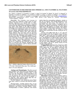

© Copyright 2026