
Physics of nail conditions: why do ingrown nails always - IOPscience
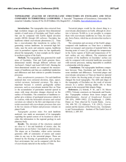
Home
Search
Collections
Journals
About
Contact us
My IOPscience
Physics of nail conditions: why do ingrown nails always happen in the big toes?
This content has been downloaded from IOPscience. Please scroll down to see the full text.
2014 Phys. Biol. 11 066004
(http://iopscience.iop.org/1478-3975/11/6/066004)
View the table of contents for this issue, or go to the journal homepage for more
Download details:
IP Address: 5.9.72.48
This content was downloaded on 26/11/2014 at 15:17
Please note that terms and conditions apply.
Physical Biology
Phys. Biol. 11 (2014) 066004 (10pp)
doi:10.1088/1478-3975/11/6/066004
Physics of nail conditions: why do ingrown
nails always happen in the big toes?
Cyril Rauch and Mohammed Cherkaoui-Rbati
School of Veterinary Medicine and Science, University of Nottingham, College Road, Sutton Bonington,
LE12 5RD, UK
E-mail: [email protected]
Received 10 June 2014
Accepted for publication 12 August 2014
Published 16 October 2014
Abstract
Although surgical treatment of nail conditions can be traced back centuries to the writings of
Paul Aegineta (625–690 AC), little is known about the physical laws governing nail growth.
Such a poor understanding together with the increasing number of nail salons in the high street
should raise legitimate concerns regarding the different procedures applied to nails. An
understanding of the physics of nail growth is therefore essential to engage with human medicine
and to understand the aetiology of nail conditions. In this context, a theory of nail plate adhesion,
including a physical description of nail growth can be used to determine the transverse and
longitudinal curvatures of the nail plate that are so important in the physical diagnosis of some
nail conditions. As a result physics sheds light on: (a) why/how nails/hooves adhere strongly, yet
grow smoothly; (b) why hoof/claw/nail growth rates are similar across species; (c) potential nail
damage incurred by poor trimming; (d) the connection between three previously unrelated nail
conditions, i.e. spoon-shaped, pincer and ingrown nails and; last but not least, (e) why ingrown
nails occur preferentially in the big toes.
Keywords: hard and growing tissues, biomechanics, dermatology, adhesion
Introduction
across all species. However, depending on the animal considered, the epidermal ridges can be more complex than in
man as they can display primary and secondary structures that
are thought to increase adhesiveness. For example, slowmoving animals like humans and cattle have only primary
lamellae (Thoefner et al 2005) whereas fast-moving
animal like horses (Pollitt 1994) or heavy animals like elephants (Benz et al 2009) have primary and secondary
lamellae (figures 1(B), (C), (D) and (E) show the horse foot as
an example).
Even though there now exists an in depth and complex
cross-species description of macroscopic/microscopic anatomical and cellular/sub-cellular structures, how nail and hoof
growth inform their shape remains unclear. This apparently
simple question is in fact central to medicine as the first
diagnosis of a nail/hoof condition by medics or vets is
necessarily a physical and visual appraisal of the shape or
form of the nail/hoof. In this context it is worth noting that
although nail cutting and hoof trimming have traditionally
been advocated to alleviate pain and reshape the nail/hoof
with time, there is little theory on which to ground these
The human nail is a keratinized structure and window to the
nail bed, held in place by lateral nail folds (the cutaneous
folded structures providing the lateral borders of the nail). It is
made of dead cells that multiply from the proximal matrix. As
a result, the nail originates from this proximal matrix, grows
longitudinally, and ends at a free edge distally. Nail adhesion
to the nail bed involves a number of well characterised
microscopic adhesive units. These units are apposed in a
pattern along longitudinal epidermal ridges (or lamellae)
stretching to the lunula, the half moon, pale convex portion of
the matrix seen through the nail (figure 1(A)). On the
underside of the nail plate there is a complementary set of
ridges as if the nail plates were held to the nail beds via a set
of longitudinal rails. A similar anatomical structure exists
Content from this work may be used under the terms of the
Creative Commons Attribution 3.0 licence. Any further
distribution of this work must maintain attribution to the author(s) and the
title of the work, journal citation and DOI.
1478-3975/14/066004+10$33.00
1
© 2014 IOP Publishing Ltd Printed in the UK
Phys. Biol. 11 (2014) 066004
C Rauch and M Cherkaoui-Rbati
Figure 1. Anatomy of nail and hoof adhesion. (A) Avulsion of the human nail showing the nail bed and in particular the epidermal ridges
interacting with the nail plate (figure reproduced from de Berker, Andre and Baran 2007). The anatomy of the epidermal structures is similar
across species. (B) Avulsion of the equine hoof capsule showing the bed and in particular the epidermal ridges interacting with the hoof
capsule. (The material was obtained from an ethically managed abattoir). (C) Internal view of epidermal ridges separated from foot (B). (D)
Internal view of dried epidermal ridges. (E) A microscopic section shows that in addition to the first lamellae (pink colour) there is a second
one (dark blue/dark purple colour pointed by the black arrow) that is connected to the hoof capsule (red colour). The hoof-lamellar interface
increases the surface area for adhesion.
Model
practices (Eliashar 2012). Furthermore, for each shape-related
condition, it is unclear whether another cutting/trimming
method exists that could be optimized. As a result, if a connection exists between the zoology of nail shapes and the
zoology of nail conditions, it is paramount to describe how
these changes have been made possible and what ‘physical’
parameters are involved. These parameters should help to
define the aetiology of nail/hoof conditions and give valuable
clues as to how they should be treated.
The present work considers the nail as an adhering solid
plate and will start by a brief hand-waiving introduction of
physical concepts used. The adhesion and growth stresses
present in nail will be modelled and incorporated into the
general balance of stresses to obtain unique solutions. The
optimization of the total energy of the nail including bending
and potential energies will be performed using the Euler–
Lagrange method to determine the nail shape equation. The
nail shape equation will be tested against specific nail conditions where changes in the shape of the affected nail occur.
We conclude that our model may provide a continuous relationship between three well known, but previously thought to
be unrelated, nail conditions namely ingrown, pincer and
spoon-shaped nails. Finally we shall also discuss the potential
importance of nail cutting in the aggravation of nail
conditions.
Below we explain the different concepts that will be used.
As a nail is a growing solid that adheres to a substrate, the
adhesion of the nail plate on its bed is necessarily involved
in the way it grows. Adhesion between cells and the
extracellular matrix is formed via specialized junctions
involving different sets of macro-molecules including more
complex ones such as focal adhesion or hemi-desmosomes
(Worth and Parsons 2008). Cellular adhesion has been
extensively modelled in different contexts. As a function of
the biology considered, the frameworks can differ, but
intermolecular bonds forming adhesions are usually treated
as Hookean springs that can either remain fixed vertically
(Dembo et al 1988, Dong and Lei 2000) and/or tilt from
their vertical position under stress (Reboux et al 2008). As a
nail adheres strongly, but needs to grow smoothly (i.e.
without stick-slip), one possibility is that this adhesion
imposes a ratchet-like mechanism on the nail so that the
‘growth’ is essentially confounded with diffusion. This is
possible if the length of nail growth per unit of time has a
magnitude that is similar to the thermal tilting of an adhesive unit. In these conditions the adhesive units should not
‘feel’ the nail to which they are bound, and a nail should
grow smoothly and adhere strongly. This ratchet-like model
2
Phys. Biol. 11 (2014) 066004
C Rauch and M Cherkaoui-Rbati
k − ~ 10s−1 (Ra et al 1999) and z 0 ~ 10 nm (Arnold
et al 2004, Cavalcanti-Adam et al 2006, Cavalcanti-Adam
et al 2007) one finds Vth ~ 0.1−1 mm/day at room temperature. The growth rate of nail/hoof/claw measured in a range of
species (table 1) falls within the range of thermal velocities
that can be predicted by this model. In addition, the very close
similarity between growth rates in-vivo suggests that physics
drives the process of growth. Indeed these in-vivo values are
not dependent on allometric properties and, as a result, do not
seem to involve the species–species metabolic rates, at least
under normal conditions.
indicates a universal form of growth for hooves and nails in
non-pathological conditions but it needs to be amended
when the growth rate of the keratinized plate is too fast. In
this case, the adhesive forces opposing the nail plate
movement and the mechanics of the plate need to be considered together. To understand clearly how mechanical
stresses arise, it is essential to understand the mechanical
impact of the nail edge as a boundary condition.
To explain this let us isolate a thin longitudinal slab of
the nail plate, from the proximal matrix to the free end. As the
pink colour underneath the slab is proportional to the number
of adhesive units involved in the adhesion of the slab, the
longer the slab the higher the adhesion. As a result, with a
constant growth force emanating from the proximal matrix,
the longer the slab the harder it will be to make it move
forward or grow. Thus, given the parabola-like shape where
the adhesion of the nail terminates, the right and left extremities of the nail of a thumb should grow faster than the
longitudinal slab positioned exactly in its center because the
latter would be longer than any other slab. Naturally, this is
never observed as the nail has solid properties. Nevertheless it
describes the set of residual longitudinal shear stresses
involved when the nail grows that result directly from the
boundary conditions. As a longitudinal shear stress promotes
a tendency toward rotation (i.e. to generate angular momentums) but because nails do not rotate locally, another stress,
transverse this time, must balance the virtual rotation primed
by the shear stress. This transverse stress resulting from the
profile of the nail edge, is expected to have an impact on the
transverse curvature of the nails, which is important in the
diagnosis of nail conditions.
Binding probability of adhesive units
Above the thermal velocity the probability that a unit
remains attached becomes a function of the stress imposed
on the unit. In this context, consider as above a single
adhesive unit that stands vertically without solicitation of
any sort. Once bound with a chemical energy, ΔE0 , the
thermal escape rate from the bound state is k − ~ e−ΔE0 / k B T f0
where, f0 , is a fundamental frequency. Upon movement of
the nail the unit tilts and the energy landscape changes. In
this context the unit has two possibilities, to remain bound
or to break and as a result the transition rate between the
2
‘bound’ and ‘unbound’ states is: f− ~ e−( ΔE0 − k el θ /2) / k B T f0 .
Considering independent and identical adhesion units, a
kinetic model can be used to describe the probability, P ,
that a unit is attached over time:
dP / dt = −f− × P + k + × (1 − P )
where k + is the binding rate. The last term on the righthand-side of equation (2) represents the probability that an
adhesive unit rebinds after unbinding. In this case, the
relaxation time relating to the adhesive unit going from a
stretched state to its resting position before rebinding is
neglected. The later relation also assumes that there is no
competition between binding sites and that a reservoir of
ligands exists so that the binding can be considered spatially
continuous. Considering a steady state regime of growth,
i.e. dP /dt = 0 , it follows:
Thermal growth rate
To provide a model, let us first focus on a single adhesive unit
that stands vertically without solicitation of any sort. Single
molecular adhesion can only last a time ~1/k − where, k −, is
the unbinding kinetic (figure 2(A)). If, when the adhesive unit
has just bonded, a movement involving a constant velocity, V ,
is now imposed on the unit and that the unit is fully compliant
mechanically, the unit will tilt up to a certain angle, θ , before
unbinding. This angle is expected to be, θ ~ V /z 0 k −
(figure 2(B)). The thermal agitation of any free, i.e. unbound,
unit can also define an angle proportional to the absolute
temperature, k el θ 2 ~ k B T , where k el is the tilt modulus of
the unit, k B the Boltzmann’s constant and T , the absolute
temperature (figure 2(C)). Equating the two relations
allows one to define a thermal velocity:
Vth ~ z 0 k − k B T / k el
(2)
−1
P ( V ) = ⎡⎣ 1 + α exp V 2 /2 ⎤⎦
(
)
(3)
where V = V /Vth and α = k − /k + (figure 2(D)). Equation (3)
shows the sensitivity of P ( V ) with regard to the growth rate
of nails.
Adhesion force developed by adhesive units
(1)
Focusing on a single adhesion unit, the force that opposes the
growth can be determined trivially as: k el Vth /z 02 k − × V P ( V ).
Consider a small element of nail surface area, the adhesion
per
unit
of
surface
of
nail
is
thus:
fadh (V ) = k el Vth /z 02 k − × ρV P ( V ) (figure 2(E)), where ρ is
the number of adhesive units per surface area of nail that will
This velocity defines a limit below which adhesion/
binding is controlled by the thermal energy. This means that
the movement of a nail plate onto adhesive units is possible
without further damage of units other than those determined
thermally if the velocity is close to, or below, Vth. Using
k el ~ 10 − 103 k B T (Reboux, Richardson and Jensen 2008),
3
Phys. Biol. 11 (2014) 066004
C Rauch and M Cherkaoui-Rbati
Figure 2. Adhesion and growth force. (A) Adhesive units bind and unbind to their ligand over time. In a steady state regime, with no
velocity involved, the bound and unbound states can be described by a basic 2 states model. (B) When the nail grows, the binding of the
adhesive units last until a certain value of the titling angle is reached, that is determined thermally or mechanically depending on the regime
considered. (C) When the adhesive unit is not bound, the tilt fluctuates around the vertical position and the use of thermodynamics allows one
to determine the deviation from the average value. (D) Representation of the probability that a bond is not consumed as a function of the
growth rate and α . (E) Representation of the force generated by a bound adhesive unit as a function of the growth rate and α .
Balance of in-plane stresses
Table 1. Hoof/nail growth rates for different animal species.
Animal
Growth rate
(mm day−1)
Horse
Sheep
Deer
Cow
0.2–0.3
0.1–0.2
0.1–0.2
0.1–0.3
Elephant
0.1–0.3
Pig
Rat
Human
0.3–0.4
0.1–0.2
0.1–0.2
The adhesive stress being defined, plate mechanics theory can
now be used to start investigating the interaction between the
shape and adhesion of nails.
The nail is held in place by an adhesion stress and a
boundary stress generated by the skin folds. However, as the
change of nail shape is expected to be slow, the deformation
of the nail bed and related impact on the adhesion stress
normal to the nail bed is unlikely to intervene actively in the
process, suggesting that we can consider the adhesion stress
constant at least for moderate deformations. In addition, as the
full characterisation of the external stresses applied by the
skin folds on the boundary of nail are, for the moment,
unknown to us, we shall only focus on a nail plate that is free
from external stresses induced by skin folds.
Consider the in-plane description and a nail plate orientated in such a way that the y-axis corresponds to the direction
of growth from the proximal to the distal parts and an x-axis
orthogonal to the y-axis along the proximal matrix
(figure 3(A)). Let us assume that the nail has a width, l , a
length, h′ (x ), with a constant thickness, e . The initial 3D
stress tensor, [σi, j ], can be reduced to a two dimensional stress
⎡ f (x, y ) fx, y (x, y ) ⎤
⎡ f (x, y ) ⎤ = ⎢ x, x
tensor:
with:
⎣ i, j
⎦ ⎢ f (x, y ) f (x, y ) ⎥⎥
y, y
⎦
⎣ y, x
References
(Butler and Hintz 1977)
(Shelton et al 2012)
(Miller et al 1986)
(Harrison et al 2007, Telezhenko
et al 2009)
(Benz, Zenker, Hildebrandt, Weissengruber, Eulenberger and
Geyer 2009)
(Johnston and Penny 1989)
(Godwin 1959)
(de Berker, Andre and Baran 2007)
0
be considered constant and fadh
= k el Vth /z 02 k − ~ 10 pN . Note
that as the nail plate is a projection of the nail bed and that
avulsion of the nail plate reveals a pattern of longitudinal
epidermal ridges involved in adhesion (figure 1), the ‘true’
total surface area available for adhesion is larger than the
visible surface of the nail plate and as a result a factor, λ ,
needs to be introduced to determine the true number of
adhesive units per unit of surface area to draw comparisons
between species. In these conditions:
0
¯ (V¯ )
fadh (V ) = fadh
λρVP
e /2
fi, j = ∫−e /2 σi, j dz where fi, j is defined as a force per unit of
length along the ‘j’-axis with direction along the ‘i’-axis
(figure 3(B)). Note that the terms on the diagonal define
(3a)
4
Phys. Biol. 11 (2014) 066004
C Rauch and M Cherkaoui-Rbati
Figure 3. Nail characteristics. (A) The nail is described by a system of axes allowing a simple analytic representation. h (x ) is the adhesion
profile i.e. the upper boundary between the white and pink parts (i.e. delimiting the yellow region from the blue one), and h′ (x ) the
most distal part of the nail (i.e. delimiting the blue region from the outside). These profiles are symmetrical and will be expressed by
polynomials involving even functions only. For a clearer representation, h0 corresponds to the limit of the green area from the proximal
matrix. (B) Representation of the set of stresses applied to the element of surface dxdy. (C) Results concerning the symmetry analysis. The
stress tensor can be used on every side of the small square dxdy to determine the conditions of symmetry with regard to the set of forces
applied to the nail. Given the balance of angular momentums we will further assume that ∂y fx,y is also equal to zero everywhere on the x-axis.
This is equivalent of considering that the shear stress along a longitudinal slab is constant (does not change much at the lowest order) along
the y-axis.
the classical surface tension whereas the non diagonal
terms define the shear stresses. Finally, the balance of
stresses on an element of surface dxdy of the nail can be
written as:
∂ x fx, x (x , y) + ∂yfx, y (x , y) = 0
further conditions follow (see appendix 1):
(4)
(5)
where H (y − h (x ) ) = 1 if y ⩽ h (x ) or zero otherwise and
where h (x ) defines the adhesion profile (see figure 3). As no
local rotation appears when nails grow, the conservation of
angular momentum imposes that:
fx, y (x , y) = fy, x (x , y)
(8)
fy, y (x , h′ (x )) = ∂ x h′ (x ) ⋅ fx, y (x , h′ (x ))
(9)
From the geometry of the nail (i.e. its symmetrical
shape), it is obvious that the stresses will have to follow some
important symmetry conditions. Figure 3(C) enounces all the
symmetries with regard to the stresses:
∂yfy, y (x , y) + ∂ x fy, x (x , y)
− fadh (V ) ⋅ H (y − h (x ) ) = 0
fx, x (x , h′ (x )) ⋅ ∂ x h′ (x ) = fx, y (x , h′ (x ))
fx, x (−x , y) = fx, x (x , y)
fy, y (−x , y) = fy, y (x , y)
fy, x (−x , y) = −fy, x (x , y)
∂yfx, y = 0
(6)
(10)
Hence the 2D stress tensor is symmetrical. To fully
define the problem, boundary conditions need to be added,
and two of those can be defined. We first assume that the
growth stress f0 (x ) is only defined at the origin on the y-axis
(i.e. proximal side) and has direction along the y-axis. This
first boundary condition leads to:
The set of equations (4)–(10) defines the physical stresses
present in a growing nail.
fy, y (x , 0) = −f0 (x )
Let us consider a solid nail growing at a constant velocity, V .
Without further assumptions and using the set of
equations (4)–(10) it is possible to determine the components
of the stress tensor analytically. To this end, let us consider
Stress solutions
(7)
Furthermore, let us assume that no force is applied distally i.e. that the nail has a free distal edge. Therefore two
5
Phys. Biol. 11 (2014) 066004
C Rauch and M Cherkaoui-Rbati
equations (5) & (7) and integrate equation (5) over the ‘y’
variable. By virtue of ∂y fx, y = ∂y fy, x = 0 (i.e. fy, x is independent of the ‘y’ variable—see equation (10)). One obtains:
fy, y (x , y) = −f0 (x ) + fadh (V )
∫0
The normal case
Let us first consider a nail that is trimmed in such a way that:
h′ (x ) = h (x ) and which has a growth profile given by:
f0 (x ) = fadh (V ) h (x ). In this case, one finds:
y
H (u − h (x )) du
− ∂ x fy, x (x ) × y
fx, x = fx, y
(11)
=0
Replacing equation (11) into the boundary condition
given by equation (9) leads to:
⎛
y ⎞
fy, y (x , y) = −f0 (x ) ⎜ 1 −
⎟
⎝
h (x ) ⎠
fy, x (x ) = fy, x (0)
+
1
h ′ (x )
∫0
x
⎡⎣ −f (u) + f (V ) h (u) ⎤⎦ du
0
adh
Equation (15) shows that the only existing stress is linked
to the growth stress. Such a stress should define the natural
longitudinal curvature, i.e. the claw shape, of any growing nail.
(12)
Provided the adhesion profile of the nail and the profile
of the distal edge, equation (12) completely determines
fx, y (x ) and, by symmetry, fy, x (x ). As fy, x (x ) is an odd
function (equation (10)), the shear stress in x = 0 has to be null
and as a result: fy, x (0) = 0 . Therefore, fx, y (x ) and, by symmetry, fy, x (x ) are now fully determined. Replacing
equation (12) into equation (11) allows us to complete the
determination of, fy, y (x, y ), as follow:
The pathological case
Let us now consider a nail that is trimmed in such a way that:
h′ (x ) = h (x ) and that has an imbalanced growth profile such
that: F (x ) = − f0 (x ) + fadh (V ) h (x ) ≠ 0 . In this case,
one shall assume also that, F (x ), is small and is an even
function of the variable ‘x’ to preserve the conditions
regarding the symmetry of the nail. As a result, F (x )
can be developed using Taylor series as follow:
∞
F (x ) = F (0) + ∑i = 1∂ x 2i F x = 0 x 2i /2i!. Naturally, each term
in the later development is expected to be small compared to
the leading term. Applying the same operation to the equation
∞
of the edge, h′ (x ) = h (x ) = h (0) + ∑i = 1∂ x 2i h x = 0 x 2i /2i!,
and replacing the Taylor series of F (x ) and h′ (x ) = h (x ) into
equations (11), (12) and (13), leads to:
⎛
y ⎞
fy, y (x , y) = −f0 (x ) ⎜ 1 −
⎟
⎝
h ′ (x ) ⎠
⎡ y
⎤
⎢ 0 H (u − h (x )) du
y ⎥
+ h (x ) fadh (V ) ⎢
−
⎥
h (x )
h ′ (x ) ⎥
⎢
⎣
⎦
y
+
∂ x h ′ (x )
h ′ (x )
∫
×
∫0
x
⎡⎣ −f (u) + h (u) f (V ) ⎤⎦ du
0
adh
F (0) x
α (x )
h (0)
F (0)
fx, x (x ) =
β (x )
h (0) ∂ x 2 h x= 0
⎛
y ⎞
fy, y (x , y) = −f0 (x ) ⎜ 1 −
⎟
⎝
h (x ) ⎠
∂ x 2 h x= 0 F (0)
γ (x )
+ yx 2
h (0)
fy, x (x ) =
(13)
Using equation (4) together with ∂y fx, y (x ) = 0
(equation (10)) allows one to determine that: ∂x fx, x = 0 , namely
that fx, x is only dependent on the variable ‘y’, i.e. is constant
along the x-axis for a given value on the y-axis. As fx, x is only a
function of the variable ‘y’, it is convenient to introduce ‘y’
using the reciprocal function, ‘h′−1’ of the edge equation defined
as h′−1 (h′ (x )) = x . Therefore, using the boundary condition
given by equation (8) allows the full determination of, fx, x :
fx, y (x )
∂ x h ′ (x )
(
= ∂yh′−1 (y) × fx, y h′−1 (y)
)
(16)
where the expression of α (x ), β (x ) and γ (x ) are given in
appendix 2 and where α (0) = β (0) = γ (0) = 1. We note here
that for nails having very flat distal edges, i.e. when:
∂ x 2 h x = 0 ≪ 1, the stress component that will dominate over
all the others in the distal part of the nail (the yellow and
blue parts in figure 3(B)) is: fx, x (x )2. This result suggests
that nails with a flat profile such as those the big toe should be
more prone to distal transverse stresses, if the difference
between the growth and adhesion stresses is not properly
balanced.
fx, x (x , y = h′ (x ))
=
(15)
(14)
As a result, provided the shapes of the adhesion profile and
edge of a nail, with the set of equations that have been determined
so far, it is possible to determine the stress tensor components
without any ambiguity. It is worth noting here that it is only when
the growth and adhesion stresses do not compensate each other,
i.e. − f0 (u ) + fadh (V ) h (u ) ≠ 0 , that fy, x and fx, x differ from
zero. In turn this could lead to some pathological conditions
2
As by definition: fx, x = fx, y / ∂x h (equation (14)), it follows that for flat
profiles the edge equation musty verifies ∂x h ≪ 1 and therefore, fx, x ≫ fx, y .
From equation (13) and ∂x h ≪ 1, it follows that fy, y ~ −f0 (x ) (1 − y / h (x ) ).
As in the distal part of a flat nail y ~ h (x ), one can assume fy, y ~ 0 or at least
very small. Note that these results do not hold when the nail has not a flat
profile.
6
Phys. Biol. 11 (2014) 066004
C Rauch and M Cherkaoui-Rbati
Figure 4. Nail shape and related conditions. (A) Transverse profile of the distal part of a nail for different sign of λ . (B) Photos of nail
conditions: pincer nail (left), ingrown nail (middle) and spoon-shaped nail (right); photos from Baran R et al (2014).
2
D
Application to ingrown nails: why the big toe?
Ventsel and Krauthammer 2001): E2 = 2 ∬ ( Cx − C0 ) dS,
where the integration is performed over the nail surface and
where, D = Ye3 /12(1 − υ), is the flexural rigidity of the nail
plate (Y , is the Young modulus assumed to be isotropic across
the nail and; υ the Poisson’s coefficient), Cx , the transverse
curvature of the nail surface along the x-axis and, C0 , the
spontaneous curvature of the nail that is identical to the curvature of the finger3. For small deformations, it is convenient
to describe the nail using the Monge gauge, i.e. by its height
z′ = w (x′, y′), with respect to a reference plane where x’
and y’ are the Cartesian coordinates in the reference plane. As
are related together by:
dx′dy′ = dS′ and dS
2⎤ ⎡
⎡
dS = ⎣ 1 + (∂w /∂x′) ⎦ ⎣ 1 + (∂w /∂y′)2⎤⎦ dS′, making use of
The ingrowing nail or ‘onychocryptosis’ is a condition
causing much discomfort and morbidity in school children/
adolescents/young adults (Khunger and Kandhari 2009) and
is diagnosed in 15% of pregnant women (Ponnapula and
Boberg 2010). Though recognized for a long time, a satisfactory treatment of onychocryptosis remains elusive, in part,
because the aetiology for ingrown nails is not understood.
Different theories were initially proposed and classified
according to whether the primary fault is based on the nail
plate or not (Haneke 2008). However a structural abnormality
of the nail plate has been ruled out (Pearson et al 1987), but it
was demonstrated instead, that a change in the transverse
curvature of the nail plate (i.e. curvature along the x-axis)
located distally promotes the condition (Pearson, Bury,
Wapples and Watkin 1987). As a transverse curvature of the
nail plate is involved, this suggests in turn, that some
mechanical consideration may be underlie the aetiology of
onychocryptosis.
It is well known that ingrown nail occurs predominantly
in the big toe. From a physical point of view one central
difference between the big toe and all other toes or fingers is
the fact that the adhesion profile of the nail plate is remarkably flat (but not straight—as otherwise the boundary
conditions defined by equation (8) and (9) would not apply).
As seen above, this means that the transverse stress fx, x is the
leading stress in the distal part of the nail. If an exaggerated
distal curvature of the nail defines the ingrown nail, it is
central to determine how the curvature is related to the
transverse stress.
To determine how the shape of the nail is affected we
concentrate on the energies method.
As fx, x is the leading stress in the distal part of the nail, the
the small deformation hypotheses: ∂y w ~ ∂y 2w ~ 0 and of
∂ x ′w ≪ 1 and e ∂ x ′2w ≪ 1; it is possible to rewrite the sum
of energies to the leading orders in the displacement along the
z’-axis as follows:
⎞2
D ⎛ ∂ 2w
E1 + E2 ~
− C0 ⎟
⎜
2 ⎝ ∂x′2
⎠
2⎤
⎡
1 ⎛ ∂w ⎞
⎟ ⎥ dS′
+ fx, x (y′) ⎢ 1 + ⎜
⎢⎣
2 ⎝ ∂x′ ⎠ ⎥⎦
⎛ ∂ 3w ⎛ ∂w ⎞3⎞
⎟ ⎟ dS′
+
o ⎜⎜ 3 ; ⎜
⎟
⎝ ∂x ′ ⎝ ∂x ′ ⎠ ⎠
∬
∬
(17)
Finally, removing the superscript prime (i.e. “ ’ ”) as all
the physical variables are now expressed in the fix Cartesian
referential, the Euler–Lagrange method determines the
equation of the nail shape (see appendix 3):
∂ x 4 w − λ (y ) ∂ x 2 w = 0
(18)
where λ (y ) = fx, x (y )/D . Using Fourier series with the following initial conditions: w x = 0 = w0 , ∂x w x = 0 = 0 and
potential energy of the nail can be simplified to E1 ~ ∬ fx, x dS
where the integration is performed over the nail surface area.
As the potential energy is transformed into a bending energy to
impose a new configuration to the nail and that, ingrown nails
are diagnosed by a change in the transverse curvature (i.e.
along the x-axis—see figure 4(B)) and not by longitudinal
curvature (i.e. not along the y-axis), it is legitimate to assume
that the only curvature involved is the one along the x-axis. In
these conditions, Kirchhoff’s theory of plate bending allows us
to rewrite the bending energy under the form (Helfrich 1973,
∂x 2w
x=0
= C0 leads to the ‘ingrown nail equation’:
w (x , y ) = w 0 +
3
C0 ⎡
cosh x λ (y) − 1⎤⎦
λ (y ) ⎣
(
)
(19)
Nails are composed of dead cells that grow from soft tissues and therefore
it is legitimate to assume that any nail should take the shape of the finger in
normal conditions.
7
Phys. Biol. 11 (2014) 066004
C Rauch and M Cherkaoui-Rbati
Shelley 1968). This condition is mostly acquired during
advanced age (Lee et al 2008). Although the aetiology is not
fully understood a change in nail growth related to a weaker
growth force with advanced age has been suggested (de Berker
et al 2007). The other well known condition, the ingrown nail
(figure 4(B) middle), is often diagnosed in school children/
adolescents/young adults (Khunger and Kandhari 2009) and
pregnant women (Ponnapula and Boberg 2010). It is remarkable that this condition occurs in patients where metabolic
growth is active. For example, in pregnancy, periods of high
sex hormone productions are well known to accelerate nail
growth (Hewitt and Hillman 1966, Ponnapula and
Boberg 2010). A last condition referred to as ‘spoon-shaped
nails’ corresponds to an inverted curvature of the nail
(figure 4(B) right). This shape can be described by the model.
Indeed, such a condition, which occurs in newborns or in brittle
nails, is expected to appear when the growth stress is small
enough to allow the curvature inversion (so that λ (y ) < 0 ) and/
or the nail is thin enough and thus brittle (Fawcett et al 2004) as
λ (y ) ∝ 1/e3. This shape is also predicted if the adhesion of the
nail drops, possibly as a result of aging.
Each condition cited above is diagnosed based on the
shape of the nail. These shapes can be modelled by a set of
physical equations suggesting that pincer, ingrown and
spoon-shaped nails are interrelated conditions forming a
continuum. In this context, physics suggests that the imperfection in nail growth, possibly due to aging and/or metabolic
changes, can precisely define the aetiology of some nail
conditions. Finally, although it seems that any condition
should recover with time, the trimming has great importance,
and should be carefully monitored, especially with the
increasing number of nail salons.
Let us assume that λ (y ) ≪ 1/l , where l is the width of
C
λ (y )
the nail, one finds: w (x, y ) ~ w0 + 20 x 2 ⎡⎣ 1 + 12 x 2 ⎤⎦. This
last relation shows that the sign of λ (y ) (i.e. the sign of
fx, x (y )) will influence the sign of the curvature as seen in
figure 4(A). As fx, x (y ) =
F (0)
h (0) ∂ x 2 h
β (h−1 (y )), the leading
x=0
term of fx, x (y ), i.e. F (0) = − f0 (0) + fadh (V ) h (0), can
change sign depending onto whether the growth stress is
larger or smaller than the adhesion stress.
Influence of trimming on nail conditions
fx, x (y ) is defined by the boundary condition given by
equation (14) and, as a result, exists only when the distal edge
of the nail is slightly curved. Therefore, cutting the nail in a
straight way, i.e. removing the slight curvature, and maintaining this profile over time should remove fx, x (y ) and
therefore improve the ingrown nail condition.
Conversely, bad trimming of the distal part of the nail
may amplify the magnitude of, fx, x (y ). To demonstrate this
point, assume in this case that the distal part is trimmed such
that h′ (x ) = h′ (0) − a (2x /l )2m . If F (x ) ≠ 0 , from
equation (14) the transverse stress can be rewritten as,
m−1
fx, x ~ [a /(h′ (0) − y )] m , showing that the magnitude of the
transverse stress can be very high. This suggests that careful
attention should be given to the different ways of trimming
nails to avoid the worsening of nail conditions involving the
transverse curvature.
Discussion
It has been known since the time of Hippocrates that a particular change in nail shapes can signify specific underlying
conditions (Myers and Farquhar 2001). From a physical point
of view, nail growth and shape are necessarily inter-related.
Therefore, it seemed important to investigate whether physics
could play a role in, and as a result explain, the aetiology of
some nail conditions.
As our daily experience of nail suggests that the nail plate
is hard and therefore should be considered as a solid we have
modelled both the adhesion and balance of stresses in this 2D
system. When diseased nails are surgically removed, their
shapes remain the same and as a result we made the implicit
assumption that the nail adapts the unbalanced stresses by
changing shape over time. To conclude, the model suggests
that the imbalance between the growth and adhesion stresses
is responsible for changing the distal transverse curvature of
nails, and confirms that it is the big toe that is predominantly
afflicted by nail conditions of mechanical origin due to its flat
profile.
This important result seems to agree relatively well with
observations in the field of nail conditions. For example, in
man, the term pincer nails (figure 4(B) left) describes an
exaggerated transverse curvature of the nail plate along the
longitudinal axis (Baran et al 2001, Cornelius and
Conclusion
We suggest that some nail conditions affecting the nail shape
can be explained by first principles. These are thus not disparate conditions but form a continuum of natural conditions.
Acknowledgements
The research was funded by the University of Nottingham
and Vertex Pharmaceuticals. The authors declare no conflict
of interests and would like to thank Professor Oliver E Jensen
for fruitful discussions; Florence Hillen and Emily Paul for
proof reading the manuscript; and Ramzi Al-Agele for providing horse materials.
Appendix A. balance of stresses and boundary
conditions
We consider the square a given by figure 3(B.1) to
determine the balance of stresses and focus on the y-axis. The
balance of stresses gives ∂y fy, y (x, y ) + ∂x fy, x (x, y ) − fadh (V )⋅
8
Phys. Biol. 11 (2014) 066004
C Rauch and M Cherkaoui-Rbati
Appendix C. Energy optimisation using the Euler–
Lagrange method
H (y − h (x ) ) = 0, where H (y − h (x ) ) = 1 if y ⩽ h (x ) or
zero otherwise. Integrating this equation over the y-axis gives:
fy, y (x , y) = fy, y (x , 0)
∫0
y
−
Consider the free energy:
⎡ ∂ f (x , y )
⎣ x y, x
− fadh (V ) ⋅ H (y − h (x ) ) ⎤⎦ dy
⎞2
D ⎛ ∂ 2w
⎜ 2 − C0 ⎟
2 ⎝ ∂x
⎠
⎡
1 ⎛ ∂w ⎞2 ⎤
⎟ ⎥ dS
+ fx, x (y) ⎢ 1 + ⎜
⎢⎣
2 ⎝ ∂x ⎠ ⎥⎦
E tot (w ) ~
(A1.a)
Where f0 (x ) = − fy, y (x, 0) as defined in the text.
Assuming further that no force is applied on the distal edge of
the nail, one finds:
{⎡⎣ f
i, j
(x , h (x )) ⎤⎦ n ⃗
}⋅e
= fy, x (x , h′ (x )) n x + fy, y (x , h′ (x )) n y
(A1.b)
⎞2
∂ 2δw
−
C
⎟
0
∂x 2
⎠
2
⎡
1 ⎛ ∂w
∂δw ⎞ ⎤
⎟ ⎥ dS
+ fx, x (y) ⎢ 1 + ⎜
+
⎢⎣
2 ⎝ ∂x
∂x ⎠ ⎥⎦
where →
n⎯ 1 + ∂x h′ (x )2 = −∂x h′ (x ) ex⃗ + ey⃗ . The balance of
stresses along the x-axis can be determined similarly:
∂ x fx, x (x , y) + ∂yfx, y (x , y) = 0
δE tot ~
(A1.c)
As one assumes that no stress is applied over the x-axis
the only relation regarding the boundary condition of the
distal edge, is:
{⎡⎣ f
i, j
(x , h (x )) ⎤⎦ n ⃗
}⋅e
−
⎛
2
∬ D2 ⎜⎝ ∂∂xw2
⎛
2
∬ D2 ⎜⎝ ∂∂xw2
+
⎡
⎞2
1 ⎛ ∂w ⎞2 ⎤
⎟ ⎥ dS (A3.b)
− C0 ⎟ + fx, x (y) ⎢ 1 + ⎜
⎢⎣
2 ⎝ ∂x ⎠ ⎥⎦
⎠
Where δEtot = Etot (w + δw ) − Etot (w ). Working the
energy difference to the first order of the small quantity δw
one finds:
x⃗
= fx, x (x , h′ (x )) n x + fx, y (x , h′ (x )) n y = 0
(A3.a)
One needs to find the function, w , that minimizes the free
energy above. Let us perform a small variation of
w → w + δw . The concomitant change in the free energy is
then:
y⃗
=0
∬
(A1.d)
⎞ ∂ 2δw
− C0 ⎟
⎠ ∂x 2
⎛ ∂w ∂δw ⎞
⎟ dS
+ fx, x (y) ⎜
⎝ ∂x ∂x ⎠
as seen in the text.
δE tot ~
Appendix B
⎛
2
∬ D ⎜⎝ ∂∂xw2
(A3.c)
Splitting equation (A3.c) into two different integrals, i.e.
over the curvature and the gradient of w one finds:
∞
1+
α (x ) =
∑∂ x
2i
F
x=0
⎛
i=1
∞
1+
∑∂ x
2i
h
x=0
2
∬ D ⎜⎝ ∂∂xw2
x 2i / F (0)(2i + 1)!
x 2i / h (0)2i!
=
i=1
∫
⎞ ∂ 2δw
dS
− C0 ⎟
⎠ ∂x 2
⎧ ⎡⎛ 2
⎫x0
⎞ ∂δw ⎤ ⎪
∂
∂w
⎥
⎨ ⎢⎜
⎬ dy
− C0 ⎟
⎪
2
⎠ ∂x ⎥⎦ ⎪
⎩ ∂x ⎢⎣ ⎝ ∂x
⎭− x
⎪
0
β (x ) =
⎡
⎢1 +
⎢⎣
∞
∑∂
x 2i
F
x=0
x
2(i − 1)
i=1
⎡∞
⎢ ∑∂ x 2 i h
⎢⎣ i = 1
x=0
⎤
x / F (0)(2i + 1)!⎥
⎥⎦
2i
/∂ x 2 h
x=0
−
⎤
(2i − 1)!⎥
⎥⎦
∫
⎧ ⎡ ⎛⎛ 2
⎫x0
⎞ ⎞⎤⎪
∂ ⎢ ∂
∂w
⎥
⎜⎜
⎨
− C0 ⎟ δw⎟⎟ ⎬ dy
2
⎪ ∂x ⎢ ∂x ⎜
⎠ ⎠ ⎥⎦ ⎪
⎩ ⎣ ⎝ ⎝ ∂x
⎭− x
⎪
0
4
+
⎡∞
⎤
⎢ ∑∂ x 2i h x= 0 x 2(i − 1) / ∂ x 2 h x= 0 (2i − 1)!⎥
⎢⎣ i = 1
⎥⎦
∞
⎡
⎤
× ⎢ 1 + ∑∂ x 2i F x= 0 x 2i / F (0)(2i + 1)!⎥
⎢⎣
⎥⎦
i=1
γ (x ) =
∞
⎡
⎤
⎢ 1 + ∑∂ x 2i h x= 0 x 2i / h (0)2i!⎥
⎢⎣
⎥⎦
i=1
∬ ∂∂xw4 δwdS
(A3.d)
And
∬ fx,x (y) ∂∂wx ∂∂δxw dS
=
∫
⎧ ∂ ⎡ ∂w ⎤ ⎫ x 0
δw ⎬ dy
fx, x (y) ⎨ ⎢
⎩ ∂x ⎣ ∂x ⎥⎦ ⎭−x
0
2
∬ fx,x (y) ∂∂xw2 δwdS
−
9
(A3.e)
Phys. Biol. 11 (2014) 066004
C Rauch and M Cherkaoui-Rbati
Where the interval [ −x 0 , xo ] is the interval of integration over
the x-axis, i.e. the extend of the projection of the nail surface
area over the x-axis contained in the Cartesian reference
plane. Using the conditions of symmetry, namely that δw and
w are even function of the variable ‘x’, from equations (A3.e)
& (A3.d) it follows that δEtot can be reduced to zero if:
∂ 4w
∂ 2w
−
f
(
y
)
=0
x
,
x
∂x 4
∂x 2
Godwin K O 1959 An experimental study of nail growth Journal of
Nutrition 69 121–7
Haneke E 2008 Controversies in the treatment of ingrown nails
Dermatol. Res. Pract. 783924
Harrison S M, Monahan F J, Zazzo A, Bahar B, Moloney A P,
Scrimgeour C M and Schmidt O 2007 Three-dimensional
growth of bovine hoof as recorded by carbon stable isotope
ratios Rapid Commun. Mass Spectrom. 21 3971–6
Helfrich W 1973 Elastic properties of lipid bilayers—theory and
possible experiments Zeitschrift Fur Naturforschung C-A
Journal of Biosciences C28 693–703
Hewitt D and Hillman R W 1966 Relation between rate of nail
growth in pregnant women and estimated previous general
growth rate Am. J. Clin. Nutr. 19 436–9
Johnston A M and Penny R H C 1989 Rate of claw horn growth and
wear in biotin-supplemented and non-supplemented pigs
Veterinary Record 125 130–2
Khunger N and Kandhari R 2009 Ingrown toenails Indian J.
Dermatol. Venereol. Leprol. 78 279–89
Lee J I, Lee Y B, Oh S T, Park H J and Cho B K 2008 A clinical
study of 35 cases of pincer nails Ann. Dermatol. 23 417–23
Miller K V, Marchinton R L and Nettles V F 1986 The growth rate
of hooves of white-tailed deer J. Wildl Dis. 22 129–31
Myers K A and Farquhar D R 2001 The rational clinical examination.
Does this patient have clubbing? JAMA 286 341–7
Pearson H J, Bury R N, Wapples J and Watkin D F 1987 Ingrowing
toenails: is there a nail abnormality? A prospective study
J. Bone Joint Surg Br. 69 840–2
Pollitt C C 1994 The basement-membrane at the equine hoof dermalepidermal junction Equine Veterinary Journal 26 399–407
Ponnapula P and Boberg J S 2010 Lower extremity changes
experienced during pregnancy J. Foot Ankle Surg. 49
452–8
Ra H J, Picart C, Feng H S, Sweeney H L and Discher D E 1999
Muscle cell peeling from micropatterned collagen: direct
probing of focal and molecular properties of matrix adhesion
Journal of Cell Science 112 1425–36
Reboux S, Richardson G and Jensen O E 2008 Bond tilting and
sliding friction in a model of cell adhesion Proc. of the Royal
Society a-Mathematical Physical and Eng. Sciences vol 464,
pp 447–67
Shelton J, Usherwood N M, Wapenaar W, Brennan M L and
Green L E 2012 Measurement and error of hoof horn growth
rate in sheep Journal of Agricultural Science 150 373–8
Telezhenko E, Bergsten C, Magnusson M and Nilsson C 2009 Effect
of different flooring systems on claw conformation of dairy
cows J. Dairy Sci. 92 2625–33
Thoefner M B, Wattle O, Pollitt C C, French K R and Nielsen S S
2005 Histopathology of oligofructose-induced acute laminitis
in heifers J. of Dairy Science 88 2774–82
Ventsel E and Krauthammer T 2001 Thin Plates and Shells: Theory,
Analysis and Applications (Boca Raton, FL: CRC Press)
Worth D C and Parsons M 2008 Adhesion dynamics:
mechanisms and measurements Int. J. Biochem. Cell Biol.
40 2397–409
(A3.f)
References
Arnold M, Cavalcanti-Adam E A, Glass R, Blummel J, Eck W,
Kantlehner M, Kessler H and Spatz J P 200 4 Activation of
integrin function by nanopatterned adhesive interfaces
Chemphyschem. 5 383–8
Baran R, Dawber R, Haneke E, Tosti A and Bristow I 2014 Informa
Health Care (Abingdon, Oxford: Taylor & Francis)
Baran R, Haneke E and Richert B 2001 Pincer nails: definition and
surgical treatment Dermatol. Surg. 27 261–6
Benz A, Zenker W, Hildebrandt T B, Weissengruber G,
Eulenberger K and Geyer H 2009 Microscopic morphology of
the elephant's hoof J. Zoo Wildl Med. 40 711–25
Butler K D Jr and Hintz H F 1977 Effect of level of feed intake and
gelatin supplementation on growth and quality of hoofs of
ponies J. Anim. Sci. 44 257–61
Cavalcanti-Adam E A, Micoulet A, Blummel J, Auernheimer J,
Kessler H and Spatz J P 2006 Lateral spacing of integrin
ligands influences cell spreading and focal adhesion assembly
Eur. J. Cell Biol. 85 219–24
Cavalcanti-Adam E A, Volberg T, Micoulet A, Kessler H,
Geiger B and Spatz J P 2007 Cell spreading and focal adhesion
dynamics are regulated by spacing of integrin ligands Biophys.
J. 92 2964–74
Cornelius C E III and Shelley W B 1968 Pincer nail syndrome Arch.
Surg. 96 321–2
de Berker D A, Andre J and Baran R 2007 Nail biology and nail
science Int. J. Cosmet. Sci. 29 241–75
Dembo M, Torney D C, Saxman K and Hammer D 1988 The
reaction-limited kinetics of membrane-to-surface adhesion and
detachment Proc. of the Royal Society of London Series BBiological Sciences vol 234, pp 55–83
Dong C and Lei X X 2000 Biomechanics of cell rolling: shear flow,
cell-surface adhesion, and cell deformability J. Biomech. 33
35–43
Eliashar E 2012 The biomechanics of the equine foot as it pertains to
farriery Veterinary Clinics of North America-Equine Practice
28 283
Fawcett R S, Linford S and Stulberg D L 2004 Nail abnormalities:
clues to systemic disease Am. Fam. Physician 69 1417–24
10


© Copyright 2026