Congo Red Amyloid Stain Kit Procedure
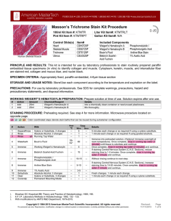
Congo Red Amyloid Stain Kit Procedure 100ml Kit Item #: KTCRE Pint Kit Item #: KTCREPT Control Slide(s) Amyloid Alpha Liter Kit Item#: KTCRELT Gallon Kit Item#: N/A Item# CSA0425P Included Components Congo Red Amyloid Stain 1% Sodium Hydroxide Modified Mayer’s Hematoxylin Human Adrenal Gland PRINCIPLE AND RESULTS: This kit is intended for use by laboratory professionals to stain routinely prepared paraffin embedded tissue specimens or frozen sections (in vitro) to identify amyloid deposits. Amyloid stain pale pink to red (Birefringent if viewed using a polarized light microscope) and nuclei blue. SPECIMEN CRITERIA: Appropriately fixed, paraffin-embedded or frozen tissue section cut at 8-10μm. STORAGE AND USAGE NOTES: Store/Use each component according to the temperature and expiration on the label. PRECAUTIONS: For use by laboratory professionals. See SDS for complete warnings, precautions, hazard and precautionary statements, and disposal information. STAINING PROCEDURE: Color coordinated steps denote stain baths that can be reused during autostainer configuration. # Action With 1 2 3 4 Deparaffinize Rinse Rinse Immerse 5 6 7 8 9 1. 2. Heat Time Mins Secs Xylene or Substitute, 2 changes Absolute Alcohol, 3 changes Running Tap Water Congo Red Amyloid Stain ºC ----- 5 1 1 60 ----- Immerse 1% Sodium Hydroxide -- -- 15-20 Immerse Dehydrate Clear Coverslip Modified Mayer’s Hematoxylin Absolute Alcohol, 3 changes Xylene or Substitute, 3 changes Permanent Mounting Media ----- 3 1 1 -- ---- Details 5 minutes each change or as required if using a xylene substitute. 1 minute each change or as required if using graded alcohols. Without rinsing, continue to next step. Differentiate until Amyloid is pink to red. Use a quick tap water rinse and microscope to check for correct differentiation. Once complete, rinse in running tap water (1 minute) and continue. Once complete, rinse in running tap water (1 minute) and continue. 1 minute each change. 1 minute each change or as required if using a xylene substitute. A.F.I.P. Manual; pg 146. Lilly; Histopathologic Technique; pg 293. With modifications by AMTS R&D Department, 1979-2015. Copyright © 1994-2015 American MasterTech Scientific Incorporated. All rights reserved. For personal use only. Reproduction, modification, storage in a retrieval system or retransmission, is strictly prohibited without prior written permission. Revised: 11/11/2015 Page 1 of 4 AUTOSTAINER CONFIGURATION AND NOTES: This stain kit in the pint size may be easily adapted for use on most open-platform autostainers using the staining procedure grid on the reverse side of this page. A minimum of 4 baths is required to perform this procedure excluding deparaffinization, hydration, dehydration, and clearing, or 15 baths to run the complete procedure. TEST YIELD: *Assumes pint kit and maximum slides per run. Actual Results may vary. S.C. denotes number of slides between “Solution Change”. Bath Type Uses Slides S.C. Bath Type Uses Slides S.C. 20ml Plastic Slide Jar 50 1000 20 250ml Glass Stain Dish 4 750 188 30ml Glass Coplin Jar 33 990 30 200ml Bath Autostainer 5 800 160 40ml Hellendahl Jar 25 1000 40 400ml Bath Autostainer 2 680 340 CE MARKINGS AND DESIGNATIONS: Catalogue Number Temperature Limitation Manufacturer American MasterTech 1330 Thurman St. Lodi, CA 95240 USA Tel 800 860 4073 Fax 209 368 4136 Batch Code Use By Representative Emergo Europe Molenstraat 15 2513 BH The Hague NL In Vitro Diagnostic Medical Device Consult Instructions Prior to Use Irritant GHS07 1. 2. Health Hazard GHS08 A.F.I.P. Manual; pg 146. Lilly; Histopathologic Technique; pg 293. With modifications by AMTS R&D Department, 1979-2015. Copyright © 1994-2015 American MasterTech Scientific Incorporated. All rights reserved. For personal use only. Reproduction, modification, storage in a retrieval system or retransmission, is strictly prohibited without prior written permission. Revised: 11/11/2015 Page 2 of 4 MULTILANGUAGE PROCEDURE PROCÉDURE DE KIT DE TACHANT EN FRANÇAIS COMPOSANTS INCLUS: Congo Red Amyloid Stain, 1% Sodium Hydoxide, Modified Mayer’s Hematoxylin LES CRITERÈS D'ÉCHANTILLONS: Sections de 8-10 microns de tissus fixés au manière appropriée, enfoncé dans la paraffine ou des sections congelées. LA PRINCIPE ET LES RÉSULTATS: Ce kit est destiné pour l'utilisation par des professionnels de laboratoire pour tacher des échantillons de tissus inclus en paraffine, lesquels sont régulièrement préparés (in vitro) pour identifier les dépôts d'amyloïde. Amyloïde tache rose pâle au rouge(Biréfringente si visualisé à l'aide d'un microscope à lumière polarisée) et les noyaux bleu. LES NOTES DE STOCKAGE ET D'UTILISATION: Utilisez chaque composante d'après la température et la date limite d'utilisation sur l'étiquette. LA PROCÉDURE DE TACHANT: Les ètapes couleur coordonnées dénotent les báins a teinture lesquels peuvent être réutilisés lors de la configuration d'Autostainer. # Temp Durée Action Avec 1 Déparaffinez Xylene ou remplaçant, 2 changements -- 5 -- 2 Rincez Alcool absolu, 3 changements -- 1 -- 3 4 Rincez Immergez L'eau du robinet courante Congo Red Amyloid Stain --- 1 60 --- 5 Immergez 1% Sodium Hydroxide -- -- 15-20 6 Immergez Modified Mayer’s Hematoxylin -- 3 -- 7 Déshydratez 1 -- Éclaircissez Alcool absolu, 3 changements Xylene ou remplaçant, 3 changements -- 8 -- 1 -- 9 Faitez une Lamelle Milieu de montage permanent -- -- -- 1. 2. ºC mins secs Détails 5 minutes pour chaque changement ou comme nécessité s'il on utilise une remplaçant de Xylene. 1 minute pour chaque changement ou comme nécessité s'il on utilise l'alcool graduée. Sans rincez, continuer à l'étape suivante. Différenciez jusqu'à amyloïde est rose à rouge. Utilisez un rinçage à l'eau du robinet rapide et un microscope pour vérifier la différenciation correcte. Une fois que c'est terminé, rincez sous l'eau du robinet courante (pour 1 minute) et continuez. Une fois que c'est terminé, rincez sous l'eau du robinet courante (pour 1 minute) et continuez. 1 minute pour chaque changement. 1 minute pour chaque changement ou comme nécessité s'il on utilise une remplaçant de Xylene. A.F.I.P. Manual; pg 146. Lilly; Histopathologic Technique; pg 293. With modifications by AMTS R&D Department, 1979-2015. Copyright © 1994-2015 American MasterTech Scientific Incorporated. All rights reserved. For personal use only. Reproduction, modification, storage in a retrieval system or retransmission, is strictly prohibited without prior written permission. Revised: 11/11/2015 Page 3 of 4 PROCEDIMIENTO PARA KIT DE TINCIÓN EN ESPAÑOL COMPONENTES INCLUIDOS: Congo Red Amyloid Stain, 1% Sodium Hydoxide, Modified Mayer’s Hematoxylin CRITERIOS DE MUESTRAS: Secciones de tejido 8-10μm apropiadamente fijadas, embebidas en parafina. PRINCIPIO Y RESULTADOS: Este kit está diseñado para su uso por profesionales de laboratorio para teñir muestras de tejido embebidas en parafina preparadas de forma rutinaria (in vitro) para identificar los depósitos de amiloide. Amiloide se tiñe de rosa pálido a rojo (Birrefringente si ve usando un microscopio de luz polarizada) y los núcleos azul. NOTAS SOBRE ALMACENAMIENTO Y USO: Guarde/Use cada componente de acuerdo con la temperatura y caducidad en la etiqueta. PROCEDIMIENTO DE TINCIÓN: El color de pasos coordinados denota baños de tinción que pueden ser reutilizados durante la configuración de tinción automática. # Acción Con 1 Desparafine 2 Enjuague 3 4 Enjuague Sumerja 5 Ta Tiempo ºC min s Xileno o sustituto, 2 cambios -- 5 -- Alcohol absoluto, 3 cambios -- 1 -- Corriente de agua grifo Congo Red Amyloid Stain --- 1 60 --- Sumerja 1% Sodium Hydroxide -- -- 15-20 6 Sumerja Modified Mayer’s Hematoxylin -- 3 -- 7 Deshidrate Alcohol absoluto, 3 cambios -- 1 -- 8 Clarifique Xileno o sustituto, 3 cambios -- 1 -- 9 Cubreobjetos Medios de montaje permanente -- -- -- 1. 2. Detalles 5 minutos cada cambio o según sea necesario si se utiliza un sustituto de xileno. 1 minuto cada cambio o según sea necesario si se utiliza alcoholes graduados. Sin enjuagar siga al siguiente paso. Diferenciar hasta amiloide es de color rosa o rojo. Utilice un enjuague de agua grifo rápido y el microscopio para verificar si hay diferenciación correcta. Una vez terminado, enjuague con corriente de girfo (1 minuto) y continúe. Una vez terminado, enjuague con corriente de agua grifo (1 minuto) y continúe. 1 minuto cada cambio. 1 minuto cada cambio o según sea necesario si se utiliza un sustituto de xileno. A.F.I.P. Manual; pg 146. Lilly; Histopathologic Technique; pg 293. With modifications by AMTS R&D Department, 1979-2015. Copyright © 1994-2015 American MasterTech Scientific Incorporated. All rights reserved. For personal use only. Reproduction, modification, storage in a retrieval system or retransmission, is strictly prohibited without prior written permission. Revised: 11/11/2015 Page 4 of 4
© Copyright 2026
