
Prevalence of serum antibodies against the p53
Publicationof the InternationalUnion Against Cancer
Publication de I Union Internationale Contre le Cancer
Znt. J. Cancer: 58,480-487 (1994)
0 1994 Wiley-Liss, Inc.
PREVALENCE OF SERUM ANTIBODIES AGAINST THE p53 TUMOR
SUPPRESSOR GENE PROTEIN IN VARIOUS CANCERS
Katerina ANGELOPOULOU~,
Eleftherios P. DIAMANDIS1,4,Donald J.A. SUTHERLAND~,
John A. KELLEN3 and Peter S. BUNTING3
Department of Clinical Biochemistry, The Toronto Hospital, Toronto Western Division, and
Department of Clinical Biochemistry, University of Toronto; 2TorontoBayview Regional Cancer Centre,
Sunnybrook Health Science Centre, University of Toronto, Toronto, Ontario, Canada; 3Departmentof Clinical Biochemistry,
Sunnybrook Health Science Centre, University of Toronto, Ontario, Canada.
We have developed 2 new quantitative methods for measuring anti-p53 antibodies in human serum. Using these methods
we analyzed 1,392 sera from patients with various malignancies
and 230 sera from individuals without malignancy. Highest
prevalence of antbps3 antibodies was associated with ovarian
and colon cancers ( I 5%), followed by lung (8%) and breast (5%)
cancers. Prevalence in other malignancies was lower (<4%). In
hospitalizedpatients and apparently healthy individuals, prevalence was very low (<2 and I Oo/ respectively). Extremely high
antibody concentrations (> lo5 U/L) were found in 5 ovarian, 2
breast, I lung and I colon cancers. Sequential analysis of 6
positive samples has shown that the p53 antibody test may have
potential for patient monitoring. The p53 antibody-positive
sera from breast cancer patients were associated with tumors
that were steroid hormone receptor-negative (p < 0.002). We
propose that the measurement of p53 antibodies is a relatively
specific serological test for cancer, which can be performed with
easily automatable and quantitative methodologies and may be
further exploited for patient monitoring, prognosis, diagnosis
and probably screening for selected cancers.
1994 II d q -LM, 1,M
Cancer diagnosis is not possible using currently available
serological tumor markers because these serum proteins are
not sensitive and/or specific for cancer. It has been proposed
that oncogenes and tumor suppressor genes and their products
may form new biochemical tests for cancer (Diamandis, 1992).
This proposal has merit because it is currently believed that
the changes in oncogenes and tumor suppressor genes are not
merely late sequelae or epiphenomena of cancer, as is the case
with most known tumor markers, but initiating and pathogenetic events (Fearon and Vogelstein, 1990). Thus monitoring
of such events may provide novel ways for the early detection
and monitoring of cancer.
In most cancers, genetic changes are somatic and are
present exclusively in the tumor. Thus to study such changes
one must obtain tumor cells. This is possible in selected
cancers which can release tumor cells in easily obtainable
specimens, e.g., stool (colon cancer), urine (bladder cancer)
and sputum (lung cancer). In these cases, extremely sensitive
techniques may provide a means of identifying the genetic
changes in a minute number of tumor cells, which can be used
for diagnosis (Sidranskyet al., 1991,1992).
The release of measurable mutant proteins from the tumor
into the bloodstream offers another possibility for cancer
diagnosis. However, many mutant proteins are either cytoplasmic or nuclear components and are released into the circulation in either minute amounts or only during tumor cell
necrosis. In the circulation, these proteins may appear for only
short periods of time because of degradation or clearance.
Other mutant proteins are membrane components and are not
usually released into the circulation. It is thus not surprising
that some mutant proteins, e.g., the p53 tumor suppressor gene
protein, have not as yet been identified in serum, even when
the tumor is very rich in mutant p53 protein levels. Probably,
mutant p53 is released into the bloodstream discontinuously or
at minute, unmeasurable amounts or it is bound to other
proteins, degraded or cleared quickly.
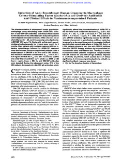
We here examine another possible way of diagnosing and
monitoring cancer based on mutant proteins by taking advantage of 3 distinct features of the immunological system: (i)
identification of non-self antigens or loss of tolerance to self
antigens, (ii) immunological amplification and (iii) stability of
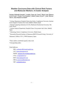
antibodies in the circulation. The concept, diagrammatically
presented in Figure 1, is similar to the one used to identify
pathogens and has not been systematically studied for cancer
diagnosis and monitoring. Antibody generation against a few
oncogene and tumor suppressor gene products, i.e., myb
(Sorokine et al., 1991), myc (Ben-Mabrec et al., 1990) and p53
(Crawford et al., 1982; Caron de Fromentel et al., 1987; Winter
et al., 1992; Davidoff et al., 1992; Schlichtholz et al., 1992), has
already been described. We present an extensive study of
antibody generation against the p53 protein in various, largely
unstudied cancer types and in groups of healthy and noncancer hospitalized patients.
MATERIAL AND METHODS
We used 2 time-resolved immunofluorometric techniques,
described in detail elsewhere (Hassapoglidou and Diamandis,
1992; Angelopoulou and Diamandis, 1993), to measure antip53 antibodies in serum. Both methods are quantitative,
non-isotopic and easily automatable. Briefly, the methods are
based on the following principles. Method A (Hassapoglidou
and Diamandis, 1992): white, opaque polystyrene microtiter
wells were coated with goat anti-mouse immunoglobulin. We
added mutant p53 antigen (produced in COLO 320 HSR+
cells) and mouse monoclonal antibody (MAb) PAb240 antip53 (mutant-specific), incubated for 3 hr and washed 6 times,
followed by patient serum (diluted 10-fold or more, as necessary) incubated for 1 hr and washed as above. We then added
alkaline phosphatase-labeled goat anti-human imrnunoglobulin, incubated for 1 hr and washed as above. The alkaline
phosphatase activity was measured with a new detection
methodology previously described by our group (Christopou10s and Diamandis, 1992; Papanastasiou-Diamandi et al.,
1992), involving the alkaline phosphatase substrate 5-fluorosalicylphosphate, Tb3+ and EDTA. This method is extremely
sensitive and can quantify analytes at attomole quantities.
Method B (Angelopoulou and Diamandis, 1993): microtiter
wells were coated as in Method A. Patient serum (undiluted or
diluted, as necessary, for samples with high antibody titers)
was then incubated in tubes with a standard amount of p53
antigen (from COLO 320 HSR+ cells). The mixture was then
added to the microtiter wells along with PAb240. After
incubation for 3 hr and washing, we added the CM-1 anti-p53
rabbit antibody (mutant and wild-type-specific), followed by 1
4To whom correspondence should be addressed, at Department of
Clinical Biochemistry, The Toronto Hospital, Toronto Western Division, 399 Bathurst Street, Toronto, Ontario M5T 2S8, Canada. Fax:
(416) 369-5605.
Received: January 24,1994 and in revised form April 6, 1994.
48 1
p53 ANTIBODIES IN VARIOUS CANCERS
0
Normal
Cell
1
Malignant Transformation
0
Malignant
m a
Immune
Surveillance
1
Malignant cell producing surface or
intracellular proteins which are
either mutant or at abnormally
high concentration.
llreak of Tolerance
Immunological Amplification
Generation of antibodies which can
be specifically detected in serum
FIGURE1 - Concept for the serological diagnosis of cancer.
Early during cancer development, alterations in oncogenes and/or
tumor suppressor genes may lead to the production of mutant
forms or abnormal levels of proteins within tumor cells or on
tumor cell membrane (black sections). Host’s immunological
system detects such altered or abnormally abundant proteins and
produces antibodies against them. Antibodies are produced in
amounts vastly higher than the immunogen (immunological amplification), circulate in the serum for long periods and could be used
to spot the cancer initiation event. The immune response can be
initiated even if the offending immunogen is only transiently
released from the tumor.
hr incubation and washing. We then added an alkaline
phosphatase-labeled goat anti-rabbit immunoglobulin, incubated for 1 hr and washed 6 times. The alkaline phosphatase
activity was measured as in Method A.
Method A is a non-competitive procedure based on the
reaction of serum antiLp53 antibodies with mutant p53 antigen
immobilized on the solid-phase through the PAb240 antibody.
Method B is essentially a p53 antigen assay (Hassapoglidou et
al., 1993),but during the incubation of serum with exogenously
added p53 antigen, any p53 antibodies present in the serum
react with p53 antigen and render it unmeasurable by the p53
assay. This is a “competitive” assay because the exogenously
added p53 binds either to the serum antibodies or to the
coating PAb240 antibody.
High performance liquid chromatography (HPLC) was performed with a Shimadzu (Kyoto, Japan) system with an
absorbance monitor at 280 nm. The mobile phase was a 0.1
mol/L Na2S04-0.1 mol/L NaH2P04solution, pH 6.8. The flow
rate was 0.5 mL/min and the HPLC was run isocratically. The
gel-filtration column was a Bio-Sil SEC-250 column, 600 x 7.5
mm (Bio-Rad, Richmond, CA). The column was calibrated
with a m.w. standard solution from Bio-Rad, containing
thyroglobulin (670 kDa), IgG (158 kDa), ovalbumin (44 kDa),
myoglobin (17 kDa) and cyanocobalamin (1.4 kDa). HPLC
fractions (0.5 mL) were collected with a fraction collector
(model FRAC-100; Pharmacia, Uppsala, Sweden).
Protein A affinity chromatography was performed manually
using the kit system MAPS, purchased from Bio-Rad. The
instructions of the manufacturer were followed throughout.
The specificity of detection of serum anti-p53 antibodies by
Methods A and B was checked in some positive sera by
Western blot analysis as follows: lysates from COLO 320
HSR+ cells were mixed with an equal volume of Tris-glycine-
SDS buffer containing 2-mercaptoethanol, denatured by heating at 90°C for 5 min and loaded onto a 4-20% polyacrylamide
gels. After electrophoresis (125 V, 90 min), proteins were
transferred to a nitrocellulose membrane (Hybond-ECL, Amersham, Arlington Heights, IL) by electroblotting at 30 V for 2
hr. The membrane was then treated overnight in a blocking
solution (5% non-fat dried milk in wash solution, consisting of
Tris-buffered saline, pH 7.6, 0.1% Tween-20). The membrane
was then cut into strips, which were probed for 1 hr at room
temperature with the human sera or the polyclonal anti-p53
antibody CM-1, diluted 1,000-foldin a 6% BSA solution. After
washing with solution the blot was incubated for 1 hr with a
goat anti-human IgG conjugated to horseradish peroxidase
(HRP), in the case of the human sera, and with a goat
anti-rabbit IgG conjugated to HRP, in the case of the CM-1.
After a final washing, antibody binding was visualized by
chemiluminescence and captured on X-ray film, using the
ECL-Western blot detection kit from Amersham.
Quantification
Because of the lack of a suitable standard solution, we
devised an arbitrary system to calibrate Methods A and B.
Among the highly p53 antibody-positive sera we selected one
and arbitrarily defined its concentration to be 10,000 UnitsiL
(U/L). This serum sample was then used at various dilutions to
construct calibration curves for assays A and B from which the
concentration of the other samples was calculated (Angelopoulou and Diamandis, 1993).
Assays for tumor markers
For the analysis of CA-125 and CEA we used commercially
available kits, i e . , the TRU-QUANT OV RIA (Biomira,
Edmonton, Canada) and the Amerlite CEA-60 assay (Kodak,
Rochester, NY). Estrogen and progesterone receptors were
measured with enzyme immunoassay kits from Abbott, North
Chicago, IL.
Statistical analysis
The chi-square (x2) test was used to determine the statistical
significance of differences in distributions and all x2 values and
corresponding p values were calculated by the statistical
software SAS (Cary, NC).
RESULTS
We collected sera from various groups of patients over a
3-year period and stored them at -70°C until analysis. Most
sera were collected within 6 months from diagnosis. No effort
was made to subclassify the patients in categories according to
disease stage, mode of treatment, duration of disease, sex, age
or any other clinical parameter. Our testing strategy was as
follows: all specimens were analyzed only by Method A, and
positive samples were identified based on a cut-off fluorescence ratio of 1.7, between fluorescence in the presence or
absence of p53 antigen, as previously described (Hassapoglidou and Diamandis, 1992). All samples positive by Method
A were also analyzed by Method B for confirmation. The
results are summarized in Table I and Figure 2.
The highest positivity rate was observed with colon and
ovarian cancer sera (15-16%). These groups of patients have
not been previously systematically studied for p53 antibody
response. Relatively high positivity rates were also obtained
with lung (8%) and breast (5%) cancer sera in accordance with
previous reports (Crawford et al., 1982; Winter et al., 1992;
Davidoff et al., 1992; Schlichtholz et al., 1992). Lower positivity
rates ( 3 4 % ) were seen in patients with pancreatic and
prostate cancer and in patients with multiple myeloma and
lymphoma. Positivity rates similar to those obtained for blood
482
ANGELOPOULOU ET AL.
donors or hospitalized patients ( < 2 % ) were obtained in
patients with hepatoma, melanoma, leukemia, Kaposi's sarcoma and testicular carcinoma.
TABLE I - SERUM ANTI-p53 ANTIBODIES IN VARIOUS PATIENT GROUPS
AND BLOOD DONORS
Positive samples'
Patient diagnosis
Samples
tested
Method A
Lung cancer
Breast cancer
Ovarian cancer
Colon cancer
Testicular cancer
Prostate cancer
Pancreatic cancer
Hepatoma
Melanoma
Multiple myeloma
Leukemia
Lymphoma
Kaposi's sarcoma
Polycytemia rubra Vera
Idiopathic thrombocytopenic
purpura
Blood donors and healthy
volunteers
Hosoitalized oatients
73
290
86
82
144
13(15.i
13 1531
O[O)
y",',","$
11 12.8
13[1S.k]
-
-
0 0)
10
-
1.50
l(0.7)
O(0)
56
l(1.8)
l(1.8)
'Samples found positive by Method A were also tested by
Method B. Percentages of positive samples are given in parentheses.
Our methods for measuring pS3 antibodies are quantitative.
Serum pS3 antibody concentrations were calculated for all
positive samples, and the results are presented in Figure 3.
Concentrations ranging from lo2 U / L to almost lo7 U/L were
obtained. The highest titers ( > lo5 U/L) were seen in the sera
of 5 ovarian, 1 lung, 2 breast and 1 colon cancer patients. We
were interested in the changes of p53 antibody titers with time
and their relation to disease progression and regression or to
therapeutic manipulations. The results for 6 patients from
whom we had serial samples are shown in Figure 4.
Patient A (70 years old) was diagnosed w'ith an ovarian
papillary invasive serous adenocarcinoma, grade 3 and stage
111, and was treated with bilateral salpingoophorectomy (BSO)
and omentectomy, but residual tumor remained. After surgery, she was treated with cisplatinum plus cyclophosphamide.
The same chemotherapy was given another 5 times over a
5-month period, as shown in Figure 4a. CA-125 levels and p53
antibody levels were monitored in 8 consecutive samples.
Patient B (62 years old) was diagnosed with ovarian papillary
serous invasive cystadenocarcinoma and was treated with BSO
and omentectomy/hysterectomy, but residual tumor remained. After surgery, she was treated with cisplatinum plus
cyclophosphamide. The same chemotherapy was repeated
over a 10-month period, as shown in Figure 4b. CA-125 levels
and pS3 levels were monitored in 10 consecutive samples.
Patient C (74 years old) was diagnosed with serous ovarian
adenocarcinoma, grade 2, stage 111, with widespread metastasis to liver and colon and presence of ascites fluid. She was
operated and treated with chemotherapy as per patient B. The
24
J
-
22
n
$?
20-
v
m,
18 -
a
UI
16-
.-d,>
.-.w
14
-
:
a
U
m
12-
E
0
tn
2m
10-
0
n
._
w
v,
8-
.-v)In
C
4
6-
0
n
lu
m
0
Q
Y
4-
2-
h
0
1
2
3
4
5
6
7
8
9
10
11
' n
= I n -
1l i 111
u
1 2
\v
13
1 4
15
1 6
17
1
Disease
F~GURE
2 -Positivity of the pS3-antibody test (Method A) in the serum of patients with various malignancies, blood donors and
patients with nonmalignant diseases. The number of patients tested per group is given in Table I.
p53 ANTIBODIES IN VARIOUS CANCERS
483
3-
5
U
L
a,
.I-+
iz
>.
-u
0
a
Disease
RGURE
3 - Concentration of p53 antibodies (Method A) in the serum of positive cancer patients, blood donors and patients with
non-malignant diseases. All antibody concentrations are expressed in arbitrary units per liter.
frequency of chemotherapy is shown in Figure 4c. Twenty-nine
consecutive samples were available for CA-125 analysis over 30
months, and from these, 13 were available for p53 antibody
levels. Patient D (71 years old) was diagnosed with carcinoma
of the fallopian tube and treated with surgery and radiation.
After a notable elevation of CA-125, she was further treated
repeatedly with carboplatin, as shown in Figure 4d. We had
available 12 samples for CA-125 analysis, 7 of which were also
available for p53 antibody levels. Patient E (63 years old) was
diagnosed with grade 3, stage I11 ovarian carcinoma and on
laparotomy was considered non-operable. She was treated
with chemotherapy (4 courses over 3 months with carboplatin
and cyclophosphamide). The changes in 7 and 5 sera for
CA-125 and p53 antibody levels, respectively, are given in
Figure 4e. Patient F (73 years old) was diagnosed with
unilateral infiltrating intraductal breast carcinoma and was
treated with radical mastectomy plus radiation. The tumor was
estrogen- and progesterone receptor-negative and a non-CEA
producer. No axillary node infiltration was found. The patient
is clinically relapse-free, but p53 antibody titers in 3 consecutive samples tend to increase with time (Fig. 4f).
It is evident from Figure 4 that the temporal patterns of
changes between the serological marker CA-125 and the
p53-antibody concentration are similar but that the latter lags
behind the CA-125 changes by approximately 1-3 months. This
delay would be expected if the relatively long serum half-lives
of antibodies and the time required for the immune system to
respond to an immunological stimulus were considered. The
p53-antibody concentration in the serum of cancer patients
appears to be dependent on tumor volume because it increases
in relapse. Our data indicate that patient immunization
represents a continuous event driven by the tumor and is not a
temporally isolated process. These preliminary observations
suggest that p53-antibody levels could be used to monitor
therapy in patients who are positive for p53 antibodies.
Among 300 cancer patient sera tested by both methods, 2
were positive by Method B and negative by Method A and 6
exhibited titers which were 2-4 times higher with Method B.
We speculated that these sera may contain antibodies of the
IgA or IgM immunoglobulin classes or other non-immunoglobulin p53 binders. To examine this possibility, we re-analyzed 12
samples with Method A but substituted the goat anti-human
immunoglobulin G antibodies with goat anti-human immunoglobulin A or M. From the 6 samples that showed differences
in p53-antibody concentrations between Methods A and B, we
found 5 that were positive for IgA and 2 that were positive for
IgM. However, in all 6 samples, the fluorescence signals with
goat anti-human immunoglobulin G were much higher. These
data suggest that, although IgA and IgM antibodies against
p53 exist in some patient sera, their concentrations or affinities
are much lower in comparison to the co-existing IgG antibodies. From the 6 samples with good agreement in p53-antibody
concentrations between Methods A and B, 2 were positive for
IgA and 1 was positive for IgM. HPLC with a molecular sieve
column of one sample that gave about 4-fold higher p53antibody concentration with Method B was performed and
fractions analyzed by both Methods A and B. This experiment
failed to identify any p53-antigen binders, which could be
detected only by Method B. Taken together, these data suggest
that the differences between titers observed between Methods
A and B in these few samples are likely due to lower-affinity
anti-pS3 IgG, IgA or IgM antibodies that are detected only by
Method B. This notion is suggested because in Method B,
low-affinity antibodies could effectively block p53 antigen
captured by the coating antibody. In Method A, low-affinity
3oooo
1500000
2oow
-1
loo00
K)oo
0
0
0
I
60
I I II II
1l11111111
I
2
I I
70
4
t
80
u
6
6
90
100
110
II I
R
an
N
Time (months)
FIGURE
4 - Monitoring 5 ovarian cancer patients ( u 4 ) and a breast cancer patient @) with CA-125 (a-e) or CEA (f) (solid lines) and
with the p53-antibody test (broken lines). Surgery was performed at time 0 in all cases; laparoscopy was performed for patient e.
Adjuvant chemotherapy was administered repeatedly, as shown by vertical lines below the X-axis. R = radiation therapy. For more
details on the patients see text.
485
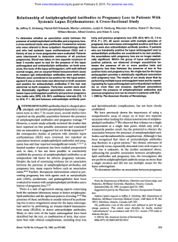
p53 ANTIBODIES IN VARIOUS CANCERS
FIGURE
5 - Western blot analysis using a mutant p53 protein as
antigen and human serum samples as sources of p53 antibodies.
Lane 1: Biotinylated m.w. markers visualized with streptavidinhorseradish peroxidase. A, phosphorylase (97.4 kDa); B, BSA
(68.0 kDa); C, ovalbumin (46.0 kDa); D, carbonic anhydrase (31.0
kDa); E, trypsin inhibitor (20.1 kDa); F, lysozyme (14.4 kDa).
Lanes 2-10: Mutant p53 protein extracted from the colorectal
carcinoma cell line COLO 320 HSR+ was separated on 4-20%
polyacrylamide gels and transferred to nitrocellulose. p53 was
then reacted with serum samples (lanes 2-9) or the specific rabbit
anti-p53 antibody CM-1 (lane 10). For more details see “Material
and Methods”. Lanes 2-4: Three different serum samples from
patients with ovarian cancer, positive for anti-p53 antibodies.
Lanes 5 and 6: Two different serum samples from patients with
colon cancer positive for anti-p53 antibodies. Lane 7: A serum
sample from a breast cancer patient positive for anti-p53 antibodies. Lanes 8 and 9: Two serum samples negative for anti-p53
antibodies. All sera were diluted 1,000-fold in 6% BSA solution
before probing. Lane 10: Probing with the CM-1 polyclonal
anti-p53 rabbit antiserum. The chemiluminescence generated by
horseradish peroxidase was captured on X-ray film (exposure 2-5
min) .
TABLE I1 - ASSOCIATION BETWEEN SERUM ANTLp53 ANTIBODIES AND
STEROID HORMONE RECEPTORS IN BREASTTUMORS
Receptor status’
ER + PR
ER
PR
E R - PR
ER
PR
[+I:
+
{-{+
I-1: f-1
p53 Antibody status
(+\
I-)
2 20%
8 [80%{
128 76%
40 i24%{
p value
<0.001
105 62%
23 [14%j
8 5%)
32 119%)
0.001
‘ER, PR, estrogen and progesterone receptors. Cut-off values
were < 10 fmolimg of total protein (Hassapoglidou et al., 1993).
antibodies could escape from the p53 antigen and pass
undetected during the 2 washing steps of the assay.
The specificity of the methodologies used was examined in 3
different experiments. (i) Some sera positive for p53 antibodies
were separated on an HPLC system using a gel filtration
column and fractions were analyzed for anti-p53 antibodies by
Methods A and B. The anti-p53 antibody-positive HPLC
fractions corresponded to a m.w. consistent with human
immunoglobulins (160-180 kDa, data not shown). (ii) Some
sera were passed through a protein A column, which is known
to bind only immunoglobulins. After elution, we tested the
eluate with Methods A and B and detected anti-p53 antibodies, further confirming that the measured moieties are human
immunoglobulins. (iii) We performed Western blot analysis
using a COLO 320 HSR+ cell lysate as a source of mutant p53
protein (Hassapoglidou et al., 1993). These data (Fig. 5) clearly
show that sera positive for p53 antibodies but not sera negative
for p53 antibodies react with a 53-kDa protein, which is also
visualized with a specific polyclonal anti-p53 antiserum (CM-1
antibody). These data taken together strongly suggest that
Methods A and B identify human immunoglobulins reacting
specifically with the p53 protein. Previous studies have established that the serum anti-p53 antibodies react with both
wild-type and mutant forms of p53 (Winter et a/., 1992;
Davidoff et a/., 1992; Schlichtholz et al., 1992).
From the 290 breast cancer patient sera analyzed, we had
estrogen and progesterone receptor data for 178. When we
classified the sera in groups, as shown in Table 11, we found
that the p53 antibody-positive sera were strongly associated
with estrogen and/or progesterone receptor-negative tumors
(p < 0.002). These findings are in agreement with the data of
Schlichtholz et al. (1992) and strongly suggest that tumors
eliciting antibody responses define a subgroup with poor
prognosis.
DISCUSSION
The prevalence of p53 antibodies in cancer patient sera has
only been occasionally studied, the cancer types examined
being few and the results qualitative (Crawford et a/., 1982;
Caron d e Fromentel et al., 1987; Winter et a/., 1992; Davidoff et
a/., 1992; Schlichtholz eta/., 1992). W e here report serum p53
antibody prevalence in a group of 1,392 cancer patients and in
230 sera from patients without malignancy or normal volunteers. Our studies were conducted using 2 new quantitative
methodologies based on different principles. Both methods
employ a detection methodology that uses alkaline phosphatase as label and time-resolved fluorometry with terbium
chelates. This detection method is among the most sensitive
reported and is suitable for measuring analytes at attomole
levels (Christopoulos and Diamandis, 1992; PapanastasiouDiamandi et al., 1992). Highest antibody prevalence was
obtained in sera from ovarian and colon cancer patients
(15-16%), 2 tumors that were not previously systematically
studied for p53-antibody generation. One report described the
presence of anti-p53 antibodies in 1 ovarian cancer patient
(Labrecque et a/., 1993). Antibody prevalence was 5 8 % in
patients with lung and breast tumors, in fair accordance with
previous reports (Crawford et al., 1982; Winter et al., 1992;
Davidoff et a/., 1992; Schlichtholz et al., 1992). We found
relatively low prevalence of anti-p53 antibodies (3-4%) in
patients with pancreatic and prostate cancer and in patients
with multiple myeloma or lymphoma. In patients with other
malignancies (hepatoma, melanoma, leukemia, Kaposi’s sarcoma and testicular carcinoma), p53-antibody prevalence was
similar to that of non-cancer patients ( < 2%).
Among the sera from the blood donor group (n = l50), we
found 1 sample that was positive by Method A only (Table I).
W e could not obtain any clinical information for this patient.
Among the 56 hospitalized patient sera, we found 1 sample
which was positive by both Methods A and B, with a p53antibody concentration of 4,000 U/L. This serum belonged to a
non-insulin-dependent diabetic who had undergone colectomy
and colestomy for complications of an abscess 3 years before
the serum sampling. No malignancy has as yet been diagnosed
in this patient.
In some patient sera, p53-antibody concentration was astronomical. These sera, which more frequently belonged to
patients with ovarian cancers, were used successfully to develop assays for the p53 antigen, as described elsewhere
(Hassapoglidou eta/., 1993). In these assays, the patient sera,
diluted 1,000- to 5,000-foId, could substitute successfully the
rabbit CM-1 anti-p53 antibody, developed by immunizing
rabbits with recombinant human p53 antigen (data not shown).
Thep.53 gene is mutated frequently in many cancers but the
reported frequencies are variable. The frequencies ofp53 gene
mutations of the cancers studied, as compiled from various
486
ANGELOPOULOU E T A L .
reports, are as follows: lung, 30-70% (Ozturk et al., 1992);
breast, 2046% (Hassapoglidou et al., 1993; Ozturk et al.,
1992); ovarian, 36-80% (Ozturk et al., 1992); colon, 20-69%
(Ozturk et at., 1992); prostate, 10-79% (Ozturk et al., 1992;
Van Veldhuizen et al., 1993); pancreatic, 40% (Ruggeri et al.,
1992); melanoma, 47435% (Stretch et al., 1991;Yamamoto and
Takahashi, 1993); multiple myeloma, 13% (Neri et al., 1993);
leukemia, 3% (Ozturk et al., 1992); lymphoma, 1 5 5 0 % (Ozturk et al., 1992). Tumors with high frequency of p53 gene
mutations appear to be associated with high prevalence of
serum p53 antibodies, but this does not appear to be the sole
contributing factor. Many tumors bearingp53 gene mutations
are not immunogenic (Winter et al., 1992; Davidoff et al., 1992;
Schlichtholz et al., 1992). Davidoff et al. (1992) have shown that
tumors which elicit an antibody response contain complexes
between heat shock protein 70 (HSP 70) and mutant p53.
Winter et al. (1992) have shown that only tumors withp53 gene
missense mutations are able to induce antibodies. No antibody
generation was observed in tumors bearing stop, splicelstop,
splice or frameshift mutations. Generally, it has been suggested that tumors bearing p53 gene mutations in exons 7-8
are not immunogenic, whereas those with mutations in exons
5-6 are (Winter et al., 1992; Davidoff et al., 1992). Although in
our study we did not examine the tumorp53 gene mutations in
patients with serum p53 antibodies, it is tempting to speculate
that the high incidence of p53 antibodies in patients with
ovarian cancer may be due to the frequent clustering of thep53
gene mutations in exon 6, as has been suggested by Teneriello
et al. (1993).
Schlichtholz et al. (1992) have shown that the p53-antibody
response may be a clinically useful indicator associated with
poor prognosis. Our finding that the p53 antibody-positive
breast tumors are associated with estrogen and progesterone
receptor-negative tumors supports this proposal (Table 11).
Because the assays used were quantitative, we were able to
monitor levels of p53 antibodies during the course of the
disease or during therapeutic manipulations in some patients
from whom we had serial samples. In these patients we have
shown that the temporal changes of p53 antibodies correlate
with disease progression or regression. This finding suggests
that the p53-antibody test may have some value for monitoring,
especially in cases where other tumor markers are normal.
It has been suggested that the appearance of anti-p53
antibodies in serum of cancer patients is a very early event,
independent of disease progression (Schlichtholz et al., 1992).
Combined with previous reports on antibody generation against
other oncogene products (Sorokine et al., 1991; Ben-Mabrec et
al., 1990) and the development of fast, quantitative and
economical methodologies for antibody measurements in serum, we propose the further exploitation of such testing for
cancer diagnosis in high-risk groups and for screening for
selected cancers. As we and others have shown, the diagnostic
specificity of the p53-antibody test is very high, approaching
100%. While the diagnostic sensitivity for cancer diagnosis is
only around 15% for ovarian and colon cancers, the test may
still be a viable alternative since the prevalence of these
cancers is relatively high and the proposed test is non-invasive.
Following the paradigm of p53, we are currently investigating
whether panel testing for antibodies against other oncogene
products will improve the diagnostic sensitivitywithout compromising specificity.
In conclusion, we here report prevalence of anti-p53 antibodies in over 1,600 patient sera and establish relatively high
positivity rates in ovarian and colon cancers. We also report
quantitative antibody concentration data and demonstrate
significant variability of antibody titers in various cancers and
association of very high antibody titers with ovarian cancer.
Furthermore, we show that antibody titers change with disease
progression or regression and that the test may have some
value in patient monitoring. We also confirm a previous report
of an association between the presence of anti-p53 antibodies
and other unfavorable prognostic indicators in breast cancer.
ACKNOWLEDGEMENTS
We thank Dr. D.P. Lane for the cell line producing PAb240,
Dr. N. Lassam for sera from melanoma patients and Dr. S.
Benchimol for helpful discussions. We also thank Ms. L.
Judisthir for secretarial assistance. This work was supported by
a grant to E.P. Diamandis from the Cancer Research Society
Inc., Montreal, Canada.
REFERENCES
ANGELOPOULOU,
K. and DIAMANDIS,
E.P., Quantification of antibodies against the p53 tumor suppressor gene product in the serum of
cancer patients. Cancer J., 6,315-321 (1993).
BEN-MABREC,
K., SOROKINE,
I., THIERRY,
D., KAWASUMI,
T., ISHII, S.,
SALMON,
R. and KOHIYAMA,
M., Circulating antibodies against c-myc
oncogene roduct in sera of colorectal cancer patients. Int. J. Cancer,
46,35-38 6990).
CARON DE FROMENTEL,
C., MAY-LEVIN,
F., MOURIESSE,
H., LEMERLE,
J., CHANDRASEKARAN,
K. and MAY, P., Presence of circulating
antibodies against cellular protein p53 in a notable pro ortion of
children with B-cell lymphoma. Int. J. Cancer, 39,185-189 (f987).
CHRISTOPOULOS,
T.K. and DIAMANDIS,
E.P., En matically amplified
time-resolved fluorescence immunoassay with texium chelates. Anal.
Chem., 64,342-346 (1992).
CRAWFORD,
L.V., Prw, D.C. and BULBROOK,
R.D., Detection of
antibodies against the cellular protein p53 in sera from patients with
breast cancer. Int. J. Cancer, 30,403408 (1982).
DAVIDOFF,
A.M., IGLEHART,
J.D. and MARKS,J.R., Immune response
to p53 is dependent upon p53 HSP70 complexes in breast cancers.
Proc. nat. Acad. Sci. (Wash.), 89,3439-3442 (1992).
DIAMANDIS,
E.P., Oncogenes and tumor suppressor genes: new biochemical tests. CRC Crit. Rev.din. Lab. Sci., 29,269-305 (1992).
FEARON,E.R. and VOGELSTEIN,
B., A genetic model for colorectal
tumorigenesis. Cell, 61,759-767 (1990).
HASSAPOGLIDOU,
S. and DIAMANDIS,
E.P., Antibodies to p53 tumor
suppressor gene product quantified in cancer patient serum with a
time-resolved immunofluorometric technique. Clin. Biochem., 25,
445-449 (1992).
HASSAPOGLIDOU,
S., DIAMANDIS,
E.P. and SUTHERLAND,
D.J.A.,
Quantification of p53 protein in tumor cell lines, breast tissue extracts
and serum with time-resolved immunofluorometry. Oncogene, 8,15011509 (1993).
LAEIRECQUE,
S., NAOR, N., THOMSON,D. and MATLASHEWSKI,
G.,
Analysis of the anti- 53 antibody response in cancer patients. Cancer
Res., 53,3468-3471 8993).
NERI, A., BALDINI,
L., TRECCA,
D., GRO,L., POLLI,E. and MAIOLO,
A.T., p53 gene mutations in multiple myeloma are associated with
advanced forms of malignancy. Blood, 81,128-135 (1993).
OZTURK,M., PONCHEL,
F. and PUISIEUX,
A., p53 as a potential target
in cancer therapy. Bone Marrow Transplant,, 9,164-169 (1992).
PAPANASTASIOU-DIAMANDI,
A., CHRISTOPOULOS,
T.K. and DIAMANDIS, E.P., Ultrasensitive thyrotropin immunoassay based on enzymatically amplified time-resolved fluorescence with a terbium chelate. Clin.
Chem., 38,545-548 (1992).
RUGGERI,
B., ZHANG,S.Y., CAAMANO,
J., DIRADO,M., FLYNN,
S.D.
and KLEIN-SZANTO,
A.J.P., Human pancreatic carcinomas and cell
lines reveal frequent and multiple alterations in the p53 and Rb-1
tumor-suppressor genes. Oncogene, 7,1503-1511 (1992).
SCHLICHTHOLZ,
B., LEGROS,V., GILLET,D., GAILLARD,
C., MARTY,
M., LANE,D., CALVO,F. and SOUSSI,T., The immune response to p53
p53 ANTIBODIES IN VARIOUS CANCERS
487
in breast cancer patients is directed against immunodominant epitopes A.L., Expression of mutant p53 in melanoma. Cancer Res., 51,
5976-5979 (1991).
unrelated to the mutational hot spot. Cancer Res., 52, 6380-6384
(1992).
TENERIELLO, M.G., EBINA,M., LINNOILA, R.I., HENRY,M., NASH,
SIDRANSKY,
D., TOKINO,T., HAMILTON,
S.R., KINDEN,K.W. and J.D., PARK,R.C. and BIRRER, M.J.,p.53 and Ki-ras ene mutations in
(1993).
VOGELSTEIN,
B., Identification of ras gene mutations in the stool of epithelial ovarian neoplasms. Cancer Res., 53,3103-!108
patients with curable colorectal tumors. Science, 256, 102-105 (1992). VAN VELDHUIZEN,
P.J., SADASIVAN,
R., GARCIA,F., AUSTENFELD,
R.L., Mutant p53 expression in prostate carciSIDRANSKY,
D., VON ESCHENBACH,
A., TSAI,Y.C., JONES,P., SUMMER- M.S. and STEPHENS,
HAYES,I., MARSHALL,
F., PAUL,M., GREEN,P., HAMILTON,
S.R., noma. Prostate, 22,23-30 (1993).
FROST,P. and VOGELSTEIN,
B., Identification of p53 gene mutations in WINTER,S.F., MINNA,J.D., JOHNSON,
B.E., TAKAHASHI,
T., GAZDAR,
bladder cancers and urine samples. Science, 252,706709 (1991).
A.F.and CARBONE,
D.P., Development of antibodies against p53 in
SOROKINE,
I., BEN-MABREC,
K., BRACONE,
A.,THIERRY,
D., ISHII,S., lung cancer patients appears to be dependent on the p53 mutation.
IMAMOTO,
F. and KOHIYAMA,
M., Presence of circulating anti-c-myb CancerRes., 52,4168-4174 (1992).
oncogene product antibodies in human sera. IntJ. Cancer, 47,665-669
YAMAMOTO,
M. and TAKAHASHI,
H., Immunohistochemical detection
(1991).
of the p53 oncoprotein in tumors of rnelanocytic origin. VirchowsArch.
STRETCH,
J.R., KEVIN,C.G., RALFKIAER,
E., LANE,D.P. and HARRIS, A Pathol. Anat. Histopathol., 422,127-132 (1993).

© Copyright 2026