
Phase I Clinical Trial Using Escalating Single-Dose
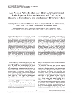
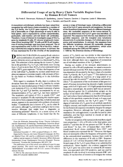
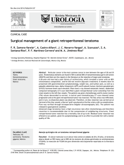
From www.bloodjournal.org by guest on February 6, 2015. For personal use only.
Phase I Clinical Trial Using Escalating Single-Dose Infusion
of Chimeric Anti-CD20 Monoclonal Antibody (IDEC-C2BS)
in Patients With Recurrent B-Cell Lymphoma
By D.G. Maloney, T.M. Liles, D.K. Czerwinski, C. Waldichuk, J. Rosenberg, A. Grillo-Lopez, and R. Levy
The B-cell antigen CD20 is expressed on normal B cells and
by nearly all B-cell lymphomas. This
nonmodulating antigen
provides an excellent target forantibody-directedtherapies.
A chimeric anti-CD20 antibody (IDEC-C2B8), consisting of
human IgG1-K constant regions and variable regions from
the murine monoclonal anti-CD20 antibody IDEC-2B8,has
been produced for clinical trials. It lyses CD20' cells in vitro
via complement and antibody-dependent cell-mediated lysis. Preclinical studies have shown that the chimeric antibody selectively depletesB cells in blood and lymph nodes
in macaque monkeys.In this phase I clinical trial, 15 patients
(3 per doselevel) with relapsed low-grade B-cell lymphoma
were treated with a single dose (IO, 50, 100. 250, or 500
mg/m2)of antibody administered intravenously. Treatmentrelated symptoms correlated with the number of circulating
CD20 cells and grade II events consistedof fever (5patients),
nausea (2), rigor (2). orthostatic hypotension (2). bronchospasm (l),and thrombocytopenia (1). No significant toxici-
ties were observed during the 3 months of follow-up. Serum
C3, IgG, and IgM levels, neutrophils, andT cells were largely
unchanged. At thethree higher dose levels, pharmacokinetics of the free antibody showed a serum half-life of 4.4 days
(range, 1.6 t o 10.5).Levels greaterthan 10 pg/mL persisted
in 6 of 9 patients for more than 14 days. No quantifiable
immune responses t o the infused antibody have been detected. CD20+ B cells were rapidly and specifically depleted
in the peripheral blood at 24 t o 72 hours and remained depleted for at least 2 to 3 months in most patients. Two-week
postinfusion tumor biopsies showed the chimeric antibody
bound t o tumor cells anda decrease in the percentage of B
cells. Tumor regressions occurredin 6 of 15 patients (2 partial and 4 minor responses). The results of this single-dose
trial have been used t o design a multiple-dose phase 1/11
study.
0 1994 by The American Societyof Hematology.
T
1 e ~ e l .Two
l ~ trials have been reported using anti-CD20 radioimmunoconjugates. Bone marrow ablative doses of I3'I-conjugated B1 (IgG2a murine MoAb anti-CD20) resulted in
complete remissions in 84% of patients.I4 The use of nonmarrow-ablative doses also resulted in partial and complete
remissions inthemajority of patient^.'^ In both of these
trials, antitumor effects were observed during the imaging
portion of the trials when trace doses of radiolabeled MoAbs
were infused with large amounts of unlabeled antibody, suggesting that the murine anti-CD20 MoAb itself may be contributing to the antitumor effect. Indeed, the relative contributions to the antitumor effect of the targeted radiotherapy,
of the cold antibody, and of the nonspecificwholebody
radiation delivered by the radioimmunoconjugates is difficult
to
Murine MoAbs have several potential limitations when
used in clinical trials. Genetic manipulation has made it possible to engineer chimeric antibodies with murine binding
HE TREATMENT OF patients with relapsed non-Hodgkin's lymphoma (NHL) remains a frustrating problem. More than 50% of patients with aggressive lymphomas
and the majority of patients with low-grade lymphomas are
not cured by current therapies. New treatments with different
mechanisms of action and toxicity profiles are needed. Previous work using patient-specific anti-idiotype monoclonal antibodies (MoAbs) has shown that NHLs are accessible to
intravenously infused antibody, and that tumor regressions,
including durable complete remissions, can be induced in
some patient^.'.^ With current technology, because of the
difficulty and time required to produce patient-specific antibodies, this approach is not feasible for general application.
In contrast, the antigen CD20, a 32-kD nonglycosylated
phosphoprotein present on the surface of nearly all B cells
provides a more universal target for imm~notherapy.~
CD20
is expressed on the surface of normal B cells from the time of
cytoplasmic pH chain expression throughout differentiation
until the antibody-secreting plasma cell stage. Importantly,
it is notexpressed on early pre-B cells, stem cells, or antigenpresenting dendritic reticulum cells5 More than 90% of Bcell NHLs express this surface protein."' It is also expressed
at a lower density on B-cell chronic lymphocytic leukemia.'
Although the function of this molecule isnot completely
defined, it may aggregate and function as a calcium channel.'
Antibodies binding to surface CD20 can induce a transmembrane signal" that can cause a variety of effects from cell
activation to blocking cell cycle progression and differentiation.".'2
The CD20 protein has multiple trans-membrane domains
and does not modulate from the cell surface in response
to antibody binding and thus provides an ideal target for
immunotherapeutic strategies not depending on internalization for their antitumor effect. Unconjugated murine MoAbs
to CD20 have been used for immunotherapy. A trial of the
1F5 murine IgG2a anti-CD20 MoAb in four patients showed
antitumor activity with minimal toxicity at the highest dose
Blood, Vol 84, No 8 (October 15). 1994 pp 2457-2466
From theDepartment of Medicine, Division of Oncology, Stanford
University Medical Center, Stanford, CA; and IDEC Pharmaceuticals, San Diego, CA.
Submitted February 9, 1994; accepted June IO, 1994.
Supported in part by Grant No. CA34233 from the US Public
Health Services, National Institutes of Health. D.G.M. is supported
by a Clinical Associate Physician Award from the General Clinical
Research Center. R.L. is an American Cancer Society Clinical Research Professor.
Address reprint requests to D.G. Maloney, MD, PhD, Stanford
University, Department of Medicine, Division of Oncology, SUMC
M207, Stanford, CA 943055306,
The publication costs of this article were defrayed in part by page
charge payment. This article must therefore be hereby marked
"advertisement" in accordance with 18 U.S.C. section 1734 solely to
indicate this fact.
0 1994 by The American Society of Hematology.
0006-4971/94/8408-0013$3.00/0
2457
From www.bloodjournal.org by guest on February 6, 2015. For personal use only.
2458
MALONEY ET AL
sites and humanconstant regions that havelower immunogenicity, longer half-life, and are able to lyse tumor cells using
human complement or antibody-dependentcell-mediated cytotoxicity (ADCC) effector cells more effectively than the
murine MoAb.'*-*'' Achimeric anti-CD20 antibody has been
producedthat contains theheavy and lightchainvariable
regions from a murine IgCl monoclonal anti-CD20
antibody
(IDEC-2B8) and human IgCl K constant regions." Stable,
high-level expressionwas obtained by transfection of the
relevant gene constructs into Chinese hamster ovary (CHO)
cells. In vitro studies showed similar binding characteristics
betweenthe
chimericand murine anti-CD20antibodies;
however, the chimeric antibody was ableto lyse CD20+
B cells using human complement or human effector cells
(ADCC) 1,000-fold more effectively than the murine antibody. Preclinical studies in macaque cynomolgus monkeys
haveshownthatrepeated
doses of thechimericantibody
depletedaround 80% of CD20' B cells intheperipheral
blood, lymph nodes, spleen, and bone marrow, with gradual
recovery over a period of several months." No toxicity was
observed in these studies. We describe here the first phase
1clinical trial of single-dose infusion with the chimeric antiCD20 antibody (IDEC-C2B8) in patients with relapsed Bcell NHL.
MATERIALS AND METHODS
Chimeric monoclonal anti-CD20 antibody. The chimeric monoclonalanti-CD20antibody(IDEC-C2B8)
has beenproducedand
provided for clinical trialsby IDEC Pharmaceuticals CO (San Diego,
CA) and supplied under an Investigational New Drug Application.
Protocol design. This was a phase I clinical trial of single-dose
IDEC-C2B8chimericanti-CD20MoAbadministeredtopatients
with relapsed B-cell NHL. Detailed informed consent was obtained
from all patients in accordance with the human subjects institutional
review board of Stanford University Medical Center. Three patients
were treated at each dose level
with a single intravenous infusion
of IO, 50, 100,250,or 500 mglm' of MoAb. Patients were evaluated
for infusional related toxicity and effect on peripheral blood B cells,
T cells, neutrophils and platelets, serum chemistries, Ig, and complement levels. In patients treated at the upper three doses, tumor biopsies were obtained 2 weeks after treatment and examined for evidence of antibody binding and B- and T-cell content. All patients
were evaluated for antitumor activity.
Patient selection. On entry to the study, patients were required
to have relapsed NHL with measurable disease after at least one
prior course of standard therapy. A tumor biopsy was performed to
document tumor cell expression of the CD20 antigen and reactivity
withIDEC-2B8orIDEC-C2B8antibodiesusing
flow cytometry.
In addition, baseline hematologic function (1,500 granulocytes and
50,000 platelets/pL), renal function (serum creatinine of <2.5 mg/
dL), quantitative serum IgG of greater than 600 mg/dL, a negative
serology to human immunodeficiency virus, a negative hepatitis B
surface antigen, and a life expectancy of at least 3 months without
to
otherseriousillnesswasrequired.Patientspreviouslyexposed
murine antibodies were required to have no evidence of a pretreatment human antimurine antibody immune response (HAMA).
Flow cytometty. CD20antigenexpression was determinedon
all cases before antibody treatment by flow cytometry of fresh or
cryopreserved tumor cell suspensions. Tumor cells were obtained
from excisional biopsies or from fine needle tumor aspirations and
stained for CD20 expression with fluorescein isothiocyanate
(FIT0
conjugated IDEC-2B8 or IDEC-C2B8 (IDEC Pharmaceuticals) and
Leu-16. an independent anti-CD20 antibody (Becton Dickinson. San
Jose, CA). Tumor cells were also analyzed for expression of surfacc
Ig light chains[FITC-goatF(ab)?-antihuman
K or X; T a p . Burlingame,CA).CD19,CD4,CD3,CD8
(FITC- orphycoerythrin
[PE]-conjugated Leul2, L e d , Leu4, and Leu2; Becton Dickinson).
and CD37 (MBI clone 6A4). Peripheral
blood samples were anaIyLed for the number of cells expressing the CD20 antigen using
two-color flow cytometry using PE or FITC conjugates of the above
reagents
Two-week posttreatment tumor biopsies were also evaluated for
B-andT-cellcontentusingthesamereagentsdescribedabove.
Antibodyboundtotumorcellsfrom
in vivoadministration was
detected by acombination of twodifferentmethods.
In the first
method, cells were stained using FITC-labeled anti-CD20 antibodies.
The presence of the unlabeled antibody blocked the binding of the
labeled antibody, resulting in decreased immunostaining of the Bcell tumor population (as
identified using antibodies to additional
B-cell antigens CD19, CD37, IgM, IgG, K or X). Second, the bound
chimeric antibody was detected directly by looking for IgM K - or
X-positive tumorcells now bearingthehuman
IgG ( K ) constant
regions of the chimeric antibody (IDEC-C2B8) using an FITC-laheled goat F(ab)> antihuman IgG y-chain-specific reagent (Tago).
An estimate of the percentage of tumorcells with the chimeric
antibodyattachedwasobtained
by comparingthestaining of the
pretreatment and the posttreatment biopsies for human IgG constant
regions.
IUEC-C2BSpharmacokinetic.s. Serum levels of the chimeric antibody were determined using anenzyme-linkedimmunosorbent
assay (ELISA). Microtiter plates were coated with a purified polyclonal goat anti-IDEC C2B8 idiotype antiserum. After washing and
blocking, posttreatment sera were serially diluted. Bound human
IgG
was then detected using a horseradish peroxidase (HRP)-conjugated
polyclonal antihuman IgG reagent, and the plates developed with the
substrate 2,2-a~inobis(3-ethylbenzthiazoline
sulfonic acid) (ABTS).
Antibody concentration was determined by comparison of the signal
from the patients sera with that obtained from known concentrations
of purified chimeric antibody diluted into normal human serum.
Measurement I$ host anti-IDEC C2B8 antibody response. Posttreatment sera from evaluations at l,2, and 3 months were analyzed
for evidence of a host antichimeric antibody immune response using
a sandwich ELISA with microtiter plates coated with IDEC C2B8,
the murine antibody 2B8, or normal murine IgG. Dilution's of the
patients sera were added and, after washing, detected with biotinlabeled IDECC2B8followed
by Avidin-HRPand the substrate
ABTS. This assay has a level of quantification of 5 pg/mL.
Studv measurements. Patientswereevaluatedforinfusional
related toxicity using the National Cancer Institute's Common Toxicity
Criteria.Hematologic,renal,andhepaticfunctionwasmonitored
before and after infusion and duringmonthly intervals after therapy.
Sera for evaluation of antibody levels and pharmacokinetics, serum
IgG and IgM levels, and CD20 expression on peripheral blood B
cells was obtained at eachfollow-upvisit.Tumor
response was
assessed by evaluation of tumor measurements fromphysical examination and from radiologic imaging studies. For 3 months after therapy, patients were evaluated at monthly intervals and then followed
at 1- to 3-month intervals until disease progression was observed.
A complete remission (CR) required complete resolution of all detectabledisease.A partial remission(PR)requiredagreater
than
50% reduction in measurable disease persisting more than 30 days.
A minor response (MR) was defined as a 25% to 50% reduction in
disease. Stable disease (SD) was defined as no significant change in
tumor measurements without progression over the
period of observa-
From www.bloodjournal.org by guest on February 6, 2015. For personal use only.
ANTI-CD20 ANTIBODY THERAPY FOR B-CELL LYMPHOMA
2459
Table 1. Patient Characteristics
Patient No./Sex/Age
FM
001lMl53
FSC
002/M/55
003N149
Dose
(mad)
Tumor Histology
Stage"
111
10
111
10
10
DlLD (mantle zonel
MACOP-B
Prior Therapy
Splenectomy
C-MOPP
m-BACOD
CHOP
XRT (total nodal)
CVP
Chl
IV
Disease Bulkt
Maximal
Response
+++
Leukemia
+
Delayed PR?
Leukemia
Splenomegaly
++
Chl
Anti-Id (MoAb)
CVP
004N163
DLCFSC 50
FSC
005N161
50
006F159
50
007F158
100
IV
FM
111
SL
IV
DLC, monocytoid B cell
IV
008lMi73
100
FM
IV
009lMl65
100
FM
IV
0 1OlMI38
250
FSC (diffuse areas)
IV
0111M146
250
DLID (mantle zone)
IV
CVP
Id-Vac
ProMACEICytaBOM
XRT
anti-Lym-l (MoAb)
ChMP
CVP
CVP
CVP
CVPIBVP
MACOP-B
XRT
XRT
Chl
anti-CD4 (MoAb)
ChlIP
Splenectomy
Chl
Velban
CVP
Anti-Id (IFN)
Anti-Id (Chll
MINWSHAP
9
~
~
1
CVP
+
Mixed
++
+++
+
PR
Splenomegaly
++
MR
++
Mixed
++
Splenomegaly
+++
MR
Splenomegaly
0 121F152
250
FSC
013lMl48
500
FM
0 14Fl65 FM
500
015ffE.8 FSC
500
MR
CHOP
IV
CHOPIXRT
C hIIP
Chl/P
VACOP-B
Fludarabine
IFN
IVb
CHOP
DHAP
CVP
gOy-Bl
IV
IV
Ctx
ProMACENOPP
XRT
IFN
+++
+
+
MR
+
PR
Abbreviations: FSC, follicular small cleaved cell; FM, follicular mixed small and large cell; DLID, diffuse lymphoma intermediate differentiation; DLC, diffuse large
cell; SL, small lymphocytic; XRT, radiation therapy; IFN, interferon a;ProMACElMOPP, etoposide, cyclophosphamide, adriamycin, methotrexate, prednisone, nitrogen
mustard, vincristine, and procarbazine; ProMACHCytaBOM. cyclophosphamide, adriamycin, etoposide, prednisone, cytarabine, bleomycin, vincristine, and methotrexate; Ctx, cyclophosphamide; CHOP, cyclophosphamide. adriamycin, vincristine, and prednisone; CVP, cyclophosphamide, vincristine, and prednisone; 9 - 6 1 . ytriumlabeled B1 (antLCD20MoAb); DHAP, cisplatin, cytarabine, and decadron; VACOP-B, VP-16, adriamycin. cyclophosphamide, vincristine, prednisone, and bleomycin; Chll
P, chlorambucil and prednisone; MINE-ESHAP, ifosphamide, novantrone, etoposide, platinum, and cytarabine; Anti-Id, anti-idiotype MoAb; Id-Vac, ldiotype vaccination;
BVP, bleomycin, vincristine, and prednisone; Mixed, regression noted in some but not a11 areas.
Clinical stage at disease diagnosis.
t Tumor bulk estimated from physical exam and CT scans of thechest, abdomen, and pelvis and graded as follows: multiple areas of adenopathy with largest mass
<5 cm (+), nodal mass >5 cm (++), extensive disease with multiple areas 2 5 cm ( + + + h
From www.bloodjournal.org by guest on February 6, 2015. For personal use only.
2460
MALONEY ET AL
Table 2. Infusional-Related Symptoms During Chimeric Anti-CD20
Antibody Therapy
Patient
Dose
Dose given
No.
(mglm')
(mg)
Pre Rx CD20
(cells/@L)
00 1
10
16
1,630 (tumor)
002
10
22
960 (tumor)
003
004
005
006
007
008
009
010
01 1
012
013
10
50
50
50
100
100
100
250
250
250
500
21
100
100
90
180
188
1,200
50
60
40
140
130
50
580 (normal
90
130
20
60
014
015
500
500
965
760
200
520
575
400
130
160
Toxicity
B)
Fever, rigor,
bronchospasm
Fever, rigor,
nausea
None
Fever
Fever, chill
Myalgia
Fever
Fever
Fever, rigor
Chill
Fever, headache
Fever
Fever orthostatic
hypot. nausea
Fever, nausea
Fever, nausea,
headache
lymphoma. One patient had an initial diagnosis of a diffuse
large-cell lymphoma; however, a relapsed node biopsy
showed a monocytoid B-cell histology. Two patients with
an initial diagnosis of a low-grade lymphoma had histologic
progression on tumor biopsies performed before antibody
therapy (1 follicular large cell and 1 diffuse large cell). The
majority of patients (12/15) presented with stage IV disease.
Patients had lived with their disease a mean of 5 years (range,
1.7 to 9.5) at the time of antibody therapy. All patients had
measurable progressive disease and had received a median
of two prior regimens (range, 1 to 5 ) of conventional therapy
within the past 1.5 years. Five patients had previously been
treated with murine MoAbs, including 2 patients who had
received radiolabeled anti-CD20 antibody therapy ( I with a
short PR and 1 with no response). All patients previously
exposed to murine antibodies were HAMA-negative at the
time of treatment.
Infusional-related toxicity. All patients completed the
planned antibody infusion. Infusional-related symptoms are
detailed in Table 2. The most frequent side effect was lowgrade fever, which was seen in 13 of 15 patients (grade I1
in 5/15). Three patients additionally developed rigors and 1
developed bronchospasm requiring transient administration
of supplemental oxygen. One patient on multiple medications for high blood pressure developed orthostatic hypotension during the antibody administration. In all patients, the
antibody infusion was temporarily discontinued when significant side effects were observed. If necessary, the patients
were treated as indicated with diphenhydramine and acetaminophen. Antibody infusions were usually restarted within
30 to 45 minutes at 50 to 100 mg/h and then escalated as
tolerated to 200 m@. No significant further toxicity except
fever was observed in any patients during the remainder of
the antibody infusion. Three of the 4 patients having more
significant reactions were also noted to have thehighest
levels of pretreatment CD20' cells in the peripheral blood
(Table 2). Interestingly, these cells could be malignant (patients no. I and2) or normal (mixed K or h phenotype, as
observed in patient no. 9).
Effect on circulating B cells. In all patients, the numbers
of B cells present in the peripheral blood before and after
anti-CD20 therapy were analyzed using two-color flow cytometry. B cells were identifiedusing the B-cell antigens
CD19 and surface Ig, neither of whichisblocked by the
binding of the chimeric antibody to CD20. Bound antibody
from in vivo administration was identified on the B cells by
finding human IgC or K bound to B cells coexpressing IgM
and h, or by the blocking of binding of a directly labeled
anti-CD20 antibody. There was a dose-dependent. rapid, and
specific depletion of the B cells in all patients, especially
those receiving doses of morethan 100 mg. In allbut 1
patient receiving the higher doses (>50 mg/m2),these depletions persisted for I to greater than 3 months. The specificity
of the B-cell depletion in a patient receiving 100 mg/m2 is
shown in Fig 1. After intravenous administration of 180 mg
of antibody, there was a rapid and complete disappearance
of B cells expressing CD20, CD1 9, K , or h surface antigens.
There was no effect on the number of T cells (CD3), and
the decrease in the numbers of lymphocytes is accounted for
entirely by the decrease in the numbers of B cells. Peripheral
blood B-cell levels for the duration of the 3-month study are
shown on all 15 patients in Table 3 . Three patients had
circulating tumor cells, as shown by a clonal expansion of
cells containing the same heavy and light chain isotype as
identified on cells of their lymphomas obtained from tumor
biopsies. Three patients had no detectable peripheral blood
B cells at the 3-month evaluation. Follow-up is available
50
0
0
20
40
80
Days since Rx
Fig 1. Depletion of peripheralblood B cells after 100 mg/mz chimeric anti-CD20 antibody. Patient no. 007 was treated with 100 m g l
m* of chimeric anti-CD20 administered intravenously on day 1. Peripheral blood mononuclear cells were analyzed by flow cytometry
for the expression of B- and T-cell antigens. There was a rapid and
specific depletion of B cells, as indicated by the loss of cells expressing K or A light chains aswell as the &cell antigens CD19 and CD20.
(m) Lym; IO) CD19; (AI CD20; (0)
CD3; (A) K; IO) L.
From www.bloodjournal.org by guest on February 6, 2015. For personal use only.
ANTI-CD20 ANTIBODY THERAPY FOR B-CELL LYMPHOMA
2461
Table 3. Dwletion of B Cells From the Blood After Infusion of Chimeric Anti-CD20Antibody
B-Cell Depletion Post-Rx CD19
Dose
Patient No.
(mglm’l
00 1
002
1,870
003
004
,150
20
1
Pre Rx CD19
(cells/pL)
10
1,250
10
10
50
005
006
50
50
007
008
009
010
011
012
013
014
015
100
100
100
250
250
250
500
500
500
3
7
1,940
2,240
60
60
50
EO
140
40
600
90
140
d3
995
10
5
6
4
130
30
10
1 mo
2 mo
3 mo
1,260
ND
ND
ND
10
5 ND
1
80
20
1
9
0
165
3
5
10
1 wk
1
180
75
6
50
8
60
130
160
290
ND
90
40
8
110
60
10
6
130
0
ND
10
120
0
120
3
0
0
0
6
0
0
20
70
0
0
5
20
180
ND
ND
4
7
0
Abbreviation: ND, not determined.
for 1 patient showing a return of B cells 7 months after
treatment.
Serum antibody pharmacokinetics. Serum antibody was
detected in all patients immediately after the intravenous
infusion. Serum levels following 100, 250, and 500 mg/m‘
infusions are shown in Fig 2. In the 9 patients receiving 100
mg/m2 or greater, the mean half-life of the antibody was 4.4
days, with a range from 1.6 to 10.5 days. Antibody levels
of greater than 10 pg/mL persisted in the serum of 6 of 9
patients for more than 14 days. In 3 of 6 patients treated
with 250 mg/m’ or 500 mg/m2, antibody levels were detected
1 month after antibody therapy. In one case, the antibody
half-life was 10.5 days and serum antibody, capable of binding to pretreatment tumor cells was present more than 1
month after treatment. One patient (patient no. 011) treated
at the 250 mg/m’ dose had a large tumor burden with splenomegaly and a short half-life of the chimeric anti-CD20 antibody (1.6 days).
IO00
Detection of host anti-IDEC-C2B8 immune response.
Sera were analyzed at monthly intervals for the 3-month
duration of the trial for evidence of an immune response.
No quantifiable antibody responses were detected against the
chimeric antibody, with a level of quantification of 5 pg/
d.
Subsequent analysis of serum from 7 patients at various
intervals to 1 year of follow-up were also negative.
Analysis of posttreatment tumor biopsyspecimens.
In
patients receiving 100 mg/m’ or greater doses of the chimeric
antibody, excisional tumor biopsies were performed 2 weeks
after antibody therapy. Tissue sections of these biopsies were
examined for histopathology and cell suspensions examined
using two-color flow cytometry to determine cellular composition and to look for evidence of the chimeric antibody
bound to the tumor cells from the in vivo administration.
Successful lymph node biopsies were obtained in 7 of these
9 patients. In 1 patient, a large node was resected; however,
histologic examination showed necrosis without any viable
.
op;o
op11
E
Sm 100
100
P
Fig 2. Pharmacokinetics of
chimeric anti-CD20 antibody in
patients receiving 100. 250,or
500 mg/m2 infusions. Serum
from 9 patients treated with a
singleinfusionof
the chimeric
anti-CD20 antibody were analyzed forIDEC-C2B8 by ELISA.
Results from each patient
treated at 100 mg/mz (A), 250
mglm’ (B), and 500 mg/m2 (C)
are shown.
-@
1000
._
E
v
m
m
g
10
10
2
0
0 5 10 15 20 25 30 35
A
100mg/m2
0
B
5 10 15 20 25 30 35
Time from infusion (days)
250 mg/m2
0
C
5 10 15 20 25 30 35
500 mg/rn2
From www.bloodjournal.org by guest on February 6, 2015. For personal use only.
MALONEY ET AL
2462
Table 4. Summary of Pretreatment and 2-Week Posttreatment Tumor Biopsies
Analysis by Flow Cytometry
Patient No.
Dose (mglm’)
007
100
Date of Bx
10/9/90
009
5/10/93
3/24/92
5/12/93
9/16/92
010
5/17/93
6/4/87
01 1
5/19/93
1/92
ooa
012
013
014
015
5/26/93
511 1/93 fna
6/1/93
10/28/92
6/1/93
511 1/93
6/9/93
6/7/93 fna
6/22/93
Lymphoma Histology
% B (CD37)
Monocytoid
21 B
79
F&DMX
FLC
No tumor
FM
Necrosis
FLC
FM
SLID
SLID
54
-
FM19
FM
FM 35
FSC
FSC23
-
FSC
% T C268
(CD3)
on Tumor Cells?*
-
43
Yes (72)
53
43
-
-
-
a0
12
-
-
63
51
79
a3
97
77
57
34
85
26
44
20
16
5
52
a3
60
-
Yes (30)
No
-
Yes (41)
27
-
Yes (83)
12
-
Yes (100)
15
40
-
Yes (93)
Abbreviations: fna, cells obtained by fine needle aspiration; FM, follicular mixed small and large cell; FLC, follicular large cell; FSC, follicular
small cleaved cell; SLID, small lymphocytic intermediate differentiation (mantle cell); F&DMX, follicular and diffuse mixed small and large cell.
* Expressed as the percentage (shown in parentheses) of lymphoma cells in the biopsy specimen that stained positive for the expression of
human IgG constant regions.
tumor. Flow cytometry also failed to detect any viable cells.
A second patient had large bilateral inguinal lymph nodes
present before therapy, but attempted surgical excisions in
both inguinal regions after treatment failed to find anylymph
nodes for biopsy. In the remaining 7 patients, lymph node
material was available for analysis. Table 4 details the histologic findings present in the 2-week posttreatment tumor
biopsies and compares the B- and T-cell content as determined by flow cytometry with tumor biopsies obtained before antibody therapy. In 3 patients, tumor biopsies were
obtained immediately before treatment, and in the remaining
patients comparisons were made to earlier cryopreserved cell
suspensions obtained during the patient’s course (from 8
months to 6 years earlier). Histologic examinations of the
posttreatment samples remained diagnostic of lymphoma in
the 7 patients. In 1 patient, the histologic appearance of
the posttreatment tumor biopsy identified large numbers of
hemosiderin-laden macrophages clogging the sinusoids of
the lymph node. When the posttreatment samples were compared with the earlier biopsies, there appeared to be a decrease in the percentage of B cells (as determined by the
expression of CD37 and CD19, both independent B-cell antigens not blocked by anti-CD20 antibodies) and a corresponding increase in the percentage of T cells seen in all but 1
patient. In addition, chimeric antibody was identified on the
surface of the tumor cell population in the 2-week posttreatment biopsy in all but one case. In some cases, the bound
antibody nearly completely saturated the available CD20
binding sites.
Effect on serum IgG, IgM, serum complement, and platelets. Sequential quantitative serum Ig levels were obtained
pretreatment and monthly during the 3-month follow-up period. As shown in Fig 3, there was no significant change in
the serum IgG (Fig 3A) or IgM (Fig 3B) levels over this
period. IgA levels also were unchanged (data not shown).
The platelet count was largely unaffected by the administration of antibody. One patient who started with a low pretreatment level developed moderate thrombocytopenia (Fig 3C).
Two patients developed transient decreases in serum complement (C3) at 24 hours after therapy (Fig 3D).
Clinical antitumor effect. Despite the fact that this trial
involved the administration of only a single infusion of antibody, tumor responses were observed. Partial tumor responses were documented in 2 patients and minor responses
observed in 4 others. An example of a partial remission is
shown in Fig 4. Patient no. 015, who had a follicular small
cleaved cell lymphoma previously treated with ProMACE/
MOPP, radiation therapy, and interferon was treated with
500 mg/m2 of antibody. Computed tomography (CT) images
of an abdominal mass pretherapy (Fig 4A and B) and 3
months posttherapy (Fig 4C and D) are shown. This tumor
mass substantially decreased after treatment, and other nodes
on CT scan and ultrasound examination as well as physical
examination disappeared or significantly decreased in size.
Disease progression was documented 8 months posttherapy,
with recurrence of axillary adenopathy. A second patient
treated with 100 mg/m’ had a greater than 50% decrease in
cervical adenopathy as well as resolution of splenomegaly
(CT scan) lasting 9 months posttherapy. Patient no. 002, with
follicular small cleaved cell lymphoma previously treated 10
years earlier with total nodal radiation and multiple courses
of alkylating agents, was found on pretreatment evaluation
to have circulating lymphoma cells and persistent thrombo-
From www.bloodjournal.org by guest on February 6, 2015. For personal use only.
2463
ANTI-CD20 ANTIBODY THERAPY FOR B-CELL LYMPHOMA
10 rnglrn2
3500 7
“4
0 250
100
Pre
4wk
mg/m2
A 500mg/m2
3 mo
2 mo
A
600 140 2
500-
-
0
8
4004
.
7
E
Q 300-
W
G
a
roo<
80-
60;
2001
5
&
,
W
407
m
m 20 7
1002
100
0:
I
Pre
I
I
I
I
post 1 day 3 day 1 wk wk2
I
I
l
I
0
0
Pre
1 mo 2 m0 3 m0
C
post
1 day
3 day
D
Fig 3. Effect of anti-CD20 antibody therapy on platelets, IgG, IgM, and serum complement Levels. After therapy with a single infusion of
chimeric anti-CD20 antibody patients were monitored for the effect on serum IgG (A), IgM (B), platelets (C), and serum complement-C3 (D).
cytopenia. He was treated with 10 mg/m’ antibody. Throughout the 3 months of follow-up there was no significant
change in disease measurement or thrombocytopenia. However, evaluation at 7 months showed a significant reduction
in disease (lymph nodes and splenomegaly), resolution of
thrombocytopenia, and disappearance of tumor cells from
the peripheral blood lasting more than 1 year from therapy.
Although no other lymphoma therapy had been administered, it is impossible to know whether the clinical improvement in this patient was spontaneous or was indeed related
to the antibody treatment. Two patients had attempted excisional biopsies of known disease sites that showed necrosis
in one instance and no tumor in a second case. Both patients
had evidence of tumor regression with a mixed response and
a minor response (25% to 50% decrease in measured lesions), the latter lasting l l months. Shorter minor responses
were also observed in 2 other patients. Additional mixed
responses were observed in several patients. One patient with
a history of low-grade lymphoma with transformation in
bone to diffuse large-cell lymphoma had complete resolution
of peripheral disease (last biopsy follicular mixed
lymphoma), but progressed 2 months after therapy with relapsed large cell lymphoma again involving bone. He received local radiation therapy to the involved bone, but remains in remission of his peripheral disease.
DISCUSSION
In this phase I clinical trial, patients with relapsed NHL
received a single infusion of chimeric anti-CD20 MoAb
IDEC-C2BSin doses ranging from 10 to 500 mg/m’.All
patients received the planned dose and no dose-limiting toxicities were identified. Symptoms were mild to moderate and
easily manageable and more commonly observed in the 3
patients with higher numbers of CD20 antigen-bearing B
cells (normal or malignant) present in the peripheral blood,
suggesting that the destruction or removal of these cells
during the early portions of the antibody infusion may contribute to the adverse events observed.
Analysis of antibody pharmacokinetics in patients receiving doses of 100 mg/m2 or greater showed a mean serum
half-life of 4.4 days, with a range from 1.7 to 10.5 days.
However, it is difficult to establish the half-life of antibody
in patients with widely different degrees of tumor burden
receiving a single nonsaturating dose of antibody. It is likely
that the true half-life will be longer once sufficient antibody
is administered to saturate all tumor and normal CD20 antigenic sites. In the majority of patients receiving these doses,
levels of greater than 10 &mL were present in the serum
2 weeks after therapy. Lower levels were identified in patients with extensive disease. Two-week postinfusion lymph
From www.bloodjournal.org by guest on February 6, 2015. For personal use only.
2464
MALONEY ET AL
Fig 4. Example of antitumor effect. Patient no. 015 with relapsedfollicularsmallcleavedcell
lymphoma was treated with 500 mglm’
chimeric anti-CD20 antibody. CT images of an abdominal mass from two contiguous images are shown in (A) and (B).The corresponding
images from a scan performed 3 months after antibody therapy are shown in (C) and (D), demonstrating partial regression ofthe tumor.
node biopsies were performed in the 9 patients receiving
doses of greater than 1 0 0 mg/m’. In 7 of the 9 biopsies.
tumor was identified on pathologic examination. However,
in 2 patients, tumor was not identified; in 1 case a large node
that had become smaller after therapy was necrotic and in a
second case inguinal lymph nodes presentbefore therapy
were not found after surgical exploration of leftandright
inguinal regions. In some of the biopsies. an increased infiltrate of macrophages was observed. Analysis of the posttreatment lymph nodes by flow cytometry identified tumor cells
coated with nonsaturating amounts of the chimeric antibody
in 6 of the 7 cases. The single exception was a patient with
a large tumor burden, splenomegaly, and a short serum halflife of the administered antibody. In the majority of cases.
a general decrease in the percentage of B cells andan increase in the percentage of T cells was observed when comparing pretreatment and posttreatment biopsies.
Treatment caused a selective elimination of the peripheral
CD20-expressing B cells in all but I patient receiving doses
of 100 mg/m’ or greater. This was observed 24 to 72 hours
after MoAb infusion. Over the 3-month follow-up phase,
peripheral blood B cells slowly returned to base line in patients treated with the lower doses and partially in patients
receiving the higher doses. There was no apparent effect on
the serum IgG, IgM, or IgA levels through 3 months of
follow-up. The CD20 antigen is not expressed on the early
B-cell precursors or on the antibody-secreting plasma cell.
The compartment of B cells/plasma cells is much larger than
the few cells observed in the peripheralblood,and
it is
possible that they were not effected by the single dose of
antibody. In addition, the long half-life of serum IgGmay
mean that late effects on serum IgG will be observed. There
did not appear to be any increasedincidence of opportunistic
infections in this group of patients during the 3 months of
follow-up. Depletion of normal B cells iscommon after
high-dose chemotherapy and autologous bone marrow reconstitution that slowly resolves 3 to 6 months after transplant.”.” Depression of Ig levels can be treatedby Ig transfusions.” It is possible that longer duration of antibody therapy
achieving saturating levels may also cause a greater antitumor effect and a greater effect on the normal B cells and,
ultimately, serum Ig.
The mechanism of the antibody-induced antitumor effect
isnot clear. Serum complement levels (C3) wereslightly
changed in only 2 patients during therapy. The chimeric
antibody is capable of lysing target tumor cell lines in vitro
by complement and antibody-dependent cell-mediated lysis.” In addition, direct effects of antibody binding to the
From www.bloodjournal.org by guest on February 6, 2015. For personal use only.
ANTI-CD20 ANTIBODY THERAPY FOR E-CELL LYMPHOMA
CD20 antigen, including growth inhibition, have been reported.'* It is likely that a combination of these mechanisms
is involved in the tumor regressions observed in these patients. Studies using murine anti-CD20 antibodies have also
noted antitumor effect^.'^ Two recent reports detailing the
use of radiolabeled anti-CD20 antibodies describe impressive clinical activity with complete or partial remissions in
the majority of patient^.'^.'' Both also note tumor regressions
associated with the imaging portion of the studies, suggesting
clinical activity of the murine antibody.
The use of a chimeric naked antibody offers some advantages over similar trials using toxin-conjugated or radiolabeled antibodies against B-cell NHL. The antibody preparation is used directly for therapy, not requiring conjugation
to drugs, toxins, or radiolabels, each of which requires extensive safety testing and may not be stable after formation of
the active conjugate. Antibody modification may interfere
with antigen binding. Radioiodinated antibodies are unstable, and undergo autolysis, and it is technically difficult to
obtain consistent conjugates for large-scale clinical trials. In
addition, significant hematologic toxicity is associated with
the use of high-dose radiolabeled conjugates, making the
application of this approach difficult in patients with impaired bone marrow function or significant involvement by
lymphoma. In some studies, immunotoxin conjugates have
been associated with significant toxicities.= In contrast, this
chimeric anti-CD20 antibody is stable and has been engineered to lyse tumor cells through interaction with the patient's own immune system.
The modest tumor responses observed in this trial occurred after the administration of a single infusion of the
chimeric antibody. Extension of these studies using multiple
doses to achieve prolonged, tumor-saturating levels may lead
to responses in patients with more extensive disease. Ultimately, extension of these studies to patients with minimal
residual disease, using antibody alone or in combination with
conventional therapies, may provide the greatest benefit.
Based on these observations of safety and tumor responses
to a single infusion of this chimeric anti-CD20 MoAb, a
phase I/II trial using four weekly doses of antibody in patients with relapsed B-cell NHL has been initiated.
REFERENCES
1. Brown SL, Miller RA, Homing SJ, Czerwinski D, Hart SM,
McElderry R, Basham T, Warnke RA, Merigan TC, Levy R:
Treatment of B-celllymphomas with anti-idiotypeantibodies
alone and in combination with alphainterferon. Blood 73:651,
1989
2. Maloney DG, Brown S, Czerwinski DK, Liles TM, Hart SM,
Miller RA, Levy R: Monoclonal anti-idiotype antibody therapy of
B-cell lymphoma: The addition of a short courseof chemotherapy
does not interfere with theantitumoreffect
nor prevent the
emergence of idiotype-negative variant cells. Blood 80:1502,
1992
3 . Meeker TC, LowderJ, Maloney DG, Miller RA, Thielemans
K, Warnke R, Levy R: A clinical trial of anti-idiotype therapy
for B cell malignancy. Blood 65:1349, 1985
2465
4. Stashenko P, Nadler LM, Hardy R, Schlossman SF: Characterization of a human B lymphocyte-specific antigen. J Immunol
125:1678,1980
5. Schriever F, Freedman AS, Freeman G, Messner E, Lee G,
Daley J, Nadler LM: Isolated human folliculardendriticcells
display a unique antigenicphenotype.JExp
Med 169:2043,
1989
6. Anderson KC, Bates MP, Slaughenhoupt BL, Pinkus CS,
Schlossman SF, Nadler LM: Expression of human B cell-associated antigens on leukemias and lymphomas: A model of human
B cell differentiation. Blood 63: 1424, 1984
7. Nadler LM, Ritz J, Hardy R, Pesando JM, Schlossman SF,
Stashenko P: A unique cell surface antigen identifying lymphoid
malignancies of B cell origin. J Clin Invest 67:134, 1981
8. Almasri NM, Duque RE, Iturraspe J, Everett E, Braylan RC:
Reduced expression of CD20 antigen as a characteristic marker
for
chronic
lymphocytic
leukemia.
Am J Hematol 40:259,
1992
9. Bubien JK, Zhou LJ,Bell PD. Frizzell RA,TedderTF:
Transfection of the CD20 cell surface molecule into ectopic cell
typesgeneratesa
Caz+ conductance found constitutively in B
lymphocytes. J Cell Biol 121:1121, 1993
10. TedderTF,Schlossman
SF: Phosphorylation of the B1
(CD20) molecule by normal and malignant human 3 lymphocytes.
J Biol Chem 263:10009, 1988
11. Golay JT, Clark EA, Beverley PC: The CD20 (Bp35) antigen is involved in activation of B cells from the GO to the G1
phase of the cell cycle. J Immunol 135:3795, 1985
12. Tedder TF, Forsgren A, Boyd AW, Nadler LM, Schlossman SF:Antibodiesreactive with the B1 molecule inhibitcell
cycle progression but not activation of human B lymphocytes.
Eur J Immunol 162381, 1986
13. Press OW, Appelbaum F, Ledbetter JA, Martin PJ, Zarling
J, Kidd P, Thomas ED: Monoclonal antibody 1F5 (antiCD20) serotherapy of human B cell lymphomas. Blood 69584,
1987
14. Press OW, Eary JF, Appelbaum FR,MartinPJ,
Badger
CC, Nelp WB, Glenn S, Butchko G, Fisher D, Porter B,Matthews
DC, Fisher LD, Bernstein ID: Radiolabeled-antibody therapy of
B-cell lymphoma with autologous bonemarrow support [see comments]. N Engl J Med 329:1219, 1993
15. Kaminski MS, Zasadny KR, Francis IR, Milik AW, Ross
CW, Moon SD, Crawford SM, Burgess JM, Petry NA, Butchko
GM, Glenn SD: Radioimmunotherapy of B-cell lymphoma with
['3'I]anti-B1 (anti-CD2O) antibody.N
Engl J Med 329:459,
1993
16. Buchsbaum DJ, Wahl RL, Normolle DP, Kaminski MS:
Therapy with unlabeled and '3'I-labeled pan-B-cell monoclonal
antibodies in nude mice bearing Raji Burkitt's lymphoma xenografts. Cancer Res 52:6476, 1992
17. Knox SI, Levy R, Miller RA, Uhland W, Schiele 3, Ruehl
W, Finston R, Day LP, Goris ML: Determinants of the antitumor
effect of radiolabeled monoclonal antibodies.
Cancer
Res
50:4935, I990
18. Liu AY, Robinson RR, Murray EJ, Ledhetter JA, Hellstrom
I, Hellstrom KE: Production of a mouse-human chimeric monoclonal antibody to CD20 with potent Fc-dependent biologic activity. J Immunol 139:3521, 1987
19. LoBuglio AF,Wheeler RH, Trang J, Haynes A, Rogers
K, Harvey EB, Sun L, Ghrayeb J, Khazaeli MB: Mouse/human
chimeric monoclonal antibody in man: Kinetics and immune response. Proc Natl Acad Sci USA 86:4220, 1989
20. Mueller BM, Romerdahl CA, Gillies SD, Reisfeld RA:
From www.bloodjournal.org by guest on February 6, 2015. For personal use only.
2466
Enhancement of antibody-dependent cytotoxicity with a chimeric
anti-GD2 antibody. J Immunol 144:1382. 1990
2 I . Reff ME, Carner K, Chambers KS, Chinn PC, Leonard JE,
Raab R, Newman RA, Hanna N, Anderson DR: Depletion of B
cells in vivo by a chimeric mouse human monoclonal antibody
to CD20. Blood 83:435, 1994
22. Fumoux F, Guigou V, Blaise D, Maraninchi D, Fougereau
M, Schiff C: Reconstitution of human immunoglobulinVH repertoire after bone marrow transplantation mimics B-cell ontogeny.
Blood 81:3153, 1993
MALONEY ET AL
23. Pedrazzini A, Freedman AS, Andersen
J. Heflin L. Anderson K, Takvorian T, Canellos GP, Whitman J , Coral F. Ritz J :
Anti-B-cell monoclonal antibody-purged autologous bone marrow
transplantation for B-cell non-Hodgkin’s lymphoma: Phenotypic
reconstitution and B-cell function. Blood 74:2203.
1989
24. Sara1 R: The role of immunoglobulin in bone marrow transplantation. Transplant Proc 23:2 128, I99 I
2.5. Vitetta ES, Stone M, Amlot P, Fay J, May R, Till M, Newman
J, Clark P, Collins R, Cunningham D: Phase I immunotoxin trial in
patients with B-cell lymphoma. Cancer Res 51:4052. 1991
From www.bloodjournal.org by guest on February 6, 2015. For personal use only.
1994 84: 2457-2466
Phase I clinical trial using escalating single-dose infusion of chimeric
anti-CD20 monoclonal antibody (IDEC-C2B8) in patients with
recurrent B-cell lymphoma
DG Maloney, TM Liles, DK Czerwinski, C Waldichuk, J Rosenberg, A Grillo-Lopez and R Levy
Updated information and services can be found at:
http://www.bloodjournal.org/content/84/8/2457.full.html
Articles on similar topics can be found in the following Blood collections
Information about reproducing this article in parts or in its entirety may be found online at:
http://www.bloodjournal.org/site/misc/rights.xhtml#repub_requests
Information about ordering reprints may be found online at:
http://www.bloodjournal.org/site/misc/rights.xhtml#reprints
Information about subscriptions and ASH membership may be found online at:
http://www.bloodjournal.org/site/subscriptions/index.xhtml
Blood (print ISSN 0006-4971, online ISSN 1528-0020), is published weekly by the American
Society of Hematology, 2021 L St, NW, Suite 900, Washington DC 20036.
Copyright 2011 by The American Society of Hematology; all rights reserved.


© Copyright 2026