
PDF (209 kB) - Oral Oncology
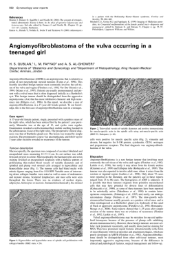
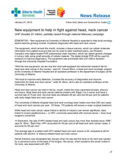
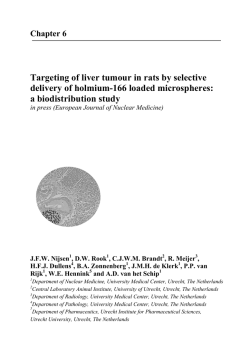
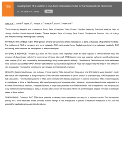
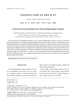
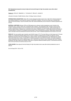
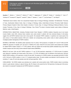
Oral Oncology (2007) 43, 1007– 1013
available at www.sciencedirect.com
journal homepage: http://intl.elsevierhealth.com/journals/oron/
Predictive value of histopathologic parameters
in early squamous cell carcinoma of oral tongue
¨ntti a,*, Timo Atula a, Jarkko Tikka b, Jaakko Hollme
´n b,
Harri Keski-Sa
¨kitie a,d, Ilmo Leivo c,d
Antti A. Ma
a
Department of Otorhinolaryngology Head and Neck Surgery, Helsinki University Central Hospital, P.O. Box 220,
FIN – 00029 HUCH, Helsinki, Finland
b
Laboratory of Computer and Information Science, Helsinki University of Technology, Espoo, Finland
c
Haartman Institute, Department of Pathology, University of Helsinki and HUSLAB, Department of Pathology,
Helsinki, Finland
Received 3 October 2006; accepted 20 November 2006
Available online 15 February 2007
KEYWORDS
Summary The clinical course of early squamous cell carcinoma of oral tongue (OTSCC) is
unpredictable and various histopathologic parameters of the primary tumour have been suggested as prognostic factors to be used in clinical decision-making. We reviewed clinicopathologic data of 73 patients diagnosed with Stage I–II OTSCC. Predictive value of pathological Tstage, depth of infiltration, grade, and mode of invasion with respect to local recurrences,
occult cervical metastases, and disease specific survival (DSS) was analysed. Depth of infiltration and pT-stage significantly predicted occult nodal disease, while only pT-stage predicted
local recurrence. Specific cut-off value for depth of infiltration separating high-risk and low-risk
patients was not found. Significant correlations between the histopathologic parameters and
DSS were not found. We conclude that depth of infiltration predicted occult nodal disease
but its value in clinical decision-making is limited because of poor specificity when using a
cut-off value that offers reasonable sensitivity for finding the patients with occult nodal disease. The risk for occult metastases and local recurrence was high in patients with pT2
tumours.
c 2006 Elsevier Ltd. All rights reserved.
Depth of infiltration;
Tumour thickness;
Tumour diameter;
Grade;
Mode of invasion;
Survival;
Occult metastases;
Oral cancer;
Tongue cancer
Introduction
* Corresponding author. Tel.: +358 50 4271589; fax: +358 9 4717
5010.
E-mail address: [email protected] (H. Keski-Sa
¨ntti).
d
Equal contribution.
The clinical course of early squamous cell carcinoma of oral
tongue (OTSCC) is fairly unpredictable. For the majority of
patients, surgical peroral excision of the primary tumour
would be adequate treatment to cure the disease. However,
1368-8375/$ - see front matter c 2006 Elsevier Ltd. All rights reserved.
doi:10.1016/j.oraloncology.2006.11.015
1008
the incidence of occult metastasis is relatively high 1–4 and
despite the advances in modern imaging technology, histopathologic examination of neck dissection specimen is still
the only reliable way to detect the subclinical metastases.
Treatment failures with even fatal outcome can occur in patients who initially have had very small primary tumour with
no clinical evidence of metastatic disease.1,4 The results of
salvage treatment of locoregional recurrences are generally
reported poor.4–6 Finding the patients who are at greatest
risk for occult cervical metastases and poor survival is
essential in the treatment planning in this patient group.
This has led to the search for various histopathologic parameters of the primary tumour that would predict the clinical
course of the disease.
The size of the primary tumour has been shown to have
prognostic significance in predicting nodal involvement
and survival in patients with OTSCC, thickness of the tumour
being more important than the greatest diameter.3,7–16 In
some studies, the morphology of the invasive front of the
tumour has had predictive value.9,17,18 Tumour grade (degree of differentiation) seems to have no value as a prognosticator.11,13,15,16,19 It has been suggested, that tumour
thickness should be used to indicate tumour size in the
TNM classification system instead of tumour diameter.15
However, controversy exists regarding the critical tumour
thickness, or depth of infiltration, identifying the patients
with high-risk of nodal involvement and poor prognosis. In
previous studies of patients with OTSCC, the suggested value for critical tumour thickness (or depth of infiltration)
has varied from 3 to 10 mm, being most often 4 mm.3,7–16
In a recent review including all oral cavity tumour sites
the critical tumour thickness (or depth of infiltration) varied
from 1.5 mm to 10 mm being most often 3–5 mm.20 The
authors pointed out that the measurement technique has
not been uniform in different studies and therefore the results are not fully comparable. Therefore, more studies with
comparable measurement technique are needed to reach a
consensus on this topic.
The aim of this study was to analyse whether depth of tumour infiltration, pT-stage, mode of tumour invasion, or tumour grade predict occult metastases, survival or local
recurrence in patients with Stage I–II OTSCC at our institution. In addition, we wanted to find out if a critical depth of
infiltration can be found for clinical decision-making.
Patients and methods
Clinicopathological data of 141 patients diagnosed with
OTSCC at the Helsinki University Central Hospital (covering
an area of 1.4 million inhabitants) between 1992 and 2002
were reviewed. The local Ethics Committee approved the
study protocol. Only patients with tumours clinically defined as T1/T2 N0, original histopathological material available for review and a clinical follow-up data with a
minimum of 24 months or until death were included in the
study. Seventy-three patients were eligible for the inclusion. All patients were surgically treated with curative
intent.
Hospital records were reviewed and data on patient characteristics, treatment, histopathology, and follow-up were
collected. The dates and causes of death were provided
H. Keski-Sa
¨ntti et al.
by Statistics Finland (a national agency of population statistics). The histopathological paraffin sections of each patient
were reviewed by a single, experienced Head and Neck
pathologist (I.L.) and depth of infiltration, grade, greatest
diameter, and mode of invasion of the primary tongue tumour were re-analysed. Mode of invasion was classified into
one of three fundamental categories: pushing-border, diffuse or destructive. Depth of infiltration was measured from
the level of the adjacent normal mucosal surface to the
deepest portion of the tumour invasion in the tongue musculature using a field arrow measurement system provided by
the microscope manufacturer. Measurements were expressed at an accuracy of 0.1 mm. We also intended to analyse the resection margins. However, many patients had
first had excisional biopsies or biopsies of the resection margins making determination of the full resection margins
unreliable and therefore this parameter was omitted.
Only re-analysed histopathological data were used in the
analysis. In the case of tumours of large diameter, measuring and reconstructing the greatest tumour diameter from
histopathological sections of previously dissected surgical
specimens is less reliable than the original measurement.
Therefore original histopathologic data were used in these
cases. Re-analysis of data on tumour diameter was carried
out only when original data were not available. According
to the pathologically analysed tumour diameter, the tumour
was classified as pT1 (620 mm) or pT2 (21–40 mm).
In patients who had not had an elective neck dissection
(END), occult neck disease was defined as a histologically
proven neck metastasis detected during the follow-up without failure at the primary site. Cervical metastases in patients with recurrent primary tumour were not considered
occult metastases, because the nodal spread may have occurred after the initial treatment. In patients who had had
an END, occult neck disease was defined as presence of
microscopic disease on the histopathological examination
of the neck dissection specimen. One patient with prophylactic neck irradiation only was not taken into account when
assessing the correlations between histopathological parameters and occult metastases.
Statistical analysis
In order to compare the difference between two proportions f1 and f2, we performed the following statistical test.
If the null hypothesis is true, f1 and f2 are both estimators
for the same proportion f. The pooled estimator for f is
^f ¼ n1 f1 þ n2 f2 ;
n1 þ n2
where n1 and n2 are sample sizes. The test statistic is defined as follows
f1 f2
z ¼ rffiffiffiffiffiffiffiffiffiffiffiffiffiffiffiffiffiffiffiffiffiffiffiffiffiffiffiffiffiffiffiffiffiffiffiffiffiffiffiffiffiffiffiffiffiffiffi
;
^f 1 ^f ð1=n1 þ 1=n2 Þ
which is approximately normally distributed with zero mean
and unit variance.21
The Wilcoxon rank-sum test can be used in the comparison of two populations.21 It is especially sensitive to differences in location, thus, the null hypothesis is that the
Predictive value of histopathologic parameters in early squamous cell carcinoma of oral tongue
medians of populations are equal. Since the test is nonparametric, no assumptions on parametric distributions of populations are made.
The Pearson’s coefficient of correlation q defines the
strength of linear dependency between two random variables X and Y.21 The value of correlation coefficient is in
the range q 2 [ 1, 1] and the values of q near 1 or 1 indicate strong linear dependency. It was used to illustrate the
dependency between the depth of infiltration and the greatest diameter.
The Kaplan–Meier method was applied in the analysis of
disease specific survival (DSS) of two populations. The different follow-up times of the patients were taken into account, i.e. the patients whose follow-up was less than 5
years were censored. In the survival curves, the tick marks
represent the time when one patient was censored. The difference of two survival curves was statistically tested using
log-rank test.22
The analysis of separating patients with and without occult metastases based on the depth of infiltration was carried out using the bootstrap re-sampling procedure. The
accuracy of the statistical estimates can be assessed using
the bootstrap without making any assumptions of underlying
probability distributions.23 Let (x1, . . ., xm) and (y1, . . ., yn)
indicate the measurements of the depth of infiltration of
the patients with and without the occult metastases,
respectively. For a given cut-off point c, the proportion of
true negatives f0 is the proportion of yj, j ¼ 1; . . . ; n being
less than c and the proportion of true positives f1 is the proportion of xi, i ¼ 1; . . . ; m being greater than c. The bootstrap replications f0 and f1 are calculated from the
bootstrap samples ðx1 ; . . . ; xm Þ and ðy 1 ; . . . ; y n Þ. The bootstrap samples are randomly drawn with replacement from
the original data (x1, . . ., xm) and (y1, . . ., yn), that is, some
original data points may appear zero times, some once,
some twice, etc. in the bootstrap sample. The re-sampling
procedure is repeated B times, which produces B bootstrap
replications f0 and f1 . These replications formulate the
empirical distributions of f0 and f1. The medians of the
bootstrap replications f0 and f1 are used as estimates of f0
and f1. The accuracy of estimates can be assessed using
quantiles of the bootstrap replications. The medians and
quantiles are used, since they describe properly also skewed
distributions and distributions with outliers.
The results were considered significant when the p value
was <0.05.
Results
Seventy-three patients met the inclusion criteria. There
were 36 males and 37 females (median age 59 years, range
23–95 years). The clinical T classification was as follows:
T1:n = 35 (48%), T2:n = 38 (52%). All patients were staged
N0 after clinical and radiological assessment. The assessment of the cervical lymph nodes consisted of palpation
only in five patients. For the rest of the patients, imaging
of the neck was performed as follows: CT:n = 19, ultrasonography: n = 15, MRI: n = 34. All patients underwent resection of the primary tumour (60 without reconstruction and
13 with reconstruction of the surgical defect). In 31 patients
there was no further treatment primarily. Forty-two pa-
1009
tients received elective neck treatment. The clinico-pathological characteristics of the study cohort are presented in
Table 1.
The overall rate of occult cervical metastases was 32%
(23/72). The rates of occult metastases for patients with
pT1 and pT2 tumours were 23% (11/48) and 50% (12/24),
respectively (p < 0.05). Depth of infiltration and pT-stage
significantly predicted occult cervical metastases. The difference between proportions of the patients with occult
Table 1
Clinicopathological features of study cohort
No. of patients (%)
Total number of patients
73
Gender
Female
Male
37 (51)
36 (49)
Age (y)
Range
Median
23–95
59
Clinical T-classification
cT1
cT2
35 (48)
38 (52)
Treatment of the primary tumour
Resection
Resection + Radiotherapy
40 (55)
33 (45)
Neck treatment
Observation
Neck dissection
Neck dissection + Radiotherapy
Radiotherapy
31(43)
9 (12)
32 (44)
1(1)
Tumour site
Lateral border
Ventral surface
Tip of tongue
Dorsum
61 (84)
9 (12)
2 (3)
1 (1)
Mode of the invasive front
Pushing border
Diffuse
Destructive
34 (47)
28 (38)
11 (15)
Histological grade
Well differentiated (G1)
Moderately differentiated (G2)
Poorly differentiated (G3)
24 (33)
35 (48)
14 (19)
Pathological T-classification
pT1
pT2
49 (67)
24 (33)
Occult cervical disease
Yes
No
Not defined
23 (32)
49 (67)
1 (1)
Local recurrence
Yes
No
9 (12)
64 (88)
1010
Table 2
H. Keski-Sa
¨ntti et al.
Different cut-off points in depth of infiltration
Depth of
infiltration
Patients
with occult
Metastasis (%)
No. of
cases
62 mm
>2 mm
2 (14)
21 (36)
14
58
63 mm
>3 mm
2 (11)
21 (39)
18
54
64 mm
>4 mm
5 (18)
18 (41)
65 mm
>5 mm
66 mm
>6 mm
p value
5-year
DSS (%)
p
value
78
81
NS
<0.05
83
79
NS
28
44
<0.05
82
79
NS
6 (17)
17 (46)
35
37
<0.01
82
79
NS
10 (23)
13 (46)
44
28
<0.05
85
75
NS
0.057
Figure 1 Correlation between the depth of infiltration and
the greatest diameter is 0.63 and the 95% confidence interval is
[0.47, 0.75].
DSS, disease specific survival; NS, non significant.
metastases was significant with different cut-off values of
depth of infiltration (Table 2). Mode of invasion classified
as pushing-border resulted in significantly less occult metastases than classification as diffuse (p < 0.05) but no other
correlations existed between the different invasion mode
groups (pushing-border vs. destructive: p = 0.29, diffuse
vs. destructive: p = 0.29). Tumour grade did not have any
correlation with the presence of occult metastases.
The overall rate of local recurrence was 12% (9/73). Of
the histopathological parameters studied, only pT-stage
was found to significantly predict local recurrence. The
rates of local recurrence for pT1 and pT2 tumours were
6% (3/49) and 25% (6/24), respectively (p < 0.05).
Significant correlations between the histopathological
parameters and survival were not detected.
The differences in median depth of infiltration between
different patient groups are presented in Table 3.
A positive correlation existed between the greatest tumour diameter and depth of infiltration, as shown in Fig. 1.
Table 3
The 5-year DSS rates for patients with and without occult
metastases were 65% and 91%, respectively (Fig. 2). The
5-year DSS rates for patients with and without local recurrence were 33% and 90%, respectively (Fig. 3).
Discussion
Clinical and histopathological data of 73 patients with Stage
I–II OTSCC treated at a single tertiary care centre according
to a consensus protocol during a 10-year period were reviewed. Depth of tumour infiltration, pT-stage, tumour
grade, and mode of tumour invasion were re-examined
and their ability to predict local recurrences, occult cervical
metastases, and survival was analysed. Depth of infiltration
and pT-stage were found to correlate significantly with the
rate of subclinical nodal metastases. A specific cut-off value
for the depth of infiltration separating high-risk and low-risk
patients was not found. These results are in concordance
Median depth of infiltration
No. of
patients
Median depth
of infiltration (mm)
p-value
Occult neck disease
Yes
23
No
49
8
5
<0.05
Local recurrence
Yes
9
No
64
8
5
NS
Dead of disease
Yes
13
No
60
8
5
NS
T-Stage
pT1
pT2
4
9
<0.001
49
24
NS, non significant.
Figure 2 Cumulative disease specific survival (DSS) for
patients with occult metastases (dashed line) and without
occult metastases (solid line) evaluated using the Kaplan–Meier
method. The difference in survival was statistically significant
(p < 0.01) according to log-rank test.
Predictive value of histopathologic parameters in early squamous cell carcinoma of oral tongue
Figure 3 Cumulative disease specific survival (DSS) for
patients who had local recurrence (dashed line) and for those
who did not have local recurrence (solid line) evaluated using
the Kaplan–Meier method. The difference in survival was
statistically significant (p < 0.001) according to log-rank test.
with previous studies, in which the thickness (or depth of
infiltration) 3,7,9,10,12,13,15,16 and the greatest diameter 3,12
of the primary tongue tumour have been found to predict
occult neck disease in patients with early tongue tumours.
The high proportion of occult metastases in patients
with pT2 tumours (50%) in the present study emphasizes
the importance of elective neck treatment in this patient
group. Most institutions advocate END as it allows definite
pathologic staging of the cervical lymph nodes. It is recommended, that levels I–IV should be removed when treating
patients with OTSCC.24,25 In our study, two patients with a
very superficial pT1 tumour (depth of infiltration 1.3 mm
and 1.5 mm) had occult metastasis pointing out that the
risk of subclinical metastasis has to be considered in practically all patients diagnosed with OTSCC. Therefore, pathologic staging of the cervical lymph nodes seems to be
warranted also in patients with T1 tumours, for whom sentinel lymph node biopsy may prove to be the most suitable
method instead of END.26
Due to limitations of depth and size of a biopsy specimen, a reliable estimation of the histopathological parameters necessitates the evaluation of the whole tumour
block. Therefore, these parameters can reliably be applied
to clinical decision-making only when the whole primary
tumour has first been excised and assessed. The measurement technique of the tumour thickness, or depth of infiltration, in different studies has not been uniform.
Measurements from the level of normal mucosa (depth of
infiltration) 9,10,12,16 from the level of basal membrane
(depth of infiltration),14 or from the surface of the tumour
(tumour thickness) 15,27 to the deepest portion of the tumour infiltration have been used. In some studies the
method of measurement has not been defined.3,7,11,13
Hence, the results of the studies are not fully comparable.
A tongue tumour can be exofytic or more often ulcerated.
The critical aspect in the growth of a tongue tumour is
probably the deep invasion into the tongue musculature,
in which the vascular invasion and metastatic spread are
likely to occur, and not the exofytic growth. Therefore,
measuring the depth of infiltration from the level of the
1011
normal mucosa provides more accurate measure on the
depth of tumour infiltration in the tongue tissue for prognostic consideration than tumour thickness measured from
the surface of the tumour. The measurement technique
should be standardised in future studies to enable comparison of studies.
Many previous studies have stated a critical tumour
thickness (or depth of infiltration) separating the high-risk
and low-risk patients. According to our results it seems,
however, that such a clear cut-off point cannot be defined.
There is a risk of metastatic disease even in patients with
very superficial tumours and the risk seems to increase quite
linearly with increasing depth of infiltration (Fig. 4). Thus,
there is probably no clinically relevant depth of infiltration
a tumour must reach before it can metastasize. In the
present study, 5.5 mm depth of infiltration as a cut-off point
had the best overall accuracy in separating patients with
and without subclinical nodal disease (Fig. 5). As shown in
Figure 5, with the cut-off point at 5.5 mm, approximately
65% of both patient groups with and without occult
metastases would be classified correctly. Then, however,
a considerable proportion of occult metastases are overlooked. If the cut-off point is moved downwards to increase
the sensitivity, the specificity falls rapidly resulting in ‘‘at
risk’’ status of many true negative necks. Thus, as a tool
for clinical decision-making (elective neck treatment or
not), depth of infiltration is far from optimal.
We found only pathological T-stage to predict local
recurrences. A considerable proportion of patients with
pT2 tumours had locally recurrent disease (25%) and the
outcome of these patients was quite miserable (Fig. 3).
Assessment of also the resection margins would have been
important, as increasing tumour size leads more often to
close or involved resection margins, as demonstrated by
Sutton et al. 28 In their study, the incidence of local recurrences was highly dependent on status of the resection
margins. Van Es et al. 29 concluded that when excision of
Figure 4 Empirical cumulative distribution functions of depth
of infiltration for the patients without (solid line) and with
occult metastases (dashed line). The steady increase of the
curve of patients with occult metastases indicates that risk of
occult metastases increases steadily with increasing tumour
thickness. The significance of difference between the distributions was tested using Kolmogorov–Smirnov test (p < 0.05).
1012
H. Keski-Sa
¨ntti et al.
while only pT-stage predicted local recurrences. Significant
correlations between histopathological parameters and survival were not found. A specific depth of infiltration separating high-risk and low-risk patients was not found. The
value of depth of infiltration in clinical decision-making is
limited because of poor specificity when using a cut-off value that offers reasonable sensitivity for finding the patients
with occult metastases.
Conflict of interest statement
None declared.
References
Figure 5 Proportions of correctly classified patients with
either true negative necks (dots) or occult metastases (circles)
with different cut-off values of the depth of infiltration used as
a decision variable to recognise patients with occult metastases. The solid line with dots indicates specificity and the dashed
line with circles indicates sensitivity. They are medians from
1000 bootstrap replications. At the crossing of the curves, the
overall accuracy is at its best. The dotted lines represent the
16% and 84% quantiles obtained using the bootstrap method
reflecting the uncertainty.
a T1 or T2 primary tumour in oral tongue or floor of mouth is
performed with good margins, local recurrences are rare
(4%) and not linked to any other histopathologic variable.
As discussed previously, we had to omit the assessment of
resection margins, because these data could not be measured accurately afterwards and original data was not available in many cases.
In our study, mode of invasion classified as pushing-border resulted in significantly less occult metastases than that
classified as diffuse; no other significant differences were
found between the proportions of patients with occult
metastases in different invasion mode groups, however.
This may be due to small sample size; only 11 patients
had tumours with invasion mode classified as destructive.
In the studies by Fukano et al. 9 and Sawair et al. 30 the invasion mode was found to significantly predict nodal metastases. With respect to tumour grade, our study confirmed
many former studies, in which the grade of the primary tumour does not seem to have predictive value,11,13,15,16,19
though opposite findings have also been reported.3,12 Lack
of a significant correlation, however, seems to be the general experience of head and neck cancer surgeons and
pathologists alike, and the present results tend to reaffirm
that view. The strength of present data lies in consistent
criteria of grading applied by a single pathologist throughout
the study, as well as the well-documented clinical data of a
patient material from a single institution practise.
In the present study, none of the histopathologic parameters examined had prognostic significance with respect to
survival. This contradicts many previous studies, in which
the tumour thickness or depth of infiltration, 7,8,11,14–16,27
diameter,8,14 and invasion mode 18 have been found to have
prognostic value.
In conclusion, depth of infiltration and pT-stage were
found to significantly predict occult cervical metastases,
1. Haddadin KJ, Soutar DS, Oliver RJ, Webster MH, Robertson AG,
MacDonald DG. Improved survival for patients with clinically
T1/T2, N0 tongue tumors undergoing a prophylactic neck
dissection. Head Neck 1999;21(6):517–25.
2. Teichgraeber JF, Clairmont AA. The incidence of occult
metastases for cancer of the oral tongue and floor of the
mouth: treatment rationale. Head Neck Surg 1984;7(1):15–21.
3. Byers RM, El-Naggar AK, Lee YY, Rao B, Fornage B, Terry NH,
et al. Can we detect or predict the presence of occult nodal
metastases in patients with squamous carcinoma of the oral
tongue? Head Neck 1998;20(2):138–44.
4. Keski-Sa
¨ntti H, Atula T, To
¨rnwall J, Koivunen P, Ma
¨kitie A.
Elective neck treatment versus observation in patients with T1/
T2 N0 squamous cell carcinoma of oral tongue. Oral Oncol
2006;42(1):96–101.
5. Yii NW, Patel SG, Rhys-Evans PH, Breach NM. Management of
the N0 neck in early cancer of the oral tongue. Clin Otolaryngol
1999;24(1):75–9.
6. Kowalski LP. Results of salvage treatment of the neck in
patients with oral cancer. Arch Otolaryngol Head Neck Surg
2002;128(1):58–62.
7. Fakih AR, Rao RS, Borges AM, Patel AR. Elective versus
therapeutic neck dissection in early carcinoma of the oral
tongue. Am J Surg 1989;158(4):309–13.
8. Brown B, Barnes L, Mazariegos J, Taylor F, Johnson J, Wagner
RL. Prognostic factors in mobile tongue and floor of mouth
carcinoma. Cancer 1989;64(6):1195–202.
9. Fukano H, Matsuura H, Hasegawa Y, Nakamura S. Depth of
invasion as a predictive factor for cervical lymph node
metastasis in tongue carcinoma. Head Neck 1997;19(3):
205–10.
10. Asakage T, Yokose T, Mukai K, Tsugane S, Tsubono Y, Asai M,
et al. Tumor thickness predicts cervical metastasis in patients
with stage I/II carcinoma of the tongue. Cancer 1998;82(8):
1443–8.
11. Al-Rajhi N, Khafaga Y, El-Husseiny J, Saleem M, Mourad W,
Al-Otieschan A, et al. Early stage carcinoma of oral tongue:
prognostic factors for local control and survival. Oral Oncol
2000;36(6):508–14.
12. Kurokawa H, Yamashita Y, Takeda S, Zhang M, Fukuyama H,
Takahashi T. Risk factors for late cervical lymph node metastases in patients with stage I or II carcinoma of the tongue.
Head Neck 2002;24(8):731–6.
13. Okamoto M, Nishimine M, Kishi M, Kirita T, Sugimura M,
Nakamura M, et al. Prediction of delayed neck metastasis in
patients with stage I/II squamous cell carcinoma of the tongue.
J Oral Pathol Med 2002;31(4):227–33.
14. Gonzalez-Moles MA, Esteban F, Rodriguez-Archilla A, Ruiz-Avila
S, Gonzalez-Moles S. Importance of tumour thickness measurement in prognosis of tongue cancer. Oral Oncol 2002;38(4):
394–7.
Predictive value of histopathologic parameters in early squamous cell carcinoma of oral tongue
15. Po Wing Yuen A, Lam KY, Lam LK, Ho CM, Wong A, Chow TL, et al.
Prognostic factors of clinically stage I and II oral tongue
carcinoma-A comparative study of stage, thickness, shape,
growth pattern, invasive front malignancy grading, Martinez–
Gimeno score, and pathologic features. Head Neck 2002;24(6):
513–20.
16. O-charoenrat P, Pillai G, Patel S, Fisher C, Archer D, Eccles S,
et al. Tumour thickness predicts cervical nodal metastases and
survival in early oral tongue cancer. Oral Oncol 2003;39(4):
386–90.
17. Bryne M, Koppang HS, Lilleng R, Kjaerheim A. Malignancy grading
of the deep invasive margins of oral squamous cell carcinomas
has high prognostic value. J Pathol 1992;166(4): 375–81.
18. Bundgaard T, Rossen K, Henriksen SD, Charabi S, Sogaard H,
Grau C. Histopathologic parameters in the evaluation of T1
squamous cell carcinomas of the oral cavity. Head Neck
2002;24(7):656–60.
19. Woolgar JA. Histopathological prognosticators in oral and oropharyngeal squamous cell carcinoma. Oral Oncol 2006;42(3):
229–39.
20. Pentenero M, Gandolfo S, Carrozzo M. Importance of tumor
thickness and depth of invasion in nodal involvement and
prognosis of oral squamous cell carcinoma: a review of the
literature. Head Neck 2005;27(12):1080–91.
21. Milton JS, Arnold JC. Introduction to probability and statistics
– principles and applications for engineering and computing
sciences. Second ed. McGraw-Hill; 1990.
22. Kalbfleisch JD, Prentice RL. The statistical analysis of failure
time data. New york: Wiley; 1980.
1013
23. Efron B, Tibshirani RJ. An introduction to the bootstrap. Chapman & Hall/CRC; 1993.
24. Byers RM, Weber RS, Andrews T, McGill D, Kare R, Wolf P.
Frequency and therapeutic implications of ‘‘skip metastases’’
in the neck from squamous carcinoma of the oral tongue. Head
Neck 1997;19(1):14–9.
25. Wei WI, Ferlito A, Rinaldo A, Gourin CG, Lowry J, Ho WK, et al.
Management of the N0 neck – reference or preference. Oral
Oncol 2006;42(2):115–22.
26. Ross GL, Soutar DS, Gordon MacDonald D, Shoaib T, Camilleri I,
Roberton AG, et al. Sentinel node biopsy in head and neck
cancer: preliminary results of a multicenter trial. Ann Surg
Oncol 2004;11(7):690–6.
27. O’Brien CJ, Lauer CS, Fredricks S, Clifford AR, McNeil EB, Bagia
JS, et al. Tumor thickness influences prognosis of T1 and T2
oral cavity cancer – but what thickness? Head Neck
2003;25(11):937–45.
28. Sutton DN, Brown JS, Rogers SN, Vaughan ED, Woolgar JA. The
prognostic implications of the surgical margin in oral squamous
cell carcinoma. Int J Oral Maxillofac Surg 2003;32(1):30–4.
29. van Es RJ, van Nieuw Amerongen N, Slootweg PJ, Egyedi P.
Resection margin as a predictor of recurrence at the primary
site for T1 and T2 oral cancers. Evaluation of histopathologic
variables. Arch Otolaryngol Head Neck Surg 1996;122(5):
521–5.
30. Sawair FA, Irwin CR, Gordon DJ, Leonard AG, Stephenson M,
Napier SS. Invasive front grading: reliability and usefulness in
the management of oral squamous cell carcinoma. J Oral
Pathol Med 2003;32(1):1–9.


© Copyright 2026