
Diclofenac sodium pellets for flexible pediatric drug dosing
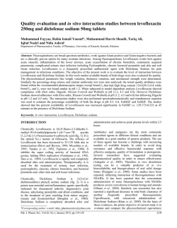
Available online at www.scholarsresearchlibrary.com
Scholars Research Library
Der Pharmacia Lettre, 2015, 7 (1):122-133
(http://scholarsresearchlibrary.com/archive.html)
ISSN 0975-5071
USA CODEN: DPLEB4
Diclofenac sodium pellets for flexible pediatric drug dosing:
Preparation, characterization and evaluation
Swetha Arella1, Subrahmanyam P. V. R. S.2, Kiran Thadkala1, Soumya Das3, Dharmajit
Patnaik3 and Jithan Aukunuru1,*
1
Mother Teresa College of Pharmacy, Hyderabad, Telangana, India
2
MSN Laboratories India Ltd., Hyderabad, Telangana, India
3
Vikas College of Pharmacy, Suryapet, Telangana, India
_____________________________________________________________________________________________
ABSTRACT
The objective of the current investigation was to formulate Eudragit RS100 based sustained release pellets,
containing Diclofenac sodium intended for flexible dosing in pediatric patients. Diclofenac sodium is a type II antiinflamatory agent. Pellets in the form of microspheres were prepared by O/O emulsion solvent evaporation method
with different stabilizer concentrations and at different speeds of emulsification while maintaining constant amounts
of Diclofenac sodium. Span 80 was used as the stabilizer. Drug excipient compatibility study was performed prior
to formulation development and only compatible excipients were used in the fabrication of microspheres. Prepared
microsphere formulations were characterized for percentage yield, particle size analysis, entrapment efficiency,
invitro release behavior, differential scanning colorimetry (DSC), scanning electron microscopy (SEM), in vivo
drug release and in vitro in vivo correlation (IVIVC). SEM studies showed that the microspheres were spherical
with rough surface morphology. The drug loaded microspheres showed 50-80% entrapment efficiency. The invitro
release profile showed a slow and steady release pattern for Diclofenac sodium. A 100% Diclofenac sodium was
released within a period of 12 hrs during this time. The drug release was found to follow diffusion controlled
mechanism. The n value of Korsmeyer Peppas equation indicated non Fickian type of diffusion. DSC results
indicated that the physical state of the drug was changed upon fabrication. In rats, the pellets released the drug for
over a period of 12 hrs after oral administration. A 100% IVIVC was achieved with the optimized formulation. As a
result of these experiments, it was concluded that, novel sustained release oral pellets comprising of diclofenac
sodium were successfully prepared using eudragit RS100 as the polymer and using emulsion solvent evaluation
method.
Key words: Pellets, Microspheres, Diclofenac sodium, Eudragit RS100, O/O Emulsion solvent evaporation method.
_____________________________________________________________________________________________
INTRODUCTION
Previously acute or chronic illness was treated with drug formulations such as tablets, capsules, creams, pills,
suppositories, simple injections, sustained release tablets, etc. Recent innovations resulted in several formulations
such as fast dissolving tablets, targeted nanoparticles, floating tablets, parenteral sustained release implants and
microspheres, nasal formulations, respiratory formulations, colon specific formulations etc [1]. These formulations
would eventually replace the currently available formulations in a pharmacy. In this regard, we propose pellets as
one more such delivery system. These pellets can be developed as proprietary technologies where in once
122
Scholar Research Library
Jithan Aukunuru et al
Der Pharmacia Lettre, 2015, 7 (1):122-133
______________________________________________________________________________
formulated can be used for a variety of drugs. In this study, we developed such technology for diclofenac sodium
and these pellets can be used as a flexible padietric dosage form. The pellets are prepared in the form of
microspheres. Diclofenac microspheres were previously prepared and reported [2,3].
Pellets can be defined as small, free flowing, spherical particulates manufactured by the agglomeration of fine
powders or granules of drug substances and excipients typically from about 0.5 mm to 1.5 mm by using
appropriate processing equipment or method. Hot melt extrusion, spheronization are commonly used to prepared
pellets. On the other hand, microspheres, other forms of pellets are of size 1-1000 µm. These are prepared by
solvent evaporation technique or spray drying techniques. In this study, we have used microspheres as pellets and
used further for sustained release oral release of diclofenac sodium, especially focusing at padietric purposes.
Further, pellets can be conveniently used for flexible dosage adjustments [4]. Oral route of administration is most
widely used route of administration of drugs. It is convenient, predominant and widely accepted. The formulations
administration via the oral route are commonly divided into immediate release and modified release systems.
Modified release systems are used for sustained release and drug targeting. Oral drug delivery systems (ODDS) are
commonly divided into immediate release and modified release systems. These modified release dosage forms
achieve a desired pharmacokinetic profile, to enhance patient compliance and to minimize adverse drug reactions.
Microencapsulation technique is widely used to prepare ODDS, especially microparticles (microspheres). In this
technique, a thin coat is applied to core material [5,6]. These formulations are generally called as microcapsules.
The other way of forming microspheres is embedding the drug in the matrix form which is of micro in size. These
are called as micromatrices. Micromatrices and microcapsules are together called as microspheres. Based on the
polymer, these microspheres can be conveniently used as powders, or used as formulations that have been
transformed into tablets and capsules. Several polymers such as xanthine gum, guar gum, gumarabic, gum karaya,
olibanum gum, methyl cellulose, polylactides-co-glycolides can be conveniently used to prepare these
microspheres. Depending on the nature of the polymer any technique can be used to prepare microspheres [7,8].
Microspheres are used as carriers with dispersed drug particles to allow for sustained action and also minimize the
adverse effects.The most significant feature of microspheres is their microscopic size that allows for a huge surface
area. This large surface area of microspheres results in sites of adsorption and desorption, chemical reactions,
lightscattering, etc [9,10]. The objective of this study is to prepare diclofenac pellets in the form of microspheres
and investigate them for the formulation development and characterization. The intention was to develop padietric
sustained release delivery system for diclofenac sodium.
MATERIALS AND METHODS
Materials:
Diclofenac sodium as a gift sample from MSN Laboratories Pvt. Ltd, Hyderabad, India, Eudragit RS100, Paraffin
Liquid (light), Span 80, Acetone are from SD Fine chemical Ltd, Mumbai, India.
Preformulation Studies with the Drug
Preformulation testing is the first step in the rationale development of dosage forms of a drug substance. It can be
defined as an investigation of physical and chemical properties of a drug substance alone and when combined with
excipients [11,12]. The overall objective of preformulation testing is to generate information useful to the formulator
in developing stable and strong formulations. The preformulation studies with the drug obtained were performed
using conventional and reported techniques. The UV-Visible spectrum, solubility, flow properties, melting point and
drug crystallinity were determined [13,14].
Methods:
Microspheres were prepared by O/O emulsion solvent evaporation method. A mixed solvent system (MSS) of
acetonitrile and dichloromethane and distilled water were used for the preparation of microspheres as internal
organic phase and aqueous phase. Liquid paraffin and Span 80 were used as external oily phase and surfactant. Nhexane was added as a non-solvent to the processing medium to solidify the microspheres.This method for
preparation of microspheres was reported to overcome the problem of low encapsulation efficiency of water soluble
drugs prepared by conventional o/o emulsion solvent evaporation method [15-17]. The composition and ratio of
compounds was showed in the following table no: 1
123
Scholar Research Library
Jithan Aukunuru et al
Der Pharmacia Lettre, 2015, 7 (1):122-133
______________________________________________________________________________
Table: 1 Formulation design of Microspheres
Formulation code
Materials
Diclofenac sodium (mg)
Eudragit RS 100 (mg)
Span-80 (%)
Acetone
Petrolieum ether
Liquid Paraffin (ml)
Stirring Speed (rpm)
F1
F2
F3
F4
F5
F6
50
100
0.5
10
4
50
750
50
100
1
10
4
50
750
50
100
2
10
4
50
750
50
50
0.5
10
4
50
1000
50
50
1
10
4
50
1000
50
50
2
10
4
50
1000
CHARACTERIZATION OF MICROSPHERES
PERCENTAGE YIELD:
Percentage practical yield is calculated to know about percentage yield or efficiency of any method, thus it helps in
selection of appropriate method of production. The prepared microspheres of all batches were accurately weighed.
The measured weight of prepared microspheres was divided by the total amount of all the excipients and drug used in
the preparation of the microspheres, which give the total percentage yield of floating microspheres [18]. It was
calculated by using following equation.
% Yield = actual weight of product/total weight of excipients and drug × 100
DRUG ENTRAPMENT EFFICIENCY:
Microspheres equivalent to 50 mg of the drug were taken for evaluation. Microspheres formulation was dissolved in
aliquot amount of methanol by continuous shaking, in a 10 mL volumetric flask and the volume was made up to the
mark. The solution was filtered and the absorbance was measured after suitable dilution. The amount of drug
entrapped was estimated spectrophotometrically (UV 1700, Shimadzu, Japan) at Diclofenac Sodium of wavelength
i.e., 275nm and against appropriate blank [19]. The amount of drug entrapped in the microspheres was calculated by
the following formula:
Amount of drug actually present (DC)
% Drug entrapment =
X 100
Theoretical drug load expected
(DC- Actual Drug Content)
PARTICLE SIZE DETERMINATION:
The mean particle size was determined by using optical microscope. In this method 25 particles size was determined
by using stage micro meter. The average particle size was determined in this method. The eye piece was adjusted
and the stage micrometer was adjusted according to the eye piece. Calibration factor was calculated by the
following formula:
Calibration factor =stage micrometer/eye piece
A minute quantity of prepared microspheres was spread on a clean glass slide. Then particle size of the
microspheres were measured, from which average particle size was calculated which was then multiplied with the
obtained calibration factor .In this way, the average particle size was calculated for all the six batches.
SCANNING ELECTRON MICROSCOPY (SEM):
The most widely used procedures to visualize microparticles are conventional light microscopy (LM) and scanning
electron microscopy (SEM). It images the sample surface of a solid specimen by using a focused beam of highenergy electrons. The signal contains information about surface topography, texture, external morphology of
fractured or sectioned surface, chemical composition, crystallographic information, and electrical conductivity.
In order to examine the particle surface morphology and shape, Scanning Electron Microscopy (SEM) was used.
Microspheres were scanned and examined under Electron Microscope. Dry microspheres were spread over a slab.
The sample was shadowed in a cathodic evaporator with gold layer 20 nm thick [20]. Photographs were taken using
an S-3700N Scanning Electron Microscope (Hitachi) operated at 20 kV.
124
Scholar Research Library
Jithan Aukunuru et al
Der Pharmacia Lettre, 2015, 7 (1):122-133
______________________________________________________________________________
IN-VITRO RELEASE STUDY:
The drug release rate from microspheres was determined by using USP dissolution apparatus Type II (basket-type).
A weighed amount of microspheres equivalent to 50 mg of drug (Diclofenac Sodium) was weighed and placed in a
non reacting mesh that had a smaller mesh size than the microspheres. Dissolution medium used was 0.1 N HCl (pH
1.2, 750 ml) for first 2 hours and maintained at 37 ± 0.5°C at a rotation speed of 100 rpm. 5 ml of sample was
withdrawn at each 15 min interval for the first hour followed by 30 min interval, later this interval was extended to 1
h. Sample was then passed through a 5 µm membrane filter, and analyzed spectrophotometrically at 275 nm
determine the concentration of Diclofenac Sodium present in the dissolution medium respectively. The initial
volume of dissolution medium was maintained by adding 5 ml of fresh dissolution media after each withdrawal. The
dissolution study was continued with using phosphate buffer (pH 6.8 ± 1, 900ml) for next 10 h. The cumulative %
drug release was calculated using standard calibration curve [21].
RELEASE KINETICS:
The matrix systems were reported to follow the Peppas release rate and the diffusion mechanism for the release of the
drug. To analyze the mechanism for the release and release rate kinetics of the dosage form, the data obtained was
fitted in to, Zero order, First order, Higuchi matrix and Peppas model. In this by comparing the r-values obtained, the
best-fit model was selected [22].
DRUG- EXCIPIENT COMPATIBILITY STUDIES [23]
FOURIER TRANSFORM INFRA-RED SPECTROPHOMETER (FT-IR STUDIES):
The FT-IR spectra of drug, Eudragit RS 100 and optimized formulation diclofenac sodium microspheres were
obtained. Sample about 5 mg was mixed thoroughly with 100 mg potassium bromide IR powder and compacted
under vacuum a pressure of about 12 Psi for 3 minutes. The resultant disc was mounted in a suitable holder in Perkin
Elmer IR spectrophotometer and the IR spectrum was recorded from 4000 cm-1 to 625 cm-1 in a scan time of 12
minutes.
DIFFERENTIAL SCANNING CALORIMETRY (DSC):
Differential Scanning colorimetry is used to determine drug excipient compatibility studies, and also used to observe
more phase changes such as glass transition, crystallization, amorphous forms of drugs and polymers. Thermal
properties of the powder samples were investigated with a DSC. The physical state of drugs and polymer was
analyzed by Differential Scanning calorimeter (Schimadzu). Approximately 10 mg of sample was analyzed in an
open aluminum pan, and heated at scanning rate of 10°C/min between 0°C and 400°C. Magnesia was used as the
standard reference material.
IN VIVO STUDIES
To determine drug release into systemic circulation in vivo and in vitro in vivo correlation with the pellets, the
microspheres were administered orally into male wistar rats at 0 time. All the animal studies were conducted as per
the guidelines of CPCSEA, India. The protocol was approved by Institutional Animal Ethics Committee of Vikas
College of Pharmacy, Suryapet. Wistar rats (weighted 180-220 g) were used as experimental animals. Twelve rats
were randomly divided into two groups with six rats in each group. Prior to the experimentation the rats were fasted
for 12 h with free access to water. The next day, diclofenac sodium sustained release microspheres and diclofenac
oral suspension prepared using sodium CMC as suspending agent were given to the two groups of rats via the oral
route. All the formulations contained 50 mg of drug. About 0.5 ml blood samples were collected via the orbit vein at
0.125, 0.25, 0.5, 1, 2, 3, 4, 6, 8, 10, 12 and 24 hr after administration. The collected blood samples were placed in
heparinized tube and then separated immediately by centrifugation at 3000 rpm for 10 min and stored at -20ºC prior
to the analysis. The plasma samples were then extracted and the diclofenac was analyzed using a LCMS method as
described earlier [24]. The method was validated over a concentration range of 0.1- 50 mcg/ml. The peak areas of
drug was determined. The plasma concentration time profile for all the batches was determined and plotted using
Excel.
The plasma data and the in vitro release data obtained using F5 formulation was used in the development of IVIVC.
A Level A IVIVC, point-to-point relationship between in vitro dissolution and the in vivo input rate, was studied.
Mean in vitro dissolution and mean in vivo concentration time data were used to build a correlation. The procedure
of developing an IVIVC consisted of following steps: calculation of cumulative in vitro percentage dissolved,
calculation of cumulative in vivo absorbed from concentration-time data by deconvolution, and modeling the
125
Scholar Research Library
Jithan Aukunuru et al
Der Pharmacia Lettre, 2015, 7 (1):122-133
______________________________________________________________________________
relationship between cumulative in vivo absorbed and cumulative in vitro absorbed. Cumulative in vivo drug
absorbed was determined using a method as previously described [25].
RESULTS AND DISCUSSION
Preformulation study for diclofenac sodium has been performed to know the drug physical properties so as to design
it to a suitable formulation. The solubility of pure drug Diclofenac in 10 mg/10 ml of solvent was carried out and it
reveals that it is freely soluble in water, slightly soluble in ethanol, chloroform and dichloromethane. It is soluble in
methanol. The melting point of the pure drug (Diclofenac sodium) was determined at 283-285 °C. The drug
entrapment efficacy of microspheres for F1 to F6 was in the range of 50-80%. Highest entrapment efficacy was
observed with F5 formulation, with a percentage entrapment of 80% for Diclofenac sodium. The results of
percentage drug entrapment efficiency are shown in the table no 2. From the encapsulation efficiency values it was
observed that increase in the speed of rotation from 750 rpm to 1000 rpm at constant surfactant concentration,
resulted in higher encapsulation efficiency. This may be due to the formation of larger emulsion droplets at low
speed ensuring enough drug diffusion out of the microspheres before they harden. From the encapsulation efficiency
values it was observed that by keeping the speed of rotation constant, there was a significant decrease in
encapsulation efficiency of the drugs with increase in concentration of surfactant for the secondary emulsion. This
may be due to the fact that increase in surfactant concentration proportionally increases miscibility of acetone with
light liquid paraffin (processing medium) which may increase the extraction of drug into the processing medium.
Percentage yield of all the formulations was calculated and reported in the table. 3. Percentage yield in the range of
42% to 80% was observed for the formulations F1-F6. Maximum yield was obtained from formulation F5 with a
yield of 80%. The mean particle size of the developed formulations of microspheres was found to be in the range of
88.62 to 296 µm given in table 3. Minimum size was obtained from batch F5 having 1% span 80 concentration at a
stirring speed of 1000 rpm. It was found that the mean particle size was decreased with an increase in the stirring
speed and stabilizer concentration. Surface morphology of the microspheres was examined by scanning electron
microscopy (SEM). The microspheres of optimized formulation were examined. The SEM results showed that the
microspheres were spherical in nature with rough surface morphology. In addition, micropores were observed on
the surface of microspheres at higher magnifications. SEM pictures are shown in figure 1, from figure it was
concluded that the average particle size was found to be in a micron range (µm). The cumulative present drug
release of F1 to F6 formulations at various time intervals was calculated, the cumulative present drug release in all
formulations was plotted against time in figure 1, among all the batches slow and constant release was observed with
all formulation. But F5 was considered the optimized formulation and was used further because it produced ideal
sustained release. It was observed that Diclofenac sodium release was at the end of the release study this may be
due to reason that release from the microspheres depends on the core: coat ratio i.e., drug: polymer ratio, which
resulted in low cumulative percentage drug release of diclofenac sodium from the microspheres. The in-vitro release
data obtained from optimized Formulation F5 was fitted in various kinetic dissolution models such as zero order,
first order, Higuchi model and Korsmeyer-Peppas model. The Peppas model is widely used to confirm whether the
release mechanism is Fickian diffusion, non-Fickian diffusion or zero order. ‘n’ value could be used to characterize
different release mechanisms. Optimized formulation F5 is following Higuchi model release mechanism for both the
drugs (Diclofenac sodium), with first order release kinetics and it follows non Fickian diffusion when it applied to
the Korsmeyer-Peppas model for mechanism of drug release. The results are shown in table no 4. Drug polymer
compatibility studies were carried out by using FTIR spectral studies to establish the possible interaction in the
formulations. The FTIR spectrum of Diclofenac sodium, Eudragit RS100 & their physical mixture is shown in Figs
3,4,5,6. The following characteristic peaks were observed with Diclofenac sodium as well as the physical mixture
containing. As the identical principle peaks were observed in all the cases, hence it shall be confirmed that
interactions do not exist between the drug and polymer. The physical mixture retained the integrity of drugs and as a
reason these polymer was selected for further studies. From the above spectra of Diclofenac sodium, physical
mixture and polymers and formulation F5, it was observed that all characteristic peaks of diclofenac sodium were
present in the combination spectrum and there is no shift in peaks, thus indicating compatibility of the diclofenac
sodium and polymer. There is no physical and chemical interaction of drug and polymers. Hence there is no drug
and excipient incompatibility, Thus drug & excipients are compatable. DSC studies were performed to understand
the nature of the encapsulated drug in the matrix. The physical state of drug in the polymer matrix would also
influence its release characteristics. To probe this effect, DSC analysis was performed. Diclofenac sodium and
optimized formulation F5 as shown in the Fig 7 and 8. The DSC thermogram of diclofenac sodium exhibits an
endothermic peak at 230oC, corresponding to its melting transition point. There was no peak detected in the
temperature ranges of both the drugs in the optimized formulation (Diclofenac sodium, Eutragit, formulation
126
Scholar Research Library
Jithan Aukunuru et al
Der Pharmacia Lettre, 2015, 7 (1):122-133
______________________________________________________________________________
microspheres). The absence of drug peak may be due to conversion of drugs from crystalline state to semicrystalline
or amorphous state.The absence of detectable crystalline domains in the optimized formulation clearly indicates that
the drug Diclofenac sodiumc in amorphous or disordered-crystalline form of a molecular dispersion in the polymer
matrix. The study successfully developed diclofenac sodium pellets in the form of microspheres so as to form a
flexible sustained release padietric dosage form. The preparation technique is based on previous publications from
our group [26,27].
The in vitro data was conveniently supported using the in vivo data. The in vivo data obtained for optimized
formulation and oral suspension is shown in Fig 5. As we observe, the drug release in the plasma was sustained for
over a period of 12 hours. IVIVC is a tool to develop strong formulation development programmes for a variety of
formulations. In near future, IVIVC may become mandatory as part of formulation development [28]. We previously
developed and used IVIVC for the development of oral osmotic pump [29], parenteral sustained release dosage form
[30] and nanosuspension [25]. The feasibility of developing a Level A IVIVC for diclofenac sustained release oral
pellets of our study was evaluated by plotting fraction dissolved in vitro with respect to fraction absorbed in vivo. A
100% IVIVC was achieved in our study.
Table: 2 Drug entrapment efficiency Data of all the formulations
Formulation
F1
F2
F3
F4
F5
F6
Entrapment efficiency
60% ±2
50%±3
50%±4
63%±2
80%±2
56%±4
Table: 3 Particle size and Percentage yield Data of microspheres
Formulation
F1
F2
F3
F4
F5
F6
Particle size
(µm)
184.56±3.78
98.35±4.25
230±2.32
296±4.28
88.62±3.69
170.33±2.51
Percentage (%)
Yield
67.5 ±5
42±4
54±3
74.2±2
80±3
72.5±5
Table: 4 Regression coefficient (r2) values of different kinetic models and diffusion exponent (n) of Peppas model for Diclofenac sodium
Zero order
Formulation
Code
2
F5 (Diclofenac sodium)
R
0.9608
First order
2
R
0.9671
Higuchi
2
R
0.9819
Peppas
2
R
0.9504
N
0.684
Fig 1 SEM potographs of microspheres of optimized formulation
127
Scholar Research Library
Jithan Aukunuru et al
Der Pharmacia Lettre, 2015, 7 (1):122-133
______________________________________________________________________________
Fig 2 Comparison of cumulative percentage drug release (Diclofenacsodium) of all the formulation
Fig: 3A FTIR Spectra of pure drug (Diclofenac sodium )
128
Scholar Research Library
Jithan Aukunuru et al
Der Pharmacia Lettre, 2015, 7 (1):122-133
______________________________________________________________________________
Fig: 3B FTIR Spectra of Excipient (Eudragit RS100)
Fig: 3C FTIR Spectra of Mixture (Diclofenac sodium and Eudragit RS100)
129
Scholar Research Library
Jithan Aukunuru et al
Der Pharmacia Lettre, 2015, 7 (1):122-133
______________________________________________________________________________
Fig: 3D. FTIR Spectra of optimised Formulation F5
Fig: 4A Thermogram of pure drug of Diclofenac Sodium
130
Scholar Research Library
Jithan Aukunuru et al
Der Pharmacia Lettre, 2015, 7 (1):122-133
______________________________________________________________________________
Fig: 4B DSC thermogram of optimized formulation(F5)
Fig 5. In Vivo Plasma Profile of Diclofenac Sodium after Administration of Conventional Oral Suspension and Pellets
131
Scholar Research Library
In Vitro Release
Jithan Aukunuru et al
Der Pharmacia Lettre, 2015, 7 (1):122-133
______________________________________________________________________________
In Vitro Release
Fig 6. IVIVC with Oral Suspension and Oral Pellets of Diclofenac Sodium
CONCLUSION
From the study it is evident that promising sustained release microspheres of Diclofenac sodium may be developed
by O/O emulsion solvent evaporation technique by using Eudragit RS100 as polymer.
REFERENCES
[1]GA Shazly, HM Tawfeek, MA Ibrahim, SH Auda, M El-Mahdy. Digest Journal of Nanomaterials and
Biostructures, 2013, 8, 1281 – 1293.
[2]C Devarapalli, SR Gutta, S Paparaju, M Jayavarapu, N Samireddy, R Kommu. International Journal of
Inventions in Pharmaceutical Sciences, 2013, 1, 1-5.
[3]AK Saha, SR Dey. Brazilian Journal of Pharmaceutical sciences, 2013, Vol. 49, No 4.
[4]N Chella, KK Yada, R Vempati. Journal of Pharmaceutical Sciences and Research, 2010, 2, 884-888.
[5]P Sipos, R Rajko, K Pinty-Hodi, I Eros, et al. Pharmaceutics, 2011, 3, 830-847.
[6]MP Shah, Patel PK, Lin S, PL Madan. Asian Journal of Pharmaceutical Sciences, 2011, 6, 241-250.
[7]B Patel, V Modi, K Patel, M Patel. International Journal for Research in Management and Pharmacy (IJRMP),
2012, 1.
[8]GV Kumar, KA Babu. International Journal of Pharma and Bio Sciences, 2011, 2
[9]GD Basarkar, GN Shirsath, SB Patil. Bulletin of Pharmaceutical Research, 2013, 3, 14-22.
[10]KM Manjunatha, MV Ramana, D Satyanarayana. Indian Journal of Pharmaceutical Sciences, 2007, 69, 384389.
[11]JP Raval, DR Naik, PS Patel. International Journal of Drug Formulation and Research, 2011, 2, 247.
[12]YS Pathare, S Hiray, AH Fayeza. The Pharma Innovation – Journal, 2013, 2, 92.
[13]P. Venkatesan, VS Janardhan, R Manavalan, K Valliappan. International Journal on Pharmaceutical and
Biomedical Research (IJPBR), 2011, 2, 107-117.
[14]C Pavanveena, K Kavitha, SN Anil Kumar. International Journal of applied Pharmaceutics, 2010, 2.
[15]Q Wang, W Jie, W Wang, A Wang. Journal of Biomaterials and Nanobiotechnology, 2011, 2, 250-257.
[16]HA Ahad, SK Chitta, KKB Reddy, et al. International Journal of Pharmaceutical Sciences Review and
Research, 2010, 1.
[17]S Rawat, S Bisht, P Kothiyal. American Journal of Advanced Drug Delivery, 2013, 1, 596.
[18]RBD Reddy, K Mallishwari, G Prasad, D Prasanna. International Journal of Pharmaceutical and Clinical
Research, 2012,4, 81-88.
132
Scholar Research Library
Jithan Aukunuru et al
Der Pharmacia Lettre, 2015, 7 (1):122-133
______________________________________________________________________________
[19]A Senthil, HR Thakkar, T Shiva Kumar, DA Jain. International Journal of Preclinical and Pharmaceutical
Research, 2011,2, 45-51.
[20]KG Albhar, VS Wagh, BB Chavan. Der Pharmacia Lettre, Scholars Research Library, 2012,4, 110-127.
[21]MA Kassem, MIA El-Assal, AA Al-Badrawy. International Research Journal of Pharmaceutics, 2012,2, 82-90.
[22]SK Senthilkumar, B Jaykar, S Kavimani. International Journal of Biological & Pharmaceutical Research,
2011, 2, 80-84.
[23] A Yurdasiper, F Sevgi. Journal of Chemical and Pharmaceutical Research, 2010,2, 704-721.
[24]Y Cui, X Lin, TT guan, Y Zhang, X Tang. Biomed Chromatogr, 2010, 24, 406.
[25]RK Devara, P Reddipogu, S Kumar, B Rambabu, A Jithan. Indian Drugs, 2014, 51,29.
[26]S Sappidi, K Thadkala, J Kota, J Aukunuru. Der Pharmacia Lettr, 2014, 6, 213.
[27]A Tadudari, K Thadkala, RK Devara, J Aukunuru. Int J Pharm Tech Res, 2014, 6, 1170.
[28]M Zhu, Y Tu, H Liu, S Hueng. CJTCMP, 2014, 29,537.
[29]S Patha, P Dara, SK Yamsani, R Thadkapally, J Aukunuru. Indian Drugs. 2012, 49,23.
[30]A Navitha, S Jogala, C Krishnamohan, J Aukunuru. J Adv Pharm Technol and Res, 2014, 5,84.
133
Scholar Research Library
© Copyright 2026