
Download Full Text - Harvard University
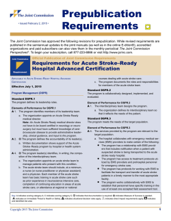
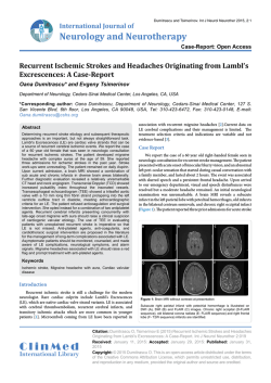
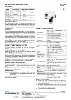
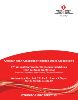
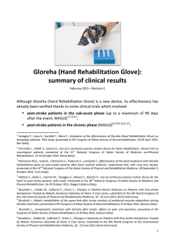
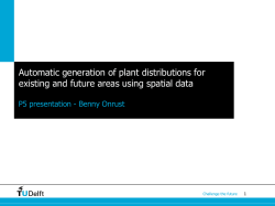
The TeleStroke Mimic (TM)‐Score: A Prediction Rule for
Identifying Stroke Mimics Evaluated in a Telestroke Network
The Harvard community has made this article openly available.
Please share how this access benefits you. Your story matters.
Citation
Ali, S. F., A. Viswanathan, A. B. Singhal, N. S. Rost, P. G.
Forducey, L. W. Davis, J. Schindler, et al. 2014. “The TeleStroke
Mimic (TM)‐Score: A Prediction Rule for Identifying Stroke
Mimics Evaluated in a Telestroke Network.” Journal of the
American Heart Association: Cardiovascular and Cerebrovascular
Disease 3 (3): e000838. doi:10.1161/JAHA.114.000838.
http://dx.doi.org/10.1161/JAHA.114.000838.
Published Version
doi:10.1161/JAHA.114.000838
Accessed
February 6, 2015 10:54:04 AM EST
Citable Link
http://nrs.harvard.edu/urn-3:HUL.InstRepos:13890751
Terms of Use
This article was downloaded from Harvard University's DASH
repository, and is made available under the terms and conditions
applicable to Other Posted Material, as set forth at
http://nrs.harvard.edu/urn-3:HUL.InstRepos:dash.current.terms-ofuse#LAA
(Article begins on next page)
ORIGINAL RESEARCH
The TeleStroke Mimic (TM)-Score: A Prediction Rule for Identifying
Stroke Mimics Evaluated in a Telestroke Network
Syed F. Ali, MD; Anand Viswanathan, MD; Aneesh B. Singhal, MD; Natalia S. Rost, MD; Pamela G. Forducey, PhD; Lawrence W. Davis, MD;
Joseph Schindler, MD; William Likosky, MD; Sherene Schlegel, BSN; Nina Solenski, MD; Lee H. Schwamm, MD; on Behalf of Partners
Telestroke Network
Background-—Up to 30% of acute stroke evaluations are deemed stroke mimics (SM). As telestroke consultation expands across
the world, increasing numbers of SM patients are likely being evaluated via Telestroke. We developed a model to prospectively
identify ischemic SMs during Telestroke evaluation.
Methods and Results-—We analyzed 829 consecutive patients from January 2004 to April 2013 in our internal New England–based
Partners TeleStroke Network for a derivation cohort, and 332 cases for internal validation. External validation was performed on
226 cases from January 2008 to August 2012 in the Partners National TeleStroke Network. A predictive score was developed using
stepwise logistic regression, and its performance was assessed using receiver-operating characteristic (ROC) curve analysis. There
were 23% SM in the derivation, 24% in the internal, and 22% in external validation cohorts based on final clinical diagnosis.
Compared to those with ischemic cerebrovascular disease (iCVD), SM had lower mean age, fewer vascular risk factors, more
frequent prior seizure, and a different profile of presenting symptoms. The TeleStroke Mimic Score (TM-Score) was based on
factors independently associated with SM status including age, medical history (atrial fibrillation, hypertension, seizures), facial
weakness, and National Institutes of Health Stroke Scale >14. The TM-Score performed well on ROC curve analysis (derivation
cohort AUC=0.75, internal validation AUC=0.71, external validation AUC=0.77).
Conclusions-—SMs differ substantially from their iCVD counterparts in their vascular risk profiles and other characteristics.
Decision-support tools based on predictive models, such as our TM Score, may help clinicians consider alternate diagnosis and
potentially detect SMs during complex, time-critical telestroke evaluations. ( J Am Heart Assoc. 2014;3:e000838 doi: 10.1161/
JAHA.114.000838)
Key Words: cerebrovascular disease • stroke mimics • telestroke • thrombolysis
T
elestroke has grown significantly in the past decade and
has entered mainstream care for patients with acute
stroke.1 Defined as the use of telecommunication technologies to provide medical information and services to stroke
patients,2 telestroke has been adopted and implemented by
multiple different types of healthcare organizations across the
United States and abroad.3 Telestroke enables stroke patients
From the Massachusetts General Hospital/Harvard Medical School, Boston,
MA (S.F.A., A.V., A.B.S., N.S.R., L.H.S.); INTEGRIS Health, Oklahoma City, OK
(P.G.F., L.W.D.); Yale-New Haven Stroke Center, New Haven, CT (J.S.); Swedish
Medical Center, Seattle, WA (W.L., S.S.); University of Virginia, Charlottesville,
VA (N.S.).
Correspondence to: Lee H. Schwamm, MD, Department of Neurology, MGH
Stroke Services, Massachusetts General Hospital, Harvard Medical School,
MGH, 55 Fruit St, Boston, MA 02114. E-mail: [email protected]
Received March 13, 2014; accepted May 13, 2014.
ª 2014 The Authors. Published on behalf of the American Heart Association,
Inc., by Wiley Blackwell. This is an open access article under the terms of the
Creative Commons Attribution-NonCommercial License, which permits use,
distribution and reproduction in any medium, provided the original work is
properly cited and is not used for commercial purposes.
DOI: 10.1161/JAHA.114.000838
to be remotely evaluated, thereby allowing optimal treatment
and management in medically underserved areas, removing
geographical disparities in access to expert care.1,4 Decisionanalytic models demonstrate that telestroke is cost-effective
from both a societal and a hospital perspective.5,6
While a significant percentage of stroke patients are now
provided initial care through telestroke consultations,4 its
ease of use, greater availability, and cost effectiveness have
led to a high number of consults by emergency department
(ED) physicians. However, it is estimated that 5% to 30% of ED
patients suspected of having an acute stroke end up with
a diagnosis of a stroke mimic (SM). Seizures, migraine,
psychogenic disorders, and toxic/metabolic causes are the
most common nonvascular conditions that mimic stroke.7–14
The use of telestroke consultations to evaluate SMs, and the
use of intravenously administered tissue plasminogen activator (tPA) in these cases, while safe,12,14–16 may lessen the
cost effectiveness of this method of stroke evaluation.
These observations motivated us to perform a retrospective analysis of all the patients managed in our large national
Journal of the American Heart Association
1
TeleStroke Mimic (TM)-Score
Ali et al
Methods
Patient Population
We report data from two mutually exclusive sources: our
internal New England–based Partners TeleStroke Network and
our Partners National TeleStroke Network (PNTN). The Partners TeleStroke Network comprises 2 hubs (Massachusetts
General Hospital and Brigham and Women’s Hospital) currently
serving 31 spoke hospitals in Massachusetts, Maine, and New
Hampshire. Partners TeleStroke Network was established in
2000 as one of the first telestroke systems in the country. All
telestroke consults are entered into a database via an online
web-based portal by the neurologist, with the data stored
centrally at a secure server at Massachusetts General Hospital.
There were 8839 consecutive patients entered into the
database from January 2004 to April 2013. Patients were
excluded from this analysis if they were evaluated by telephone
only (n=7044), leaving 1795 video-telestroke consults for
evaluation. After excluding those with unverified age data
(n=463) or incomplete medical comorbidity data on admission
(n=171), there were 1161 video-telestroke consults available
for analysis. Age was calculated and verified by subtracting the
patient date of birth (DOB) from the date of consultation. Due
to the urgent nature of telestroke consults, not all patients had
a documented DOB in the database. To address any nonrandom incompleteness of age data, we compared patients with
documented DOB and those missing DOB.
Of the 1161 patients with complete data included in the
analysis, 70% (n=829) were randomly selected for the internal
derivation cohort and the remaining were used for internal
validation cohort. We extracted data on demographics (age,
race, ethnicity, and gender), baseline clinical characteristics
(hypertension, diabetes mellitus, hyperlipidemia, coronary
artery disease, atrial fibrillation, heart failure, previous stroke,
smoker, prior seizure, prior myocardial infarction), medication
use (antiplatelet or anticoagulant use), initial symptoms
documented by the referring emergency physician (presence
of “weakness,” “speech problem,” or “altered level of
consciousness”), and the evaluation performed by the
telestroke consultant (initial National Institutes of Health
Stroke Scale [NIHSS], diagnosis, and intravenous tPA eligibility and administration).
For external validation, we included 226 video-telestroke
consults from the external PNTN from January 2008 through
August 2012. The PNTN centers included four Hubs serving a
total of 22 spokes in their geographic regions: Yale–New
Haven Hospital, New Haven, Conn; Swedish Medical Center,
Seattle, Wash.; INTEGRIS Health, Oklahoma City, Okla., and
DOI: 10.1161/JAHA.114.000838
the University of Virginia Health System, Charlottesville, Va.
All PNTN Hub sites used the same shared database for
recording patient information and documenting treatment
timelines and decision-making. The data are stored on a
secure, HIPAA-compliant central server located in Boston,
Mass. housed behind the Massachusetts General Hospital
firewall. Each PNTN Hub site granted permission to share
de-identified data on patient encounters. Analysis of this
de-identified data in our stroke database is approved by our
Institutional Review Board.
TeleStroke Evaluation, Workflow, and Diagnostic
Classification
Hospitals that wish to participate in the PNTN as referring
spoke sites must first execute contracts with a PNTN Hub
hospital, and obtain the necessary approved equipment for
teleconsultation, adapt their acute stroke protocols, and
engage in training for telestroke consultations. All Hub
neurologists in our network are board certified in vascular
neurology or have extensive experience in stroke. Spoke sites
are instructed to contact the Hub by phone for any cases in
which they suspect acute ischemic stroke and for advice or
treatment guidance. Typically, spokes will have performed an
initial assessment including basic diagnostics, blood work,
and often brain imaging. If the clinical scenario requires
videoconferencing and image review to be properly
addressed, a secure, web-based system of high-quality
commercial videoconferencing is initiated and image transfer
via secure Internet protocols is performed. These methods
have been described in detail in prior publications. They
permit interactive dialogue, neurologic examination, discussion of risks and benefits of the available treatment options,
and documentation of the consult in a secure, web-enabled
SQL database that can be used for reporting and analysis. At
the conclusion of the teleconsultation, the spoke and Hub
physicians come to agreement on the triage disposition of the
patient (eg, remain at the spoke, discharge to home, transfer
to the Hub, etc.). At this time, the Hub neurologist documents
his/her findings and assigns a diagnosis based on review of
all the available clinical and imaging data, picking this
diagnosis from a list of prespecified options, or writing it in
as a free text diagnosis. For these analyses, diagnoses were
classified as either ischemic cerebrovascular disease (iCVD) if
they were acute ischemic stroke (<9-hour duration), subacute
ischemic stroke (>9-hour duration) or transient ischemic
attack; or as a SM for all other diagnoses.
Statistical Analysis
Baseline demographics, clinical characteristics, initial presentation, and intravenous administration of tPA were compared
Journal of the American Heart Association
2
ORIGINAL RESEARCH
telestroke network with a goal of developing a score to
identify SMs based on presenting characteristics.
TeleStroke Mimic (TM)-Score
Ali et al
Results
Patients Characteristics
There were 829 patients with video-telestroke consults in the
derivation cohort, 332 patients in the internal validation
DOI: 10.1161/JAHA.114.000838
cohort, and 226 in the external validation cohort. When
we compared those with DOB missing versus present in
the record, there were no significant differences in any
measured variables, including demographics, baseline clinical
characteristics, medication use, initial symptoms documented
by the emergency physician, NIHSS, diagnoses, or use of
intravenous tPA.
Derivation, internal validation, and external validation
cohorts were similar in these characteristics (Table 1) without
any significant differences between the derivation and internal
validation cohorts other than the presence of facial weakness.
When compared to the derivation cohort, patients in the
external validation cohort were younger, with more often a
history of hypertension, diabetes, smoking and prior seizure,
less often atrial fibrillation, and more often the presence of
altered level of consciousness (Table 1). The percentages of
SM were not different in the 3 cohorts (22.9% versus 24.1%
versus 21.7%).
Derivation Cohort
Mean age of patients in the derivation cohort was observed
to be 68.3Æ16.2 years; 46.9% of patients were male and
88.2% were white. Seventy-seven percent of patients had
iCVD (acute ischemic stroke, 66.8%; transient ischemic
attack, 8.6%; subacute ischemic stroke, 1.7%). The most
common alternative diagnoses in SM group were seizure/
epilepsy (3.2%), headache/migraine with or without aura
(2.9%), encephalopathy (2.6%), conversion disorder (1.5%),
hemorrhagic stroke (1.0%), and all other miscellaneous
diagnoses combined (11.7%). Vascular comorbidities were
frequent in the derivation cohort, with hypertension being the
most common in 52.2%, hyperlipidemia in 23.4%, diabetes
mellitus in 18.3%, coronary artery disease in 15.6%, atrial
fibrillation in 18.2%, and a history of seizure in 3.0% of
telestroke patients.
Among clinical presentations, facial weakness was documented in 59.3%, limb weakness in 69.5%, difficulty with
speech in 63.1%, and altered level of consciousness in 41.0%
of patients. The median NIHSS was 6 (interquartile range 3 to
13), and 18.5% of the telestroke patients were in hypertensive crisis (BP>180/110) at the time of the telestroke
consult. Forty percent of patients were treated with intravenous tPA.
Patients with the diagnosis of SM as compared to those
with iCVD differed significantly in many characteristics, as
shown in Table 2. SM patients were approximately 10 years
younger; were significantly less likely to have a history of
hypertension, atrial fibrillation, and heart failure; and were
more likely to have a history of seizure. SM patients
presented with a 4-point-lower median NIHSS (7.0 versus
3.0). They were less likely to have facial weakness, limb
Journal of the American Heart Association
3
ORIGINAL RESEARCH
between patients diagnosed with iCVD or SM in the derivation
cohort. Chi-square test was used to compare categorical
variables, and an independent-sample t test or Wilcoxon rank
sum test was used to compare mean or median differences for
continuous variables in baseline characteristics between
groups. Variables significant in univariate testing at a level of
P<0.10 were entered into a stepwise logistic regression
analysis in the order of their significance of association. Those
that remained significant in multivariable testing were then
included in the prediction model scoring system. NIHSS was
analyzed both as a continuous variable and as a categorical
variable. In addition, the NIHSS was plotted as a distribution,
and was found to be skewed with an inflection point at an NIHSS
of 14 points. Based on this distribution, a dichotomous variable
(NIHSS >14) was also created and both versions were tested in
the multivariable model separately. In the stepwise logistic
regression model, factors with P<0.05 were retained. We also
explored interactions between these variables, and included
any interaction terms that were significant at the 0.05 level.
The results of the multivariable analysis were then used
to develop a prediction model for SMs using a standard
regression coefficient-based scoring method.17,18 Integer
scores were assigned by dividing factors’ coefficients by the
model constant and rounding up to the nearest integer. The
TeleStroke Mimic Score (TM-Score) for any individual patient
was determined by summing the points assigned for each
factor present. Scores were divided into deciles and displayed
accordingly. Model discrimination was assessed by the
receiver-operating characteristic area under curve, which is
equivalent to the c-statistic.19 The performance of the model
was validated by comparing the receiver-operating characteristic curve analysis in the derivation set with that in the internal
validation and external validation sets.20 The resulting continuous distributions of total scores across all patients in the
2 validation cohorts were then stratified into the decile groups
of the derivation cohort. Separate graphs were plotted with a
trend line showing the relationship between the score and
likeliness of being diagnosed as a SM in the derivation cohort
and comparing that with internal and external validation
cohorts. Finally, we constructed a simple graphic nomogram
for use by physicians performing telestroke consults to
quantify the likelihood of any individual case being a SM. All
statistical analyses were performed using version 20.0 of the
IBM Software Package for Statistical Analysis (SPSS) software
package for statistical analysis (SPSS).
TeleStroke Mimic (TM)-Score
Ali et al
ORIGINAL RESEARCH
Table 1. Characteristics of Those Patients Evaluated by Telestroke Consultation, Comparing Those in the Derivation Cohort to
Those in the Internal Validation and External Validation Cohorts, Respectively
Variable
Derivation
Cohort (n=829)
Int. Validation
Cohort (n=332)
P Value
Ext. Validation
Cohort (n=226)
P Value
Age, y
68.3Æ16.2
67.8Æ16.8
0.580
65.5Æ16.0
0.020
Gender—male
46.9%
48.8%
0.564
46.0%
0.809
Ethnicity—Hispanic
7.0%
8.4%
0.398
Race—white
88.2%
86.4%
0.417
87.2%
0.679
Hypertension
52.2%
51.2%
0.752
63.3%
0.003
Diabetes mellitus
18.3%
18.1%
0.917
25.7%
0.014
Hyperlipidemia
23.4%
20.5%
0.282
26.5%
0.327
Coronary artery disease
15.6%
15.4%
0.932
20.4%
0.086
Atrial fibrillation
18.2%
12.7%
0.023
12.4%
0.039
Heart failure
5.9%
8.7%
0.082
7.5%
0.375
Previous stroke
24.7%
24.7%
0.992
30.1%
0.103
Smoker
5.8%
4.8%
0.512
12.8%
0.001
Seizure
3.0%
2.7%
0.781
6.2%
0.025
Prior MI
7.2%
5.7%
0.354
6.2%
0.586
6 (3 to 13)
5 (2 to 11)
0.246
5 (2 to 11)
0.190
Facial weakness
59.3%
51.8%
0.022
63.3%
0.285
Limb weakness
69.5%
65.4%
0.173
72.1%
0.442
Speech problem
63.1%
60.2%
0.366
65.9%
0.431
Altered mental status
41.0%
39.8%
0.694
50.9%
0.008
Medical history
Clinical presentation
NIHSS*
Signs at presentation
Hypertensive crisis
Diagnosis—SM
18.5%
21.2%
0.285
20.6%
0.274
22.9%
24.1%
0.668
21.7%
0.694
Ext. indicates external; Int., internal; MI, myocardial infarction; NIHSS, National Institutes of Health Stroke Scale; SM, stroke mimics.
*Median (interquartile range).
weakness, speech problems, or hypertensive crisis (blood
pressure >180/110) at presentation (Table 2).
Univariate and Multivariable Analysis
Factors significant in univariate correlates of SM in the
derivation set included age, hypertension, atrial fibrillation,
heart failure, history of seizures, initial NIHSS, presenting
symptoms of facial weakness, limb weakness, speech problems, hypertensive crisis, and prior use of antiplatelet and
anticoagulation medicines. In the stepwise logistic regression
model for SM, the variables listed in Table 3 were retained as
independent predictors (P<0.05) of SM. NIHSS was not
significant in logistic regression when entered as a continuous
variable but was significantly associated with iCVD when
entered in the dichotomous form (NIHSS >14, Figure 1)
(Table 3).
DOI: 10.1161/JAHA.114.000838
Development of the Prediction Rule: TM Score
To develop a prediction rule, we selected the 6 variables that
remained significant after stepwise logistic regression (age,
atrial fibrillation, hypertension, seizure, facial weakness, and
NIHSS >14) and calculated an integer score proportional
to their beta coefficients on logistic regression. Age was
modeled as a continuous variable, while the other 5 variables
were modeled as dichotomous variables. For each patient, all
applicable scores were summed to attain a total TM Score for
that patient. There was a graded decrease in the likelihood of
having a SM disorder with increasing TM Score. Deciles were
used to plot the data with a corresponding percentage of
patients with SM in each point category and a trend-line was
plotted (Figure 2). The TM Score performed well on receiveroperating characteristic curve analysis for predicting the
likelihood of SM (area under the curve 0.75Æ0.02) (Figure 3).
Journal of the American Heart Association
4
TeleStroke Mimic (TM)-Score
Ali et al
iCVD (n=639)
Stroke Mimic (n=190)
P Value
Age, y
70.5Æ15.0
61.1Æ18.2
Gender—male
48.4%
42.1%
0.130
Ethnicity—Hispanic
6.7%
7.9%
0.580
Race—white
88.6%
86.8%
0.516
Hypertension
56.2%
38.9%
<0.0001†
Diabetes mellitus
19.1%
15.8%
0.302
Hyperlipidemia
24.6%
19.5%
0.145
Coronary artery disease
16.6%
12.1%
0.134
Atrial fibrillation
21.6%
6.8%
<0.0001†
Heart failure
6.9%
2.6%
0.029†
Previous stroke
25.8%
21.1%
0.181
Smoker
5.3%
7.4%
0.289
Seizure
2.2%
5.8%
0.011†
Prior MI
7.2%
7.4%
0.937
NIHSS*
7 (3 to 15)
3 (1 to 8)
<0.0001†
NIHSS <14
25.2%
9.6%
<0.0001†
Facial weakness
66.8%
34.2%
<0.0001†
Limb(s) weakness
73.6%
55.8%
<0.0001†
Speech problem
66.4%
52.1%
<0.0001†
Altered LOC
40.7%
42.1%
0.727
Hypertensive crisis
22.5%
15.8%
0.006†
Antiplatelet
41.9%
34.2%
0.056†
Anticoagulation
9.2%
5.3%
0.082†
<0.0001†
Medical history
Clinical presentation
Signs at presentation
Medications
Ext. indicates external; iCVD, ischemic cerebrovascular disease; Int., internal; LOC, level of consciousness; MI, myocardial infarction; NIHSS, National Institutes of Health Stroke Scale.
*Continuous NIHSS; median (interquartile range).
†
Stepwise logistic regression model.
Table 3. Factors Significantly Associated With Stroke Mimics in Derivation Cohort on Stepwise Logistic Regression Model
Multivariable Analysis
SM Predictors
b
Adjusted OR (95% CI)
Points
Age (per y)
À0.026
0.98 (0.96 to 0.99)
+0.2/y
Atrial fibrillation
À0.781
0.48 (0.26 to 0.90)
+6
Hypertension
À0.399
0.67 (0.46 to 0.96)
+3
Seizure
0.885
2.70 (1.10 to 6.67)
À6
Facial weakness
À1.245
0.32 (0.22 to 0.45)
+9
NIHSS >14
À0.591
0.56 (0.31 to 0.98)
+5
NIHSS, National Institutes of Health Stroke Scale; OR, odds ratio; SM, stroke mimics.
DOI: 10.1161/JAHA.114.000838
Journal of the American Heart Association
5
ORIGINAL RESEARCH
Table 2. Demographics and Clinical Characteristics of Patients With Ischemic Cerebrovascular Disease (iCVD) vs Stroke Mimics
(ie, Those Without Ischemic Cerebrovascular Disease) in the Derivation Cohort Over the Study Period of 9 Years
TeleStroke Mimic (TM)-Score
Ali et al
60%
% Stroke Mimics
50%
40%
30%
20%
Discussion
10%
Using a national network of 52 spoke hospitals supported by
6 Hub hospitals in 7 states and evaluating a cohort of 1387
patients, we developed and validated a clinical prediction rule
to calculate the likelihood of a patient being “tele”-SM. A
model based on the 6 factors that were associated with
mimics allowed us to readily differentiate between SMs and
patients with true iCVD on telestroke consults with reasonable model performance. The described predictive model
represents a real-time clinical decision-making aid to prompt
consideration of the possibility of a SM and is not intended as
a diagnostic classification tool.
To our knowledge, this is the largest cohort study to date
evaluating SM in a national telestroke network. We observed a
similar rate of SMs in our telestroke cohort as that reported
for patients presenting directly to stroke centers. In one of the
earliest studies on SM patients by Harbison et al., the rate
was 27% in a series of 487 consecutive patients who were
directly admitted to a stroke service over a 6-month period.21
Hand et al. reported that among 350 consecutive patients
0%
0–4
5–9
10 – 14
15 – 19
≥ 20
NIHSS
Figure 1. Stroke mimic patients classified according to initial
NIHSS. NIHSS indicates National Institutes of Health Stroke
Scale.
Model Validation
Validation sets consisted of an internal validation cohort and
an external validation cohort. When the TM-Score was applied
to the internal validation cohort, the area under the curve
was observed to be 0.72Æ0.03, only a slight degradation in
performance. While applying the TM-Score to the external
validation cohort, we observed a greater area under curve of
0.77Æ0.03, showing that the prediction rule holds true on
both internal and external validation (Figure 3). Similar to the
derivation cohort, using the same deciles, we divided the data
in the internal and external validation cohort and plotted as a
80
% Likelihood of Stroke Mimic
70
60
50
40
30
20
10
0
0
5
10
15
20
25
30
35
40
TM - Score
Figure 2. Scatter plot of the data with a trend line showing the relationship between TM-Score and the
likelihood of having a stroke mimic in derivation cohort. TM-Score indicates TeleStroke Mimic Score.
DOI: 10.1161/JAHA.114.000838
Journal of the American Heart Association
6
ORIGINAL RESEARCH
trend line on the same graph. As shown by Figure 4, the 3
lines depict a similar trend and were not significantly different.
For the ease of telestroke physicians using TM-Score during
their consult, we have included a nomogram showing the
relationship between TM-Score and the likelihood of having a
SM based on the prediction rule (Figure 5).
70%
TeleStroke Mimic (TM)-Score
Ali et al
ORIGINAL RESEARCH
Internal Derivation Cohort
Internal Validation Cohort
External Validation Cohort
Figure 3. Response operator curve in the derivation cohort for the proposed score. ROC indicates receiver-operating characteristic.
with focal brain dysfunction of sudden onset presenting to an
urban teaching hospital, 31% were SMs.9 A recent series from
a single stroke center found that 27% of patients referred
from the ED as a stroke code did not have a cerebrovascular
disorder, and the proportion of inpatient SMs was even
higher.22 Other studies have reported lower rates of SMs. In a
study by Libman et al., among 411 consecutive patients
presenting to the ED, only 19% had a SM. It should be noted
for this study that the ED physicians were specifically trained
to identify stroke features since they were participating in an
acute intervention trial.10 The SM rate was only 4.8% in a
sample of 637 patients who were admitted to a stroke
department after an initial evaluation, performed by a
neurologist, that included computed tomography imaging.23
Several clinical factors that predict the presence of SM
have been identified, and studies have noted that patients
with SMs are younger and have lower vascular risk factors.9,10,12,15,24 The current study, however, is one of the first
to explore the differences in demographics and clinical
characteristics in patients managed during video-telestroke
DOI: 10.1161/JAHA.114.000838
consultations. We observed similar differences between SMs
and those with cerebrovascular disease as those that were
reported in the literature. Our results showed that SM
patients were around 10 years younger with significantly
fewer vascular risk factors. They were also observed to have
fewer focal neurological symptoms and a lower median NIHSS
at the time of consult. Chang et al. also reported a lower focal
weakness and a lower median NIHSS in patients with SMs.25
Commensurate with the available literature, the SM group had
a significantly higher percentage of patients with a medical
history of seizure.9,12,14
Recognizing patient characteristics that differentiate iCVD
patients from SM patients can be extremely useful when
evaluating patients during telestroke consults. One interesting
finding from our data is the very strong association between
absence of facial weakness and a SM diagnosis. This suggests
that patients with conditions that mimic stroke often do not
have facial weakness, and this may be an important feature to
focus on during the evaluation of patients with strokelike symptoms during telestroke evaluation. Similarly, the
Journal of the American Heart Association
7
TeleStroke Mimic (TM)-Score
Ali et al
ORIGINAL RESEARCH
80
ood of Stroke Mimic
% Likeliho
70
60
50
40
30
20
10
0
0
5
10
15
25
20
30
35
40
TM -Score
Figure 4. Scatter plot with a trend line showing the relationship between TM-Score and the likelihood of
having a stroke mimic in derivation cohort (solid blue line), internal validation cohort (dotted red line), and
external validation cohort (dashed green line). TM-Score indicates TeleStroke Mimic Score.
presence of a seizure disorder raises the likelihood that the
current symptoms may be due to seizures with a postictal
Todd’s paralysis. However, a careful evaluation is still required
because some of these patients will have had a new ischemic
cerebrovascular event. Advanced imaging may be useful in
these patients to definitively exclude ischemia. It is possible
Figure 5. Recommended final prediction nomogram for Telestroke stroke mimics. TM-Score=(Age
multiplied by 0.2)+6 (if Hx of atrial fib)+3 (if Hx of HTN)+9 (if facial weakness)+5 (if NIHSS >14)À6 (if Hx of
seizure). TM-Score indicates TeleStroke Mimic Score.
DOI: 10.1161/JAHA.114.000838
Journal of the American Heart Association
8
TeleStroke Mimic (TM)-Score
Ali et al
DOI: 10.1161/JAHA.114.000838
However, the factors in our prediction rule were recorded
regularly and are relatively unlikely to be influenced by the Hub
neurologist judgment. The stroke neurologist’s clinical diagnosis (iCVD versus SM) is the reference standard used in these
analyses to classify patients. We had no alternative method
available to further validate this diagnosis since only a fraction
of patients were transferred to the Hub hospital postconsult for
further evaluation or definitive magnetic resonance imaging.
Since our approach to intravenous tPA eligibility is to err on the
side of a diagnosis of ischemic stroke unless an alternative
diagnosis is convincingly present, it is likely that some patients
without true iCVD were classified as having iCVD; this result is
more likely rather than the reverse, ie, patients with true iCVD
would be classified as mimics. As this study included data from
the spokes associated with these 6 urban tertiary care
hospitals, it is possible that the proportion of SMs at other
hospitals could differ. Therefore, we encourage the replication
of this approach in other telestroke systems, especially those
that serve very rural populations. Lastly, it is possible that SMs
evaluated in person might have different characteristics than
those seen over telestroke, where a physician has already
applied some judgment as to the likelihood of iCVD. Therefore,
our findings should also be replicated in non-telestroke
environments before being applied in these paradigms.
In conclusion, we believe that as telestroke consultation
expands, increasing numbers of SM patients will be evaluated.
These SM patients differ substantially from their counterpart
iCVD patients in their vascular risk profiles and other
characteristics. Decision-making support tools based on
predictive models, such as the TM-Score we have proposed,
may help clinicians consider alternative diagnoses and
potentially identify SMs during complex, time-critical telestroke evaluations.
Acknowledgments
We would like to thank all the staff at our regional and Partners
National TeleStroke Network sites for their continued effort and
support. We gratefully acknowledge the contributions of John
Johnson and Juan Estrada for their leadership and guidance in the
technical and administrative aspects of the program, respectively.
Disclosures
Syed F. Ali, Anand Viswanathan, Aneesh B. Singhal, Natalia S.
Rost, Pamela G. Forducey, Lawrence W. Davis, Joseph
Schindler, William Likosky, Sherene Schlegel, and Nina
Solenski all report no disclosures. Dr Schwamm serves as a
consultant to the Massachusetts Department of Public Health,
and is the Medical Director of the Mass General TeleHealth
program and the Partners TeleStroke Center. Massachusetts
General Hospital provides telehealth services to hospitals in
Journal of the American Heart Association
9
ORIGINAL RESEARCH
that this tool will also prove useful in detecting SMs in real
time over telestroke, and potentially even in those patients
who present in person to stroke centers. While it is possible
that this tool is of greater value in a telestroke environment
where some additional bedside clues may not be appreciated,
most of the factors in the score are objective elements that
are unlikely to be different when assessed in person versus
remotely. The main difference would be facial weakness,
which has been shown to be of good inter-rated reliability on
studies comparing in person to remote NIHSS.26 If one were
to speculate on useful operational cutoffs for how best to use
the score prospectively, based on our data it would be
reasonable to use a score of ≤5 or the lack of facial weakness
to strongly raise suspicion of a mimic (65% chance based on
the score) and a score of ≥20 to strongly support the
diagnosis of stroke. These cutoffs might be most useful in the
decision of whether or not to mix intravenous tPA in advance
of brain imaging, or to delay treatment until magnetic
resonance imaging evidence of stroke, to most efficiently
use these expensive resources.
As telestroke is rapidly being adopted,4 neurologists can
anticipate an increased incidence of telestroke patients
presenting with SM features. It may be beneficial to have a
simple prediction rule that can be used to heighten awareness. However, a major issue with prediction rules is that
physicians have found prediction rules difficult to implement
in real-time use.27 The prediction rule presented herein is
based on the information easily available at the time of the
initial ED evaluation. All the variables used to calculate the
score are intuitive and biologically plausible, and the method
of calculating the TM-Score is straightforward. We have also
produced a nomogram, which could be printed and carried on
a pocket-sized card and used to estimate the likelihood of a
patient being a SM. If independently validated in other
centers, the TM-Score may help future ED physicians at
spokes and tele-neurologists at Hubs to be able to better
prioritize and triage consultations, thus improving the overall
system performance of telestroke consultations and saving
valuable resources.
There are inherent limitations in the interpretations of the
current study design. First, it is a retrospective analysis of a
prospectively collected dataset. There may be incomplete data
capture, and risk factors might have been abstracted with some
variability across sites despite common definitions and a single,
web-based data report form. We may be missing some
important patient characteristics that could be associated with
SMs such as symptoms of vertigo, ataxia, dizziness, and visual
changes that could add discriminating power to our prediction
rule. The data were not subject to external audits, so there may
be inaccuracies in data abstraction. Random measurement
error and misclassification can lead to dilution bias and
underestimation of the effects of the tested risk factors.
TeleStroke Mimic (TM)-Score
Ali et al
References
12. Winkler DT, Fluri F, Fuhr P, Wetzel SG, Lyrer PA, Ruegg S, Engelter ST.
Thrombolysis in stroke mimics: frequency, clinical characteristics, and
outcome. Stroke. 2009;40:1522–1525.
13. Uchino K, Massaro L, Hammer MD. Transient ischemic attack after tissue
plasminogen activator: aborted stroke or unnecessary stroke therapy?
Cerebrovasc Dis. 2010;29:57–61.
14. Chernyshev OY, Martin-Schild S, Albright KC, Barreto A, Misra V, Acosta I,
Grotta JC, Savitz SI. Safety of tpa in stroke mimics and neuroimaging-negative
cerebral ischemia. Neurology. 2010;74:1340–1345.
15. Tsivgoulis G, Alexandrov AV, Chang J, Sharma VK, Hoover SL, Lao AY, Liu W,
Stamboulis E, Alexandrov AW, Malkoff MD, Frey JL. Safety and outcomes
of intravenous thrombolysis in stroke mimics: a 6-year, single-care
center study and a pooled analysis of reported series. Stroke. 2011;42:
1771–1774.
2. Perednia DA, Allen A. Telemedicine technology and clinical applications. JAMA.
1995;273:483–488.
16. Guillan M, Alonso-Canovas A, Gonzalez-Valcarcel J, Garcia Barragan N, Garcia
Caldentey J, Hernandez-Medrano I, Defelipe-Mimbrera A, Sanchez-Gonzalez V,
Terecoasa E, Alonso de Lecinana M, Masjuan J. Stroke mimics treated with
thrombolysis: further evidence on safety and distinctive clinical features.
Cerebrovasc Dis. 2012;34:115–120.
3. Park S, Schwamm LH. Organizing regional stroke systems of care. Curr Opin
Neurol. 2008;21:43–55.
17. Moons KG, Harrell FE, Steyerberg EW. Should scoring rules be based on odds
ratios or regression coefficients? J Clin Epidemiol. 2002;55:1054–1055.
4. Silva GS, Farrell S, Shandra E, Viswanathan A, Schwamm LH. The status of
telestroke in the United States: a survey of currently active stroke
telemedicine programs. Stroke. 2012;43:2078–2085.
18. Tu JV, Naylor CD. Clinical prediction rules. J Clin Epidemiol. 1997;50:743–744.
1. Hess DC, Audebert HJ. The history and future of telestroke. Nat Rev Neurol.
2013;9:340–350.
5. Nelson RE, Saltzman GM, Skalabrin EJ, Demaerschalk BM, Majersik JJ. The
cost-effectiveness of telestroke in the treatment of acute ischemic stroke.
Neurology. 2011;77:1590–1598.
6. Switzer JA, Demaerschalk BM, Xie J, Fan L, Villa KF, Wu EQ. Cost-effectiveness
of hub-and-spoke telestroke networks for the management of acute ischemic
stroke from the hospitals’ perspectives. Circ Cardiovasc Qual Outcomes.
2013;6:18–26.
7. Brunser AM, Illanes S, Lavados PM, Munoz P, Carcamo D, Hoppe A, Olavarria VV,
Delgado I, Diaz V. Exclusion criteria for intravenous thrombolysis in stroke
mimics: an observational study. J Stroke Cerebrovasc Dis. 2013;22:1140–1145.
8. Hemmen TM, Meyer BC, McClean TL, Lyden PD. Identification of nonischemic
stroke mimics among 411 code strokes at the University of California, San
Diego, stroke center. J Stroke Cerebrovasc Dis. 2008;17:23–25.
9. Hand PJ, Kwan J, Lindley RI, Dennis MS, Wardlaw JM. Distinguishing between
stroke and mimic at the bedside: the brain attack study. Stroke.
2006;37:769–775.
10. Libman RB, Wirkowski E, Alvir J, Rao TH. Conditions that mimic stroke in the
emergency department. Implications for acute stroke trials. Arch Neurol.
1995;52:1119–1122.
11. Ay H, Buonanno FS, Rordorf G, Schaefer PW, Schwamm LH, Wu O, Gonzalez
RG, Yamada K, Sorensen GA, Koroshetz WJ. Normal diffusion-weighted mri
during stroke-like deficits. Neurology. 1999;52:1784–1792.
DOI: 10.1161/JAHA.114.000838
19. Hanley JA, McNeil BJ. The meaning and use of the area under a receiver
operating characteristic (roc) curve. Radiology. 1982;143:29–36.
20. Metz CE. Basic principles of roc analysis. Semin Nucl Med. 1978;8:283–298.
21. Harbison J, Hossain O, Jenkinson D, Davis J, Louw SJ, Ford GA. Diagnostic
accuracy of stroke referrals from primary care, emergency room physicians,
and ambulance staff using the face arm speech test. Stroke. 2003;34:
71–76.
22. El Husseini N, Goldstein LB. “Code stroke”: hospitalized versus emergency
department patients. J Stroke Cerebrovasc Dis. 2013;22:345–348.
23. Vroomen PC, Buddingh MK, Luijckx GJ, De Keyser J. The incidence of stroke
mimics among stroke department admissions in relation to age group. J Stroke
Cerebrovasc Dis. 2008;17:418–422.
24. Scott PA, Silbergleit R. Misdiagnosis of stroke in tissue plasminogen activatortreated patients: characteristics and outcomes. Ann Emerg Med.
2003;42:611–618.
25. Chang J, Teleb M, Yang JP, Alderazi YJ, Chapple K, Frey JL, Restrepo L. A model
to prevent fibrinolysis in patients with stroke mimics. J Stroke Cerebrovasc Dis.
2012;21:839–843.
26. Shafqat S, Kvedar JC, Guanci MM, Chang Y, Schwamm LH. Role for
telemedicine in acute stroke. Feasibility and reliability of remote administration of the NIH stroke scale. Stroke. 1999;30:2141–2145.
27. Laupacis A, Sekar N, Stiell IG. Clinical prediction rules: a review and suggested
modifications of methodological standards. JAMA. 1997;277:488–494.
Journal of the American Heart Association
10
ORIGINAL RESEARCH
the New England region, including telestroke. Dr Schwamm is
the Principal Investigator of an National Institute of Neurological Disorders and Stroke–funded SPOTRIAS center clinical
trial, MR WITNESS, for which Genentech provides alteplase
and additional funding. His work is supported in part by Health
Resource Services Administration Requisition 09-HRS9923AB (Stroke and Traumatic Brain Injury Telehealth Services).

© Copyright 2026