
Assessment of in vitro prophylactic efficacy of gallic acid fabricated
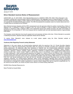
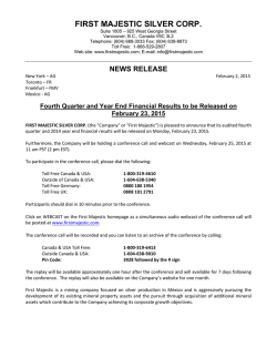
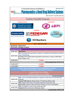
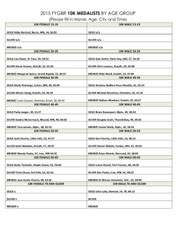
Available online www.jocpr.com
Journal of Chemical and Pharmaceutical Research, 2015, 7(1):356-361
Research Article
ISSN : 0975-7384
CODEN(USA) : JCPRC5
Assessment of in vitro prophylactic efficacy of gallic acid fabricated
silver nanoparticles
Muthukrishnan Lakshmipathy1 and Anima Nanda2*
1
Department of Biomedical Engineering, Faculty of Bio & Chemical Engineering, Sathyabama
University, Chennai, Tamilnadu
2
Faculty of Bio & Chemical Engineering, Sathyabama University, Chennai, Tamilnadu
_________________________________________________________________________________________
ABSTRACT
The nanoscale dimension harbors dynamic physiochemical properties quite different from those of bulk counterpart
relative to its size and large surface area to volume. Despite its wide application, research on fabrication using
eco-friendly agents had been a major breakthrough in gaining control over size. Gallic acid, a phytochemical
compound embodies characteristic features for an efficient reducing and capping agent. Silver nanoparticles
(AgNPs) were synthesized using gallic acid and its biomedical application (antibacterial and antiproliferative
activity) validated. Aqueous chemical reduction method was used to synthesize AgNPs and characterized by UV-vis
spectrophotometer, X-ray diffraction and microscopic analyses. The antimicrobial susceptibility of AgNPs was
tested using Kirby Bauer’s disc diffusion method and the anti-proliferative effect on HEp-2 cells by MTT dye
reduction assay. AgNPs were synthesized rapidly in less than a minute with narrow peak showing λmax at 424nm,
with crystalline nature and uniformly dispersed spherical shaped particles of size < 30nm. The antibacterial study
of AgNPs revealed significant susceptibility toward a panel of Gram positive and Gram negative clinical isolates at
all concentrations (10µg, 20µg and 30µg) on par with antibiotics. Further, the AgNPs showed potent antiproliferative activity on HEp-2 cells with IC50 < 1mg/mL concentration accompanied by morphological
disturbances and membrane damage. The strong affinity toward intracellular proteins and thiol formation accounts
for its toxicity which may further be extended for varied biomedical applications as a broad spectrum therapeutic
agent.
Keywords: Silver nanoparticles, Gallic acid, Antimicrobial assay, MTT, Cyto-toxicity
_________________________________________________________________________________________
INTRODUCTION
Nanotechnology constitutes one of the ever expanding technologies impacting diverse areas such as economy, food
and feed, agriculture, medicine and environment. This nanotechnological venture has anticipated the demand for
commercial application of nanomaterials such as silver, gold, copper, zinc, platinum and titanium [1]. Engineering
of such materials at nanoscale enhances the physicochemical and opto-electric properties compared with its bulk
counterpart [2]. Such nanostructured materials are now extending its footage in biomedical field as scaffolds for
drug delivery, bio-labeling and as therapeutic agent [3,4]. Recently, there is a growing interest in developing ecofriendly approach for synthesizing metal nanoparticles using biocatalysts, phytochemicals and micro organisms.
Though microbe-mediated synthesis [5] served as an alternative to chemical method, the exact mechanism by which
reduction and capping occurs remain unclear. Alongside there is a growing research interest in using phytochemical
constituents such as metabolites as reducing and capping agent [6]. One of its kinds, gallic acid, found in plants is
being used for synthesizing nanoparticles of different sizes by redox reaction [7]. It is noteworthy to mention that
such phenolic derivatives constitute a large class of natural antioxidants protecting cells from oxidative damage
conferring bioavailability [8]. Accordingly, this study has been designed to investigate on the gallic acid mediated
356
Muthukrishnan Lakshmipathy and Anima Nanda
J. Chem. Pharm. Res., 2015, 7(1):356-361
______________________________________________________________________________
synthesis of silver nanoparticles and its antibacterial and anti-proliferative activity toward clinical pathogens and
Human Epidermoid Larynx Carcinoma (HEp-2) cells.
EXPERIMENTAL SECTION
Materials
Silver nitrate (99.9% pure, AR grade) and Gallic acid (C6H2(OH)3COOH; M.wt. 170.12 g/mol) were procured from
HiMedia, India. Penicillin-streptomycin solution, trypsin-ethylenediaminetetraacetic acid-glucose (TPG) solution,
Dulbecco’s Minimum Essential Medium (DMEM) and fetal bovine serum were purchased from Sigma-Aldrich (St
Louis, MO, USA). The antibiotic discs of standard dose were supplied by HiMedia, India.
Methods
Synthesis of silver nanoparticles (AgNPs)
To 100mL of 1x10⁻³ M silver nitrate (AgNO3) placed in a 250mL Erlenmeyer flask, 10mL of deionised milliQ
water containing 0.01 g of gallic acid was added to the solution under stirring condition. In the same experimental
set up, 1M NaOH was added drop-wise to adjust the pH of the solution to 11.
Characterization
The synthesized nanoparticles were characterized by UV–Vis spectroscopy (Shimadzu UV-1800) to track the
absorption spectra of the reaction mixture. X-ray diffraction patterns were recorded with a Rikagu-SMART lab
Diffractometer, Japan using Cu-Kβ radiation (k = 1.54 Ǻ) operated at 40 kV and 100 mA. FESEM analysis was
performed on a Carl Zeiss-Supra 55, Germany at an accelerating voltage of 20 kV. For XRD and FESEM analyses,
freeze dried samples were preferred as reported [9].
Antimicrobial assay – Disc diffusion method
The antimicrobial activity of the synthesized nanoparticles was tested using the standard disc diffusion method [10].
Fresh log phase test cultures of bacterial pathogens (108 cfu/mL) standardized using 0.5 McFarland's standard were
uniformly spread over MHA plate using a sterile swab (HiMedia, India). The antimicrobial susceptibility of AgNPs
prepared at various concentrations (10µg, 20µg, 30µg / disc) along with antibiotics (Amoxicillin - 10µg,
Tetracycline - 30µg and Vancomycin - 30µg) was tested. The plates were then incubated at 37 °C for 18 − 22 h.
Cyto-toxicity testing of AgNPs
Cell viability was measured using the MTT dye-reduction assay [11] to determine the cytotoxic effect of the AgNPs
at various concentrations. In brief, cells were seeded onto 96-well culture plates with various concentrations of
AgNPs (100, 10, 1, 0.1, 0.01, 0.001mg/mL) dissolved in DMSO. All cultures were incubated for 24 h in an
incubator (37°C; 5% CO2). After 24 h of incubation, 20µL of MTT (5mg/mL PBS) was added to each well, and the
plate was incubated for a further 4 h at 37°C. The resulting formazan crystals were dissolved in 80µL DMSO
(Himedia, India) with gentle shaking and absorbance measured at 570 nm with an ELISA reader. The experiment
was performed in triplicates and the results were given as the mean of three independent experiments. The
concentration of AgNPs showing 50% reduction in cell viability ie., half-maximal inhibitory concentration [IC50]
values were then calculated. The data were expressed as mean ± standard deviation (SD) of three independent
experiments.
RESULTS AND DISCUSSION
In the present study, gallic acid was used as reducing and stabilizing agent in the synthesis of silver nanoparticles.
The NPs synthesized were observed from the presentation color of the reaction mixture to brown in less than a
minute. The particles present a narrow band with absorbance maxima (λ max) at 424nm as shown in Figure 1. The
surface Plasmon resonance of silver nanoparticles in the range of 420 to 460nm determines the narrow range of size
of the particles [12]. It is obvious that the interference of phenolic group was responsible for reducing Ag+ to Ag0
with enol group offering stability [13].
The typical X-ray diffraction pattern shown in figure 2 revealed the crystalline nature of the AgNPs with 2θ indices
at 33.03, 44.14, 64.37, 77.38 corresponding to (111), (200), (220) and (311) planes of silver (JCPDS No.04-0783)
[14]. The results of FESEM micrograph (Figure 3) show the narrow size distribution of silver nanoparticles with
size < 30nm and spherical to nearly spherical in shape. The particles were uniformly arranged and well dispersed
without any agglomeration. AgNPs obtained was 97% pure as shown by the Energy Dispersive X-ray spectrum
(EDX) signals (Figure 4). This is in consistent with the results obtained from the narrow particle distribution in
XRD.
357
Muthukrishnan Lakshmipathy and Anima Nanda
J. Chem. Pharm. Res., 2015, 7(1):356-361
______________________________________________________________________________
Figure 1. UV-visible spectrum of silver nanoparticles showing broad peak (λmax) at 424nm attributing to surface plasmon resonance
(SPR)
Figure 2. X-ray diffraction pattern of AgNPs and corresponding 2θ values
Figure 3. Size and morphology of AgNPs by FESEM analysis (Scale bar corresponds to 20nm)
358
Muthukrishnan Lakshmipathy and Anima Nanda
J. Chem. Pharm. Res., 2015, 7(1):356-361
______________________________________________________________________________
Figure 4. Energy Dispersive X-ray diffraction of AgNPs showing strong signals from silver (97%w) and weak signals from carbon
The antibacterial effect of silver nanoparticles toward panel of Gram positive and Gram negative clinical isolates
was found significant compared to antibiotics. The Gram negative organisms which were less susceptible to
amoxicillin, tetracycline and vancomycin showed maximum susceptibility to AgNPs treated cells (Figure 5) in a
dose dependent manner (10µg, 20µg and 30µg / disc). It is evident that the surface charge of AgNPs plays an
important role in anchoring them to the bacterial cell wall. As a result, the integrity of the cell wall gets disrupted
leading to the formation of pits and subsequent oozing out of cellular components and cell death [15]. Indeed, there
is a slight difference in the antibacterial sensitivity of Gram-positive and Gram-negative clinical isolates exposed to
AgNPs. It is noteworthy to mention that the Gram-positive membrane is much thicker compared to Gram-negative
bacteria. Further, the charged AgNPs has a greater affinity toward the Gram-negative strains and gets accumulated
thereby increasing the permeability [16]. It was found that silver, a soft acid has a natural tendency to react with
base. As the biological system is made up of sulphur and phosphorous, these AgNPs react with soft bases and
destroy the genetic material terminating DNA replication and paralyzes the microbe [17].
Figure 5. Antibacterial susceptibility of AgNPs at different concentrations along with antibiotics
The cytotoxicity assay remains one of the toxicological assays used to screen the level of toxicity induced by range
of compounds such as chemicals, metabolites, drugs etc. MTT assay is the most widely used to measure the
reducing potential of the mitochondrial dehydrogenase enzyme in differentiating viable from toxic ones. The results
showed that AgNPs treated Vero and HEp-2 cells showed significant cytotoxicity in a dose dependent manner in
24h. At 0.001mg/mL concentration, the viability of the cells accounted to 97% and 98% respectively for Vero and
HEp-2 cells (Fig. 6). Whereas with the increase in concentration of AgNPs (0.01, 0.1, 1, 10, 100 mg/mL) the
percentage of viable cells decreased to 15% and 22% for Vero and HEp-2 cells. The half maximal inhibitory
359
Muthukrishnan Lakshmipathy and Anima Nanda
J. Chem. Pharm. Res., 2015, 7(1):356-361
______________________________________________________________________________
concentration (IC50) for Vero cells was calculated to be less than 0.1mg/mL and less than 1mg/mL for HEp-2 cells.
The increase in concentration to bring about 50% inhibition in HEp-2 cells validates the cancerous nature of the
cells [18]. Alongside, the morphological changes associated with rounding of cells, reduction in size and
aggregation could well be appreciated on par with the normal and healthy cells. The ROS generated and the stress
induced by AgNPs might account for cellular damage leading to apoptotic cell death [19]. It was also observed
from the study that the adhesion property was lost in AgNPs treated cells. This property remains vital for cellular
functions such as growth, differentiation, migration and tissue regeneration. This alteration in the membrane
potential serves to be the foremost factor responsible for inducing apoptosis induced by AgNPs. However, the
mechanism by which AgNPs brings about the apoptotic cell death remain unclear but earlier reports on silver
nanoparticles’ tendency to activate cascade of genes and their regulation would account for determining the fate of
the cell [20].
Figure 6. Anti-proliferative activity of AgNPs on HEp-2 and Vero cells determined using MTT assay
* The data were expressed as mean ± standard deviation (SD) of three independent experiments
CONCLUSION
The present study validates the therapeutic antimicrobial and anti-proliferative potential of gallic acid fabricated
silver nanoparticles. A strong electrostatic attraction of AgNPs and ionization toward functional biomolecules
interferes with the normal functions and brings about cell death. Fabrication of such NPs using metabolic products
of biological origin may find its extended application as a multifaceted therapeutic agent with improved specificity.
Acknowledgements
The authors acknowledge Department of Biotechnology (DBT), Government of India for the financial support. The
authors are grateful to Hon’ Chancellor, Managing Directors and Department of Biomedical Engineering,
Sathyabama University for providing infrastructural facilities. Technical Support from Dr. M. Bavanilatha,
Associate Professor, Biotechnology Department, Sathyabama University is highly appreciated.
REFERENCES
[1] SWP Wijnhoven; WJGM Peijnenburg; CA Herberts; WI Hagens; AG Oomen; EHW Heugens; B Roszek; J
Bisschops; I Gosens; D Van De Meent; S Dekkers; WH De Jong; M van Zijverden; AJAM Sips; RE Geertsma,
Nanotoxicol., 2009, 3, 109–138.
[2] JP Wise Sr; BC Goodale; SS Wise; GA Craig; AF Pongan; RB Walter; D Thompson; AK Ng; AM Aboueissa; H
Mitani; MJ Spalding; MD Mason, Aquat. Toxicol., 2010, 97, 34–41.
[3] A Ito; K Tanaka; K Kondo; M Shinkai; H Honda; K Matsumoto; T Saida; T Kobayashi, Cancer Sci., 2003, 94,
308-313.
[4] E Hood, Environ. Health Perspect., 2004, 112, A747–A749.
[5] A Nanda; M Saravanan, Nanomedicine., 2009, 5, 452-456.
[6] H Mülpfordt, Experientia., 1982, 38(9), 1127–1128.
[7] X Tian; W Wang; G Cao, Mater. Lett., 2007, 61(1), 130–133.
360
Muthukrishnan Lakshmipathy and Anima Nanda
J. Chem. Pharm. Res., 2015, 7(1):356-361
______________________________________________________________________________
[8] GA Martınez-Castanon; N Nino-Martınez; F Martınez-Gutierrez; JR Martınez-Mendoza; F Ruiz, J. Nanopart.
Res., 2008, 10, 1343–1348.
[9] M Lakshmipathy; A Nanda, Int. J. ChemTech Res., 2013, 5(3), 1162–1168.
[10] AW Bauer; WMM Kirby; JC Sherris; M Turck, Amer. J. Clin. Pathol., 1966, 45(4), 493-496.
[11] T Mosmann, J. Immunolog. Meth., 1983, 65(1–2), 55–63.
[12] C Sonnichsen; T Franzl; T Wilk; G von Plessen; J Feldmann, New J. Phys., 2002, 4, 931–938.
[13] W Wang; Q Chen; C Jiang; D Yang; X Liu; S Xu, Colloids Surf A Physicochem Eng Asp., 2007, 301, 73–79.
[14] L Muthukrishnan; A Nanda, J. Phar. Res., 2013, 6, 725-729.
[15] I Sondi; B Salopel-sondi, J. Colloid Interface Sci., 2004, 275, 177-182.
[16] Y Matsumura; K Yoshikata; S Kunisaki; T Tsuchido, Appl. Environ. Microbiol., 2003, 69, 4278–4281.
[17] DW Hatchett; S Henry, J. Phys. Chem., 1996, 100, 9854–9859.
[18] MA Franco-Molina; E Mendoza-Gamboa; CA Sierra-Rivera, RA Gomez-Flores; P Zapata-Benavides; P
Castillo-Tello; JM Alcocer-Gonzalez; DF Miranda-Hernandez; RS Tamez-Guerra; C Rodriguez-Padilla, J. Exp.
Clin. Cancer Res., 2010, 29, 148.
[19] PV AshaRani; G Low Kah Mun; MP Hande; S Valiyaveettil, ACS Nano., 2009, 3(2), 279-290.
[20] P Bernardi; L Scorrano; R Colonna; V Petronilli; FD Lisa, Eur. J. Biochem., 1999, 264(3), 687-701.
361
© Copyright 2026