
Isolation and characterization of endophytic fungi from medicinal
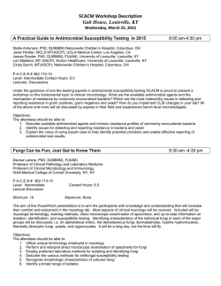
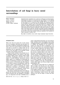
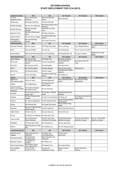
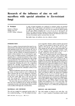
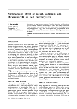
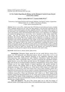
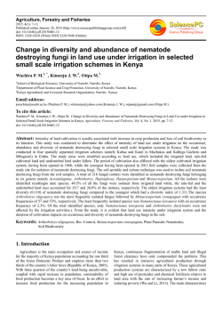
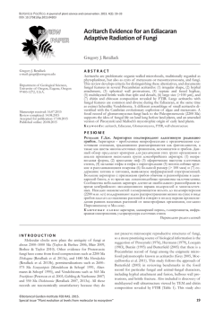
Available online www.jocpr.com
Journal of Chemical and Pharmaceutical Research, 2015, 7(1):757-762
Research Article
ISSN : 0975-7384
CODEN(USA) : JCPRC5
Isolation and characterization of endophytic fungi from medicinal plant,
buah makassar(Makassar fruit: Brucea javanica)
Nur Amin1, Fitrianti2 and Muh. Danial Rahim1
1
Department of Plant Protection, Faculty of Agriculture, Hasanuddin University, Makassar, South Sulawesi
– Indonesia
2
Undergraduate Student of the Department Plant Protection, Faculty of Agriculture, Hasanuddin
University, Makassar, South Sulawesi – Indonesia
_____________________________________________________________________________________________
ABSTRACT
Brucea javanica (L.) Merr. is a member of Simaroubaceae which call as Makassar Fruit and having medicinal
potency. In the last few years fungal endophytes have been detected in hundreds of plants including medicinal
plants. The current study aimed to isolate and characterization of endophytic fungi from roots, stems, leaves and
fruit of Makassar fruit B. javanica. A total of 4 (four) genera of endophytic fungi were identified, which
Trichoderma sp. isolated from roots and stems, Fusarium sp and Penicillium sp from fruits and Aspergillus sp from
leaves.
Key words: Endophytic Fungi, Brucea javanica, Makassar Fruit, Medicinal Plant
_____________________________________________________________________________________________
INTRODUCTION
The term endophyte was coined by the German scientist, Heinrich Anton De Bary in 1884, and used to define fungi
and bacteria occurring inside plant tissues without causing any apparent symptoms in the host (Schulz et al, 2006).
In the last few years fungal endophytes have been detected in hundreds of plants including important agricultural
commodities as wheat [1], bananas [2], maize [3]; cocoa plant [4]. A number of authors have documented that the
presence of endophytic fungi in medicinal plant, such as Aquilaria sinensis [5], Acorus calamus rhizomes [6],
Juniperus communis L. Horstmann [7], Withania somnifera [8,9] , the Chinese medicinal plant, Tripterygium
wilfordii [10] and Brucea sp. [11,12,13,14].
Brucea javanica (L.) Merr. is a member of Simaroubaceae which call as Makassar Fruit (Indonesia), lada hitam
(Malaysia) as well as "Ya-Dan-Zi" and "Ya-Tan-Tzu" (China). The bitter fruits of medicinal plant Brucea javanica
(L.) Merr. is widely used in traditional medicine for various ailments [15]. Bruceine A, a natural quassinoid
compound extracted from the fruits of B. javanica (L.) Merr., with a wide spectrum of biological effects, such as
having potential antibabesial [16], antitrypanosomal [11,15]; antimalarial [17,18,19] and cytotoxicity against human
cancer cell lines [20,21]. Other biological activities such as anti-protozoan [22] and anti-inflammatory Hall et al.
[23] activities have also been studied regarding its use among the traditional medicine practitioners.
757
Nur Amin et al
J. Chem. Pharm. Res., 2015, 7(1):757-762
______________________________________________________________________________
Figure 1. Makassar Fruit (Brucea javanica)
EXPERIMENTAL SECTION
Sample Collection
Healthy plants with roots, leaves, stems and fruits of B.javanica taken and collected from various places of Takalar
Region, Province of South Sulawesi, Indonesia. The samples were collected by aseptic procedures and brought to
the laboratory of Plant Protection Department, Faculty of Agriculture, Hasanuddin University, Indonesia and
processed within 24 hours of collection.
Isolation of Endophytic Fungi
Endophytic fungi were isolated according the protocols described by Petrini 1986, which were slightly modified
based on preliminary tests. The roots, leaves, stems and fruits of B.javanica taken from the field were washed twice
in distilled water then surface sterilized by immersion for 1 minute in 70% (v/v) ethanol, 5 minutes in sodium
hypochlorite (2.5 % (v/v) available chlorine) and 30 seconds in 70% (v/v) ethanol and then washed three times in
sterilized distilled water for 1 minute each time. After surface sterilization, the samples were cut into 5 mm pieces
and aseptically transferred to plates containing potato dextrose agar (PDA, pH 6.8, containing (g/l): potato 200;
dextrose 20; agar 15.), which had been autoclaved for 15 minutes at 121ºC and then aseptically supplemented with
100 mg/ml of chloramphenicol (Pfizer) to suppress bacterial growth. Aliquots from the third wash were plated onto
PDA to check that surface sterilization had been effective and they were then incubated at 28ºC. Any fungi present
was isolated, purified and then maintained at 4ºC on PDA slopes for further identification. For tentative
identification, microscopic slides of each fungal endophyte were prepared, examined under light microscope
(Olympus, USA) and identified with reference to [24,25].
RESULTS AND DISCUSSION
Endophytic fungi isolated from roots, leaves, stems and fruits of B.javanica were identified 4 genera such as
Trichoderma sp., (roots and stems), Fusarium sp. (fruits and stems), Aspergillus sp. (leaves) and Penicilium sp.
(Fruits) (Table 1).
Trichoderma sp. characterized by green colored colonies and there is also a white spot (Figure 2a,b,c). Trichoderma
sp. have half-round to oval conidia, upright branched conidiophores and conidia at the end is fialit.
Fusarium sp. characterized by very rapid growth of hyphae and orange, dark red clamidospora, which stick to the
walls of petridishes if the age of 3 days. Microscopic identification results showed that the crescent-shaped
makrokonidia with 3-5 septa and clamidospora is round and slightly oval (Figure 3a, 3b).
758
Nur Amin et al
J. Chem. Pharm. Res., 2015, 7(1):757-762
______________________________________________________________________________
Table 1. Diversity of endophytic fungi from roots, leaves, stems and fruits of B.javanica
No
Genera
Macroscopic characterization on PDA media
1.
Trichoderma
sp
Green colored colonies and there is also a white
spot
2.
Fusarium sp.
The colony of cotton white and fine texture
3.
Penicillium sp.
Coloni white
4.
Aspregillus sp
Blackish brown
Microscopic characterization
- Conidia half-round to oval
- Upright
branched
conidiophores
- Conidia at the end fialit
- Macro and microconidia are presence
- Hyphae have septae
- Microconidia is crescent and have septae
- Finger-shaped conidiophores
- There are 2-3 hyphae per branch
- Conidia round, slightly oval
- Smooth conidiophores where
hyphae perpendicular
- Conidia round
- At the end of hyphae forming Globusa
Sources
Roots and
Stems
Fruits and
Stems
Fruits
Leaves
B
A
C
Figure 2: Endophytic Fungi Trichoderma sp, (a) Colony performance on the PDA (b) conidia and (c) Conidiophores
Fusarium sp
B
A
D
C
Figure 3: Endophytic Fungi Fusarium sp, (a) Colony performance on the PDA (b) macroconidia , (c) hyphae, (d) conidia
759
Nur Amin et al
J. Chem. Pharm. Res., 2015, 7(1):757-762
______________________________________________________________________________
The colony performance on the PDA media by Penicillium sp. is white colony, irregular mycelium, colony growth
flat, thick, flat edge of the colony . Microscopic characteristic is Finger-shaped conidiophores, there are 2-3 hyphae
per branch. On the whole surface is covered vesicles fialid formed and the formed conidia fialid in sequence .
Conidia are round , hyaline , and arrayed 1 cells.
B
A
C
D
Figure 4: Endophytic Fungi Penicillium sp, (a) Colony performance on the PDA (b) conidia , (c) hyphae, (d) conidiophore
A
B
C
D
Figure 4: Endophytic Fungi Aspergillus sp, (a) Colony performance on the PDA (b) conidia , (c) hyphae, (d) conidiophore
The fungus Aspergillus sp. on morphological appearance is blackish brown on PDA media. Feature of microscopy :
spherical conidia, upright hyphae, conidiophores smooth, long and formed freely. Aspergillus sp conidia carrier has
a large head and a dense, round and black. Microscopic observations above adapted to the description [24], which
760
Nur Amin et al
J. Chem. Pharm. Res., 2015, 7(1):757-762
______________________________________________________________________________
states that the vesicle surface is covered fialid that produce conidia. Conidia arranged 1 cells and has a variety of
colors. Conidial heads are black and round.
DISCUSSION
The results of the isolation and identification of endophytic fungi from Makassar fruit isolated 4 genera, which
Trichoderma sp. isolated from roots and stems, Fusarium sp and Penicillium sp from fruits and Aspergillus sp from
leaves.
According to Liang et al [26], a total of 83 endophytic fungi strains were isolated from the root, stem, leaf and fruit
of Brucea javanica. 34 strains were obtained from the stem,32 strains were obtained from the leaf, 15 strains were
isolated from the root and 2 strains came from the fruit. These 73 strains which had been identified attribute to 5
orders, 6 families and 12 genera. The other medicinal plant Tripterygium wilfordii isolated endophytic fungi such as
Colletotrichum gloeosporioides, Guignardia sp., Glomerella cingulata, Pestalotiopsis spp., Phomopsis spp. and
Phyllosticta sp. [10].
In the current study have been isolated Trichoderma sp. from roots and stems of Makassar Fruit B. javanica. This
fungus produced a wide range of phenolic substances such as glycosides as strong antioxidant and anti-mutagenic
activities thus, can be used against cancers [27]. Glycosides are usually compounds which have been isolated in
plants, made up of one or more sugars combined with an alcohol, a phenol, or a complex molecule such as a steroid
nucleus. Many plants contain cardiac glycosides, which occur both in monocotyledons and in dicotyledons and have
been documented as one of the chemicals in 20 well documented poisonous plants and equally possesses
antimicrobial properties [28].
The ability of an endophyte to produce some metabolites but not others has been described by Selim et al. [29];
where different endophytes in a plant may produce different secondary metabolites hence play different functions in
the plant and that the total number of metabolites in a plant extract maybe a contribution of all the endophytes that
live in the plant. The available secondary metabolites have anti-pathogenic properties and their roles in plant defense
have been elaborated in the works of Hari et al and Figen [27, 28], and could be responsible for the function of the
plant extracts in defense against plant and animal pathogens.
Liang et al [26] and Kumar, D.S.S and Hyde, K.D [10] get the different result with the current study in term of the
amount and tipe of endophytic fungi. According to Nur Amin et al [4] the different result influenced by biotic and
abiotic factors. Biotic factors consist of varieties and species of host. While abiotic factors that influence the
weather factors that temperature , relative humidity and moisture content of the soil and cultivation techniques.
CONCLUSION
A total of 4 (four) genera of endophytic fungi were identified, which Trichoderma sp. isolated from roots and stems,
Fusarium sp and Penicillium sp from fruits and Aspergillus sp from leaves.
REFERENCES
[1] Larran, S., Perello, A., Simon, M.R., Moreno, V. World Journal of Microbiology & Biotechnology., 2002, 18,
683–86.
[2] Nur Amin. Untersuchungen uber die Bedeutung endophytischer Pilze fur die biologische Bekampfung des
wandernden Endoparasiten Radopholus similis (Cobb) Thirne an Bananen. 1994, PhD-Thesis, 112 p. Bonn
University.
[3] Nur Amin. International Journal of Current Microbiology and Applied Science. 2013, 2 (8) : 148-54.
[4] Nur Amin., Salam, M., Junaid, M., Asman and Baco, MS. International Journal of Current Microbiology and
Applied Science. 2014, 3 (2) : 459 – 67
[5] Jin, L.C and Pei, G.X. J Zhejiang Univ Sci B. 2011, 12 (5): 385–92.
[6] Barik, B.P., Tayung, K., Jagadev, P.N., Dutta, S.K. 2010. Eur. J. Biol. Sci. 2010, 2 (1) : 8-16 .
[7] Kusari, S., Lamshoeft,M and Spiteller, M. J. Appl. Microbiol. 2009, 107(3): 1019-30.
[8] Khan, R; Saleem, S, Chodhary, MI; Khan SA and Ahmad, A. Vet Parasitol. 2008, 20 : 158 (4) : 288-94
[9] Kumar, S; Nutan, K and Peter, P. Springer Plus. 2013, 2 : 37.
[10] Kumar, D.S.S. and Hyde, K.D. Fungal Diversity. 2004, 17: 69-90.
[11] Subeki, Matsuura, H., Takahashi, K., Nabeta, K., Yamasaki, M., Maede, Y and Katakura, K. J Nat Prod.
2007, 70 (10) :1654-57
[12] Lu, J.B, Shu, S.Y and Cai, J.Q. Zhongguo Zhong Xi Yi Jie He Za Zhi. 1994, 14 ( 10 ), 610-11
761
Nur Amin et al
J. Chem. Pharm. Res., 2015, 7(1):757-762
______________________________________________________________________________
[13] Lau, F.Y., Chui, C.H., Gambari, R., Kok, S.H., Kan, K.L., Cheng, G.Y., Wong, R.S, Teo, Cheng, C.H, Wan,
T.S., Chan, A.S and Tang, J.C. Int J Mol Med. 2005, 16 ( 6 ) : 1157-62 .
[14] Kim, I.H., Takashima, S., Hitotsuyanagi, Y., Hasuda, T and Takeya, K. J Nat Prod . 2004, 67 ( 5 ): 863-8.
[15] Bawm, S., Matsuura, H., Elkhateeb, A., Nabeta, K., Subeki, Nonaka, N., Oku, Y., Katakura, Vet Parasitol.
2008, 20 : 158(4):288-94
[16] Yamada, K., Subeki., Nabeta, K., Yamasaki, M., Katakura, K and Matsuura, H. Biosci. Biotechnol.Biochem.
2009, 73
[17] Pavanand, K., Nutakul, W., Dechatiwongse, T., Yoshihira, K., Yongvanitchit, K., Scovill, J. P., FlippenAnderson, J. L., Gilardi, R. & George, C. 1986, Planta Med. 52, 108-111.
[18] O’Neill, M. J., Bray, D. H., Boardman, P., Chan, K. L., Phillipson, J. D., Warhurst, D. C. and Peters, W. 1987,
J. Nat. Prod. 50, 41-48.
[19] Kim H-S., Shibata Y., Ko N., Ikemoto N., Ishizuka Y., Murakami N., Sugimoto M., Kobayashi M., Wataya Y.
Parasitology International. 2000, 48 :271-274.
[20] Zhao, L., Li, C., Zhang, Y., Wen, Q and Ren, D. Anticancer Agents Med Chem. 2014, 14 (3) : 440-58.
[21] Hong, Z., Jing, Y.Y., Fan, Z., Li H.W., Wen, Z., Sha, S and Chun, F.W. Evidence-Based Complementary and
Alternative Medicine. 2011, 1-14
[22] Sawangjaroen, N and Kitja, S. Journal of Ethnopharmacology. (2005), 98 :67–72
[23] Hall, I.H., Lee, K.H., Imakura, Y., Okano,M and Johnson, A. J. Pharm. Sci. 1983, 72, 1282–1284.
[24] Barnet, H.L and Hunter, B.B, Illustrated genera of imperfect fungi. 1998, 4th ed. APS Press. St. Paul.
Minnesota. p. 218
[25] Dugan, F.M, The Identification of Fungi: An Illustrated Introduction With key, Glossary and Guide to
Literature. 2006, The American Phytopathological Society, St. Paul. Minnesota. p. 184.
[26] Liang., Z.N, Zhu, H, Lai, K.P and Chen, L. Zhong Yao Cai. 2014, 37(4):564-8.
[27] Hari, O., Anjana, S., Naseer, M and Santosh K. Journal of Chemical and Pharmaceutical Research. 2014, 6 (5)
:756-760.
[28] Figen, M.T.Ÿ. Journal of Cell and Molecular Biology. 2006, (13) 5:13-17.
[29] Selim, K., El-Beih, A., Abdel-Rahman, T and El-Diwany, A. Current Research in Environmental & Applied
Mycology. 2012, 2(1) : 31-82.
762
© Copyright 2026