
Mesoaxial complete syndactyly and synostosis with hypoplastic
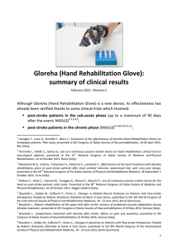
8686 Med Genet 1998;35:868-874
Department of
Medical Biology and
Genetics, Cumhuriyet
University, Medical
Faculty, Sivas, Turkey
E F Percin
I Sezgin
Department of
Orthopaedics,
Cumhuriyet
University, Medical
Faculty, Sivas, Turkey
S Percin
Department of
Radiodiagnostics,
Cumhuriyet
University, Medical
Faculty, Sivas, Turkey
H Egilmez
DNA/Cell Bank and
Gene Research
Laboratory
(TUBITAK),
Hacettepe University,
Medical Faculty, Child
Health Institute,
Sihhiye 06100, Ankara,
Turkey
F Ozbas
A N Akarsu
Correspondence to:
Dr Akarsu.
Received 30 October 1997
Revised version accepted for
publication 23 February
1998
Mesoaxial complete syndactyly and synostosis
with hypoplastic thumbs: an unusual combination
or homozygous expression of syndactyly type I?
E Ferda Percin, Sitki Percin, Hulusi Egilmez, Ilhan Sezgin, Filiz Ozbas, A Nurten Akarsu
Abstract
Syndactyly type I is an autosomal dominant condition with complete or partial
webbing between the third and fourth fingers or the second and third toes or both.
We report here a previously undescribed
phenotype of severe mesoaxial syndactyly
and synostosis in patients born to affected
parents. The characteristic features of
these severe cases are (1) complete syndactyly and synostosis of the third and
fourth fingers; (2) severe bone reduction
in the proximal phalanges of the same fingers; (3) hypoplasia of the thumbs and
halluces; (4) aplasialhypoplasia of the
middle phalanges of the second and fifth
fingers; and (5) complete or partial soft
tissue syndactyly of the toes. We report on
three offspring with this phenotype from
two different branches of a syndactyly type
I family, suggesting that they may be
homozygous for this condition. SSCP and
linkage analysis indicated that neither
HOXD13 nor other relevant genes in the
chromosome 2q31 region was responsible
for this phenotype.
(7 Med Genet 1998;35:868-874)
Keywords: syndactyly type I; HOXD 13; chromosome
2q31; homozygous phenotype
Isolated syndactyly is one of the most common
congenital malformations affecting the hands
or feet or both. To date, many attempts have
been made to classify isolated syndactylies, but
none of them have covered all the types of syndactyly reported so far. According to the most
widely used classification, isolated syndactylies
have at least five forms, all being inherited as an
autosomal dominant trait with variable expressivity and complete penetrance.'
In this classification, type I syndactyly
(zygodactyly), which comprises complete or
partial webbing between the third and fourth
fingers or the second and third toes or both, is
the most common form. Sometimes only the
17
33-36
37
38
Figure 1 The large six generation Turkish syndactyly type I pedigree. Solid symbols are severely affected cases (homozygous?) and half shaded symbols
are affected persons with only soft tissue syndactyly between the second and third toes. DNA =DNA available. Asterisks below symbols indicate that they
had been examined.
869
Mesoaxial complete syndacyly and synostosis with hypoplastic thumbs
second to fourth fingers and toes, hypoplastic
thumbs and halluces, and mesoaxial synostosis
in at least three affected members of a large
syndactyly type I family.
Materials and methods
The family in this study was from Sivas, Turkey. The pedigree consists of a total of 57 people of whom 13 are affected in seven
generations (fig 1). Seventeen family members
had already died by the time of the study.
Twenty-four family members (12 affected)
Figure 2 Subject 42 infig 1 is an example of heterozygous were examined by one of us (FP, SP, or HE).
syndactyly type I. Partial soft tissue syndactyly is present on
Radiographic evaluation was performed in five
the left and the rightfoot is also mildly affected.
affected cases. Systemic evaluation, routine
hands are affected and sometimes only the laboratory tests, abdominal ultrasonography,
feet. Type II syndactyly (synpolydactyly) echography, and x ray examination of the vershows various degrees of duplication along tebral column were performed only for two
with syndactyly of the third and fourth fingers sibs (subjects 55 and 57 in fig 1) who are
and fourth and fifth toes. Type III syndactyly is severely affected with complete syndactlyly
complete or partial webbing between the and synostosis. A total of 22 blood samples
fourth and fifth fingers. Complete syndactyly were obtained and DNA was extracted from
of all fingers is classified as type IV syndactyly. relevant family members for molecular evaluaThe only type associated with metacarpal and tion. SSCP analysis5 was performed to check
metatarsal synostosis is type V syndactyly. point mutations in the HOXD 13 coding
In the latter, the abnormality usually affects region. Primers and amplification conditions
the fourth and fifth fingers and toes and were as previously reported.4 Amplified prodoccasionally the third and fourth fingers and ucts were also separated on a 6% polyacrylatoes.
More recently, Winter and Tickle' have pro- mide gel for quick detection of potential size
posed an alternative embryological classifi- differences between affected and normal
cation of the syndactylies mainly based on the cases. The family was also tested for linkage to
fact that the pattern formation is normal or the chromosome 2q31 region where the
abnormal.' Although the genetic background synpolydactyly (SPD) gene has been mapped.6
of isolated syndactyly is clear, the gene(s) Tightly linked DNA markers, D2S 1238,
involved have not been identified, except for D2S2307, and D2S2314, were used for
type II syndactyly (synpolydactyly). The ex- linkage and haplotype analysis. Two point
pansion of alanine in the HOXD 13 gene was linkage was performed using the MLINK
found to be responsible for the synpolydactyly component of the LINKAGE package
(Fastlink version 2.20) under the assumption
phenotype.34
Here we report another example of meso- of both autosomal dominant and autosomal
axial deformity which appears with an unusual recessive modes of inheritance with full
combination of complete syndactyly of the penetrance.
Table 1 Physicalfindings of the three severely affected cases
Case Hands
1
2
3
Four fingers on both hands
Hypoplasia of thumbs
Complete syndactyly between 2nd and 3rd fingers on left
Partial syndactyly between 2nd and 3rd fingers on right
Complete syndactyly between 3rd and 4th fingers
Distal structures of 4th finger not palpable in the
synostotic complex
Flexion contracture of the 5th fingers
Bilateral simian line
Four fingers on both hands
Hypoplasia of thumbs
Complete syndactyly between 3rd and 4th fingers
Distal structures of 4th finger not palpable in the synostotic
complex
Flexion contracture of the 3rd finger on the right
Mild syndactyly between 2nd and 3rd fingers on the left
Clinodactyly of 5th fingers
Bilateral simian line
Four fingers on both hands
Bilateral hypoplasia of 2nd fingers
Complete syndactyly between 3rd and 4th fingers
Hypoplastic nails on postaxial fingers
Four flexion lines on synostotic complex between 3rd and
4th fingers whereas only one flexion line on 2nd and 5th
fingers (fig 5C)
Clinodactyly of 5th fingers
Feet
Metatarsal varus and valgus deviation of toes
Enlarged 1st and 2nd toes
Complete syndactyly between 2nd and 3rd, partial
syndactyly between 1st and 2nd toes on the right
Complete syndactyly between 4th and 5th toes on the left
Clinodactyly of 4th and 5th toes on the right and 3rd, 4th,
and 5th toes on the left
Enlarged 1 st and 2nd toes
Complete syndactyly between 2nd and 3rd toes on the left
Mild syndactyly between 2nd and 3rd toes on the right
Clinodactyly of 3rd and 4th toes on the right and 4th and 5th
toes on the left
Small and broad halluces
Bilateral syndactyly between 2nd and 3rd toes
Hypoplastic nails of big toes
870
Percin, Percin, Egilmez, et al
Table 2 Radiographic findings in the hands and feet of the three cases
Case 1 (DOB 17.3.93)
Hands
Radius
Ulna
Capitate
Hamate
Trapezoid
Metacarpal bones
Right
Left
Right
N
N
N
N
N
N
+
+
+
1st
2nd
3rd
Hypoplasia
N
(Distal fusion between 3rd and 4th)
Hypoplasia
N
(Distal fusion between 3rd and 4th)
N
Hypoplasia
N
(Severe hypoplasia, deformation in 3rd
and 4th)
N
Hypoplasia
N
Severe hypoplasia (Hypoplasia,
deformation)
Hypoplasia
N
Hyperplasia
N
N
Hypoplasia
Deformed
(Complete synostosis between 3rd and
4th)
Hypertrophy
Hypertrophy
N
Aplasia
(Complete synostosis between 3rd and
4th)
Aplasia
(Complete synostosis between 3rd and
4th)
Aplasia
(Complete synostosis between 3rd and
4th)
2nd
3rd
(Hypoplasia, deformation)
Hypoplasia
Hypoplasia
(Hypoplasia, complete synostosis
between 3rd and 4th)
(Hypoplasia, deformation)
Hypoplasia
Hypoplasia
(Hypoplasia, complete synostosis
between 3rd and 4th)
(Hypoplasia, deformation)
Hypoplasia
(Hypoplasia, deformation)
(Hypoplasia, complete synostosis
between 3rd and 4th)
4th
5th
N
N
N
N
N
N
N
N
N
N
N
N
Hypoplasia of 1st toe, varus deformity in
all metatarsals
N
N
N
N
Hypoplasia
N
N
N
N
Hypoplasia
N
N
N
N
Varus deformity in all metatarsals
4th
5th
Proximal phalanges
Case 2 (DOB 1.2.94)
1st
2nd
3rd
4th
Middle phalanges
5th
1st
2nd
3rd
4th
5th
Distal phalanges
Feet
Tibia
Fibula
Talus
Calcaneus
Navicular
Cuneiform
Cuboid
Metatarsal
1st
I
II
III
1st
Hypoplasia
Hypoplasia
N
N
Hypoplasia of 1st toe, varus deformity in
all metatarsals
2nd
3rd
4th
5th
Proximal phalanges
Middle phalanges
Distal phalanges
1st
2nd
3rd
4th
5th
2nd
3rd
4th
5th
1st
2nd
3rd
4th
5th
Deformed
N
N
N
N
N
N
N
Hypoplasic, deformed
N
N
N
N
Hypoplasia
N
N
N
N
N
N
N
N
N
Aplasia
Aplasia
Aplasia
N
Aplasia
N
N
N
N
N
N
N
N
N
N
N
N
N
N
N: normal. +: present. ---: absent (consistent with bone age). DOB: date of birth.
Results
CLINICAL FINDINGS
subjects with similar soft tissue syndactyly in
their toes with autosomal dominant inheritance with reduced penetrance and variable
Two sibs with severe syndactyly were diagnosed in the Department of Orthopaedics at expressivity.
The other two cases (subjects 17 and 51)
Cumhuriyet University in Sivas, Turkey. The
parents of the affected children were consan- with a more severe form of syndactyly were
guineous (fig 1) and found to be affected with similar to the probands. The parents of subject
soft tissue syndactyly between the second and 51 were consanguineous and had second and
third toes (fig 2). Neither physical examination third toe syndactyly. However, accurate infornor x ray evaluation showed any abnormality in mation about consanguinuity and complete
the hands of either parent. Dermatoglyphic physical examination could not be obtained for
changes in the hands, especially the absence of subject 17, whose hands and feet are severely
triradii at the base of the fingers (zygodactyly), affected, since the entire branch of the family
can be a sign of involvement of the hands. moved to Germany many years ago.
The comparative physical and x ray findings
However, the dermatoglyphics were normal in
the hands of the parents of the affected of cases with severe syndactyly are summarised
children. Pedigree analysis showed seven more in tables 1 and 2.
871
Mesoaxial complete syndacyly and synostosis with hypoplastic thumbs
Table 2 Continued
A
Case 3 (DOB 1.1. 91)
Left
Right
Left
N
N
N
N
Carpal bones complete
except scaphoid bone
N
N
Carpal bones complete
Minimal hypoplasia
N
N
N
N
N
N
between 3rd and 4th)
Minimal hypoplasia
N
N
N
N
N
N
(Complete synostosis
between 3rd and 4th)
Deformation and synostosis
with 3rd
N
N
N
Aplasia
Severe hypoplasia
N
(Complete synostosis
between 3rd and 4th)
Severe hypoplasia
(Complete synostosis
between 3rd and 4th)
Severe hypoplasia
N
Severe hypoplasia
Severe hypoplasia
N
Severe hypoplasia
(Hypoplasia, complete
synostosis between 3rd and
4th)
(Hypoplasia, complete
synostosis between 3rd and
4th)
N
Hypoplasia
Hypoplasia
N
N
N
N
N
N
N
N
N
Varus deformity in all
metatarsals
N
N
N
N
N
N
N
N
N
Varus deformity in all
metatarsals
N
N
N
N
N
N
N
N
N
Varus deformity in all
metatarsals
N
N
N
N
N
N
N
N
N
N
N
N
N
N
N
N
N
N
N
Hypoplasia
Aplasia
Aplasia
Aplasia
Hypoplasia
Hypoplasia
Hypoplasia
Hypoplasia
Hypoplasia
N
N
N
N
N
+
Hypoplasia
N
Hyperplasia
N
N
Hypoplasia
N
Severe hypoplasia
Aplasia
(Hypoplasia, deformation)
Hypoplasia
(Hypoplasia, deformation)
(Hypoplasia, complete
synostosis between 3rd and
4th)
(Complete synostosis
I
., "
A
I
.,
.,
-,
.7
I
j
s;t
x
t
w
.)
*. l0
Hypoplasia
Aplasia
Aplasia
Aplasia
Hypoplasia
Hypoplasia
Hypoplasia
Hypoplasia
Hypoplasia
Case 1 (subject S5,fig 1)
The proband is a 4 year old male with normal
mental and physical development. He had four
fingers on each hand with partial or complete
syndactyly involving all four extremities (fig 3A,
B). Both hands displayed soft tissue syndactyly
mainly between the second and third fingers.
The fourth fingers were not distinguishable
individually; only small, dysmorphic, and rudi- figure 4 (A-D) Hands andfeet of subject 57.
mentary bone particles were palpable within the
mass next to the third finger bilaterally. The synostosis of the third and fourth metacarpal
thumbs were short and hypoplastic. The fifth bones along with a severely hypoplastic
finger had a flexion contracture deformity with biphalangeal thumb were present. The middle
clinodactyly on both sides. The nails were nor- phalanges were absent in the second
mal. A simian line was observed bilaterally.
fingers and hypoplastic in the fifth fingers.
X rays of the hands showed a similar The distal phalanges of all fingers were
picture on both sides (fig 3G). Partial hypoplastic and dysmorphic. Normal tubular
872
Percin, Percin, Egilmez, et al
Figure 5 (A-E) Hands and feet of subject 51. (C) Note
fourflexion lines on synostotic complex.
shapes were lost in the proximal phalanges
of the synostotic complex and replaced by
some cuboid structures owing to severe
bone retardation. Complete synostosis of
the third and fourth fingers was the main
finding in the distal part of this complex. Bone
age was compatible with that of a 3 year old
child.
The feet showed varus deformity in the
metatarsal region but all toes were in the valgus
position (fig 3B). The feet were dissimilar apart
from short and enlarged first and second toes.
On the right foot the first three toes were
webbed. Syndactyly was partial between the
first two toes but complete between the second
and third. The fourth and fifth toes were
normal except for clinodactyly in both. On the
left foot, there was complete syndactyly only
between the fourth and fifth toes. Clinodactyly
was also present in the last three toes. X rays of
the feet showed severe growth retardation of
the tarsal bones especially the navicular and
first and second cuneiform bones in the left
foot. The proximal and distal phalanges of the
big toes were found to be hypoplastic and
deformed on both sides with variable severity
(fig 3D).
Case 2 (subject 57,fig 1)
The proband's 2 year old sister had four fingers
on each hand. Her psychomotor development
was normal. The phenotypic appearance was
quite similar to the proband's except for complete syndactyly between the second and third
fingers (fig 4A). Complete syndactyly mainly
involved the third and fourth fingers and there
was hypoplasia of both thumbs. X rays of the
hands showed hypoplasia of the first metacarpal bones (fig 4C). Like the proband, there was
symmetrical aplasia of the middle phalanx,
severe hypoplasia of the second fingers, and
clinodactyly of the fifth fingers. The normal
tubular shape was lost in the proximal phalanx
of the third finger and replaced by two
deformed, shapeless bony structures. Compared to the proband the phenotypic expression was less severe in the feet, the prominent
manifestation being soft tissue syndactyly
between the second and third toes on the left
(fig 4B, D).
She had a dizygotic male twin sib (fig 1, subject 56). He had mild soft tissue syndactyly in
the feet between the second and third and
fourth and fifth toes. The hands were normal.The phenotype of case 3 (fig 1, subject 51,
Mesoaxial complete syndacyly and synostosis with hypoplastic thumbs
873
.-:
77.'
missing fourth finger instead of the duplication
seen in synpolydactyly. Therefore, we considered that the possibility of a lack of function
type mutation in the HOXD 13 could produce
this unique phenotype. However, we could not
observe any changes in the HOXD 13 coding
region by means of SSCP analysis and size
detection in polyacrylamide gels. On the other
hand, the region harbouring HOXD 13 has a
number of genes each of which is closely
related to limb development (for example,
EVX2, DLX1, DLX2, etc). We performed
linkage analysis to see if this syndactyly type I
family is also linked to this region. Under the
assumption of autosomal dominant inheritance, negative lod scores were obtained with
DNA markers D2S 1238, D2S2307, and
D2S2314 from the 2q31 region (table 3). The
most informative DNA marker, D2S2314,
excluded the disease phenotype for at least 8
cM outside the critical region (exclusion
area=0.08 at Z%-2). The same data were
evaluated assuming autosomal recessive inheritance with full penetrance. Neither homozygosity nor shared haplotype for the entire
region was observed in the affected cases.
Discussion
Here we present an unusual combination of
mesoaxial complete syndactyly with hypoplasia
of the thumbs and halluces in children born to
two affected parents with type I syndactyly.
Figure 6 Illustration of bone structures in complete
Type I syndactyly is the most common form of
mesoaxial syndactyly of both hands and feet. A normal
syndactyly involving the third and fourth
hand andfoot are shown on the left. The filled structures
fingers as well as the second and third toes.
represent the affected bones. The middle phalanges are
usually and randomly lost whereas there is hypoplasia of the Involvement of the feet only is common in type
distal phalanges in the feet. Note metacarpal synostosis.
I. The family reported here has type I syndacHypoplasia of the first cuneiform and navicular bone is
tyly with only involvement of the feet segregatobserved in only one case (subject 55, see text).
ing in an autosomal dominant fashion in at
fig 5A-E, tables 1 and 2) was similar to those of least three generations.
the above mentioned cases except for the
We have identified two marriages between
severe hypoplasia of the thumbs.
two affected people. These produced a total of
The abnormalities of the severely affected five offspring, three of whom show a more
cases are illustrated in fig 6.
severe phenotype. The characteristic findings
in these subjects are: (1) complete syndactyly
MOLECULAR STUDIES
in the third and fourth fingers; (2) severe bone
Previously it has been shown that a gain of reduction in the proximal phalanges of the
function type mutation in the upstream exon of same fingers; (3) hypoplasia of the thumbs and
HOXD 13 produced the synpolydactyly halluces; (4) aplasia/hypoplasia of the middle
phenotype.3 In synpolydactyly, the abnormal- phalanges of the second and fifth fingers; and
ity mainly involves the mesoaxial line, starting (5) complete or partial soft tissue syndactyly of
at the metacarpal level, and does not change the toes. This is the first report of these findings
the proximal-distal and anterior-posterior pat- in a type I syndactyly family and could
tern except for duplication of the third finger represent the homozygous state. To the best of
and the fifth toes.7 8 Generally, similar features our knowledge, homozygosity for type 1
also exist in the phenotype of the cases with syndactyly has not been reported before. More
severe syndactyly, with the exception of the
severe expression is expected in autosomal
dominant
disorders and a number of examples
Table 3 Lod scores between syndactyly type I and DNA markers from chromosome 2q31
have been published. These severe cases born
(synpolydactyly region) assuming autosomal dominant mode of inheritance. The respective
map order of the markers and the position of candidate genes are centromere D2S1238to two affected parents are good candidates for
DLX1/DLX21EVX2/D2S2307-D2S2314/HOXD cluster-telomere.4 Approximate
for type I syndactyly.
homozygosity
distance between D2S1238 and HOXD cluster is 2.4 cM.' Exclusion area was determined
On the other hand, metacarpal synostosis
according to the recombination fraction (cM) at which the lod score was $ -2
seen in one affected case raises the possibility
Recombination fraction (0)
that these severe cases are type V syndactyly
cosegregating in a type I syndactyly family.
Type V syndactyly is the only type which
Marker
0.001
Exclusion (cM)
0.05
0.10
0.20
0.30
0.40
includes metacarpal and metatarsal synostosis
D2S1238
-7.50
-2.34
-1.43
-0.59
6
-0.20
-0.03
between the fourth and fifth digits in
usually
D2S2307
-4.04
-0.80
-0.35
-0.04
1
0.03
0.02
both hands and feet.' Occasionally, synostosis
-9.42
D2S2314
-2.77
-1.71
-0.81
-0.41
-0.18
8
is seen between the third and fourth digits. The
874
Percin, Percin, Egilmez, et al
mode of inheritance is autosomal dominant. In phenotypic manifestations could have a differour family, only one sib is affected with synos- ent non-allelic mutation. This would diminish
tosis. Additionally, none of the other family the chance of observing many homozygous
members has metacarpal or metatarsal synos- cases in the population. Since the family
tosis, supporting autosomal dominant inherit- reported here is highly inbred, both parents are
ance. Therefore, the possibility of these cases expected to share the same molecular abnorbeing a phenotypic variation of type V syndac- mality which could therefore produce homotyly is less likely.
zygous features of syndactyly type I.
Usually, these types of complex syndactyly
Since the gene(s) for type I syndactyly has
are classified under different syndromes, for not yet been mapped, we could not test the
example, hand-foot-uterus (HFU) syndrome homozygosity at a molecular level. However,
(OMIM 140000). Hypoplastic thumbs and our molecular studies indicate that neither
halluces are one of the prominent features of HOXD 13 nor other relevant genes in the
hand-foot-uterus syndrome. In our cases, region are responsible for this phenotype.
thumb and hallux hypoplasia is present, but
there were no urinary tract or genital findings The authors wish to thank the family members who participated
which are seen in HFU syndrome. Similarly, in this study. We would like to express our deepest appreciation
to Dr Robin M Winter, Institute of Child Health, London, UK,
severe soft tissue syndactyly and the bone syn- for
his comments and critical review of this manuscript before
ostosis described in our cases are not character- submission.
istic features of HFU syndrome.
1 Temtamy SA, McKusick VA. The genetics of hand malforAutosomal recessive inheritance causing this
mations. Birth Defects 1978;XIV(3):301-2.
unusual phenotype should also be considered 2 Winter
RM, Tickle C. Syndactylies and polydactylies;
embryological overview and suggested classification. Eur .7
owing to the highly inbred nature of the
Hum Genet 1993;1:96-104.
pedigree. The OMIM catalogue (http:// 3 Muragaki Y, Mundlos S, Upton J, Olsen BR. Altered growth
and branching patterns in synpolydactyly caused by mutawww3.ncbi.nlm.nih.gov/omim/) gives 82 entions in HOXD13. Science 1996;272:548-51.
tries for the autosomal recessive syndactylies. 4 Akarsu
AN, Stoilov I, Yylmaz E, Sayly BS, Sarfarazi M.
Genomic structure of HOXD1 3 gene: a nine polyalanine
However, none of these are isolated entities nor
duplication causes syndactyly in two unrelated families.
do they show the characteristic features obHum Mol Genet 1996;5:945-52.
served in our severe syndactyly cases. There- 5 Hayashi K. PCR-SSCP: a method for detection of
mutations. GATA 1992;9:73-9.
fore, the phenotype reported here with no 6 Sarfarazi
M, Akarsu AN, Sayly BS. Localization of the syndactyly type II (synpolydactyly) locus to 2q31 region and
associated abnormality appears to be a new
identification
of tight linkage to HOXD8 intragenic
entity, possibly representing homozygosity for
marker. Hum Mol Genet 1995;4:1453-8.
type I syndactyly. On the other hand, webbing 7 Sayli BS, Akarsu AN, Sayli U, Akhan 0, Ceylaner S,
Sarfarazi M. A large Turkish kindred with syndactyly type
between the second and third toes is very
II (synpolydactyly). 1. Field investigation, clinical and
pedigree data. JMed Genet 1995;32:421-34.
frequent in the population and often found in
AN, Akhan 0, Sayli BS, Sayli U, Baskaya G,
spouses. One might expect that homozygosity 8 Akarsu
Sarfarazi M. A large Turkish kindred with syndactyly type
II (synpolydactyly). 2. Homozygous phenotype? Y Med
is therefore not very rare in the general populaGenet 1995;32:435-41.
tion. However, if genetic heterogeneity exists in 9 McKusick
VA. Mendelian inheritance in man. 10th ed.
Baltimore: The Johns Hopkins University Press, 1992.
syndactyly type I, the spouses with similar
© Copyright 2026