
Download - Region Örebro län
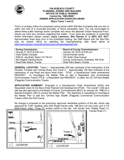
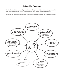
Annals of Otology, Rhinology & Laryngology I2O(9):6O8-614.
© 2011 Annals Publishing Company. All rights reserved.
Otosclerosis: Thirty-Year Foliow-Up After Surgery
Ylva Dahlin Redfors, MD; Claes Möller, MD, PhD
Objectives: The aims of this study were to evaluate the hearing outcomes 28 to 30 years after stapedectomy in patients
with surgically confirmed otosclerosis, and to evaluate inner ear involvement.
Methods: A retrospective clinical study was performed. Sixty-five consecutive patients who underwent stapedectomy
at a tertiary referral center between 1977 and 1979 were included in the study. Medical records, including preoperative
and postoperative audiograms, were reviewed, and a long-term follow-up clinical examination and pure tone audiometry
were performed. The hearing outcome was compared with that of a reference population (ISO 7029) in terms of age and
gender.
Results: Thirty years after stapedectomy, 66% of the patients' ears studied showed a moderate to profound hearing loss.
The deterioration was mainly caused by a sensory hearing loss. The hearing loss was significantly greater than that in the
reference population for both air and bone conduction thresholds at the early and late stages of the disease. A large majority of the patients (88%) had bilateral otosclerosis.
Conclusions: Patients with otosclerosis have a sensorineural hearing loss that cannot be explained by age. Otosclerosis
should be regarded as a middle and inner ear disease. Almost all patients with otosclerosis are in need of ongoing audiological rehabilitation and hearing aids.
Key Words: hearing threshold, long-term follow-up, otosclerosis, sensorineural hearing loss, stapedectomy.
of otosclerosis is unknown, the disease remains incurable. The treatments that can be implemented are
symptomatic: hearing aids or surgery to compensate
for hearing loss, and medical treatment with sodium
fluoride or bisphosphonates to reduce the progression of cochlear otosclerosis.''*
INTRODUCTION
Otosclerosis is a mysterious disease that has confused and interested clinicians and researchers for
centuries. In 1704, Antonio Maria Valsalva reported findings of a stapes fixation caused by ankylosis.
The medical entity of hearing loss associated with
stapes fixation was first described as a specific disease of the ear by Adam Politzer in 1893. Earlier, the
ankylotic stapes was generally regarded as a complication of catarrhal inflammation of the middle ear
and the hearing loss was considered a nervous deafness, because the tympanic membrane was intact.'
The etiologic and pathophysiologic mechanisms remain unknown despite intensive research. Different theories have been postulated; they include viral,^'^ endocrine, hormonal,'* and autoimmune factors.^ Genetic factors appear to play a vital role, and
several genes have been localized, but no true candidate gene has as yet been identified .^"'^ It is still
not known whether the postulated autosomal dominant inheritance with reduced penetrance is the only
mode of transmission, because approximately half
of cases appear to be sporadic.'^ Because the cause
Otosclerosis has a prevalence of approximately 0.3%'^ in Western countries, with a peak onset
in the third decade. Clinical otosclerosis is twice
as common in women. It has traditionally been regarded as a middle ear disease, and during the past
50 years the development of surgical methods has
been successful. In 1958, stapedectomy was introduced by Shea.'^ Later, many modifications were
made, and stapedotomy is now the most widely
used technique.'^ Many studies have shown good
surgical results, including closure of the air-bone
gap (ABG), on both a short- and long-term basis (up
to 32 years).'^'^ The inner ear component in otosclerosis has been extensively studied and debated.
Histologie studies have questioned the relationship
between histologie cochlear otosclerosis and sensorineural hearing loss.^^^'2' Audiological studies
From the Department of Otolaryngology, Department of Clinical Sciences, Sahlgrenska .Academy, University of Gothenburg, Gothenburg (Dahlin Redfors), and the Department of Audiology, Örebro University Hospital, School of Health Science, Örebro University/
Swedish Institute of Disability Research, Örebro (Möller), Sweden. This work was supported by the Gothenburg Medical Society Research Fund, the Swedish Association of Hard of Hearing People Research Fund, Amiöf's Research Fund, and the Acta Otorhinolaryngologica Research Fund.
Correspondence: Ylva Dahlin Redfors, MD, Dept of Otolaryngology, Sahlgrenska Academy, Grona straket 9, SE-413 45 Gothenburg,
Sweden.
608
Dahlin Redfors & Möller, Thirty-Year Follow-Up After Surgery for Otoselerosis
have reported various results. Several studies have
reported a stable bone conduction (BC) threshold,
whereas others have demonstrated a sensory hearing
loss progression of various degrees. The sensorineural hearing loss has been interpreted as being age-related in the majority of cases,^2-24 in contrast, a 2006
study by Topsakal et aP^ demonstrated a preoperative sensorineural hearing loss disproportionate to
age. The term cochlear otoselerosis, which was previously only used to describe histologie otoselerosis with the replacement of the endosteal layer of
the cochlea, has subsequently been used in conjunction with clinical otoselerosis to refer to sensorineural hearing loss that is presumably caused by otoselerosis, in contrast to age-related hearing loss. In
this study, the term cochlear otoselerosis refers to
sensorineural hearing loss disproportionate to age in
patients with otoselerosis. The question of the nature of the sensorineural hearing loss in otoselerosis
remains unclear, and as there is a long lifetime expectancy with the disease, it is important to pursue
long-term follow-up studies of audiological function. It is also of great importance to improve clinical expression and phenotype studies.
The aims of the study were 1) to analyze hearing
in a group of consecutive patients who underwent
stapedectomy for otoselerosis 28 to 30 years earlier,
2) to evaluate the degree of cochlear involvement in
otoselerosis by comparing hearing loss in patients
with otoselerosis with that in an age-matched reference population, and 3) to compare long-term hearing deterioration in operated ears with that in nonoperated otosclerotic ears. The study was reviewed and
approved by the Regional Ethical Review Board,
Gothenburg, Sweden,
MATERIALS AND METHODS
Patient Selection. The study included consecutive patients who underwent stapedectomy between
1977 and 1979 at the Department of Otolaryngology, Sahlgrenska University Hospital (Gothenburg,
Sweden), a tertiary referral center. We included
only patients born in 1930 or later who underwent
stapedectomy between 1977 and 1979 for otoselerosis (ie, progressive conductive hearing loss, impedance measurements indicating stapes fixation,
and peroperative findings of stapes fixation). Ninety-three patients were invited to participate, and 65
subjects agreed to enter the study after giving their
informed consent. The study population consisted of
42 women (65%) and 23 men (35%), all Caucasian,
The mean age at surgery was 36 years (range, 20 to
48 years; SD, 6,6 years). The mean age at follow-up
was 65 years (range, 48 to 77 years; SD, 6,6 years):
for women 66 years (range, 48 to 77 years) and for
609
men 64 years (range, 54 to 73 years). The ear that
underwent surgery between 1977 and 1979 is referred to as the study ear, and the other is referred to
as the control ear.
Surgical Methods. In 62 of the 65 patients, stapedectomy was the primary form of surgery for the
study ear. Three patients had had stapes mobilization before stapedectomy, A wire prosthesis and fascia to cover the oval window niche were used in the
majority of cases, A Teflon wire prosthesis and perichondrium were used in 7 of the 65 cases. Surgery
was performed by 11 surgeons,
Preoperative and Postoperative Audiometric Data
(1977 to 1979). The patients' medical records were
reviewed. An audiogram| was obtained less than 1
month before stapedectorny, except for 2 subjects (6
and 14 months before stapedectomy). Postoperative
audiograms were obtained 1 to 34 months after stapedectomy (mean, 6,5 months; SD, 5,8 months), Preoperative and postoperatiye audiograms were available for 61 of the 65 patients: frequencies from 0,25
to 8 kHz were tested for air conduction (AC), and
frequencies from 0,5 to 4 IkHz for BC, The ABG was
calculated, as well as the pure tone average (PTA)
for AC and BC at 0,5, l' 2, and 4 kHz, The guidelines from the Committee on Hearing and Equilibrium26 regarding the 4-t(^ne PTA for 0,5, 1, 2, and 3
kHz could not be followed, because 3 kHz was not
included in preoperative and postoperative testing.
Follow-Up Data (2007 to 2008). During the follow-up, an otomicroscopic examination and a structured interview (by Y.D.R.) were performed, as well
as an audiological assessment, Audiological assessments were carried out with a clinical audiometer
(AC 30, Interacoustics) calibrated according to the
norms of the Internatiorial Standards Organization,
A pure tone audiogram including frequencies from
0,5 to 8 kHz was obtained. For AC and BC thresholds, the PTA values and ABG (0,5, 1, 2, 4 kHz)
were calculated, Stapediàl reflexes were tested in the
nonoperated ears (at 0,5] 1, and 2 kHz), with a maximum stimulation of 110 dB, During the follow-up,
a nonoperated control ear was considered to have
otoselerosis if the audiogram showed a conductive
or mixed hearing loss with a significant ABG (10 dB
at 2 or more frequencies or 15 dB at 1 frequency) or
with pathological stapedial reflexes.
The stapedial reflexes were considered pathological if no reflexes could be measured, provided that
the hearing loss did not exceed 30 to 40 dB if conductive and 50 dB if sensorineural on the stimulus
side, or if a biphasic reflex pattern was noted,2'' The
hearing was compared with that of a reference group
from the ISO 7029 according to age and gender,28
Dahlin Redfors & Möller, Thirty-Year Follow-Up After Surgery for Otosclerosis
610
O
0.5
Frequency (kHz)
1
2
4
6
8
10
20
—•— Preoperative AC
30
•»s
40
50
60
- • - Preoperative BC
—ki • Postoperative AC
*S
^—-••'"^
^"^^^^
- • - Postoperative BC
70
Fig 1. Audiograms comparing air conduction (AC) and bone
conduction (BC) thresholds for each frequency. A) Preoperative
and postoperative data. B) Postoperative and follow-up data. C)
Study ears compared with 23 control ears with otosclerosis without stapedectomy. Control ears are shown both with and without
correction according to Carhart.^'
80
90
100
0.5
1
Frequency (kHz)
2 , 4
6
0.6
8
10'
10
00
^M--m ^
•---•'
\'N.
— ^ Postoperative AC
•''
•••'.:-
2 40
50
60
70
X
S—•
H » - Postoperative BC
- * • Follow-up AC
- • - Follow-up BC
V
80
90
B
8
0-
o
20
,-X 30
Frequency (kHz)
1
2
4
6
100
The BC value in the. otosclerotic ears without surgery (23 of 65) was corrected according to the Carhart effect (5 dB at 0.5 kHz, 10 dB at 1 kHz, 15 dB
at 2 kHz, and 5 dB at 4 kHz).22.29
Statistical Analysis. Patients with otosclerosis
were compared with an otologically normal population .2^ For each variable reflecting hearing deviation, a z-score was calculated. The z-scores were
based on the 10th, 25th, 50th, 75th, and 90th percentiles presented for the normal material for each
frequency and age at 10-year intervals. The 90th
percentile was assigned the value of 1.28 (according
to the normal distribution), the 75th percentile was
assigned 0.67, the median was 0, the 25th percentile was -0.67, and the 10th percentile was -1.28. To
obtain z-scores between and beyond the given percentiles, we used a linear regression model to interpolate and extrapolate the association between the
z-score and the variable of hearing deviation. These
linear regressions were piecewise linear, with breakpoints in the medians for each age and frequency. A
z-score described how the values for the hearing deviation variable for patients with otosclerosis were
related to those of the normal population of the same
age and gender and at the same frequency.
Fisher's test for paired comparisons was used to
test whether the difference between z-scores after
20'
J 30S 40'
S 50'
—•—
-•—* •
•••••
-•-
Study ear AC
Study ear BC
Control ear AC
Control ear BC
Control ear BC
with correction
ß 70
80
90
~ 100
Stapedectomy and at follow-up was not zero (ie, to
test whether there was a difference between scores).
Fisher's test for paired comparisons was also used to
test whether there was a statistical difference between
the preoperative and follow-up PTA scores regarding AC and BC thresholds. Statistical analysis comparing study ears and otosclerotic control ears was
performed in 23 patients with bilateral otosclerosis
who only underwent surgery on the study ear by use
of Fisher's test for paired comparisons. Preoperative
and postoperative audiograms of the patients who
declined to participate in the study were reviewed
and compared with those of the study group. No differences were found regarding age, gender, or preoperative and postoperative hearing outcomes.
RESULTS
The follow-up periods were 28 to 30 years. At
follow-up, 57 of the 65 patients (88%) had bilateral
otosclerosis and 8 (12%) had unilateral otosclerosis.
Among patients with bilateral otosclerosis, 23 (35%
of the total) had a nonoperated otosclerotic control
ear. At the time of follow-up, 12 of the 65 study ears
(18%) had had 1 revision, 4 (6%) had had 2 revisions, and 4 (6%) had had 3 revisions.
The mean preoperative AC PTA in the study ear
was 53 dB, the mean preoperative BC PTA was 27
dB, and the mean ABG was 26 dB. The mean post-
Dahlin Redfors & Mötler, Thirty-Year Follow-Up After Surgery for Otosclerosis
AC pure
tone average
BC pure
tone average
TABLE 1. AUDIOMETRIC DATA FOR STUDY EARS
Air-bone gap
AC PTA
53 ± 11
32 ±13
51 ± 19
Preoperative
Postoperative
Follow-up
I
Preoperative
m
611
Postoperative
Follow-up
p < 0.0001
Fig 2. Comparison of audiometric data for study ears.
operative AC PTA was 32 dB and the mean BC PTA
was 22 dB, making the mean postoperative ABG 10
dB. The mean improvements were 21 dB in the AC
PTA, 6 dB in the BC PTA, and 16 dB in the ABG.
The improvement in AC threshold was most pronounced in the lower frequencies of 500 and 1,000
Hz (Fig 129).
The mean follow-up PTA was 51 dB for AC and
36 dB for BC, with a mean ABG of 15 dB. The mean
AC PTA had deteriorated by 19 dB compared with
the postoperative value. The mean BC PTA had decreased to 36 dB, compared with the postoperative
value of 22 dB (Fig 1). The mean 30-year AC PTA
showed no significant difference from the mean preoperative value (p > 0.30). The BC PTA demonstrated a significant deterioration in threshold compared
with preoperative values (p < 0.0001; Fig 2 and Table 1). A summary of all of the data is presented in
Table 2.
At the time of the follow-up, 66% of the study ears
showed a moderate to profound hearing loss as calculated by the PTA. No major differences were seen
BCPTA
ABG
27 ±10
22 ± 9
36 ±14
26 ± 9
10±7
15±9
Data are mean ± SD in decibels.
AC — air conduction; PTA — pure tone average for 0.5. 1. 2. and 4
kHz; BC — bone conduction; ABG — air-bone gap.
regarding the BC PTA when the revision groups (ie,
those with 0, 1,2, and 3 revisions) were compared
before and after stapedectomy and at follow-up. In
terms of AC, no major differences were seen compared with the whole cohort, except for two findings.
The first finding was that in 4 of the 8 patients in the
groups with 2 and 3 revisions, no hearing gain was
measured after stapedectomy. These patients had
early revision. The second finding was an increase
in AC threshold at follow-up in the group with 3 revisions. This increased PTA was due to the fact that
1 individual developed a severe hearing loss (AC
PTA of 101 dB). The patient's hearing loss had deteriorated during the 1980s and 1990s in both ears,
with a BC PTA that was equal between the ears. The
control ear only underwent stapedectomy once.
The control ears with otosclerosis that were not
treated by surgery had an AC PTA of 46 dB and a
BC PTA of 34 dB at follow-up. After correction according to Carhart,29 the corresponding figure for
BC was 25 dB (Fig 3 and Table 3). The mean PTA
values for postoperative and follow-up AC and BC
for each frequency were compared with those of a
normal population with no history of ear disease,
controlled for age and gender. The z-scores for both
AC and BC thresholds, after stapedectomy and at
the 30-year follow-up, were significantly worse than
those of the reference group ISO 7029 (p < 0.001).
The differences were significantly larger for AC
than for BC and for postoperative values than for
TABLE 2. SUMMARY OF AUDIOMETRIC MEAN DATA FOR STUDY EARS BY FREQUENCY
Preoperative
Postoperative
Follow-up
Difference*
AC (dB HL)
BC (dB HL)
ABG (dB)
AC (dB HL)
BC (dB HL)
ABG (dB)
AC (dB HL)
BC (dB HL)
ABG (dB)
AC (dB HL)
BC (dB HL)
ABG (dB)
0.5 kHz
I kHz
2 kHz
4 kHz
6 kHz
8 kHz
57
25
32
28
20
8
41
26
15
13
54
24
30
26
18
8
44
26
18
18
8
10
50
33
17
31
25
6
52
43
50
27
23
42
26
16
67
48
19
25
22
3
58
50
54
54
6
7
*Difference between postoperative and follow-up values.
9
21
18
3
77
23
35
PTA
53
27
26
32
22
10
51
36
15
19
14
5
Dahlin Redfors & Möller, Thirty-Year Eollow-Up After Surgery for Otosclerosis
612
Control ear
Study
ear AC
Control
ear AC
Study
ear BC
Control
ear BC
BC v\íith
correction
TABLE 4. COMPARISON OF STUDY GROUP WITH
REFERENCE POPULATION
z-Score
No. of
Mean ±SD
Range
Pts
Postoperative
AC
Follow-up AC
Postoperative
BC
Follow-upBC
*Flsher's test.
-o
• Preoperative
• Postoperative
• Follow-up
Fig 3. Comparison of audiometric data for study ears
and control ears with and without correction according
to Carhart.29
follow-up values (p < 0.001; Table 4).
At follow-up, there were no significant differences in PTA between the study ears and the 23 untreated otosclerotic control ears in terms of either AC or
BC. After the correction according to Carhart, a significantly better BC threshold was noted for the otosclerotic ears without surgery (p < 0.001; Fig 3 and
Table 3). The mean z-score for the untreated otosclerotic control ears was significantly lower than
that for the normal population for both AC and BC
with the Carhart correction. No gender differences
were noted in PTA before or after stapedectomy or
at follow-up, and as a result, all of the patients were
analyzed as one group.
DISCUSSION
The aims of the present study were to assess hearing in patients with otosclerosis 28 to 30 years after
stapedectomy and to evaluate sensorineural hearing
loss in patients with otosclerosis by comparing their
hearing thresholds with those of a normal population
and by comparing operated versus nonoperated otosclerotic ears. Our study shows that hearing gained
by surgery is lost at the follow-up and that the sensorineural hearing loss associated with otosclerosis
cannot be explained simply by age. After the Carhart correction, the untreated otosclerotic ears had
TABLE 3. COMPARISON OF AUDIOMETRIC DATA
FOR STUDY EARS AND CONTROL EARS WITH AND
WITHOUT CORRECTION ACCORDING TO CARHART^«
Study Ear PTA
AC
BC
Preoperative
53±13
3O±I2
Postoperative 35 ± 12 24 ± 9
Follow-up
54 ± 1 9 40 ± 1 5
Data are mean ± SD.
CC — Carhart correction.
Control Ear PTA
BC With
AC
BC
CC
23±I3
23 ± 1 5
46 ± 2 1
20±10 1 2 ± 1 0
34 ± 1 5
25 ± 1 5
Two-Sided
p Value*
61
-3.80 ±1.51 -7.6 to-1.0
<0.001
58
61
-2.82 ±1.28 -6.3 to-0.8
-2.54 ±1.17 -6.7 to-0.6
<0.001
<0.001
58
-1.73±1.18 -4.7to0.4
<0.001
a significantly better BC threshold than did the operated otosclerotic ears, reflecting better-preserved
sensorineural function.
The cohort of individuals included in this study
was chosen in an attempt to have the longest possible follow-up period, with as few dropouts as possible. Of the 93 eligible subjects, 65 (70%) participated in the study. All patients underwent stapedectomy, the standard procedure of the 1970s, whereas the standard procedure of today is stapedotomy.
In comparing the postoperative results of the two
techniques, no significant difference is seen in the
middle frequency range.^^ Although a significant
improvement in the higher frequencies has been reported in favor of the stapedotomy technique after
operation and at shorter follow-up periods,''^•^""^^ ^
20-year follow-up study by Aarnisalo et aP^ reported no difference between the techniques (except at
the single frequency of 2 kHz). As a result, in our
opinion, the finding of long-term hearing deterioration after surgery in our study is probably valid even
today in ears that undergo stapedotomy.
In the present study, 31 % had one ( 12 of 65), two
(4 of 65), or three (4 of 65) revision surgeries. Other studies have reported lower revision rates — approximately 11%33-35 _ but they are not completely
comparable, because their follow-up periods were
considerably shorter and ranged from 1 to 26 years.
The higher rate could also be due in part to the fact
that the operations in our series were performed by
11 surgeons, both senior and in training. Another explanation could be that these surgeons were more
likely to perform revision surgery than were surgeons in other studies. No major differences were
seen in BC or AC thresholds at follow-up between
the different revision groups (according to number
of revisions), and as a result, the somewhat higher
number of revisions in the present study did not affect the final hearing outcome.
Many studies have demonstrated that stapes surgery is a successful means of improving hearing,
which we also found to be true. However, in a longterm perspective, we found that the hearing gained
Dahtin Redfors & Möller, Thirty-Year Follow-Up Afier Surgery for Otosclerosis
by surgery was lost. The indications for stapedectomy, as well as the preoperative and postoperative
hearing levels for AC, BC, and ABG, were comparable to those in other studies.32.33 j o our knowledge,
one other study has followed a cohort with a uniform follow-up time of approximately 30 years.'^
They followed 58 of 322 patients over a period of 33
years and found that the long-term ABG was largely maintained and that the patients' hearing had deteriorated during the time period. These results are
similar to our results.
In our study, at the time of follow-up, 66% of the
participants displayed a moderate to profound hearing loss. The deterioration was mainly due to a loss
of cochlear function, whereas the ABG had only deteriorated by 5 dB during the 30-year period. To assess whether the sensorineural hearing losses in the
operated ears were mainly due to age-related hearing loss, we made a comparison with the International Standardized Database, ISO 7029. At the present
time, there are two International Standardized Databases for hearing threshold levels as a function of
age: ISO 1999 database A (equal to ISO 7029), and
ISO 1999 database B (otologically unselected population). Database A, which was chosen, describes
the normal hearing distribution in an otologically
normal population "free from all signs or symptoms
of ear disease and from obstructing wax in the ear
canals, and who has no history of undue exposure
to noise ."2^(p') Because the population was otologically normal, with no history of middle or external
ear disease, the assumption was made that the AC
threshold was equal to the BC threshold. The database describes hearing loss as a function of age and
gender. A literature search revealed very few studies that used an accessible reference population for
comparing results. We have found only the study by
Topsakal et al .25 The statistical analysis comparing
the study ears and the ISO control group with regard
to both AC and BC on the basis of the described zscore revealed a highly significant difference both
after operation and at the 30-year follow-up. This
means that patients with otosclerosis have a significantly more pronounced hearing loss, compared
with a normal-hearing population, measured by both
AC and BC thresholds and both after operation and
after 28 to 30 years.
Our results are in agreement with the study by
Topsakal et a\P who compared preoperative BC
613
thresholds after the correction according to Carhart
with ISO 7029. Our study indicates that the sensorineural hearing loss, both in the study ears and
in the control ears with otosclerosis, is more pronounced in comparison to that of the normal population. When age-related hearing loss pt-ogresses with
age, the differences decrease. However, it should be
noted that the otosclerotic ears still have a significantly larger hearing loss than the reference population. The true nature of the sensorineural hearing
loss progression seen in otosclerotic patients can be
and has been the subject of a great deal of debate. In
our opinion, the sensorineural hearing loss seen in
elderly otosclerotic patients is a combination of otosclerosis and age-related hearing loss.
Different approaches have been used in attempts
to investigate the cochlear component in otosclerosis.20 One approach was to compare otosclerotic
ears that underwent surgery with the other, nonoperated control ears.2236,37 j ^ our study, 23 patients had
an otosclerotic ear that had not undergone surgery
at follow-up. The hearing result revealed no statistical differences in AC compared with the operated
ears, and the same applied to BC until correction for
the Carhart effect was made. With the Carhart correction, the BC threshold was significantly better in
the otosclerotic ears that did not undergo surgery.
This is in contrast to the theory that surgery enables
sounds to reach the cochlea and thus protects it from
premature degeneration due to inactivity.^'^ On the
basis of our results, we suggest further studies to address audiological rehabilitation, hearing aid use,
and quality of life in these patients.
CONCLUSIONS
Thirty years after stapedectomy, 66% of the subjects displayed a moderate to profound hearing loss
in the study ear. Hearing deterioration was mainly caused by sensorineural loss. The sensorineural
hearing loss was significantly greater than that in the
ISO reference population, both early and late in the
course of the disease. The difference in sensorineural function could not be explained by age. Otosclerosis should be regarded as a middle and inner
ear disease. Surgery did not affect the audiological
function in the long term, as there was no statistical
difference between operated and nonoperated otosclerotic ears after 30 years. In our opinion, all patients with otosclerosis require ongoing audiological
rehabilitation.
Acknowledgments: The authors thank Helena Johansson, Kristina Björnham, Margareta Magnusson, Ann-Christin Hermansson, Eivor
Palsson, Jonas Carlsson, and Camilla Johansson for their work on the study.
REFERENCES
I. Mudry A, Adam Politzer (1835-1920) and the description of otosclerosis. Otol Neurotol 2006;27:276-81.
2. McKenna MJ, Mills BG, Galey FR, Linthicum FH Jr.
Filamentous structures morphologically similar to viral núcleo-
614
Dahlin Redfors & Möller, Thirty-Year Follow-Up After Surgery for Otoselerosis
capsids in otosclerotic lesions in two patients. Am J Otol 1986;7 :
25-8.
3. Lolov S, Edrev G, Kyurkchiev S. Antimeasles immunoglobulin G and virus-neutralizing activity in sera of patients
with otoselerosis. Adv Otorhinolaryngol 2007;65:107-13.
4. Lippy WH,BerenholzLP,SchuringAG,Burkey JM. Does
pregnancy affect otoselerosis? Laryngoscope 2005;115:1833-6.
5. Yoo TJ, Tomoda K, Stuart JM, Kang AH, Townes AS.
Type II collagen-induced autoimmune otospongiosis. A preliminary report. Ann Otol Rhinol Laryngol 1983;92:103-8.
6. Tomek MS, Brown MR, Mani SR, et al. Localization of
a gene for otoselerosis to chromosome 15q25-q26. Hum Mol
Genet 1998;7:285-90.
7. Van Den Bogaert K, Govaerts PJ, Schatteman I, et al.
A second gene for otoselerosis, 0TCS2, maps to chromosome
7q34-36. Am J Hum Genet 2001;6B:495-500.
8. Chen W, Campbell CA, Green GE, et al. Linkage of
otoselerosis to a third locus (0TSC3) on human chromosome
6p21.3-22.3. J Med Genet 2002;39:473-7.
9. Brownstein Z, Goldfarb A, Levi H, Frydman M, Avraham KB. Chromosomal mapping and phenotypic characterization of hereditary otoselerosis linked to the 0TSC4 locus. Arch
Otolaryngol Head Neck Surg 2006;132:416-24.
10. Van Den Bogaert K, De Leenheer EM, Chen W, et al. A
fifth locus for otoselerosis, 0TSC5, maps to chromosome 3q2224. J Med Genet 2004;41:450-3.
11. Thys M, Van Den Bogaert K, Iliadou V, et al. A seventh
locus for otoselerosis, OTSC7, maps to chromosome 6ql3-16.1.
Eur J Hum Genet 2007;15:362-8.
12. Bel Hadj Ali I, Thys M, Beltaief N, et al. A new locus
for otoselerosis, 0TSC8, maps to the pedcentromeric region of
chromosome 9. Hum Genet 2008;123:267-72.
13. Larsson A. Otoselerosis. A genetic and clinical study.
Acta Otolaryngol Suppl 1960;154:l-86.
14. Uppal S, Bajaj Y, Coatesworth AP. Otoselerosis 2: the
medical management of otoselerosis. Int J Clin Pract 2010;64:
256-65.
15. Declau E, van Spaendonck M, Timmermans JP, et al.
Prevalence of otoselerosis in an unselected series of temporal
bones. Otol Neurotol 2001;22:596-602.
16. Shea JJ Jr. Fenestration of the oval window. Ann Otol
Rhinol Laryngol 1958;67:932-51.
17. Fisch U. Stapedotomy versus stapedectomy. Otol Neurotol 2009;30:1160-7.
18. Karhuketo TS, Lundmark J, Vanhatalo J, Rautiainen M,
Sipilä M. Stapes surgery: a 32-year follow-up. ORL J Otorhinolaryngol Relat Spec 2007;69:322-6.
19. Ramsay H, Kärkkäinen J, Palva T. Success in surgery for
otoselerosis; hearing improvement and other indicators. Am J
Otolaryngol 1997;18:23-8.
20. Nelson EG, Hinojosa R. Questioning the relationship between cochlear otoselerosis and sensorineural hearing loss: a
quantitative evaluation of cochlear structures in cases of otosele-
rosis and review of the literature. Laryngoscope 2004; 114:121430.
21. Schuknecht H, Kirchner JC. Cochlear otoselerosis: fact
or fantasy. Laryngoscope 1974;84;766-82.
22. Browning GG, Gatehouse S. Sensorineural hearing loss
in stapedial otoselerosis. Ann Otol Rhinol Laryngol 1984;93:136.
23. Ramsay HA, Linthicum FH Jr. Mixed hearing loss in otoselerosis: indication for long-term follow-up. Am J Otol 1994;
15:536-9.
24. Pirodda E, Modugno GC, Buccolieri M. The problem
of the sensorineural component in otosclerotic hearing loss: a
comparison between operated and non-operated ears. Acta Otolaryngol 1995;115:427-32.
25. Topsakal V, Fransen E, Schmerber S, et al. Audiometric
analyses confirm a cochlear component, disproportional to age,
in stapedial otoselerosis. Otol Neurotol 2006;27:781-7.
26. Committee on Hearing and Equilibrium guidelines for
the evaluation of results of treatment of conductive hearing loss.
American Academy of Otolaryngology-Head and Neck Surgery
Foundation, Inc. Otolaryngol Head Neck Surg 1995;113:1867.
27. Arlinger S, ed. Nordisk lärobok i audiologi. Bromma,
Sweden; CATegnér AB, 2007:192-5.
28. International Organization for Standardization. Acoustics — statistical distribution of hearing thresholds as a function
of age. ISO 7029. 2nd ed. Geneva, Switzerland: International
Standards Organization, 2000.
29. Carhart R. Clinical application of bone conduction audiometry. Arch Otolaryngol 1950;51:798-808.
30. Persson P, Harder H, Magnusson B. Hearing results in
otoselerosis surgery after partial stapedectomy, total stapedectomy and stapedotomy. Acta Otolaryngol 1997;117:94-9.
31. Kiirsten R, Schneider B, Zrunek M. Long-term results after stapedectomy versus stapedotomy. Am J Otol 1994;15:8046.
32. Spandow O, Söderberg O, Bohlin L. Long-term results in
otosclerotic patients operated by stapedectomy or stapedotomy.
Scand Audiol 2000;29:186-90.
33. Aarnisalo AA, Vasama JP, Hopsu E, Ramsay H. Longterm hearing results after stapes surgery: a 20-year follow-up.
Otol Neurotol 2003;24:567-71.
34. Birch L, Elbr0nd O, Pedersen U. Hearing improvement
after stapedectomy: up to 19 years' follow-up period. J Laryngol Otol 1986;100:l-7.
35. Kos MI, Montandon PB, Guyot JP. Short- and long-term
results of stapedotomy and stapedectomy with a Teflon-wire
piston prosthesis. Ann Otol Rhinol Laryngol 2001;110:907-ll.
36. Willis R. The fate ofthe non-operated ear in otoselerosis.
Otolaryngol Head Neck Surg 1989; 100:224-6.
37. Karjalainen S, Kärjä J, Härmä R, Vartiainen E. Hearing
in otosclerotic ears not subjected to operation. J Laryngol Otol
1984;98:255-7.
Copyright of Annals of Otology, Rhinology & Laryngology is the property of Annals Publishing Company and
its content may not be copied or emailed to multiple sites or posted to a listserv without the copyright holder's
express written permission. However, users may print, download, or email articles for individual use.
© Copyright 2026