
archives of neurology_Feb-2_15
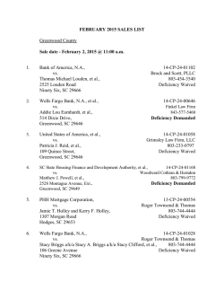
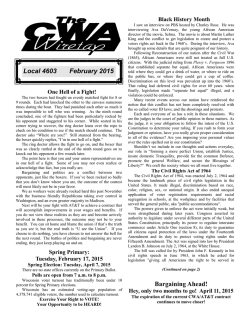
OBSERVATION Isolated Mitochondrial Myopathy Associated With Muscle Coenzyme Q10 Deficiency Seema R. Lalani, MD; Georgirene D. Vladutiu, PhD; Katie Plunkett, MS; Timothy E. Lotze, MD; Adekunle M. Adesina, MD, PhD; Fernando Scaglia, MD Background: Primary coenzyme Q10 (CoQ10) deficiency is rare. The encephalomyopathic form, described in few families, is characterized by exercise intolerance, recurrent myoglobinuria, developmental delay, ataxia, and seizures. Objective: To report a rare manifestation of CoQ10 deficiency with isolated mitochondrial myopathy without central nervous system involvement. Methods: The patient was evaluated for progressive muscle weakness. Comprehensive clinical evaluation and muscle biopsy were performed for histopathologic analysis and mitochondrial DNA and respiratory chain enzyme studies. The patient began taking 150 mg/d of a CoQ10 supplement. Results: The elevated creatine kinase and lactate levels with abnormal urine organic acid and acylcarnitine profiles in this patient suggested a mitochondrial disorder. Skeletal muscle histochemical evaluation revealed ragged red fibers, and respiratory chain enzyme analyses showed P Author Affiliations: Departments of Molecular and Human Genetics (Drs Lalani and Scaglia and Ms Plunkett), Neurology (Dr Lotze), and Pathology (Dr Adesina), Baylor College of Medicine, Houston, Tex; and Departments of Pediatrics, Neurology, and Pathology, State University of New York, Buffalo (Dr Vladutiu). partial reductions in complex I, I + III, and II + III activities with greater than 200% of normal citrate synthase activity. The CoQ10 concentration in skeletal muscle was 46% of the normal reference mean. The in vitro addition of 50 µmol/L of coenzyme Q1 to the succinate cytochrome-c reductase assay of the patient’s skeletal muscle whole homogenate increased the succinate cytochrome-c reductase activity 8-fold compared with 2.8fold in the normal control homogenates. Follow-up of the patient in 6 months demonstrated significant clinical improvement with normalization of creatine kinase and lactate levels. Conclusions: The absence of central nervous system in- volvement and recurrent myoglobinuria expands the clinical phenotype of this treatable mitochondrial disorder. The complete recovery of myopathy with exogenous CoQ10 supplementation observed in this patient highlights the importance of early identification and treatment of this genetic disorder. Arch Neurol. 2005;62:317-320 RIMARY COENZYME Q10 (CoQ10) deficiency (Mendelian Inheritance in Man 607426) is rare and is characterized by significant clinical heterogeneity. The clinical spectrum varies from encephalomyopathy,1-4 familial cerebellar ataxia,5 and Leigh encephalopathy6 to widespread multisystem disease. 7 The encephalomyopathic form, described in 4 families,1-4 is characterized by exercise intolerance, recurrent myoglobinuria, developmental delay, ataxia, and seizures. Herein, we describe a patient with exercise intolerance, ragged red fibers, muscle CoQ10 deficiency, and associated muscle carnitine deficiency with no evidence of recurrent myoglobinuria or central nervous system involvement. Treatment with CoQ10 supplementation resulted in significant clinical improve- (REPRINTED) ARCH NEUROL / VOL 62, FEB 2005 317 ment and normalization of serum creatine kinase and lactate values. This report extends the clinical spectrum of CoQ10 deficiency to include isolated primary myopathy without ataxia, seizures, or cognitive impairment. REPORT OF A CASE The patient was initially evaluated at 11.5 years of age for progressive muscle weakness. He was a previously healthy, developmentally normal child, born at 34 weeks’ gestation to healthy, nonconsanguineous parents. Insidious onset of exercise intolerance and proximal muscle weakness began 4 months prior to evaluation, manifested by difficulty ascending stairs and lifting heavy objects. This was preceded by constitutional fatigue for several months. WWW.ARCHNEUROL.COM ©2005 American Medical Association. All rights reserved. METHODS A B C D E F A skeletal muscle biopsy was performed at 11.5 years of age. Cryostat sections of flash-frozen muscle were stained with hematoxylin-eosin, the modified Gomori trichrome stain, succinate dehydrogenase, cytochrome-c oxidase, reduced nicotinamide adenine dinucleotide dehydrogenase, and adenosine triphosphatase activities at pH 4.3 and 4.6. Muscle was fixed in 3% glutaraldehyde for electron microscopy. A blood specimen for mitochondrial DNA mutation analysis was obtained from the proband. Mitochondrial DNA mutation analysis in the proband’s skeletal muscle and blood specimens were performed to analyze common point mutations (myopathy, encephalopathy, lactic acidosis, and strokelike episodes A3243G and T3271C; myoclonic epilepsy and ragged red fibers A8344G and T8356C; neuropathy, ataxia, and retinitis pigmentosa T8993G and T8993C; cardiomyopathy G8363A; and Leber heredity optic neuropathy G11778A, G3460A, T14484C, and G14459A), deletions, and duplications. The respiratory chain (RC) enzyme analysis in skeletal muscle was performed using standard spectrophotometric analyses.8-11 The carnitine palmitoyltransferase activity was quantified using the isotopeexchange method of Norum.12 Fatty acid–oxidation enzyme activities were evaluated by spectrophotometric assays with chain-length specific substrates of -oxidation. The CoQ10 level was analyzed in skeletal muscle by using highperformance liquid chromatography with UV detection (275 nm) and using coenzyme Q9 as an internal standard.13,14 RESULTS Figure. Histochemical and electron microscopy images. A, Mild variation in fiber size (hematoxylin-eosin, original magnification ϫ 400). B, Modified Gomori trichrome (original magnification ϫ400) with slightly granular fibers suggestive of ragged red fibers (arrows). C, Periodic acid–Schiff (original magnification ϫ 400) with many fibers showing increased subsarcolemmal glycogen content (arrow). D, Sudan black (original magnification ϫ 400) with increased lipid droplets. E, Electron microscopy (original magnification ϫ 5600) with increased lipid droplets (thin arrows) and subsarcolemmal accumulation of glycogen (thick arrow). F, Subsarcolemmal aggregate of mitochondria, some of which have electron dense bodies (arrows) (original magnification ϫ 22 400). He lost weight over this interval and complained of lower extremity muscle cramps. There was no history of unexplained fever, rash, ataxia, hearing loss, or seizures. Family history was unremarkable for neuromuscular disorders. On physical examination he was noted to have significantly reduced proximal muscle strength at the shoulders and the hips, with mild wasting of the shoulder muscles. The Gower maneuver was noted when arising from a seated position. There was no evidence of ophthalmoplegia or ataxia. His creatine kinase level was elevated at 359 U/L (reference range, 55-215 U/L), his lactate level was 33.33 mg/dL (3.7 mmol/L) (reference range, 1.80-18.01 mg/dL [0.2-2.0 mmol/L]), urinalysis did not reveal myoglobinuria, and urine organic acid analysis detected abnormal metabolites including ethylmalonic acid, methylsuccinic acid, hexanoylglycine, and lactic acid. The plasma acylcarnitine profile exhibited elevations of butyrylcarnitine, pentanoylcarnitine, hexanoylcarnitine, octanoylcarnitine, and decanoylcarnitine, with no evidence of plasma total carnitine depletion. Nerve conduction velocity measurements were normal, but the electromyogram showed low-amplitude polyphasic units consistent with myopathy. (REPRINTED) ARCH NEUROL / VOL 62, FEB 2005 318 Skeletal muscle histochemical evaluation revealed rare pale-staining myofibers with the cytochrome-c oxidase stain and scattered ragged red fibers with the Gomori trichrome stain (Figure). On electron microscopy, there was an increase in the number of mitochondria, although no abnormally shaped or enlarged mitochondria were found. A prominent increase in lipid droplets and subsarcolemmal and intermyofibrillar accumulation of free glycogen were found. In muscle, total and free carnitine values were 2.3 SDs below the normal reference mean. Fatty acid–oxidation enzymes and carnitine palmitoyltransferase activities in muscle were normal. Common mitochondrial DNA point mutations and deletions were not detected in skeletal muscle or lymphocytes. The RC enzyme analyses showed partial reductions in complex I, I + III, and II + III activities (Table) with greater than 200% of normal citrate synthase activity, suggestive of increased mitochondrial content and corroborating histochemical findings. Results of magnetic resonance imaging and magnetic resonance spectroscopy of the brain were normal. An echocardiogram demonstrated normal cardiac function. The patient was treated with 150 mg/d of a CoQ10 supplement and 100 mg/kg per day of carnitine for 3 months. On a 3-month follow-up visit, a remarkable improvement in muscle strength was noted with increased proximal muscle strength and absent Gower sign. Based on his considerable improvement with antioxidant therapy, the CoQ10 concentration was analyzed by high-performance liquid chromatography in the original skeletal muscle specimen and found to be 46% of the normal reference mean. The in vitro addition of 50 µmol/L of coenzyme Q1 to WWW.ARCHNEUROL.COM ©2005 American Medical Association. All rights reserved. the succinate cytochrome-c reductase assay of the patient’s skeletal muscle whole homogenate increased the succinate cytochrome-c reductase activity 8-fold compared with a 2.8-fold increase in the normal control homogenates. Citrate synthase activity was not influenced by the addition of coenzyme Q1 to the assay in patients or in controls (data not shown). The CoQ10 supplementation was increased to 300 mg/d at the 3-month visit and within 6 months of therapy, both the creatine kinase (140 U/L) and lactate (5.4 mg/dL [0.6 mmol/L]) levels normalized, with sustained clinical improvement. COMMENT Our findings in this case study suggest that CoQ10 deficiency and the concomitant significant reductions in complex I, I + III, and II + III enzymatic activities in the RC were responsible for the mitochondrial disorder observed in our patient. The observed muscle carnitine deficiency is most likely related to an increased reduced nicotinamide adenine dinucleotide–nicotinamide adenine dinucleotide ratio associated with respiratory chain defects.15 The increased ratio could impair -oxidation at the level of 3-hydroxyacyl-coenzyme A dehydrogenases, with a subsequent accumulation of acylcoenzyme A -oxidation intermediates. These intermediates, released as carnitine esters, are transported into plasma and eliminated in urine, leading to secondary carnitine deficiency.16,17 A recent study of 13 patients with childhood-onset cerebellar ataxia and marked CoQ10 deficiency suggested a cutoff for primary CoQ10 deficiency in muscle at 55% of the normal reference mean.18 Our patient’s muscle CoQ10 activity was 46% of the normal reference mean. This result, in conjunction with the in vitro augmentation of residual muscle complex II + III activity with the addition of exogenous coenzyme Q1 to the assay and the successful clinical outcome with CoQ10 therapy, suggests primary CoQ10 deficiency in our patient. However, the molecular elucidation of this disorder will be required to confirm a primary defect in the ubiquinone biosynthetic pathway in all of these cases. We could hypothesize that the partial deficiency of CoQ10 observed in our proband perhaps accounts for the late clinical manifestation and isolated muscle involvement. However, detailed review of the reported cases indicates no clear correlation between the observed in vitro muscle or fibroblast CoQ10 levels and the severity in phenotype and/or age of onset in the affected individuals. This is illustrated by the presence of undetectable CoQ10 levels in fibroblasts in 2 siblings, one with widespread multisystem involvement and the other with a milder form of the disease.7 In another report of childhood-onset cerebellar ataxia and marked CoQ10 deficiency,18 patients who exhibited muscle CoQ10 concentrations of 2.9 µg/g and 14.8 µg/g, respectively, had a similar phenotype of ataxia and cerebellar atrophy by 9 years of age with no developmental delay or seizures. Coenzyme Q10 plays an important role in the mitochondrial RC by acting as a redox carrier, transferring reducing equivalents from complex I and complex II to (REPRINTED) ARCH NEUROL / VOL 62, FEB 2005 319 Table. Respiratory Chain Analysis of Patient’s Skeletal Muscle Tissue Enzyme NADH dehydrogenase (complex I) Succinate dehydrogenase (complex II) NADH cytochrome-c reductase (complex I + III) Cytochrome-c oxidase (complex IV) Succinate cytochrome-c reductase (complex II + III) Succinate cytochrome-c reductase and coenzyme Q1 Citrate synthase‡ Coenzyme Q10 Enzyme Activity Reference, (µmol/min-1 per g-1)* Mean ± SD 5.1 (35)† 0.53 (61) 0.35 (43)† 14.74 ± 4.48 0.87 ± 0.21 0.81 ± 0.20 1.53 (63) 0.46 (45)† 2.43 ± 0.70 1.03 ± 0.31 3.74 (363) 2.56 ± 0.72 36.54 (232) 9.11 (46) 15.74 ± 4.44 19.81 ± 2.61 Abbreviation: NADH, reduced nicotinamide adenine dinucleotide. *Data represent the mean of 2 independent analyses on different muscle homogenates. Figures in parentheses represent percentage of normal reference mean. †Residual enzyme activity greater than 2 SDs below the normal reference mean. ‡Citrate synthase activity was not influenced by the addition of coenzyme Q1 to the assay (data not shown). complex III.19 Coenzyme Q10 allows the extrusion of protons from the matrix to the intermembrane space along with the electron flow through the RC.20 Deficiency of CoQ10 impairs the proton transfer across the inner mitochondrial membrane, thus affecting generation of adenosine triphosphate and all adenosine triphosphate– dependent metabolic processes. Although the antioxidant treatment for RC defects has no proven efficacy, treatment of ubiquinone deficiency might represent an exception. A defective incorporation of tritium ( 3 H)mevalonate into CoQ10 in fibroblasts initially suggested a specific site of impairment of endogenous CoQ10 synthesis.7 Rötig et al7 reported very low concentrations of labeled decaprenyl-diphosphate in patients’ fibroblast extracts, consistent with a deficiency of trans-prenyltransferase; however, no mutations in the gene encoding trans-prenyltransferase were identified, suggesting that another gene involved in this pathway may be affected. Recently, mutations in the trans-prenyltransferase gene have been identified in 2 siblings with mild intellectual retardation, profound deafness, optic atrophy, valvulopathy, and obesity who had CoQ10 deficiency in fibroblasts but not in skeletal muscle.21 At least 4 different clinical manifestations of CoQ10 deficiency have been described: the encephalomyopathic form with myoglobinuria, ataxia, and seizures1-4; a predominantly cerebellar disease with ataxia and cerebellar atrophy5,18; a widespread multisystem involvement with hypertrophic cardiomyopathy, ataxia, optic nerve atrophy, deafness, generalized amyotrophy, and nephrotic syndrome7; and Leigh encephalopathy with growth retardation, ataxia, deafness, and lactic acidosis.6 The clinical heterogeneity found among patients with CoQ10 deficiency suggests that a number of biochemical and molecular defects may be involved in causing different clinical phenotypes. The isolated myopathy with absence of central nervous system involvement and recurrent myoglobinuria WWW.ARCHNEUROL.COM ©2005 American Medical Association. All rights reserved. in our patient expands the clinical phenotype of CoQ10 deficiency. The complete recovery of myopathy with exogenous CoQ10 supplementation observed in this patient highlights the importance of early identification and treatment of this genetic disorder, perhaps offering a similar prognosis to patients affected with the myopathic form of this condition. Functional studies to identify the possible defect of ubiquinone synthesis in our patient are currently underway. This case demonstrates the need for detailed biochemical assessment of mitochondrial function in the diagnostic evaluation of isolated myopathies. Accepted for Publication: March 11, 2004. Correspondence: Seema Lalani, MD, Department of Molecular and Human Genetics, Baylor College of Medicine, One Baylor Plaza, Room T828, Houston, TX 77030 ([email protected]). Author Contributions: Study concept and design: Lalani, Vladutiu, and Scaglia. Acquisition of data: Lalani, Vladutiu, Plunkett, Lotze, Adesina, and Scaglia. Analysis and interpretation of data: Lalani, Vladutiu, Adesina, and Scaglia. Drafting of the manuscript: Vladutiu and Lalani. Critical revision of the manuscript for important intellectual content: Vladutiu, Lalani, Plunkett, Lotze, Adesina, and Scaglia. Obtained funding: Lalani and Vladutiu. Study supervision: Lalani, Vladutiu, and Scaglia. Funding/Support: This study was supported by the Doris Duke Clinical Scientist Development Award (Dr Lalani), The Children’s Guild of Buffalo, Buffalo, NY (Dr Vladutiu), and Baylor College of Medicine Mental Retardation Research Center, Houston, Tex (Dr Scaglia). Acknowledgment: We thank the family of this patient for participating in the study. REFERENCES 1. Ogasahara S, Engel AG, Frens D, Mack D. Muscle coenzyme Q deficiency in familial mitochondrial encephalomyopathy. Proc Natl Acad Sci U S A. 1989;86: 2379-2382. 2. Sobreira C, Hirano M, Shanske S, et al. Mitochondrial encephalomyopathy with coenzyme Q10 deficiency. Neurology. 1997;48:1238-1243. (REPRINTED) ARCH NEUROL / VOL 62, FEB 2005 320 3. Boitier E, Degoul F, Desguerre I, et al. A case of mitochondrial encephalomyopathy associated with a muscle coenzyme Q10 deficiency. J Neurol Sci. 1998; 156:41-46. 4. Di Giovanni S, Mirabella M, Spinazzola A, et al. Coenzyme Q10 reverses pathological phenotype and reduces apoptosis in familial CoQ10 deficiency. Neurology. 2001;57:515-518. 5. Musumeci O, Naini A, Slonim AE, et al. Familial cerebellar ataxia with muscle coenzyme Q10 deficiency. Neurology. 2001;56:849-855. 6. Van Maldergem L, Trijbels F, DiMauro S, et al. Coenzyme Q-responsive Leigh’s encephalopathy in two sisters. Ann Neurol. 2002;52:750-754. 7. Rötig A, Appelkvist EL, Geromel V, et al. Quinone-responsive multiple respiratorychain dysfunction due to widespread coenzyme Q10 deficiency. Lancet. 2000; 356:391-395. 8. King TE, Howard RL. Preparation and properties of NADH dehydrogenase from cardiac muscle. In: Estabrook R, Pullman M, eds. Methods in Enzymology: Oxidation and Phosphorylation. New York, NY: Academic Press; 1967:275-294. 9. King TE. Preparation of succinate dehydrogenase and reconstitution of succinate oxidase. In: Estabrook R, Pullman M, eds. Methods in Enzymology: Oxidation and Phosphorylation. New York, NY: Academic Press; 1967:322-331. 10. Yonetan T. Cytochrome oxidase: beef heart. In: Estabrook R, Pullman M, eds. Methods in Enzymology: Oxidation and Phosphorylation. New York, NY: Academic Press; 1967:332-335. 11. Srere PA. Citrate synthase. In: Lowenstein J, ed. Methods in Enzymology: Oxidation and Phosphorylation. New York, NY: Academic Press; 1969:3-11. 12. Norum K. PalmitoylCoA: carnitine palmityltransferase. Biochim Biophys Acta. 1964; 89:95-108. 13. Laaksonen R, Riihimaki A, Laitila J, Martensson K, Tikkanen MJ, Himberg JJ. Serum and muscle tissue ubiquinone levels in healthy subjects. J Lab Clin Med. 1995;125:517-521. 14. Rousseau G, Varin F. Determination of ubiquinone 9 and 10 levels in rat tissues and blood by high-performance liquid chromatography with ultraviolet detection. J Chromatogr Sci. 1998;36:247-252. 15. Wijburg FA, Feller N, Scholte HR, Przyrembel H, Wanders RJ. Studies on the formation of lactate and pyruvate from glucose in cultured skin fibroblasts: implications for detection of respiratory chain defects. Biochem Int. 1989;19:563-570. 16. Kler RS, Jackson S, Bartlett K, et al. Quantitation of acyl-CoA and acylcarnitine esters accumulated during abnormal mitochondrial fatty acid oxidation. J Biol Chem. 1991;266:22932-22938. 17. Scaglia F, Longo N. Primary and secondary alterations of neonatal carnitine metabolism. Semin Perinatol. 1999;23:152-161. 18. Lamperti C, Naini A, Hirano M, et al. Cerebellar ataxia and coenzyme Q10 deficiency. Neurology. 2003;60:1206-1208. 19. Ernster L, Dallner G. Biochemical, physiological and medical aspects of ubiquinone function. Biochim Biophys Acta. 1995;1271:195-204. 20. Mitchell P. Protonmotive redox mechanism of the cytochrome b-c1 complex in the respiratory chain: protonmotive ubiquinone cycle. FEBS Lett. 1975;56:1-6. 21. Giurgea I, de Lonlay P, Cretien D, Rustin P, Munnich A, Rotig A. Transprenyltransferase (TPT) mutation in patients with coenzyme Q10 biosynthesis deficiency and mitochondrial disorder [abstract]. Am J Hum Genet. 2003;73:170. WWW.ARCHNEUROL.COM ©2005 American Medical Association. All rights reserved.


© Copyright 2026