
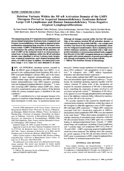
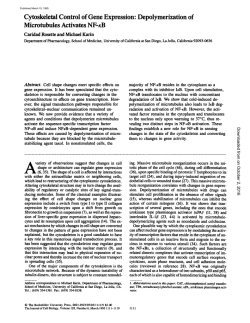
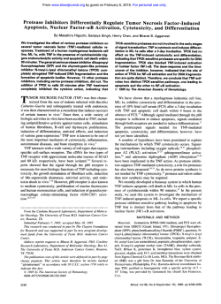
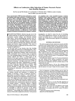
PDF (1 MB)
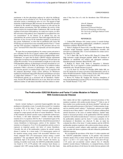
ARTICLE
UBE2L3 Polymorphism Amplifies NF-kB Activation
and Promotes Plasma Cell Development, Linking
Linear Ubiquitination to Multiple Autoimmune Diseases
Myles J. Lewis,1,6,* Simon Vyse,1,6 Adrian M. Shields,1 Sebastian Boeltz,1 Patrick A. Gordon,2
Timothy D. Spector,3 Paul J. Lehner,4 Henning Walczak,5 and Timothy J. Vyse1,*
UBE2L3 is associated with increased susceptibility to numerous autoimmune diseases, but the underlying mechanism is unexplained. By
using data from a genome-wide association study of systemic lupus erythematosus (SLE), we observed a single risk haplotype spanning
UBE2L3, consistently aligned across multiple autoimmune diseases, associated with increased UBE2L3 expression in B cells and monocytes. rs140490 in the UBE2L3 promoter region showed the strongest association. UBE2L3 is an E2 ubiquitin-conjugating enzyme,
specially adapted to function with HECT and RING-in-between-RING (RBR) E3 ligases, including HOIL-1 and HOIP, components of
the linear ubiquitin chain assembly complex (LUBAC). Our data demonstrate that UBE2L3 is the preferred E2 conjugating enzyme
for LUBAC in vivo, and UBE2L3 is essential for LUBAC-mediated activation of NF-kB. By accurately quantifying NF-kB translocation
in primary human cells from healthy individuals stratified by rs140490 genotype, we observed that the autoimmune disease risk
UBE2L3 genotype was correlated with basal NF-kB activation in unstimulated B cells and monocytes and regulated the sensitivity of
NF-kB to CD40 stimulation in B cells and TNF stimulation in monocytes. The UBE2L3 risk allele correlated with increased circulating
plasmablast and plasma cell numbers in SLE individuals, consistent with substantially elevated UBE2L3 protein levels in plasmablasts
and plasma cells. These results identify key immunological consequences of the UBE2L3 autoimmune risk haplotype and highlight
an important role for UBE2L3 in plasmablast and plasma cell development.
Introduction
UBE2L3 is strongly associated with systemic lupus erythematosus (SLE) in genome-wide association studies and
other genetic studies,1–4 as well as multiple autoimmune
diseases (Table S1).5–11 UBE2L3 is an E2 ubiquitin-conjugating enzyme, also known as UbcH7. Although UBE2L3
was one of the first E2 conjugating enzymes to have its
structure determined,12 its cellular functions have remained largely unknown. Because E2 enzymes appeared
to be substitutable in ubiquitination assays, it was initially
assumed that there was redundancy and lack of specificity
between E2 enzymes. E2 enzymes have greater specificity
than was first apparent, and they function with only
selected E3 ligases in vivo. E2 enzymes have a critical role
in determining ubiquitin (Ub) chain type.13 Because E2 enzymes act as ubiquitin shuttles, the kinetics of transfer of
Ub from E2 to substrate in the case of RING E3 ligases, or
onto the E3 in the case of HECT enzymes, might limit
the speed of polyUb chain formation. Klevit and coworkers
showed that UBE2L3 is incapable of conjugating ubiquitin
onto free lysine and directly onto the target substrate, as is
necessary for standard RING E3 ligases.14 UBE2L3 is therefore restricted to HECT-like E3s and co-operates with only a
highly restricted set of dual RING E3 ligases with a RBR
motif (RING-in-between-RING) and seven of the nine
HECT E3 ligases.15
Linear ubiquitination, which involves sequential
bonding of a ubiquitin moiety onto the Met-1 (M1) residue
of ubiquitin,16 is mediated by the 600 kDa E3 complex
LUBAC (linear ubiquitin chain assembly complex),
composed of HOIL-1, HOIP, and Sharpin.17–21 LUBAC
forms linear (M1) Ub chains on NEMO to activate the
IKK complex. Deficiency of HOIL-1 or Sharpin inhibits
phosphorylation and degradation of the NF-kB sequestration protein IkBa, leading to impaired activation of
NF-kB. HOIL-1-deficient mice have defective NF-kB responses,17 and rare human loss-of-function mutations in
HOIL-1 led to defective TNF signaling and abnormal IL-1
responses.22 Sharpin deficiency diminishes NF-kB activation, while increasing proinflammatory TNF-induced cell
death, responsible for chronic proliferative dermatitis in
Sharpin-deficient Cpdm mice.19 Thus LUBAC has been
shown to be critical for NF-kB activation downstream of
the TNF receptor 1 (TNFR1) and CD40. Presence of HOIP
in B cells was necessary for CD40 signaling,23 and reduced
immunoglobulin levels and impaired peritoneal B-1 cell
development were observed in mice with conditional
HOIP deficiency in B cells.24 HOIL-1 and HOIP are both
RBR E3 ligases, so we hypothesized that UBE2L3 would
1
Department of Medical and Molecular Genetics, King’s College London, London SE1 9RT, UK; 2Rheumatology Department, King’s College Hospital, London SE5 9RS, UK; 3Department of Twin Research and Genetic Epidemiology, King’s College London, London SE1 9RT, UK; 4Cambridge Institute for Medical
Research, University of Cambridge, Cambridge CB2 0XY, UK; 5Centre for Cell Death, Cancer and Inflammation, UCL Cancer Institute, University College
London, London WC1E 6DD, UK
6
Present address: Centre for Experimental Medicine and Rheumatology, William Harvey Research Institute, Barts and The London School of Medicine and
Dentistry, Queen Mary University of London, London EC1M 6BQ, UK
*Correspondence: [email protected] (M.J.L.), [email protected] (T.J.V.)
http://dx.doi.org/10.1016/j.ajhg.2014.12.024. Ó2015 The Authors
This is an open access article under the CC BY license (http://creativecommons.org/licenses/by/4.0/).
The American Journal of Human Genetics 96, 221–234, February 5, 2015 221
be required for LUBAC to function in vivo. Recombinant
HOIP and HOIL-1 or Sharpin can generate polyubiquitin
chains with UBE2L3 in biochemical ubiquitination assays,16,20 although this might not be limited to UBE2L3,
as hinted by the fact that the promiscuous E2 enzymes
UBE2D1, UBE2D2, and UBE2D3 (UbcH5a, UbcH5b, and
UbcH5c) can substitute for UBE2L3 in these assays.16 In
this study we set out to investigate the relative importance
of UBE2L3 to LUBAC function in vivo.
Subjects and Methods
Individuals and Genotyping
The study was approved by the UK National Research Ethics Service and institutional review boards of collaborators’ institutions
prior to the commencement of the study. All study participants
provided written consent at the time of sample collection. Samples
from 4,946 individuals with SLE of European ancestry and 1,286
control subjects collected from multiple sites as part of an ongoing
GWAS in SLE were genotyped on the Illumina Human Omni1Quad BeadChip platform. Quality-control analysis of genotyping
was carried out in accordance with Illumina’s Technical Note on
Infinium Genotyping Data, excluding SNPs with poor clustering
separation or call rate <0.95. Additional controls from the Health
and Retirement study (dbGaP accession phs000428.v1.p1) genotyped on the Illumina Human Omni2.5-Quad platform were
included in the analysis. Additional quality-control checks were
made for individual missingness, SNP missingness, autosomal heterozygosity, identity-by-descent (via PLINK algorithm), HardyWeinberg equilibrium, and population structure (via EIGENSTRAT
algorithm) with a panel of 50 ancestry informative markers. After
quality-control analysis, 4,036 SLE-affected individuals and 6,959
control subjects with 696,085 SNPs were imputed with 1000
Genomes reference data via IMPUTE2.2 algorithm across region
21.60 Mb to 22.20 Mb on chromosome 22 spanning UBE2L3. To
ensure reliability, imputed SNPs with an information score less
than 0.9 were discarded. Single marker analysis was performed
with SNPTEST (v.2.2) with four principal components as covariates in logistic regression. All SLE-affected individuals fulfilled
the American College of Rheumatology (ACR) classification
criteria for the diagnosis of SLE. SLE subphenotype data were available on a subset of 1,751 SLE-affected individuals. Case-case subphenotype statistical analysis was performed with SNPTEST with
four principal components as covariates. Presence of lupus
nephritis was defined by renal disorder subcomponent of the
ACR diagnostic criteria.
Expression Data
Microarray data from eQTL studies were obtained for lymphoblastoid cell lines from 270 HapMap individuals (GEO database
GSE6536)25 and for CD19þ B cells and CD14þ monocytes isolated
from peripheral blood mononuclear cells (PBMCs) from 288
healthy individuals (ArrayExpress E-MTAB-945).26 For both data
sets, SNPs across region 21.60 Mb to 22.20 Mb on chromosome
22 were processed with IMPUTE2.2 to generate imputed genotype
data at rs140490. For protein studies, healthy individuals, whose
genotypes had been imputed at rs140490, were recalled from the
TwinsUK resource. PBMCs were obtained from blood samples,
and CD4þ T cells and CD19þ B cells were isolated by positive (Miltenyi) and negative (Invitrogen) selection, respectively, via mag-
netic beads and lysed with ice-cold RIPA buffer with protease
and phosphatase inhibitors (Complete Mini and PhosSTOP,
Roche). BCA assay (Pierce) was used to equalize total protein concentration. UBE2L3 protein levels were assessed by immunoblot.
Densitometry was quantified with ImageJ software (NIH) normalized to actin.
Plasmids and Cell Culture
The following plasmids on the pCMV6 vector were purchased from
Origene: untagged UBE2L3, HOIP, UBE2D1, UBE2D2, UBE2D3,
Sharpin-Myc-DDK, and empty vector (pCMV6-AC). pCMV6HOIL-1-Myc-DDK (Origene) was subcloned into pCMV6AC to remove the C terminus Myc-DDK tag. pcDNA3.1-V5-His-HOIP and
pcDNA3.1-V5-His-HOIP (p.Cys885Ser) were kind gifts of H. Walczak. pFlagCMV2-UbcH8 (UBE2L6) was obtained from Addgene.
Variant p.Cys86Ser UBE2L3 was generated by site-directed mutagenesis (Stratagene Quikchange II XL). All plasmid ORF were verified by full sequencing. HEK293 cells (ATCC) were maintained in
DMEM with 10% FBS.
Antibodies
The following antibodies were used: anti-IkBa (C-21), rabbit antip65 (sc-372), anti-actin (sc-1616), and anti-UBE2L3 (C-20) from
Santa Cruz; anti-Sharpin (4444), anti-p-IkBa (5A5), anti-JNK
(56G8), anti-p-JNK (98F2), anti-ERK1/2 (137F5), anti-p-ERK1/2
(D13.14.4E), and anti-UBE2D3 (D60E2) from Cell Signaling;
and anti-HOIL-1 (HPA024185, Sigma), anti-HOIP (SAB2102031,
Sigma), anti-tubulin (AA13, Sigma), anti-UBE2L3 (20/UbcH7, BD
Bioscience and A-640, Boston Biochem), anti-pan-UbcH5 (A-615,
Boston Biochem), anti-FLAG (Sigma), and anti-V5-AlexaFluor647
(AbD Serotec). Mouse monoclonal anti-UBE2L3 antibody was conjugated to PE via Lynx rapid RPE conjugation kit (AbD Serotec) as
per the manufacturer’s instructions.
Luciferase Reporter Assay
GloResponse NF-kB-RE-luc2P HEK293 cell line (Promega) with stably integrated pGL4.32[luc2P/NF-kB-RE/Hygro] luciferase reporter
were transiently transfected with combinations of plasmids via
Fugene 6 (Promega). Luciferase activity was assayed by Luciferase
Assay Reagent II (Promega) or One-Glo (Promega) on Berthold
Orion luminometer, normalized to cell viability measured by
CellTiterGlo assay (Promega).
RNA Interference
GloResponse NF-kB-RE-luc2P HEK293 cells were seeded at 10,000
cells/well in 96-well plates. After 24 hr, cells were transfected
with siRNA at a final concentration of 25 nmol/l with Dharmafect
reagent #1. After 72 hr, cells were stimulated with TNF and
analyzed by NF-kB luciferase reporter assay. For signaling pathway
analysis, HEK293 cells at a density of 100,000 cells/well in 24-well
plates were transfected as before and stimulated with TNF and
lysed in the presence of protease and phosphatase inhibitor cocktail for immunoblot. Western blot gel electrophoresis, transfer,
and detection was performed in parallel with identical film exposure duration. The following siRNA sequences were used: UBE2L3
sense 50 -CCGCAAAUGUGGGAUGAAA-30 , anti-sense 50 -UUUCAU
CCCACAUUUGCGG-30 ; HOIL-1 sense 50 -GCUCAGAUGCACACC
GUCA-30 and anti-sense 50 -UGACGGUGUGCAUCUGAGC-30 ;
HOIP sense 50 -GGCGUGGUGUCAAGUUUAA-30 and anti-sense
50 -UUAAACUUGACACCACGCC-30 ; and Sharpin sense 50 -CCUG
GAAACUUGACGGAGA-30 and anti-sense 50 -UCUCCGUCAAGU
222 The American Journal of Human Genetics 96, 221–234, February 5, 2015
UUCCAGG-30 . siGENOME RISC-Free Control siRNA (Dharmacon)
was used as a control.
Real-Time Quantitative PCR
HEK293 cells were transfected with control, UBE2L3, or HOIP
siRNA for 48 hr and stimulated with TNF 10 ng/ml for up to
6 hr. mRNA was isolated with Trizol reagent (Life Technologies),
reverse transcribed into cDNA with SuperScript III First-strand Synthesis kit (Life Technologies), and quantified by real-time quantitative PCR with SYBR fast qPCR kit (Kapa) on an Applied Biosystems
7900HT instrument. Data normalized against b-actin levels were
analyzed with Applied Biosystems RQ manager and GraphPad
Prism software. The following gene specific primers were used:
NFKBIA (IkBa) 50 -ATGCTCAGGAGCCCTGTAATG-30 and 50 -CCC
CACACTTCAACAGGAGT-30 ; CCL2 50 -GAAAGTCTCTGCCGCC
CTT-30 and 50 -ATTGATTGCATCTGGCTGAGCG-30 ; TNFAIP3
(A20) 50 -GCGTTCAGGACACAGACTTG-30 and 50 -TTCATCATTCC
AGTTCCGAGTATC-30 ; and VCAM1 50 -TTTGCAGCTTCTCAAGC
TTTT-30 and 50 -CCTGTGGTGCTGCAAGTC-30 .
CD20-APC-H7 (2H7) from Biolegend or BD. Cells were fixed
with BD stabilizing fixative reagent. Cells were permeabilized
with BD perm/wash buffer I and stained for UBE2L3-PE (described
above) or permeabilized with Foxp3 permeabilization buffer
(eBioscience) and stained for both UBE2L3-PE and Ki-67-Alexa488
(B56). Freshly stained cells were acquired on 5 laser BD SORP
LSRFortessa instrument. BD CS&T beads were used immediately
prior to every sample run to maintain instrument consistency
throughout the entire study. Data were analyzed with FlowJo
v.10 for the entire cohort by a single individual completely blind
to genotype. Statistical analysis was performed with one-way
ANOVA. SLE-affected individual demographics and disease characteristics are summarized in Table S2. On the same day that samples
were drawn for flow cytometry, SLE disease activity was recorded
with the SELENA modification of the SLEDAI. Measurement of
erythrocyte sedimentation rate (ESR), anti-dsDNA antibody titer,
and complement C3 and C4 levels were performed as part of
routine clinical care.
Results
NF-kB Translocation
HEK293 cells were transiently transfected for 24 hr with HOIL-1,
V5-His-HOIP, Sharpin, and/or UBE2L3. Cells were detached with
TrypLE (Life Technologies), stimulated with TNF for 30 min, fixed
with BD cytofix, permeabilized with 0.1% Triton X-100, and
stained with rabbit anti-p65 (Santa Cruz), PE-conjugated antiUBE2L3, and anti-V5-AlexaFluor647. Cells were washed, incubated with AlexaFluor488 F(ab)2 donkey anti-rabbit IgG (Jackson),
washed, stained with DAPI. 20,000 cells per condition were acquired on Imagestream X imaging flow cytometer (Amnis). For
ex vivo cell analysis, PBMCs were isolated via Histopaque from
blood samples from previously genotyped healthy individuals
(TwinsUK). CD19þ B cells were isolated by magnetic bead positive
selection (Miltenyi) and CD14þ monocytes were isolated by negative selection (Miltenyi). Endotoxin-free MACS buffer was used
throughout. Cell purity was confirmed by flow cytometry. B cells
and monocytes were cultured in RPMI and stimulated with
0.1 mg/ml CD40L (Enzo) and 10 ng/ml TNF (Axxora), respectively,
for up to 60 min. Cells were fixed and stained for p65 as above,
washed, and stained with DRAQ5 (eBioscience), and 15,000–
20,000 events per sample were acquired by Imagestream X. Data
analysis was entirely automated with IDEAS software batch function applied to the entire cohort and performed fully blinded to
genotype.
Genotyping
Previously genotyped healthy twins were selected on the basis
of imputed genotype at rs140490. Genotyping was confirmed
in these individuals as well as in all SLE-affected individuals
by TaqMan genotyping assay on Applied Biosystems 7900HT
instrument.
Flow Cytometry
Fresh PBMCs were isolated from blood obtained from 29 SLEaffected individuals and 25 healthy control subjects. Cells were
stained with LIVE/DEAD Fixable Blue Dead cell stain (Invitrogen)
to exclude dead cells, Fc receptor blocked (Human TruStain FcX,
Biolegend), and surface-stained with the following markers:
IgD-BrilliantViolet(BV)421 (IA6-2), CD19-BV510 (HIB19), CD27BV650 (O323), CD138-FITC or CD138-PE-Cy7 (MI15), CD24PerCP-Cy5.5 (ML5), CD95-PE-Cy7 (DX2), CD38-APC (HB7),
rs140490 Is Associated with SLE and the rs140409 Risk
Allele Increases Expression of UBE2L3
UBE2L3 genotype data from an ongoing SLE GWAS
(T.J.V., data not shown) was imputed with 1000 Genomes
reference data, identifying rs140490 as the most strongly
associated SNP, located at 270 bp of the promoter
region of UBE2L3 (p ¼ 8.6 3 1014, OR 1.30, 95%
CI: 1.21–1.39) (Figure 1A). Haplotype analysis from
SLE-affected individuals shows that a single risk haplotype spans the entire gene (Figure S1A) and is the
same risk haplotype across all autoimmune diseases
(Figure S1B). Case-case subphenotype analysis available
on a subset of 1,751 SLE case subjects demonstrated
that rs140490 was associated with increased risk of lupus
nephritis (p ¼ 0.0036, OR 1.27, 95% CI: 1.08–1.49), suggesting that UBE2L3 is associated with increased disease
severity.
Genotypes from eQTL studies25,26 with microarray
expression data for UBE2L3 were imputed to 1000 Genomes level, showing that the rs140490 risk allele was
strongly correlated with increased UBE2L3 expression in
EBV-transformed lymphoblastoid cell lines from HapMap
individuals25 (p ¼ 6.06 3 1025). A similarly strong linear
relationship was noted between alleles of rs140490 and
UBE2L3 expression in primary human B cells (p ¼ 1.28 3
109) and monocytes (p ¼ 2.54 3 1027)26 (Figure 1B).
Samples from genotyped healthy individuals (TwinsUK)
confirmed that rs140490 increased UBE2L3 protein levels
in CD19þ B cells isolated from peripheral blood (p ¼
0.0094) but that the risk SNP rs140490 did not significantly alter UBE2L3 protein levels in CD4þ T cells
(Figure 1C). Consistent with this, eQTL microarray data
on primary CD4þ T cells showed a lower p value (p ¼
4.4 3 104) compared to CD14þ monocytes (p ¼ 3.4 3
108) for rs7444, which is in strong LD with rs140490,
showing a reduced effect on UBE2L3 expression in CD4þ
T cells (data not shown).27
The American Journal of Human Genetics 96, 221–234, February 5, 2015 223
2
100
r
80
−log10(p−value)
60
40
5
Recombination rate (cM/Mb)
10
10
8
6
4
2
0
HIC2
PI4KAP2
RIMBP3B
TMEM191C
YDJC
RIMBP3C
PPIL2
MW [kDa]
0
B
0
T/T
G/G
T/G
rs140490
T/T
CD4+ T cells
17
52
SDF2L1
G/G
MIR301B
21.85
T/G
rs140490
1
UBE2L3
UBE2L3
Actin
Actin
CCDC116
UBE2L3
21.8
G/G
2
CD19+ B cells
20
0
3
r2=0.20
P=0.0094
CD4+ UBE2L3 protein
C
15
CD19+ UBE2L3 protein
A
21.9
21.95
Position on chr22 (Mb)
T/G
T/T
G/G
T/G
T/T
22
10.0
8.5
9.8
7.5
Log2 monocyte mRNA
8.0
Log2 B cell mRNA
Log2 LCL mRNA
9.5
9.0
8.5
8.0
P=6.05×10⁻²⁵
r2=0.35
7.0
G/G
T/G
rs140490
T/T
9.4
9.0
8.6
P=2.54×10⁻²⁷
r2=0.21
P=1.28×10⁻⁹
r2=0.072
7.5
G/G
T/G
T/T
rs140490
8.2
G/G
T/G
T/T
rs140490
Figure 1. rs140490 Is Associated with SLE and a Cell-Specific Increase in UBE2L3 Production
(A) Locuszoom plot showing SNPs around UBE2L3 imputed to 1000 Genomes level. Recombination rate was calculated from HapMap
data.
(B) rs140490 is associated with increased expression in microarray data (expressed as log base 2, bars show median and interquartile
range) from EBV-transformed lymphoblastoid cell lines, as well as increased mRNA expression in CD19þ B cells and CD14þ monocytes
isolated from PBMCs.
(C) Semiquantitative analysis of UBE2L3 protein level from immunoblot densitometry compared to actin, stratified by rs140490
genotype in CD19þ B cells and CD4þ T cells isolated from PBMCs from healthy individuals, with representative immunoblots shown
from individuals for each rs140490 genotype.
UBE2L3 Exerts Rate-Limiting Control over LUBACMediated NF-kB Activation
The effect of overproduction of UBE2L3 with different
combinations of LUBAC components (HOIL-1, HOIP,
Sharpin) was examined in NF-kB luciferase reporter cell
line. Increased synthesis of UBE2L3 alone did not alter
NF-kB activation, nor did increased production of
UBE2L3 with HOIL-1, HOIP, or Sharpin as individual components of LUBAC (Figure 2A). In contrast, increased synthesis of UBE2L3 with any of the three known functional
LUBAC combinations led to a substantial upregulation in
NF-kB activity in unstimulated cells, compared to overproduction of LUBAC without UBE2L3 (Figures 2A and 2B).
Dominant-negative variant p.Cys86Ser UBE2L3, which
substitutes the catalytic cysteine required for ubiquitin
binding, fully suppressed activation of NF-kB by LUBAC,
which suggests that UBE2L3 is essential for LUBAC func-
tion (Figure 2B) and that LUBAC depends on UBE2L3 to
activate NF-kB.
We compared UBE2L3 with other E2 enzymes to determine whether UBE2D1, UBE2D2, or UBE2D3 could regulate LUBAC-mediated activation of NF-kB, because these
have been shown to polymerize ubiquitin with LUBAC
in biochemical assays.16,19 UBE2L6 (UbcH8) was included
because it is the only other human E2 other than
UBE2L3 to possess a proline at position 87, suggesting
adaptation to function with RBR E3 ligases.14 Increased
production of HOIP with or without HOIL-1 or Sharpin
was compared in the presence of different E2 conjugating
enzymes (Figure 2C). In line with a specific role for
UBE2L3 in enabling linear ubiquitination, increased synthesis of UBE2D1, UBE2D2, UBE2D3, or UBE2L6 did not
affect NF-kB activation in the context of LUBAC, whereas
substantial upregulation of NF-kB was seen with UBE2L3.
224 The American Journal of Human Genetics 96, 221–234, February 5, 2015
B
25
NF-kB luciferase activity
NF-kB luciferase activity
20
15
10
5
NF-kB luciferase activity
+
−
−
−
−
−
+
−
−
+
−
−
E2
+
+
−
−
No E2
UBE2L3 (UbcH7)
UBE2D1 (UbcH5a)
UBE2D2 (UbcH5b)
UBE2D3 (UbcH5c)
UBE2L6 (UbcH8)
15
10
5
0
0
12
24
Time (h)
36
48
−
+
−
+
+
−
−
+
−
−
−
+
−
+
+
−
20
−
−
+
+
10
5
0
HOIL-1 −
HOIP
−
Sharpin −
−
+
+
+
+
+
−
+
15
60
E2+HOIP
10
5
0
12
24
36
+
+
−
+
+
+
−
−
−
−
+
−
−
+
+
+
+
−
+
+
+
−
−
−
−
+
−
UBE2D3
20
E2+HOIL-1+HOIP
40
30
20
10
0
12
Time (h)
−
+
+
24
36
+
+
−
+
+
+
UBE2L3
50
0
48
−
−
−
−
+
−
−
+
+
+
+
−
+
+
+
p.Cys86Ser UBE2L3
E2+Sharpin+HOIP
15
10
5
0
48
0
12
24
36
48
Time (h)
Time (h)
EV
E
D
−
+
+
EV
15
0
−
+
−
NF-kB luciferase activity
20
−
−
−
−
NF-kB luciferase activity
0
HOIL1
HOIP
Sharpin
UBE2L3
C
20
NF-kB luciferase activity
A
EV
EV + TNF
1e6
7.84%
36.2%
1e5
EV
UBE2L3norm V5−
UBE2L3hi HOIPhi
V5-HOIP
EV +TNF
1e4
1e3
1e3
1e4
1e5
1e6
LUBAC+UBE2L3
1e6
UBE2L3norm V5−
10.7%
UBE2L3hi V5(HOIP)+
39.9%
1e5
1e4
1e3
1e3
1e4
1e5
UBE2L3
1e6
Co-localisation p65 vs DAPI (Similarity score)
Figure 2. UBE2L3 Exerts Rate-Limiting Control over LUBAC-Mediated NF-kB Activation
(A) Basal NF-kB luciferase activity in HEK293 cells in response to increased production of combinations of HOIL-1, HOIP, Sharpin,
and UBE2L3. UBE2L3 enhances NF-kB activation due to increased levels of HOIL-1 and HOIP. Error bars represent standard error of
the mean.
(B) Dominant-negative variant p.Cys86Ser UBE2L3 suppresses NF-kB activity due to LUBAC.
(C) UBE2L3 is the preferred E2 ubiquitin-conjugating enzyme for LUBAC in vivo. Luciferase assay time course over 48 hr in HEK293-NFkB reporter cells transiently transfected different E2 ubiquitin-conjugating enzymes in combination with EV, HOIP alone, HOIL1þHOIP, or SharpinþHOIP, comparing the basal NF-kB response after increased production of individual E2 enzymes with LUBAC.
(D and E) Comparison of NF-kB p65 translocation measured by Imagestream analysis in HEK293 cells transiently transfected with empty
vector (EV) or HOIL-1þV5-HOIPþUBE2L3. p65 nuclear translocation is quantified by Imagestream similarity feature correlating fluorescence co-localization of AlexaFluor488-p65 with nuclear DAPI. Similarity histograms show levels of p65 translocation in EV, compared
to unstimulated HEK293 cells transfected with HOIL-1þHOIPþUBE2L3. EV-transfected cells stimulated with TNF 10 ng/ml for 1 hr acted
as a positive control. AlexaFluor647 anti-V5-tag and PE anti-UBE2L3 were used to gate cells with high levels of HOIP(V5) and UBE2L3,
showing high p65 translocation in this group compared to cells with normal HOIP and UBE2L3 levels. Representative histograms are
shown from one of three separate experiments. (D) Representative Imagestream images of HEK293 cells with median similarity level
of p65 translocation as measured in (E) showing AlexaFluor488-p65 (green), DAPI nuclear dye (pseudocolored red), PE-UBE2L3 (yellow),
and AlexaFluor647-V5(HOIP) (purple). Merged images show p65/DAPI overlap in yellow.
The American Journal of Human Genetics 96, 221–234, February 5, 2015 225
80
TNF 0.5 ng/mL
70
TNF 1 ng/mL
50
50
40
30
20
10
−
−
−
−
−
−
+
−
−
−
−
−
−
+
+
+
−
−
+
+
+
+
−
−
−
+
+
+
+
−
+
+
+
−
−
+
EV
UBE2L3
LUBAC
TNF 2 ng/mL
60
0
UBE2L3
Sharpin
HOIL-1
HOIP
p.Cys86Ser UBE2L3
p.Cys885Ser HOIP
B
40
NF-kB luciferase activity
NF-kB luciferase activity
A
UBE2L3+LUBAC
30
20
10
0
0
2
4
6
8
10
TNF stimulation time (hours)
12
14
Figure 3. UBE2L3 and LUBAC Augment Late-Phase NF-kB Activation in Response to TNF
(A) Increased production of LUBAC in combination with UBE2L3 augments NF-kB activation after 10 hr of TNF stimulation. This
augmentation of the TNF response is abrogated by variant p.Cys86Ser UBE2L3 or variant p.Cys885Ser HOIP.
(B) Time course showing NF-kB activation after TNF stimulation in response to increased synthesis of LUBAC and UBE2L3. Maximal
dysregulation of NF-kB occurs in the late-phase response to TNF. Error bars represent standard error of the mean.
This suggests that in vivo LUBAC has specific E2 requirements for NF-kB activation, with UBE2L3 exerting a
much greater influence on LUBAC compared with the
other E2 enzymes tested.
We sought to confirm that UBE2L3 in combination with
LUBAC altered NF-kB p65 nuclear translocation. p65 translocation in HEK293 cells was quantified by imaging flow
cytometry, using the similarity function to correlate colocalization of p65 with DAPI nuclear dye. In HEK293 cells
transfected with HOIL-1, His-V5-HOIP, and UBE2L3, cells
gated for high co-production of V5-tagged HOIP and
UBE2L3 showed a substantial shift in the percentage of
cells showing p65 nuclear translocation (39.9%), whereas
no significant shift in p65 translocation (10.7%) was
observed in cells with normal HOIP and UBE2L3 levels
within the same sample (Figures 2D and 2E). This confirms
that only those cells that produced high levels of both
UBE2L3 and HOIP as the catalytic active component of
LUBAC demonstrated a quantitative shift in p65 translocation in unstimulated cells.
UBE2L3 in Combination with LUBAC Augments
Late-Phase NF-kB Response to TNF
Although it is well documented that overproduction of
LUBAC increases basal NF-kB activation, no studies have
demonstrated that increased levels of LUBAC augment
NF-kB activity after TNF stimulation. After stimulation by
TNF at multiple doses, increased LUBAC and UBE2L3 synthesis resulted in much stronger NF-kB activation than
LUBAC alone (Figure 3A). Furthermore, dominant-negative variant p.Cys86Ser UBE2L3 or p.Cys885Ser HOIP
suppressed the increase in NF-kB activation seen in the
context of LUBAC and UBE2L3. The amplification in
NF-kB activation by LUBAC and UBE2L3 was noted to
be greater at the late-phase component of the biphasic
NF-kB activation response. Detailed time course analysis
of TNF stimulation was performed, which showed that
UBE2L3 and LUBAC maximally affected NF-kB activation
at 9–10 hr (Figure 3B). It has been suggested that LUBAC
is directly involved in activation of the IKK complex
through linear ubiquitination of NEMO, leading to downregulation of IkBa and release of NF-kB during the initial
activation phase of the NF-kB response.17,19,20 However,
our data show that after TNF stimulation, overproduction
of LUBAC and UBE2L3 has a more potent effect on latephase NF-kB activation. Furthermore, dominant-negative
variant p.Cys885Ser HOIP and dominant-negative
p.Cys86Ser UBE2L3 did not completely suppress NF-kB
activation, but reduced NF-kB activation to the level of
response observed in control cells stimulated with TNF,
in line with our previous observations on the effects of
LUBAC deficiency.18,19
Inhibition of UBE2L3 Impairs NF-kB Activation
To assess whether UBE2L3 is required for LUBAC-dependent NF-kB activation, we suppressed UBE2L3 via siRNA
and observed that this indeed antagonized NF-kB activation
(Figures 4A and 4B). Inhibition of UBE2L3 or HOIP led to
basally increased IkBa levels in HEK293 cells. After TNF
stimulation, inhibition of UBE2L3 or HOIP impaired phosphorylation and degradation of IkBa (Figure 4C). UBE2L3
inhibition exerted no effect on JNK or ERK phosphorylation. UBE2L3 blockade also inhibited TNF-induced gene
transcription of NFKBIA (IkBa), CCL2, TNFAIP3 (A20), and
VCAM1 measured by real-time PCR (Figure 4D), consistent
with previous studies on LUBAC inhibition.18,19 Overall the
results in Figures 2, 3, and 4 demonstrate that UBE2L3 is
critical for LUBAC function and is the preferred E2 for
LUBAC in vivo. The amount of UBE2L3 exerts rate-limiting
control over LUBAC-mediated NF-kB activation, and
together UBE2L3 and LUBAC play an important role in
late-phase NF-kB activation in response to TNF.
UBE2L3 Genotype Influences Ex Vivo NF-kB
Translocation
We postulated that it might be possible to detect the functional consequences on NF-kB from the genotypic effect
of the UBE2L3 risk haplotype at the cellular level in
226 The American Journal of Human Genetics 96, 221–234, February 5, 2015
A
B
C
D
Figure 4. Inhibition of UBE2L3 Regulates NF-kB Signaling and Target Gene Transcription
(A) Luciferase assay in HEK293-NF-kB reporter cell line showing that siRNA inhibition of HOIL-1, HOIP, Sharpin, and UBE2L3 inhibits
NF-kB activation in response to TNF. Error bars represent standard error of the mean.
(B) Immunoblot showing siRNA knockdown of HOIL-1, HOIP, Sharpin, and UBE2L3.
(C) Inhibition of UBE2L3 or HOIP in HEK293 cells leads to basal increase in IkBa levels and impaired phosphorylation of IkBa in
response to TNF, and subsequent IkBa degradation is reduced. UBE2L3 does not impair JNK or ERK phosphorylation.
(D) Inhibition of UBE2L3 or HOIP reduces transcription of NF-kB target genes measured by real-time qPCR. HEK293 cells transfected
with control, UBE2L3, or HOIP siRNA were stimulated with 10 ng/ml TNF.
The American Journal of Human Genetics 96, 221–234, February 5, 2015 227
A
B
B cells (+CD40L)
G/G 31%
G/G
T/T 69%
rs140490
T/G 47%
B cells
(+CD40L)
T/G
T/T
Monocytes (+TNF)
T/G 55%
G/G
T /T 76%
rs140490
G/G 43%
Monocytes
(+TNF)
T/G
T/T
Co-localisation p65 vs DRAQ5 (Similarity score)
50
P=0.0026
r2=0.21
40
30
20
10
0
G/G
T/G
rs140490
T/T
Monocytes (basal)
NF-kB translocation (% cells)
NF-kB translocation (% cells)
B cells (basal)
60
P=0.022
r2=0.13
40
20
0
G/G
T/G
rs140490
T/T
B cells (+CD40L)
80
70
60
50
40
30
20
G/G (n=15)
T/G (n=13)
T/T (n=12)
10
Monocytes (+TNF)
80
0
CD14+ p65 translocation (%)
D
CD19+ p65 translocation (%)
C
70
60
50
40
30
20
G/G (n=16)
T/G (n=13)
T/T (n=12)
10
0
0
15
30
45
CD40L stimulation time (mins)
60
0
15
30
45
TNF stimulation time (mins)
60
Figure 5. UBE2L3 Genotype Affects Basal and Stimulated NF-kB Translocation in Primary Human B Cells and Monocytes
Translocation of NF-kB p65 was quantified by similarity score on Imagestream analysis of CD19þ B cells and CD14þ monocytes isolated
from PBMCs from healthy individuals, stratified by genotype at rs140490.
(A) Representative similarity histograms measuring co-localization of p65 and nuclear dye DRAQ5 for B cells after 30 min of stimulation
with CD40L and for monocytes after 30 min of TNF stimulation for each rs140490 genotype. Translocated cells were defined as similarity
score > 1.5.
(B) Imagestream images for each rs140490 genotype showing CD40L-stimulated B cells and TNF-stimulated monocytes of median similarity levels of p65 translocation from histograms in (A) showing p65 (Alexa488), DRAQ5 nuclear dye, and merged images.
(C) Graphs show percent of NF-kB p65 nuclear translocated cells in unstimulated CD19þ B cells and CD14þ monocytes from genotyped
individuals.
(D) Graphs show 60 min time course of p65 translocation in response to stimulation of B cells with 0.1 mg/ml CD40L and monocytes
with 10 ng/ml TNF, stratified by rs140490 genotype.
Error bars represent standard error of the mean.
genotyped ex vivo cells. Using blood samples from healthy
individuals stratified by UBE2L3 genotype, we stimulated
CD19þ B cells and CD14þ monocytes with CD40L or
TNF, respectively, for up to 60 min. NF-kB p65 nuclear
translocation was quantified by imaging flow cytometry,
using the similarity feature (Figure 5A, representative images of B cells and monocytes in Figure 5B), demonstrating
that nuclear translocation of p65 was strongest in the T/T
genotype at rs140490 for both CD40L-stimulated B cells
and TNF-stimulated monocytes. UBE2L3/rs140490 genotype was correlated with control over basal NF-kB activation in healthy human individuals for both B cells (p ¼
0.0026, r2 ¼ 0.21) and monocytes (p ¼ 0.022, r2 ¼ 0.13)
(Figure 5C), tending to a linear effect according to genotype, consistent with the effect of rs140490 on UBE2L3
expression for these cell types. Similarly, time course analysis showed that rs140490 genotype affected the sensi-
tivity of NF-kB to CD40 stimulation in B cells (genotype
effect p ¼ 0.00014; two-way ANOVA with repeated measures) and TNF stimulation in monocytes (genotype effect
p ¼ 0.0252) (Figure 5D).
UBE2L3 Is Highly Abundant in Plasmablasts and
Plasma Cells and UBE2L3 Genotype Influences
Plasmablast Proliferation in SLE
Given the importance of NF-kB activation for multiple
stages of B cell development and survival, we hypothesized
that UBE2L3 protein levels might be differentially regulated
across different B cell subsets. We measured UBE2L3 protein
levels by intracellular flow cytometry in B cell subsets in
blood samples from healthy individuals and SLE-affected
individuals (Figures 6A–6C) and observed that UBE2L3
was 3- to 4-fold more abundant in circulating CD19mid
CD20CD27hiCD38hiIgD plasmablasts (p < 0.0001) and
228 The American Journal of Human Genetics 96, 221–234, February 5, 2015
CD38
CD27
Transitional
B
CD27
CD20
IgD
CD24
A
CD38
Naive
Memory
CD138
Plasmablast
Plasma cell
UBE2L3
P=0.0116
Control (n=23)
SLE (n=23)
3500
D
1500
P<0.0001
3000
2500
UBE2L3 MFI
P<0.0001
CD19+
UBE2L3 (MFI)
C
1000
500
2000
0
1500
Ki-67
1000
2000
ls
as
m
a
ce
l
as
ts
Pl
em
or
y
M
Pl
as
m
ab
l
ce
lls
B
ce
lls
B
ve
ai
N
on
al
B
ce
lls
ce
lls
UBEL2L3 (MFI)
E
Tr
an
si
ti
B
To
ta
ll
iv
e
Ki-67+
UBE2L3
500
0
Ki-67-
P<0.0001
1500
1000
500
0
CD95-
CD95+
Figure 6. UBE2L3 Is Highly Abundant in Peripheral Blood Plasmablasts and Plasma Cells
PBMCs isolated from healthy individuals and SLE-affected individuals were subject to ten-color flow cytometry to analyze UBE2L3 levels
in B cell subsets.
(A) Gating strategy for B cell subsets.
(B) Histograms of B cell subsets from a representative SLE-affected individual demonstrating clear shift in UBE2L3 protein levels
in CD19midCD20CD27hiCD38hiIgD plasmablast and CD20CD27hiCD38hiIgDCD138hi plasma cell populations (isotype control
light gray).
(C) UBE2L3 abundance was highly increased in plasmablasts and plasma cells compared to other B cell subsets in both SLE-affected
individuals and controls.
(D and E) UBE2L3 levels were significantly higher in (D) Ki-67þ proliferating B cells and (E) CD95þ activated B cells in SLE-affected
individuals. Histograms: isotype control, light gray; blue, Ki-67 or CD95; red, Ki-67þ or CD95þ.
Error bars represent standard error of the mean.
CD20CD27hiCD38hiIgDCD138hi plasma cells (p <
0.0001) compared to transitional, naive, and memory B
cells. UBE2L3 levels were significantly higher in plasma
cells in SLE-affected individuals compared to controls (p ¼
0.012) (Figure 6C). UBE2L3 protein levels were strongly
elevated in Ki-67þ proliferating B cells (p < 0.0001)
(Figure 6D) and also increased in CD95þ (Fas/APO-1) activated B cells (p < 0.0001) (Figure 6E), consistent with a
role for UBE2L3-regulated NF-kB activation in both B cell
proliferation and activation.
The American Journal of Human Genetics 96, 221–234, February 5, 2015 229
Ctrl
20
10
G/G
T/G
T/T
Plasmablast
6
4
2
6
G/G
T/G
2
T/G
rs140490
30
20
10
0
G/G
T/T
T/G
T/T
CD38
P<0.001
8
Plasma cell
G/G
T/T
6
4
2
G/G
6
Plasma cell
G/G
6.6%
0
T/T
4
0
T/T
1.5%
10
8
0
Plasmablast G/G
40
CD27
0
B
50
CD20
30
Plasmablasts (% of B cells)
Plasmablasts (% of B cells)
Memory
40
10
Plasma cells (% of B cells)
SLE
Memory B cells (% of B cells)
50
Plasma cells (% of B cells)
Memory B cells (% of B cells)
A
T/G
T/T
0.15%
2.5%
P<0.001
CD138
4
2
0
G/G
T/G
T/T
rs140490
Figure 7. UBE2L3 Genotype Influences Peripheral Blood Plasmablast Proliferation in SLE
(A) The UBE2L3 risk allele (rs140490 genotype) was associated with a significant expansion of peripheral blood plasmablasts and plasma
cells (expressed as percent of live B cells) from SLE-affected individuals (n ¼ 29), whereas this expansion was not seen in healthy controls
(n ¼ 25). Error bars represent standard error of the mean.
(B) Representative flow cytometry plots showing percentages of plasmablasts and plasma cells (expressed as percent of live B cells) from
SLE-affected individuals for each homozygous genotype.
Although rs140490 genotype did not affect naive, transitional, or memory cell population numbers in SLE-affected
individuals, the UBE2L3 risk allele was specifically associated with increased plasmablast and plasma cell number
(both p < 0.001, one-way ANOVA) in SLE-affected individuals, but did not influence plasmablast or plasma cell
numbers in healthy individuals (Figures 7A and 7B).
UBE2L3 genotype showed a non-significant trend to correlation with SLE Disease Activity Index (SLEDAI) recorded
at the time blood was drawn for flow cytometric analysis
(Figure S2A) (p ¼ 0.063, one-way ANOVA). UBE2L3 genotype was not correlated with blood markers of lupus activity including ESR, low serum complement C3 and C4
levels, or anti-dsDNA antibody or anti-nuclear antibody
titer (Figures S2B–S2F). This suggests that the relationship
between UBE2L3 genotype and increased plasmablast
and plasma cell numbers is not confounded by increased
SLE disease activity. Taken as a whole, these results support
the concept that by regulating basal and chronic low-level
activation of NF-kB, the abundance of UBE2L3 might play
a major regulatory role in abnormal B cell differentiation
and proliferation in autoimmune diseases.
Discussion
This study set out to understand the reasons behind the
concomitant association between UBE2L3 and multiple
autoimmune diseases. Haplotype analysis shows that the
UBE2L3 locus has an unusually simple structure, with
two haplotypes covering the majority of genetic variation
and extending across the full length of the gene. Thus a
single haplotype block is associated with SLE in our own
data and the same risk haplotype is associated with multiple other autoimmune diseases (Figure S1). A recent GWAS
of chronic hepatitis B in Han Chinese individuals found an
association with UBE2L3.28 However, the UBE2L3 haplotype associated with increased risk of chronic hepatitis B
infection is consistently protective against autoimmune
diseases (Figure S1), suggestive of a balanced polymorphism. The UBE2L3 risk haplotype is associated with
increased expression of UBE2L3 in B cells and monocytes,
which we have confirmed at the protein level in primary
human B cells, but this appears to be cell specific, because
rs140490 genotype had negligible effect on UBE2L3 protein levels in CD4þ T cells. Our data show that the SNP
230 The American Journal of Human Genetics 96, 221–234, February 5, 2015
most strongly associated with SLE is rs140490 in the promoter region of UBE2L3 270 bp upstream of the 50 start
site. Given the location of rs140490, consistent with regulation of expression, it is biologically plausible that this
might be the causal SNP. However, others have identified
rs7444 in the 30 UTR as the possible causal variant, and
rs7444 is strongly correlated with rs140490.4
A major finding of this study is proof of the rate-limiting
specificity of UBE2L3 in LUBAC-mediated NF-kB activation. UBE2L3 inhibition led to an upregulation in basal
IkBa, impaired phosphorylation of IkBa, and delayed
and reduced degradation of IkBa, similar to the pattern
observed with HOIP inhibition. A recent study has independently shown that UBE2L3 inhibition reduced linear
ubiquitination of NEMO and provides evidence confirming our observations that inhibition of UBE2L3 affects
IkBa processing.29 In our study, the striking magnitude of
the amplification of NF-kB activation in response to overproduction of UBE2L3 with LUBAC is in contrast to the
data presented by Fu et al.,29 which showed that increased
production of UBE2L3 alone exerted a modest effect on
basal NF-kB activity. Our data contradict that of Fu et al.
and show that increased UBE2L3 synthesis alone has no effect on NF-kB activity unless LUBAC components are also
overproduced. This might be because HOIP in its native
form exists in an autoinhibited state until complexed
with HOIL-1 and Sharpin after TNF stimulation.30,31 We
have also shown that dominant-negative p.Cys86Ser
UBE2L3 fully suppressed NF-kB activation by LUBAC, confirming that UBE2L3 is critical for LUBAC function. By
detailed analysis of the time course of NF-kB activation,
we found that UBE2L3 in conjunction with LUBAC had
a more potent effect in dysregulating the late phase of
TNF-induced NF-kB activation, peaking at 9–10 hr, and
that this effect could be suppressed by variant p.Cys885Ser
HOIP, confirming that the effect of UBE2L3 on NF-kB is
mediated through its interaction with HOIP. Although
this is consistent with the current mechanism for LUBAC
acting upstream of IkBa degradation, it raises the possibility that UBE2L3 and LUBAC affect late-phase negativefeedback inhibitors of NF-kB.32
Because UBE2L3 showed such a potent effect on NF-kB,
we hypothesized that this could be detectable in response
to UBE2L3 genotype. We chose the p65 translocation assay
as a measurable quantitative assay in close proximity to the
direct molecular action of UBE2L3 and to reduce the possibility that genetic variation in proteins downstream of
NF-kB would affect our ability to detect the functional consequences of rs140490. We observed that the risk allele at
rs140490 was associated with increased p65 NF-kB translocation both at baseline in B cells and to a lesser extent in
monocytes, but also in response to CD40L stimulation in
B cells and TNF activation of monocytes. There are no
known regulators of UBE2L3, but three deubiquitinating
enzymes (DUBs) have been implicated in regulating linear
ubiquitination, namely OTULIN,33 CYLD,34 and A20.35,36
A20 is encoded by TNFAIP3, which is associated with mul-
tiple autoimmune diseases,37 similar to UBE2L3. DUBs can
directly regulate E2 enzymes,38 which raises the question
as to whether OTULIN, CYLD, or A20 interact with
UBE2L3.
We used PLINK EPISTASIS to investigate UBE2L3 and
TNFAIP3 for SNP 3 SNP genetic interaction, analyzing
SNPs that reached genome-wide significance for association with SLE. This analysis identified evidence of statistical interaction between rs140490 and two SNPs flanking
TNFAIP3, namely rs80126770 (ORinteraction 1.28, p ¼
0.039) and rs6932056 (ORinteraction 1.26, p ¼ 0.047). Both
rs80126770 and rs6932056 tag the SLE-associated TNFAIP3
risk haplotype with a functional TT>A polymorphic dinucleotide as described by Adrianto et al.39 Because of the
strong LD across UBE2L3, other SNPs tagging the UBE2L3
risk haplotype (including rs5754217 and rs7444) gave
near-identical results for interaction with TNFAIP3.
This study shows the importance of UBE2L3 genotype
for both CD40-mediated B cell activation and TNF-mediated monocyte activation in primary human cells. Because
aberrant autoreactive B cell survival is a key component of
SLE pathogenesis, we postulated that UBE2L3 genotype
could influence B cell differentiation. Our data show that
UBE2L3 protein levels are substantially elevated in plasmablasts, plasma cells, and Ki-67þ proliferating B cells
(Figure 6). The UBE2L3 risk haplotype correlated with
increased plasmablast and plasma cell numbers in SLEaffected individuals (Figure 7), but not in healthy control
subjects, which suggests that UBE2L3 plays an important
role in plasmablast/plasma cell development in SLE. Dysregulated B cell development with increased numbers of
circulating plasmablasts and CD138þ plasma cells is a
well-recognized feature of SLE.40 Our data showing the effect of UBE2L3 genotype on NF-kB activation in B cells and
plasmablast and plasma cell numbers in SLE suggest that
UBE2L3 has an important functional role in B cell proliferation and terminal B cell differentiation, consistent with
the critical importance of NF-kB activation for B cell
lymphoid development41–43 and plasma cell survival.44
In summary, our study shows that LUBAC-mediated
activation of NF-kB is exquisitely sensitive to the expression level of UBE2L3, consistent with our finding that
UBE2L3 is the preferred E2 for LUBAC in vivo. The molecular basis of the association of UBE2L3 with numerous
autoimmune diseases is mediated through the direct and
measurable effect of the UBE2L3 autoimmune risk haplotype on NF-kB both basally and in response to TNF and
CD40L stimulation in primary human monocytes and B
cells, respectively (Figure 5). In turn, UBE2L3 risk alleles
are strongly correlated with increased plasmablast and
plasma cell numbers in SLE-affected individuals. The
GWAS era has uncovered hundreds of disease-associated
genetic variants, and yet very few studies have been able
to identify functional consequences of complex trait genetic variants in ex vivo cells.
Because the tagging SNP rs140490 affects NF-kB responses
in vivo and influences terminal B cell differentiation in SLE,
The American Journal of Human Genetics 96, 221–234, February 5, 2015 231
we would predict that rs140490 could have potentially
important clinical implications for prognosis in SLE, as
well as response to biologic therapies such as anti-CD20 B
cell depletion or anti-BLyS treatment. Because UBE2L3 is
highly abundant in plasmablasts and plasma cells, our study
provides primary evidence that UBE2L3 could potentially be
a therapeutic target in SLE and possibly for other autoimmune diseases (Table S1) or plasma cell diseases such as multiple myeloma. A final important point can be inferred from
the genetics of UBE2L3: the low-expressing UBE2L3 genotype is not associated with ill health, which suggests that
UBE2L3 inhibitors are likely to exhibit a safe window of
tolerability.
2.
3.
4.
Supplemental Data
Supplemental Data include two figures and two tables and can be
found with this article online at http://dx.doi.org/10.1016/j.ajhg.
2014.12.024.
5.
Acknowledgments
The authors thank the volunteers who participated in this study.
This work was funded by Arthritis Research UK. M.J.L. holds an
Arthritis Research UK Clinician Scientist Fellowship (19631) and
was previously supported by the St Thomas’ Lupus Trust. T.J.V.
was awarded funding to perform genotyping and analysis from
the George Koukis Foundation and an Arthritis Research UK Special Strategic Award. The study received support from the National
Institute for Health Research (NIHR)-funded BioResource, Clinical
Research Facility, the Flow Cytometry Core Facility, and the
Biomedical Research Centre based at Guy’s & St. Thomas’ National
Health Service (NHS) Foundation Trust, in partnership with King’s
College London. The TwinsUK study was funded by the Wellcome
Trust; European Community’s Seventh Framework Programme
(FP7/2007-2013). SNP Genotyping was performed by the Wellcome Trust Sanger Institute and National Eye Institute via NIH/
CIDR. H.W. is supported through a Wellcome Trust Senior Investigator Award. We are grateful to D.L. Morris for statistical advice.
6.
7.
8.
9.
Received: October 24, 2014
Accepted: December 17, 2014
Published: January 29, 2015
Web Resources
The URLs for data presented herein are as follows:
10.
1000 Genomes, http://browser.1000genomes.org
dbGaP, http://www.ncbi.nlm.nih.gov/gap
Gene Expression Omnibus (GEO), http://www.ncbi.nlm.nih.
gov/geo/
International HapMap Project, http://hapmap.ncbi.nlm.nih.gov/
TwinsUK, http://www.twinsuk.ac.uk/
References
1. Harley, J.B., Alarco´n-Riquelme, M.E., Criswell, L.A., Jacob,
C.O., Kimberly, R.P., Moser, K.L., Tsao, B.P., Vyse, T.J., Langefeld, C.D., Nath, S.K., et al.; International Consortium for
Systemic Lupus Erythematosus Genetics (SLEGEN) (2008).
11.
Genome-wide association scan in women with systemic lupus
erythematosus identifies susceptibility variants in ITGAM,
PXK, KIAA1542 and other loci. Nat. Genet. 40, 204–210.
Han, J.W., Zheng, H.F., Cui, Y., Sun, L.D., Ye, D.Q., Hu, Z., Xu,
J.H., Cai, Z.M., Huang, W., Zhao, G.P., et al. (2009). Genomewide association study in a Chinese Han population identifies
nine new susceptibility loci for systemic lupus erythematosus.
Nat. Genet. 41, 1234–1237.
Gateva, V., Sandling, J.K., Hom, G., Taylor, K.E., Chung, S.A.,
Sun, X., Ortmann, W., Kosoy, R., Ferreira, R.C., Nordmark,
G., et al. (2009). A large-scale replication study identifies
TNIP1, PRDM1, JAZF1, UHRF1BP1 and IL10 as risk loci for systemic lupus erythematosus. Nat. Genet. 41, 1228–1233.
Wang, S., Adrianto, I., Wiley, G.B., Lessard, C.J., Kelly, J.A.,
Adler, A.J., Glenn, S.B., Williams, A.H., Ziegler, J.T., Comeau,
M.E., et al.; BIOLUPUS and GENLES Networks (2012). A functional haplotype of UBE2L3 confers risk for systemic lupus erythematosus. Genes Immun. 13, 380–387.
Dubois, P.C., Trynka, G., Franke, L., Hunt, K.A., Romanos,
J., Curtotti, A., Zhernakova, A., Heap, G.A., Ada´ny, R., Aromaa, A., et al. (2010). Multiple common variants for celiac
disease influencing immune gene expression. Nat. Genet.
42, 295–302.
Franke, A., McGovern, D.P., Barrett, J.C., Wang, K., RadfordSmith, G.L., Ahmad, T., Lees, C.W., Balschun, T., Lee, J., Roberts, R., et al. (2010). Genome-wide meta-analysis increases
to 71 the number of confirmed Crohn’s disease susceptibility
loci. Nat. Genet. 42, 1118–1125.
Stahl, E.A., Raychaudhuri, S., Remmers, E.F., Xie, G., Eyre, S.,
Thomson, B.P., Li, Y., Kurreeman, F.A., Zhernakova, A., Hinks,
A., et al.; BIRAC Consortium; YEAR Consortium (2010).
Genome-wide association study meta-analysis identifies seven
new rheumatoid arthritis risk loci. Nat. Genet. 42, 508–514.
Jostins, L., Ripke, S., Weersma, R.K., Duerr, R.H., McGovern,
D.P., Hui, K.Y., Lee, J.C., Schumm, L.P., Sharma, Y., Anderson,
C.A., et al.; International IBD Genetics Consortium (IIBDGC)
(2012). Host-microbe interactions have shaped the genetic
architecture of inflammatory bowel disease. Nature 491,
119–124.
Tsoi, L.C., Spain, S.L., Knight, J., Ellinghaus, E., Stuart, P.E.,
Capon, F., Ding, J., Li, Y., Tejasvi, T., Gudjonsson, J.E., et al.;
Collaborative Association Study of Psoriasis (CASP); Genetic
Analysis of Psoriasis Consortium; Psoriasis Association Genetics Extension; Wellcome Trust Case Control Consortium
2 (2012). Identification of 15 new psoriasis susceptibility loci
highlights the role of innate immunity. Nat. Genet. 44,
1341–1348.
Hinks, A., Cobb, J., Marion, M.C., Prahalad, S., Sudman, M.,
Bowes, J., Martin, P., Comeau, M.E., Sajuthi, S., Andrews, R.,
et al.; Boston Children’s JIA Registry; British Society of Paediatric and Adolescent Rheumatology (BSPAR) Study Group;
Childhood Arthritis Prospective Study (CAPS); Childhood
Arthritis Response to Medication Study (CHARMS); German
Society for Pediatric Rheumatology (GKJR); JIA Gene Expression Study; NIAMS JIA Genetic Registry; TREAT Study; United
Kingdom Juvenile Idiopathic Arthritis Genetics Consortium
(UKJIAGC) (2013). Dense genotyping of immune-related disease regions identifies 14 new susceptibility loci for juvenile
idiopathic arthritis. Nat. Genet. 45, 664–669.
Okada, Y., Wu, D., Trynka, G., Raj, T., Terao, C., Ikari, K., Kochi, Y., Ohmura, K., Suzuki, A., Yoshida, S., et al.; RACI consortium; GARNET consortium (2014). Genetics of rheumatoid
232 The American Journal of Human Genetics 96, 221–234, February 5, 2015
12.
13.
14.
15.
16.
17.
18.
19.
20.
21.
22.
23.
24.
25.
26.
arthritis contributes to biology and drug discovery. Nature
506, 376–381.
Zheng, N., Wang, P., Jeffrey, P.D., and Pavletich, N.P. (2000).
Structure of a c-Cbl-UbcH7 complex: RING domain function
in ubiquitin-protein ligases. Cell 102, 533–539.
Ye, Y., and Rape, M. (2009). Building ubiquitin chains: E2 enzymes at work. Nat. Rev. Mol. Cell Biol. 10, 755–764.
Wenzel, D.M., Lissounov, A., Brzovic, P.S., and Klevit, R.E.
(2011). UBCH7 reactivity profile reveals parkin and HHARI
to be RING/HECT hybrids. Nature 474, 105–108.
Sheng, Y., Hong, J.H., Doherty, R., Srikumar, T., Shloush, J.,
Avvakumov, G.V., Walker, J.R., Xue, S., Neculai, D., Wan,
J.W., et al. (2012). A human ubiquitin conjugating enzyme
(E2)-HECT E3 ligase structure-function screen. Mol. Cell. Proteomics 11, 329–341.
Kirisako, T., Kamei, K., Murata, S., Kato, M., Fukumoto, H., Kanie, M., Sano, S., Tokunaga, F., Tanaka, K., and Iwai, K. (2006).
A ubiquitin ligase complex assembles linear polyubiquitin
chains. EMBO J. 25, 4877–4887.
Tokunaga, F., Sakata, S., Saeki, Y., Satomi, Y., Kirisako, T.,
Kamei, K., Nakagawa, T., Kato, M., Murata, S., Yamaoka, S.,
et al. (2009). Involvement of linear polyubiquitylation of
NEMO in NF-kappaB activation. Nat. Cell Biol. 11, 123–132.
Haas, T.L., Emmerich, C.H., Gerlach, B., Schmukle, A.C.,
Cordier, S.M., Rieser, E., Feltham, R., Vince, J., Warnken, U.,
Wenger, T., et al. (2009). Recruitment of the linear ubiquitin
chain assembly complex stabilizes the TNF-R1 signaling complex and is required for TNF-mediated gene induction. Mol.
Cell 36, 831–844.
Gerlach, B., Cordier, S.M., Schmukle, A.C., Emmerich, C.H.,
Rieser, E., Haas, T.L., Webb, A.I., Rickard, J.A., Anderton, H.,
Wong, W.W., et al. (2011). Linear ubiquitination prevents
inflammation and regulates immune signalling. Nature 471,
591–596.
Ikeda, F., Deribe, Y.L., Ska˚nland, S.S., Stieglitz, B., Grabbe, C.,
Franz-Wachtel, M., van Wijk, S.J., Goswami, P., Nagy, V.,
Terzic, J., et al. (2011). SHARPIN forms a linear ubiquitin ligase
complex regulating NF-kB activity and apoptosis. Nature 471,
637–641.
Tokunaga, F., Nakagawa, T., Nakahara, M., Saeki, Y., Taniguchi,
M., Sakata, S., Tanaka, K., Nakano, H., and Iwai, K. (2011).
SHARPIN is a component of the NF-kB-activating linear
ubiquitin chain assembly complex. Nature 471, 633–636.
Boisson, B., Laplantine, E., Prando, C., Giliani, S., Israelsson,
E., Xu, Z., Abhyankar, A., Israe¨l, L., Trevejo-Nunez, G., Bogunovic, D., et al. (2012). Immunodeficiency, autoinflammation
and amylopectinosis in humans with inherited HOIL-1 and
LUBAC deficiency. Nat. Immunol. 13, 1178–1186.
Hostager, B.S., Kashiwada, M., Colgan, J.D., and Rothman, P.B.
(2011). HOIL-1L interacting protein (HOIP) is essential for
CD40 signaling. PLoS ONE 6, e23061.
Sasaki, Y., Sano, S., Nakahara, M., Murata, S., Kometani, K.,
Aiba, Y., Sakamoto, S., Watanabe, Y., Tanaka, K., Kurosaki, T.,
and Iwai, K. (2013). Defective immune responses in mice
lacking LUBAC-mediated linear ubiquitination in B cells.
EMBO J. 32, 2463–2476.
Stranger, B.E., Nica, A.C., Forrest, M.S., Dimas, A., Bird, C.P.,
Beazley, C., Ingle, C.E., Dunning, M., Flicek, P., Koller, D.,
et al. (2007). Population genomics of human gene expression.
Nat. Genet. 39, 1217–1224.
Fairfax, B.P., Makino, S., Radhakrishnan, J., Plant, K., Leslie, S.,
Dilthey, A., Ellis, P., Langford, C., Vannberg, F.O., and Knight,
27.
28.
29.
30.
31.
32.
33.
34.
35.
36.
37.
38.
39.
40.
J.C. (2012). Genetics of gene expression in primary immune
cells identifies cell type-specific master regulators and roles
of HLA alleles. Nat. Genet. 44, 502–510.
Raj, T., Rothamel, K., Mostafavi, S., Ye, C., Lee, M.N., Replogle,
J.M., Feng, T., Lee, M., Asinovski, N., Frohlich, I., et al. (2014).
Polarization of the effects of autoimmune and neurodegenerative risk alleles in leukocytes. Science 344, 519–523.
Hu, Z., Liu, Y., Zhai, X., Dai, J., Jin, G., Wang, L., Zhu, L., Yang,
Y., Liu, J., Chu, M., et al. (2013). New loci associated with
chronic hepatitis B virus infection in Han Chinese. Nat.
Genet. 45, 1499–1503.
Fu, B., Li, S., Wang, L., Berman, M.A., and Dorf, M.E. (2014).
The ubiquitin conjugating enzyme UBE2L3 regulates
TNFa-induced linear ubiquitination. Cell Res. 24, 376–379.
Smit, J.J., Monteferrario, D., Noordermeer, S.M., van Dijk,
W.J., van der Reijden, B.A., and Sixma, T.K. (2012). The E3
ligase HOIP specifies linear ubiquitin chain assembly through
its RING-IBR-RING domain and the unique LDD extension.
EMBO J. 31, 3833–3844.
Stieglitz, B., Morris-Davies, A.C., Koliopoulos, M.G., Christodoulou, E., and Rittinger, K. (2012). LUBAC synthesizes linear
ubiquitin chains via a thioester intermediate. EMBO Rep. 13,
840–846.
Renner, F., and Schmitz, M.L. (2009). Autoregulatory feedback
loops terminating the NF-kappaB response. Trends Biochem.
Sci. 34, 128–135.
Keusekotten, K., Elliott, P.R., Glockner, L., Fiil, B.K.,
Damgaard, R.B., Kulathu, Y., Wauer, T., Hospenthal, M.K.,
Gyrd-Hansen, M., Krappmann, D., et al. (2013). OTULIN
antagonizes LUBAC signaling by specifically hydrolyzing
Met1-linked polyubiquitin. Cell 153, 1312–1326.
Takiuchi, T., Nakagawa, T., Tamiya, H., Fujita, H., Sasaki, Y.,
Saeki, Y., Takeda, H., Sawasaki, T., Buchberger, A., Kimura, T.,
and Iwai, K. (2014). Suppression of LUBAC-mediated linear
ubiquitination by a specific interaction between LUBAC and
the deubiquitinases CYLD and OTULIN. Genes Cells 19,
254–272.
Tokunaga, F., Nishimasu, H., Ishitani, R., Goto, E., Noguchi,
T., Mio, K., Kamei, K., Ma, A., Iwai, K., and Nureki, O.
(2012). Specific recognition of linear polyubiquitin by A20
zinc finger 7 is involved in NF-kB regulation. EMBO J. 31,
3856–3870.
Verhelst, K., Carpentier, I., Kreike, M., Meloni, L., Verstrepen,
L., Kensche, T., Dikic, I., and Beyaert, R. (2012). A20 inhibits
LUBAC-mediated NF-kB activation by binding linear polyubiquitin chains via its zinc finger 7. EMBO J. 31, 3845–3855.
Vereecke, L., Beyaert, R., and van Loo, G. (2009). The ubiquitin-editing enzyme A20 (TNFAIP3) is a central regulator of
immunopathology. Trends Immunol. 30, 383–391.
Nakada, S., Tai, I., Panier, S., Al-Hakim, A., Iemura, S., Juang,
Y.C., O’Donnell, L., Kumakubo, A., Munro, M., Sicheri, F.,
et al. (2010). Non-canonical inhibition of DNA damagedependent ubiquitination by OTUB1. Nature 466, 941–946.
Adrianto, I., Wen, F., Templeton, A., Wiley, G., King, J.B.,
Lessard, C.J., Bates, J.S., Hu, Y., Kelly, J.A., Kaufman, K.M.,
et al.; BIOLUPUS and GENLES Networks (2011). Association
of a functional variant downstream of TNFAIP3 with systemic
lupus erythematosus. Nat. Genet. 43, 253–258.
Odendahl, M., Jacobi, A., Hansen, A., Feist, E., Hiepe, F.,
Burmester, G.R., Lipsky, P.E., Radbruch, A., and Do¨rner, T.
(2000). Disturbed peripheral B lymphocyte homeostasis in
systemic lupus erythematosus. J. Immunol. 165, 5970–5979.
The American Journal of Human Genetics 96, 221–234, February 5, 2015 233
41. Sha, W.C., Liou, H.C., Tuomanen, E.I., and Baltimore, D. (1995).
Targeted disruption of the p50 subunit of NF-kappa B leads to
multifocal defects in immune responses. Cell 80, 321–330.
42. Doi, T.S., Takahashi, T., Taguchi, O., Azuma, T., and Obata, Y.
(1997). NF-kappa B RelA-deficient lymphocytes: normal
development of T cells and B cells, impaired production of
IgA and IgG1 and reduced proliferative responses. J. Exp.
Med. 185, 953–961.
43. Cariappa, A., Liou, H.C., Horwitz, B.H., and Pillai, S. (2000).
Nuclear factor kappa B is required for the development of marginal zone B lymphocytes. J. Exp. Med. 192, 1175–1182.
44. Annunziata, C.M., Davis, R.E., Demchenko, Y., Bellamy, W.,
Gabrea, A., Zhan, F., Lenz, G., Hanamura, I., Wright, G.,
Xiao, W., et al. (2007). Frequent engagement of the classical
and alternative NF-kappaB pathways by diverse genetic abnormalities in multiple myeloma. Cancer Cell 12, 115–130.
234 The American Journal of Human Genetics 96, 221–234, February 5, 2015
© Copyright 2026