
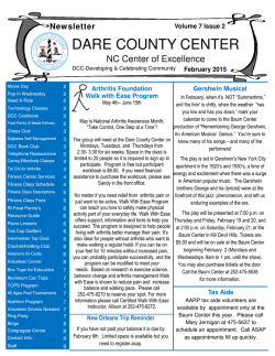
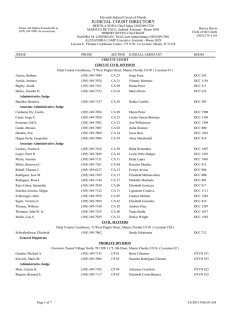
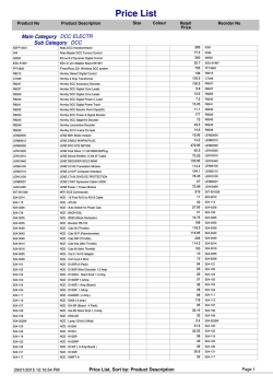
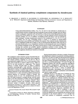
DCC Tumor Suppressor Gene Is Inactivated in Hematologic
From www.bloodjournal.org by guest on February 6, 2015. For personal use only.
DCC Tumor Suppressor Gene Is Inactivated in Hematologic Malignancies
Showing Monosomy 18
By Emilio Porfiri, Lorna M. Secker-Walker, A. Victor Hoffbrand. and J o h n F. Hancock
DCC (deleted in colorectal cancer) is a candidate tumor
suppressor gene recently identified on chromosome band
18q21. Loss of one DCC allele or decreasedDCC expression
occurs in more than 70% of colorectalcancers, suggesting
that DCC inactivation constitutes a critical event in the development of these tumors. Using polymerase chain reaction amplificationof cDNA, w e have studiedDCCexpression
in bone marrow from 4 patients with leukemia (1 chronic
myeloid leukemia-blastic crisis, case 1 ; 1 acute myeloid
leukemia, case 2; 1 T-cell acute lymphoblastic leukemia
[ALL], case 3; 1 6-cell ALL, case 4) showing loss of one
DCC allele due to monosomy 18. W e also studied DCC
expression in multiple control samples, including normal
lymphocytes,normaltonsillar tissue, and leukemias without
18q abnormalities. Four primer pairs consistentlyamplified
the predicted DCC sequences from cDNA prepared from all
control samples. However, in samples with monosomy 18,
DCC transcripts were either not detected (case 1) or detected at a very low level (cases 2, 3, and 4). Southern
analysis showed no structural rearrangement of the remaining DCC locus in all leukemia samples. Thus, loss of
DCC expressionwas demonstrated in associationwith loss
of one DCCallele in all cases tested. These results suggest
that, as for colorectal tumors, the inactivation of DCC can
have a role in the developmentof hematologicmalignancies.
0 1993 by The American Society of Hematology.
A
region, has also been described in 38% of breast carcinomas,
indicating that DCC inactivation may be important in the
development of noncolonic tumors.R
The predicted amino acid sequence suggests that DCC encodes a transmembrane phosphoprotein with several homologies to cell adhesion proteins of the neural cell adhesion
molecule (N-CAM) family.3 Proteins of this group are involved in mediating cell to cell interactions and their roles
include regulation of morphogenesis and immune recognition.' N-CAM proteins are expressed in lymphocytes, particularly natural killer (NK) cells, although they do not regulate their cytolytic activity."
Although uncommon, the loss of one chromosome 18 has
been described in acute myeloid leukemia (AML)." Recent
publications suggest that this occurs as part of a complex
karyotype showing multiple chromosomal abnormalities."
Monosomy 18 has also been described in chronic myeloid
leukemia (CML) in association with the t(9:22) translocation
and rarely in acute lymphoblastic leukemia (ALL).''
We report here an investigation of the occurrence of DCC
inactivation in hematologic malignancies showing monosomy
18.
CCUMULATION of multiple genetic abnormalities
causing activation of dominantly acting oncogenes and
loss of function of tumor suppressor genes has been described
in many human cancers. These observations have suggested
a model of tumor development in which loss of tumor suppressor function and oncogene activation cooperate to trigger
neoplastic transformation.' The paradigm of tumor suppressor gene inactivation envisages loss of both copies of the
gene because a single, normal allele can be sufficient to exert
tumor suppressor function,' although inactivation by dominant negative mutation may also occur.'
Recent studies on colorectal cancer have identified a putative tumor suppressor gene, DCC (deleted in colorectal
cancer), located on chromosome band I8q2 I .' Loss or alteration of one DCC allele due to 18q abnormalities occurs
in more than 70% of colorectal tumors. DCC is expressed in
most normal tissues and in a selection of noncolonic tumors,
but 15 of 17 (88%) colon carcinoma cell lines and 13 of 16
(8 1%) colorectal carcinomas showed either absence or significant reduction of DCC t r a n ~ c r i p t i o n .A
~ . significant
~
decrease of DCC expression was also observed in CokFu' and
SW4806 colon carcinoma cell lines, both showing loss of one
DCC allele. Reintroduction of one normal chromosome 18
in these cell lines restored DCC expression and resulted in
significant loss of t ~ m o r i g e n i c i t y .In
~ , ~addition, downregulation of DCC expression in Rat-1 cells, using an antisense
RNA strategy, resulted in anchorage-independent growth,
faster growth rate, and the cells gave rise to tumors in nude
mice.7 Allelic loss of chromosome 18q, involving the DCC
From the Department of Huematology. Ro.val Free Hospital School
of Medicine, London. UK.
Submitted October 2, 1992; accepted December 17. 1992.
Supported by a grant to E.P. and J.F.H. from the Medical Research
Coimcil, UK (Grant No. G9209270CA) and by a grant to L.M.S.-W
from Kay Kendall Leukaemia A m d .
Address reprint requests to Emilio Poi$ri, MD, ONYX Pharmaceuticals, 3031 Research Dr, Bldg A, Richmond, CA 94806.
The publication costs of this article were defiayed in part by page
charge payment. This article must therefore be hereby marked
"advertisement" in accordance with 18 U.S.C. section I734 solelv to
indicate this fact.
0 1993 by The American Society ofHematology.
0006-4971/93/8ll0-0007$3.00/0
2696
MATERIALS AND METHODS
Clinical samples. Bone marrow samples from patients with leukemia and showing monosomy 18 were identified during routine
diagnostic cytogenetics and stored frozen in liquid nitrogen or at
-70°C. Tonsillar tissue, obtained from routine tonsillectomy, was
snap frozen and stored in liquid nitrogen. Control lymphocytes were
isolated from the peripheral blood of a healthy donor. In addition,
3 further samples of ALL and 1 sample of AML without loss o f D C C
alleles were included in this study. Approval was obtained from the
Institutional Review Board for these studies. Patients and volunteers
were informed that blood or bone marrow samples were taken for
research purposes and that their privacy would be protected.
C>)togenefic
analysis. Bone marrow or peripheral blood was cultured for 24 hours and prepared for cytogenetic analysis using standard
techniques. Slides were made by edge flaming and were stained with
trypsin giemsa.13
RNA and DNA pwificafion. Mononuclear cells were isolated by
Fycoll-Hypaque (FH:
Lymphoprep: Nyegaard, Oslo, Norway) density
separation and lysed in guanidine-thiocyanate buffer. DNA and RNA
were isolated after ultracentrifugation ofthe lysate on a CsCl c ~ s h i o n . ' ~
Reverse transcriptase-pol~merarasechain reaction (RT-PCR). Two
micrograms of total RNA was reverse transcribed. The synthesis of
Blood, Vol 81, No 10(May 15). 1993: pp2696-2701
From www.bloodjournal.org by guest on February 6, 2015. For personal use only.
DCC INACTIVATION IN LEUKEMIA
2697
chain termination using modified T7 DNA polymerase (Sequenase;
US Biochemical Corp, Cleveland, OH).
Southern blot analysis. Five micrograms of genomic DNA was
digested overnight with EcoRI or Pst I and electrophoresed in a 0.8%
agarose gel. DNA was blotted onto a nylon membrane and hybridized
for 18 hours at 65°C with the [~~-'*P]-labeled1.6-kb DCC cDNA
probe.13Afterwards, hybridization blots were washed for 45 minutes
at 65°C in 45 mmol/L NaCI, 4.5 mmol/L Na citrate, 1X Denhardt's
solution, 0. I % SDS, and autoradiographed.
Table 1 . Oligonucleotide Primers
DCC1
DCC2
DCC3
DCC4
DCC5
Actin-1
Actin-2
G6PD-1
G6PD-2
5-gtcgac ATTTACCGATGCTCAGCTCGAAA
5'-TTCCGCCATGGTTTTTA A ATC A2
5'AGCCTCATTTTCAGCCACACA2
5'-gaattcCATCAACCTCTATATTCTGTTCTGTTC
5-atgcgaattcGTTCAAGGGAGGAGTCCAAC
5-TGCTATCCAGGCTGTGCTAT
5'-G ATGGAGTTG A AGGTAGTTT
5'-tagga ATTCATCATCATGGGTGCATCG'
RESULTS
5-tagaagctTGTTTGCGGATGTCAGCCACTGT'
We investigated DCC expression in bone marrow samples
from 4 patients with leukemia, 1 with CML-blastic crisis
(CML-BC) (case I), 1 with AML (case 2), I with T-ALL (case
3), and 1 with B-ALL (case 4), showing loss of chromosome
18q due to monosomy 18. These 4 samples were identified
the first-strand cDNA was primed with random hexamers and performed at 42°C for 1 hour using 40 U of AMV reverse tran~criptase'~ among 48 CML-BCs, 120 AMLs, and 194 adult ALLs that
(Boehringer, Mannheim, Germany). DNA polymerase I (Boehringer)
showed karyotypic abnormalities during routine diagnostic
was used for second-strand synthesis after RNase H treatment of the
cytogenetics. The clinical characteristics of the patients and
DNA-RNA hybrid.15One-twentieth of the cDNA was used for PCR
the full cytogenetic data are summarized in Table 2. Monamplification in a 100 pL reaction mix including I00 pmol of each
osomy 18 was detected in all cells karyotyped in 3 samples
primer, 4 U of Taq polymerase (Amersham Int, Amersham, UK),
(patients 1, 3, and 4), whereas loss of chromosome 18 was
0.2 mmol/L dNTPs, 10 mmol/L Tris-HCI, pH 8, 50 mmol/L KCI,
detected only in 25% of the cells karyotyped in the sample
1.5 mmol/L MgClz, and 0.01%gelatin. Five oligonucleotide primers
taken from patient 2. DCC expression was also studied in
(2 sense and 3 antisense) were designed using the DCC cDNA senormal lymphocytes, normal tonsillar tissue, and 3 ALL
quence,' a primer pair was designed using the /3 actin sequence,16
samples without chromosome 18 abnormalities. In addition,
and a further primer pair, specific for glucose 6-phosphate dehydrogenase (G6PD) cDNA,17was also obtained (Table 1). The PCR rewe studied DCC expression in an AML sample with karyoaction was denatured at 95°C for 3 minutes, followed by 40 cycles
typic loss of chromosome 18p and the centromeric region of
ofdenaturation at 94°C for 1 minute, annealingat 58°C (DCCprimI8q, in which the telomeric region of 18q,including the DCC
ers) or 57°C (actin primers and G6PD primers) for I minute, and
locus. was translocated onto chromosome 17p, thus
polymerization at 72°C for 2 to 4 minutes. The reaction was then
der( I7)t( 17;18)(pI 1; q 12) (patient C2).
held at 72°C for 10 minutes. One-tenth (10 p L ) of each PCR reaction
In most normal tissues, including colonic mucosa, the
was electrophoresed in an agarose gel stained with ethidium bromide
expression
of DCC is
We therefore used RT-PCR to
and the PCR products were visualized under UV light. In addition
amplify DCC transcripts from cDNA. The primer pair DCC 1 1/100 of each PCR reaction was electrophoresed and blotted onto a
DCC4 consistently amplified the predicted 1,114-bp DCC
nylon membrane (Hybond N; Amersham) using vacuum blotting.
fragment from cDNAs derived from tonsillar tissue and from
Blots were hybridized at 65 "C for I8 hours with a 1.6-kbDCCcDNA
the AML sample with chromosome 18q abnormalities not
probe labeled by nick translation with [cx-~~PI~CTP."
After hybridization, blots were washed for 60 minutes at 65°C in 15 mmol/L
involving loss of the DCC locus (lanes C, and Cz, Fig 1). In
NaC1, 1.5 mmol/L Na citrate, I X Denhardt's solution, 0. I % sodium
addition, the same DCC fragment was amplified from cDNAs
dodecyl sulfate (SDS) and autoradiographed.
prepared from normal lymphocytes and 3 ALL samples
The DCC cDNA probe used in these experiments was generated
without alterations of chromosome 18 (data not shown).
by PCR amplification from tonsillar cDNA using the primer pairs
However, this primer pair failed to amplify the expected DCC
DCCI-DCC3 and DCC2-DCC5 (Table 1) under the conditions specfragment
from any of the cDNAs derived from tumor samples
ified above. These primer pairs amplified overlapping DCC cDNA
showing
monosomy
18 (lanes 1, 2, 3, and 4, Fig 1).
fragments spanning from nt 625 to nt 2250 of the DCC cDNA seThe primer pair DCC2-DCC3 was designed to amplify a
quence. After digestion with the appropriate restriction enzymes, the
233-bp DCC cDNA fragment (Fig 2A) and was previously
PCR products were ligated together, cloned into the pGEM 3Zf(+)
vector (Promega Corp, Madison, WI), and sequenced by dideoxy
used by Fearon et a13 to investigate the level of DCC expresSome of the primers carry extra nucleotides containing restriction sites
to facilitate cloning (lower-case letters).
Table 2. Clinical and Cytogenetic Characteristics of 4 Patients Whose Tumors Showed Deletion of Chromosome 18
~
Case
SexlAae
Diagnosis
1
2
F/41
MI71
CML-myeloid blastic crisis
MDS-AML
3
4
MI39
MI53
T-ALL
B-ALL
44,XY.-2.-4,del(5)(ql3).+9,inv(9)(p22q22)c,del(l2)(pl
11,- 18[5]/
44,XY .deI(7)(q22q34),-9,inv(9)c,del(l2)(q22q24),-16,add(17)(q25)[15]
45,XY,der(5)t(l;5)(qI 2;~15),der(8)t(8;9)(p21
; q l 1)t(3;9)(pll ;q21).der(9)t(3;9)(pll; q l l ) , - 1 8 [ 9 ]
47,XY.+7,t(8;14)(q24;q32),t(9;12)(~22;q13),+12,-18[4]/48,XY,idem,+3[10]
C,'
F/24
AML
46,XX,t(6;9)(p23;q34),+8,der(17)t(l7;18)(pll;ql2),- 1 8
KaNOtVDe
45,XX,t(3;13)(~25;q13),t(9;22)(q34;ql
1),-18[11]
* Clinical characteristics of a control patient (lanes C2, Figs 1 through 5) whose tumor showed abnormalities of chromosome 18q not involving
DCC alleles.
From www.bloodjournal.org by guest on February 6, 2015. For personal use only.
2698
PORFlRl ET AL
A
spanning nt 625 to nt 1738. could be amplified from the
monosomy 18 tumors as two scparate fragments. The primer
pair DCCI-DCC3 amplified the expected 594-bp fragment.
from nt 625 to nt 1218. from all the control cDNAs (lanes
C , and C?. Fig 3) and no PCR product was amplified from
the cDNA of the monosomy I8 sample that gave no product
with primer pair DCC2-DCC3 (lane I. Fig 3). A very low
level of amplification was obtained in the remaining 3 monosomy 18 leukemia bone marrows (lanes 2. 3. and 4. Fig 3)
that was detectable only by Southern blotting an aliquot of
the PCR reactions. We next used primer pair DCC2-DCC4
to amplify a 753-bp DCC fragment, from nt 986 to nt 1738.
The expected fragment was obtained from the control cDNAs
(lanes C , and C 2 . Fig 4). but not from cDNAs derived from
two monosomy 18 samples (lanes I and 4, Fig 4). This sequence was amplified only at low level from the remaining
two monosomy 18 leukemia samples (lanes 2 and 3. Fig 4).
To confirm the integrity of the RNA used to prepare the
cDNAs. we performed RT-PCR experiments using a primer
pair designed to amplify a @-actincDNA fragment of 446 bp
(actin-I-actin-?, Table I ) . The expected @-actin cDNA sequence was consistently amplified, with equivalent efficiency.
from all the control cDNAs and from all cDNAs derived
from leukemia bone marrows showing monosomy 18 (Fig
DCCl
DCC2 DCC3
I
I
DCC4
I
I
nt 1
nt 2250
1114 bp
M
C
1
1
3
C p 2
4
Co
3000 bp1114bp-
C
cp
2
3
4
co
1114 bp-
A
D C C l DCC2
Fig 1. (A) The primers DCCl -DCC4 (Table 1) amplify a 1.114bp DCC fragment comprising nt 625 to nt 1738. (B)Agarose gel
electrophoresis of the PCR products amplified using the primer pair
DCCl -DCC4. Lane M, molecular size markers (size indicated alongside the gel); lane C,, control cDNA derived from tonsillar tissue:
lane C, control cDNA derived from an AML sample without deletions of chromosome 18q21 (patient C, Table 2); lanes 1, 2, 3,
and 4, cDNAs derived from leukemia samples with monosomy 1 8
(patients 1, 2, 3, and 4, Table 2); lane Co, no cDNA. (C) Southern
blot analysis of the agarose gel presented in (B).The probe used
for Southern blotting was a 1.6-kb DCC cDNA fragment generated
as described in Materials and Methods.
sion in normal tissues and in colon carcinoma cell lines.'
Using this primer pair we could amplify the expected DCC
fragment from control cDNAs. We could not amplify the
233-bp fragment from cDNA derived from one monosomy
18 sample (lane I , Fig 2) and we amplified the same fragment
only at a very low level from the other 3 monosomy 18
cDNAs (lanes 2,3, and 4. Fig 2). To determine the sensitivity
of this primer pair in detecting DCC mRNA, one of the control samples was diluted with H 2 0 before amplification. A
signal equivalent to that seen in lanes 2, 3. and 4 of Fig 2
was obtained when the cDNA was diluted l:103. which is
consistent with the DCC mRNA in these samples being reduced to 0.1% of the control (data not shown).
The 233-bp DCC fragment amplified by the primer pair
DCC2-DCC3 is a segment of the 1, I 14-bp fragment amplified
by the primer pair DCCI-DCC4 from all control cDNAs.
We therefore studied whether the rest of this DCC region,
DCC4
I
I
233bp
ntl
233
DCC3
I
I
nt 2250
bP
C
233 bp-
c,
1
c2
2
3
4
co
m -+
Fig 2. (A) The primers DCC2-DCC3 (Table 1) amplih a 233-bp
DCCfragment comprising nt 986 to nt 1218. (B)Agarose gel electrophoresis of the PCR products amplified using the primer pair
DCC2-DCC3. Lane M, molecular size marker (size indicated alongside the gel). Samples loaded as in Fig 1. (C) Southern blot analysis
of the agarose gel presented in (B);the probe was as in Fig 1C.
From www.bloodjournal.org by guest on February 6, 2015. For personal use only.
DCC INACTIVATION IN LEUKEMIA
2699
DISCUSSION
A
DCCl DCC2 DCC3
I
I
DCC4
I
I
nt 1
nt 2250
5 9 4 bp
C
c1
1
c 2 2
3
4 c 0
The loss or significant decrease of DCC expression has
been observed in more than 80% of colorectal cancers and
colon carcinoma cell
but abnormalities of DCC
expression have not been reported previously in any noncolonic tumor. We were interested in the possibility that DCC
inactivation may be involved in the development of hematologic malignancies with documented loss of one DCC allele
and we therefore studied expression of DCC in a set of leukemias showing loss of one chromosome 18. Using RT-PCR.
four primer pairs consistently amplified the predicted DCC
sequences from cDNAs prepared from normal tonsillar tissue.
normal lymphocytes. and leukemia samples without abnormalities of the DC'C loci. The same primer pairs failed to
amplify any DCCtranscripts from cDNA prepared from one
leukemia sample showing monosomy 18 (patient 1). In a
further two monosomy 18 leukemia samples (patients 2 and
3). the primer pairs DCCI-DCC3 and DCC3-DCC4 amplified, although at a very low level. overlapping DCC fragments
spanning nt 675 to nt 1738. This 1.1 14-bp region was amplified as a single fragment in all control cDNA by the primer
pair DCCI-DCC4. but could not be amplified from the 2
monosomy 18 cDNAs. probably because of the low level of
DCc'transcripts present in these samples. although a reduced
5 9 4 bp-
A
D C C l D C C 2 DCC3
Fig 3. (A) The primers DCCl -DCC3 (Table 1) amplify a 594-bp
DCCfragment comprising nt 625 to nt 121 8. (B) Agarose gel electrophoresis of the PCR products amplified using the primer pair
DCCl -DCC3. Lane M, molecular size marker (size indicated alongside the gel). Samples loaded as in Fig 1. (C) Southern blot analysis
of the agarose gel presented in (B); the probe was as in Fig 1C.
5A). Furthermore, in similar RT-PCR studies, a primer pair
specific for the G6PD cDNA amplified the expected 162-bp
fragment from control cDNA and from all cDNAs prepared
from the monosomy 18 samples (Fig 5B). The G6PD gene
is a widely expressed housekeeping gene whose mRNA constitutes less than 0.1% of total mRNA of mammalian cells."
Thus, an mRNA species, which is normally expressed at a
very low level. was preserved in all cDNA preparations. indicating that the loss of DCC expression we detected in
the monosomy 18 tumors was caused by a selective loss
of DCC transcripts and was not due to general RNA degradat i on.
Finally. we investigated whether there were any structural
rearrangements in the remaining DCC locus to which the
abnormalities of DCC expression could be related. Southern
blot analysis using the 1.6-kb DCC cDNA showed I I EtnRI
fragments in control and monosomy I8 samples. Similarly,
no evidence of deletions or rearrangements of the D C C
region were detected with Psf I genomic restriction. However, it should be emphasized that the probe used in this
analysis is not a full-length cDNA and thus rearrangements
outside the region investigated by this probe would not be
detected.
I
I
DCC4
I
I
nt 2 2 5 0
ntl
753 bp
B
M C i
1
C2
2
c2
2
3
4 C o
5 9 3 bp-
C
c1
1
3
4
co
753 bp-
Fig 4. (A) The primers DCC2-DCC4 (Table 1) amplify a 753-bp
DCC fragment comprising nt 9 8 6 to nt 1738. (B) Agarose gel electrophoresis of the PCR products amplified using the primer pair
DCCP-DCC4. Lane M, molecular size marker (size indicated alongside the gel). Samples loaded as in Fig 1. (C) Southern blot analysis
of the agarose gel presented in (B); the probe was as in Fig 1C.
From www.bloodjournal.org by guest on February 6, 2015. For personal use only.
PORFlRl ET AL
2700
B
c1
1
c2
2
3
efficiency of the primer pair DCCI-DCC4 in this PCR reaction cannot be excluded.
In the remaining monosomy 18 sample (patient 4). the
primer pairs DCCl -DCC3 and DCC2-DCC3 generated a low
level of amplification of DCC sequences spanning nt 625 to
nt 1218, whereas the primer pairs DCCI-DCC4 and DCC2DCC4 failed to detect DCC transcripts. Because these primer
pairs worked with comparable efficiency in control cDNAs.
it is possible that a DC‘C mRNA lacking the DCC4 primer
sequence was expressed in this tumor. This mRNA may represent a normal. alternative spliced form of the DCC mRNA
or an aberrantly spliced mRNA resulting from mutation of
a splice donor or acceptor site. Indeed. expression ofabnormal
DCC mRNA has previously been described in a colon carcinoma in which an intronic point mutation generated a potential 3’ splice acceptor site.’
The low level ofDCCexpression detected in 3 monosomy
18 cDNAs (patients 2, 3, and 4) could have originated from
the small fraction of normal hematopoietic cells present in
the leukemia samples. Consistent with this view, the lowest
RT-PCR signals were detected in samples from patients 3
and 4 in which the fraction of malignant cells was 95% and
98%. respectively. A stronger signal was detected in the leukemia sample taken from patient 2. whose bone marrow
showed an 82% substitution with blasts. Interestingly. in this
patient, loss of chromosome 18 was found in only 25% of
the cells karyotyped. The significantly reduced RT-PCR signal
detected in this sample argues that leukemic cells showing
two apparently normal chromosome I8 were also expressing
a low level of DCC and that submicroscopical alterations of
the DCC loci affecting the expression of DCC may have been
present in the majority of the leukemic cells. The sample that
gave no signal with any primer pair was taken from a patient
with CML-BC. Because this is a neoplastic transformation
of a pluripotent stem cell. we would expect the blast and the
nonblast cell population to be derived from the same clone.
Therefore, this sample probably contained no normal cells,
i
Fig 5. (A) Agarose gel electrophoresis analysis
of the PCR productsgenerated by using the primers
actin-1 -actin-2 (Table 1). Samples loaded as in Fig
1. (B) Agarose gel electrophoresis analysis of the
PCR productsgenerated by usingthe primers GGPD1-G6PD-2 (Table 1). Samples loaded as in Fig 1.
which is consistent with the total absence of DCC message
we observed.
In the development of colorectal tumors. loss or alteration
of DCC alleles occurs in 47% of late adenomas and in 73%
of carcinomas.” Interestingly, in one of the monosomy I8
samples (patient I). loss ofchromosome I8 and. presumably.
loss of DCCexpression were late events that took place during
the evolution of a CML to blastic phase. Of the remaining
three monosomy I8 samples. one was taken during the AML
transformation of a myelodysplastic syndrome (patient 2).
another during the lymphoblastic transformation of a lowgrade B lymphoma (patient 4). and the remaining sample
was taken from a patient with chemotherapy-resistant ALL
(patient 3). It is tempting to speculate that. as for colorectal
carcinogenesis, DCC inactivation constituted a late event in
leukemogenesis in at least 3 ofthese patients and contributed
to tumor progression.
In the 4 leukemia samples we studied. monosomy I8 and
DCC inactivation were associated with other chromosomal
abnormalities that cause genetic alterations known to play a
critical role in leukemogenesis.” Current views regarding the
molecular pathogenesis of cancer stress the necessity of multiple genetic abnormalities for tumor development.” Accordingly, loss of DCC function in leukemias showing monosomy I8 would constitute one of the steps contributing to
neoplastic progression.
In conclusion. our observations suggest, for the first time.
the involvement of DCC inactivation in the development of
hematologic malignancies. Further studies are needed to investigate the incidence of DCC inactivation in leukemias and
lymphomas and to identify the molecular mechanism by
which DCC may participate in the control of growth and/or
differentiation of hematopoietic cells.
ACKNOWLEDGMENT
The authors thank Dr L. Foroni for helpful discussion and advice.
From www.bloodjournal.org by guest on February 6, 2015. For personal use only.
DCC INACTIVATION IN LEUKEMIA
REFERENCES
I . Marshall CJ: Tumor suppressor genes. Cell 46:3 13, I99 1
2. Kern SE, Pietenpol JA, Thiagalingam S, Seymour A, Kinzler
KW, Vogelstein B: Oncogenic forms of p53 inhibit p53 regulated
gene expression. Science 256327, 1992
3. Fearon ER, Cho KR, Nigro JM, Kern SE, Simons JW, Ruppert
JM, Hamilton SR, Presinger AC, Thomas G, Kinzler KW, Vogelstein
B Identification of a chromosome 189 gene that is altered in colorectal
cancer. Science 247:49, 1990
4. Kikuchi-Yanoshita R, Konishi M, Fukunari H, Tanaka K, Miyaki M: Loss of expression of the DCC gene during progression of
colorectal carcinomas in familial adenomatous polyposis and nonfamilial adenomatous polyposis patients. Cancer Res 52:3801, 1992
5. Tanaka K, Oshimura M, Kikuchi R, Seki M, Hayashi T, Miyaki
M: Suppression of tumorigenicity in human colon carcinoma cells
by introduction of normal chromosome 5 or 18. Nature 349:340,
1991
6. Goyette MC, Cho K, Fasching CL, Levy DB, Kinzler KW,
Paraskeva C, Vogelstein B, Stanbridge EJ: Progression of colorectal
cancer is associated with multiple tumor suppression gene defects
but inhibition oftumorigenicity is accomplished by correction of any
single defect via chromosome transfer. Mol Cell Biol 12: 1387, 1992
7. Narayanan R, Lawlor KG, Schaapveld RQJ, Cho KR, Vogelstein B, Bui-Vinh Tran P, Osborne MP, Telang NT: Antisense RNA
to the putative tumor suppressor gene DCC transforms Rat-I fibroblasts. Oncogene 7553, 1992
8. Devilee P, van Vliet M, Kuipers-Dijkshoorn N, Pearson PL,
Comelisse CJ: Somatic genetic changes on chromosome 18 in breast
carcinomas: Is the DCC gene involved? Oncogene 6:3 11, 199 I
9. Edelman GM: Morphoregulatory molecules. Biochemistry 27:
3533, 1988
10. Lanier LL, Chang C, Azuma M, Ruitenberg JJ, Hemperly JJ,
Phillips JH: Molecular and functional analysis of human natural
killer cell-associatedadhesion molecules (N-CAM/CD56).J Immunol
146:4421, 1991
2701
1 I. Mitelman F Catalog of Chromosome Aberration in Cancer
(ed 4). New York, NY, Liss, 1991, p 1341
12. Fenaux P, Jonveaux P, Quiquandon I, Lai JL, Pignon JM,
Loucheux-Lefebvre MH, Bauters F, Berger R, Kerckaert J P P53
gene mutations in acute myeloid leukemia with 17p monosomy.
Blood 78:1652, 1991
13. Secker-Walker LM, Cooke HMG, Browett PJ, Shippey CA,
Norton JD, Coustan-Smith E, Hoffbrand AV: Variable Philadelphia
breakpoints and potential lineage restriction of bcr rearrangement in
acute lymphoblastic leukemia. Blood 72:784, 1988
14. Sambrook J, Fritsch EF, Maniatis T Molecular Cloning: A
Laboratory Manual (ed 2). Cold Spring Harbor, NY, Cold Spring
Harbor Laboratory, 1989
15. Gubler U, Hoffmann BJ: A simple and very efficient method
for generating cDNA libraries. Gene 25:263, 1983
16. Nakajima-Iijima S, Hamada H, Reddy P, Takanuga T: Molecular structure of the human cytoplasmic &actin gene: Intraspecies
homology of sequences in the introns. Proc Natl Acad Sci USA 82:
6133, 1985
17. Boehm T, Spillantini MG, Sofroniew MV, Surani MA, Rabbits
TH: Developmentally regulated and tissue specific expression of
mRNAs encoding the two alternative forms of the LIM domain oncogene rhombotin: Evidence for thymus expression. Oncogene 6:
695, 1991
18. Persico MG, Toniolo D, Nobile C, DUrso M, Luzzatto L
cDNA sequences of human glucose 6-phosphate dehydrogenase
cloned in pBR322. Nature 294:778, 1981
19. Vogelstein B, Fearon ER, Hamilton SR, Kern SE, Presinger
AC, Leppert M, Nakamura Y,White R, Smits AMM, Bos J L Genetic
alterations during colorectal tumour-development. N Engl J Med
319525, 1988
20. Sawyers CL, Denny CT, Witte ON: Leukemia and the disruption of normal hematopoiesis. Cell 64:337, 199I
2 I. Fearon ER, Vogelstein B: A genetic model for colorectal tumorigenesis. Cell 6 I :759, 1990
From www.bloodjournal.org by guest on February 6, 2015. For personal use only.
1993 81: 2696-2701
DCC tumor suppressor gene is inactivated in hematologic
malignancies showing monosomy 18
E Porfiri, LM Secker-Walker, AV Hoffbrand and JF Hancock
Updated information and services can be found at:
http://www.bloodjournal.org/content/81/10/2696.full.html
Articles on similar topics can be found in the following Blood collections
Information about reproducing this article in parts or in its entirety may be found online at:
http://www.bloodjournal.org/site/misc/rights.xhtml#repub_requests
Information about ordering reprints may be found online at:
http://www.bloodjournal.org/site/misc/rights.xhtml#reprints
Information about subscriptions and ASH membership may be found online at:
http://www.bloodjournal.org/site/subscriptions/index.xhtml
Blood (print ISSN 0006-4971, online ISSN 1528-0020), is published weekly by the American
Society of Hematology, 2021 L St, NW, Suite 900, Washington DC 20036.
Copyright 2011 by The American Society of Hematology; all rights reserved.


© Copyright 2026