
Title page (i) the title of the paper. Nephroprotective effects of
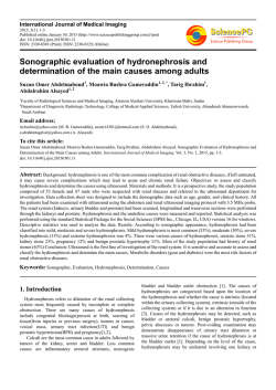
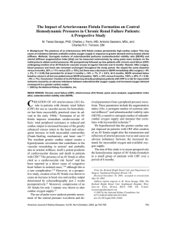
Title page
(i) the title of the paper.
Nephroprotective effects of polydatin against ischemia/reperfusion injury: A role for the PI3K/Akt signal
pathway
(ii) the full names of the authors.
Hong-Bao Liu 1, 2, †, Qiu-Hong Meng 3, †, Chen Huang 1, †, Jian-Bo Wang 3, *, Xiao-wei Liu 1, *
(iii) the addresses of the institution.
1
Department of Nephrology, Xijing Hospital, Fourth Military Medical University, Xi'an, 710032, China;
2
State Key Laboratory of Cancer Biology, Department of Medical Genetics and Developmental Biology, Fourth
Military Medical University, Xi'an, 710032, China
3
Institute of Materia Medica, School of Pharmacy, Fourth Military Medical University, Xi'an, 710032, China;
(iv) * Address correspondence to Jian-Bo Wang, Institute of Materia Medica, School of Pharmacy, Fourth Military
Medical University, Xi'an 710032, China. E-mail: [email protected]. And Xiao-wei Liu is the
co-correspondence author, Department of Nephrology, Xijing Hospital, Fourth Military Medical University, Xi'an,
710032, China. E-mail: [email protected].
(v) † Equal contributors.
Abstract:
Background: Oxidative stress and inflammation are involved in the pathogenesis in renal ischemia/reperfusion
(I/R) injury. It has been demonstrated that polydatin processed the anti-oxidative, anti-inflammatory and
nephroprotective properties. However, whether it has beneficial effects and the possible mechanisms on renal I/R
injury remains unclear.
Methods: Adult BALB/c mice equally divided into sham group, vehicle group (I/R injury), polydatin low dose
group (I/R + polydatin 10mg/kg at post-I/R 0,1,2,3,4,5,6 d), polydatin middle dose group (I/R + polydatin 20 mg/kg)
and polydatin high dose group (I/R + polydatin 40 mg/kg) following clamping of bilateral renal pedicles for 30
minutes (except sham group). For assessing the role of PI3K/Akt pathway in regulating the therapeutic effects of
polydatin in acute renal I/R injury mice, wortmannin, a specific inhibitor of PI3K/Akt, was intraperitoneally
injected into mice to block the activation of Akt.
Results: Compared with vechile control, the administration of polydatin significantly improved the renal function,
accelerated the mitogenic response and reduced cell apoptosis in renal I/R injury models, strongly suppressed the
I/R-induced up-regulation of the expression of tumor necrosis factor-α, interleukin-1β, cyclooxygenase-2, inducible
nitric oxide synthase, prostaglandinum E-2 and nitric oxide levels, dramatically decreased contents of
malondialdehyde, but increased the activity of superoxide dismutase, glutathione transferase, glutathione
peroxidase and catalase, and the level of glutathione. Further investigation showed that polydatin up-regulated the
phosphorylation of Akt in kidneys of I/R injury dose-dependently. However, all beneficial effects of polydatin
mentioned above were counteracted when we inhibited PI3K/Akt pathway with its specific inhibitor, wortmannin.
Conclusions: Our data reveal that polydatin exhibited prominent nephroprotective effects against renal I/R injury,
at least in part, through PI3K/Akt-dependent molecular mechanisms. Therefore, polydatin is promising to be a good
drug for prevention and treatment of acute renal I/R injury in the clinical practice.
Keywords:
Polydatin; Kidney; ischemia; hypoxia; AKT; oxidative stress; inflammation
1. Introduction
Renal ischemia/reperfusion (I/R) injury still remains to be a major medical problem due to the lack of more
effective treatment [1, 2]. Inflammatory response and oxidative stress are involved in the pathogenesis in I/R injury
[3]. It is likely to be an important therapeutical strategy to implement antioxidant and anti-inflammatory agents to
treat renal diseases after I/R injury.
Polydatin (C20H22O8, resveratrol glucoside with a 3,4′,5-trihydroxystibene-3-β-mono-D-glucoside molecular
structure, has also been named piceid, Figure 1A), a natural stilbene compound extracted from the dried roots of the
perennial herb Polygonum Cuspidatum Sieb. Et Zucc, which has been widely used in traditional Chinese medicine
for its multiple pharmacological activities, including its strong antioxidative effects, anti-inflammatory reactions,
and improvement of microcirculation [4]. Mounting studies thus far have focused on the beneficial effects of
polydatin in prevention of I/R-induced oxidative stress and inflammation. Our previous studies have demonstrated
that polydatin exerts cardioprotective effects by activating protein kinase C and mito KATP -dependent signaling and
the direct anti-oxidative stress mechanisms in myocardial I/R rat models [5]. Recent years, polydatin has been
suggested to have the properities of nephroprotective effects in diabetes and urate nephropathy [6-9]. However,
little work has been done on its underlying possible mechanism as a drug in treating I/R-induced renal dysfunction.
The phosphatidylinositol 3-kinase (PI3K) family is a group of revolutionary conserved signal transduction
molecule, which can activate its downstream signaling protein, serine/threonine kinase Akt (also known as protein
kinase B, PKB) to participate the regulation of cell proliferation, survival, apoptosis and various of biological
responses including oxidative stress, inflammation and chemotaxis [10, 11]. We [12] and other authors [13]
reported that I/R can induce the activation of PI3K/Akt, which promotes the proliferation and viability of renal
tubular epithelial cells. However, whether PI3K/Akt signaling pathways participate in the mechanism of actions of
polydatin in I/R injury models is unclear.
Therefore, in this study we tested the potential protective effects of polydatin on renal I/R injury models both
in vitro and in vivo. And by the intervention with wortmannin, the specific PI3K/Akt inhibitor, we explored the role
of PI3K/Akt pathway in regulating the therapeutic effects of polydatin in acute renal I/R injury.
2. Materials and methods
2.1 Induction of I/R-AKI
Male BALB/c mice (weighing 20~25g, 7~9 weeks of age) were obtained from the Experimental Animal Center
of the Forth Military Medical University (Xi’an, China), and bred in an experimental animal room of
specific-pathogen-free (SPF) grade. The experiment procedures were in line with the national institutes of health
guide for care and use of laboratory animals (NIH publication No. 85-23, revised 1985) and with the European
communities council directive of 24 November 1986 (86/608/EEC). All mice were provided with food and water
ad libitum in a 12:12-h light/dark cycle (lights on at 6 am., and off at 6 pm.). They were allowed to adapt to new
surroundings for at least 5 days prior to any experimentation. All efforts were made by us to minimize the suffering
of animals in present study.
For the establishment of renal I/R injury models, operation were performed in BALB/c mice by clamping
bilateral renal pedicles for renal ischemia 30 minute time, followed by releasing clamps to allow blood reperfusion
as described previously [14]. Briefly, the bilateral renal pedicles of mice were carefully bluntly dissected, and then
occluded using non-traumatic vascular clamps. The similar operation was performed on mice in sham group, with
the pedicles also dissected but not clamped. Animals were ethically sacrificed at 12 h, 1 d, 3 d, 5 d and 7 d after
renal I/R injury respectively, and whole blood and kidneys were harvested for further analysis.
The animals were randomly divided into five experimental groups as follows: (1) Sham group; (2) Vehicle group:
I/R injury mice with saline vehicle (dimethyl sulfoxide, DMSO 1%) intraperitoneally injected; (3) Polydatin low
dose group (Polydatin-L group): I/R injury mice with polydatin (Weijia Technology Company, Xi’an, China)) of 10
mg/kg intraperitoneally injected. (4) Polydatin middle dose group (Polydatin-M group): I/R injury mice with
polydatin of 20 mg/kg intraperitoneally injected; (5) Polydatin high dose group (Polydatin-H group): I/R injury
mice with polydatin of 40 mg/kg intraperitoneally injected. For group 3 to 5, the initial dose of polydatin was given
before the incision sutured after I/R completed, and then continued with daily injections for 6 days. For evaluated
the PI3K/Akt pathway in vivo, 1 mg/kg of wortmannin (Sigma), the specific inhibitor of PI3K/Akt signaling, was
given 30 min before operation, intraperitoneally, and then continued with daily injection for 2 days. In studies in
vivo polydatin and wortmannin were first dissolved in DMSO (Sigma), and then diluted by physiological saline
with a final concentration of 1% (vol/vol) DMSO for intraperitoneally application.
2.2 Blood physiochemical assays
The whole blood drawn from the heart or the retro-ocular vein plexus was centrifuged at 4 ℃, 3000 g, for 10 min
to obtain the serum sample. The level of blood urea nitrogen (BUN) and serum creatinine (Scr) was measured by
the automatic biochemistry analyzer (Beckman; Fullerton, CA).
2.3 Histological score of kidney injuries
Kidney samples were fixed overnight in 10% phosphate-buffered formalin and then embedded in paraffin. Renal
sections were next prepared and then subjected to Hematoxylin and eosin (H&E) staining to assess the histological
injury. Evaluation of histological score of kidney injuries (HSK) was performed by a renal pathologist under
blinded conditions. HSK was graded using a 4-point quantitative scale as described previously [15]: 0 represented
normal histology; 1 represented mild damage [less than 1/3 of nuclear loss (necrosis) in a tubular cross section]; 2
represented moderate damage [more than 1/3 and less than 2/3 of a tubular cross section shows nuclear loss
(necrosis)]; 3 represented severe damage [more than 2/3 of nuclear loss (necrosis) per tubular cross section]. We
calculated the total score of per kidney section by adding up all 10 scores with a possible maximum injury score of
30.
2.4 Immunohistochemical Staining
The tissue sections were subject to immunohistochemical staining for proliferating cell nuclear antigen (PCNA, a
marker of mitogenesis) 12 h, 1 d, 3 d, 5 d, 7 d after I/R injury. For immunohistochemical staining, we used the
rabbit specific horseradish peroxidase-diaminobenzidine (HRP-DAB) detection immunohistochemical kit (ab64261,
Abcam). After being deparaffinated, hydrated, and peroxidase-blocked, 4 μm sections of kidneys were incubated
overnight at 4 ℃ with a rabbit polyclonal FL-261 antibody (1:200, sc-7907, Santa Cruz Biotechnology, Santa Cruz,
USA). Then sections were incubated with a biotinylated secondary antibody, goat anti rabbit IgG (H +L), for 10
min. Control experiments were performed by omitting either the primary or secondary antibody. Then we
developed the sections using an enzymatic conversion of the DAB, visualizing the color of specific antibodiy
binding sites to change into brown. After all sections were counterstained with hematoxylin (Sigma), they were
clearned and finally coverslipped for observation. We randomly selected 10 sections from the corticomedullary area
per kidney, counting the number of positive nuclei in high-power fields (HPF, 620 magnification). Then we
calculated the mean number of PCNA positive cells of per kidney. The tubular cell apoptosis in kidneys after I/R
were detected by terminal deoxynucleotidyl transferase dUTP nick end labeling (TUNEL) assay following the
manufacturer instructions (In Situ Cell Death Detection Kit; Roche China, Ltd.). Meanwhile, we stained all cell
nuclei with DAPI (Sigma, USA). We examined TUNEL-stained sections for screening positive nuclei with a
fluorescence microscope, and selected 10 random fields in renal cortex and outer medullar area in every kidney,
then counted them at 640 magnification.
2.5 Western blot
As previously described [1], routinely, we carried out western blot analyses to detect target protein levels in
kidney tissues at 3 day after I/R injury with or without polydatin
(10, 20, 40 mg/kg) and wortmannin (1 mg/kg)
treatment and cells with or without OGD/R treatment in presence or absence of polydatin (10, 20, 40 μM) and
wortmannin (1 μM). The primary antibodies included rabbit polyclonal antibodies against phospho-Akt (P-Akt, Ser
473, 1:1000, Cell Signaling, Danvers, MA, USA), total Akt (T-Akt, 1:1000, Cell Signaling, Danvers, MA, USA),
cyclooxygenase-2 (COX-2, 1:500, Abcom, USA), inducible nitric oxide synthase (iNOS, 1:1000, Abcom, USA),
and
β-actin
(1:2000,
Abcom,
USA).
The
primary
proteins
were
detected
by using
horseradish
peroxidase-conjugated secondary antibodies (Abcam, USA), and the immune complexes were finally developed by
using the enhanced chemiluminescence Plus kit (Amersham, Freiburg, Germany).
2.6 Real-time PCR (RT-PCR) analysis
Total RNA was extracted from renal tissues using Trizol according to the manufacturer’s instructions (Takara,
Japan). We obtained complementary DNA (cDNA) by reverse transcribing four micrograms of total RNA by using
the PrimeScript RT Master Mix (Takara, Japan) as instructed. Real-time PCR amplifications were performed by
using qPCR technique of SybrGreen assay on the ABI 7500 system (Applied Biosystems, USA). PCR primers
(Takara, Japan) for all analyzed genes are as follows: tumor necrosis factor-α (TNF-α), amplicon size 122 bp,
forward, 5’-GTG GAA CTG GCA GAA GAG GC-3’ and reverse, 5’-AGA CAG AAG AGC GTG GTG GC -3’;
interleukin-1β (IL-1β), amplicon size 230 bp, forward, 5’- GCC CAT CCT CTG TGA CTC AT-3’ and reverse, 5’AGG CCA CAG GTA TTT TGT CG -3’; COX-2, amplicon size 121 bp, sense: 5’-CCT GGT CTG ATG ATG TAT
GC -3’; antisense: 5’-GTA TGA GTC TGC TGG TTT GG -3’; iNOS, amplicon size 108 bp, forward, 5’-TCC ATG
ACT CCC AGC ACA -3’ and reverse, 5’-CCA TCT CCT GCA TTT CTT CC -3’; GAPDH, amplicon size 211 bp,
forward, 5’-CAT CAA CGG GAA GCC CAT C -3’ and reverse, 5’- CTC GTG GTT CAC ACC CAT C-3’. PCR
conditions were as follows: 94 ℃ for 5 min; 35 cycles at 94 ℃ for 40 s, 58 ℃ for 40 s, and 72 ℃ for 60 s; final
elongation at 72 ℃ for 10 min. The relative expression levels were calculated using the 2-ΔΔCt method as reported.
2.7 oxygen-glucose deprivation (OGD)
The human proximal tubular epithelial cells, HK-2 (ATCC-CRL-2190, Manassas, VA), were seeded in high
glucose DMEM (Hyclone, USA) containing 10% fetal bovine serum (FBS; Gibco, USA), incubated in humidified
cell culture incubator containing a gas mixture composed of 21% O2, 74% N2 and 5% CO2 at 37℃ for 48 h. OGD
followed by reoxygenation (OGD/R) was used to simulate an in vitro model of I/R injury [16]. Specifically, the
cells in OGD group were incubated in glucose-free DMEM (Gibco, USA) without serum and placed in a hypoxic
chamber (Billups-Rothenberg, USA) filled with an anoxic gas mixture (95% N2 / 5% CO2) for 6 h. Meanwhile,
cells in normal control was incubated in high glucose DMEM supplemented with 10% FBS and placed in a
normoxic incubator. At the end of OGD, the plates were taken out from the hypoxic chamber, cells were transferred
to high glucose DMEM containing 10% FBS and continued to incubate for 24 h under normoxic conditions to
generate reoxygenation. In some groups, polydatin (10, 20, 40 μM) was continuously applied from 30 min before
OGD to the end of reoxygenation. To determine the involvement of PI3K/Akt pathway, the PI3K/Akt inhibitor,
wortmannin (1 μM) were continuously applied from 30 min before OGD to the end of reoxygenation. In studies in
vitro polydatin and wortmannin were first dissolved in DMSO (Sigma), and then diluted by DMEM with a final
concentration of 1‰ (vol/vol) DMSO for cell treatment.
2.8 Cell apoptosis assay
We performed apoptosis assays by using an Annexin V-fluorescein isothiocyanate (FITC) apoptosis detection kit
(catalog No. 556419; BD Pharmingen) in accordance to the manufacturer's instructions. Briefly, HK-2 cells in the
dish (105 cells/well) were collected and then resuspended in binding buffer. Annexin V-FITC and PI were added
into the single-cell suspension, which was then incubated in dark place for 15 min. Finally, cells were analyzed by
a FACSCalibur flow cytometer (Becton Dickinson, BD Biosciences, USA).
2.9 Enzyme linked immunosorbent assay (ELISA)
The levels of TNF-α, IL-1β, prostaglandinum E-2 (PGE-2) and nitric oxide (NO) in renal tissue homogenate
were measured by ELISA assay using a commercially available ELISA kit (R&D Systems, USA) referring to the
manufacturer’s recommendation. And the quantification of all these factors was implemented by using BCA
protein assay reagent (Pierce, USA). Optical density values at 450 nm were measured with wavelength correction
set to 570 nm. All standards and samples were measured in duplicate.
2.10 Measurement of renal oxidative indexes
Renal tissue samples were weighed and homogenized (1:10, w/v) in 50 mmol/L phosphate buffer (PH 7.4) in an
ice-bath, and centrifuged at 1500 g for 20 min at 4 ℃. The supernatant was used to measure the activity of
malondialdehyde (MDA), superoxide dismutase (SOD), glutathione transferase (GST), glutathione peroxidase
(GPx), catalase (CAT) and the content of glutathione (GSH) followed the commercial kit instructions by using a
spectrophotometer (Spectrophotometer DU640, Beckman Coulter, Fullerton, CA) with the associated detection kits
(Jiancheng, Nanjing, China). All of the levels are expressed as U/mg protein or nmol/mg protein, respectively.
2.11 Statistical analysis
All of the values in present study were expressed as means ± SD. Differences between data means were
compared by use of analysis of variance (ANOVA) or Student’s t-test by the SPSS statistical software package
(SPSS, Inc., Chicago, IL, USA). A threshold of statistical significance was set at P < 0.05 for all analyses.
3. Results:
3.1 Polydatin improved the renal function in renal I/R injury mice
First, we detected whether polydatin can improve the renal function of mice after I/R injury. For this purpose,
BUN and Scr levels were examined at day 12 h, 1 d, 3 d, 5 d, 7 d after I/R injury in mice with different doses of
polydatin administration, respectively. Compared with sham group, the renal functions of mice in vechile group and
polydatin groups were all worsened significantly, which suggesting that the renal I/R models were successfully
established in the present study (Figure 1B and C). The impaired renal function in mice of vechile group
self-recovered significantly at 7 day after I/R operation. Howver, compared with vechile (saline) group, the
administration of all three doses (10, 20 and 40 mg/kg) of polydatin significantly improved the impaired renal
function of mice after I/R injury, but respectively at day 5 and 3 after I/R. These results suggested that polydatin
can accelerate the recovery of renal function in mice afer I/R injury in dose dependent manner. (Figure 1B and C).
Histological examinations including HSK (Figure 1D), PCNA (Figure 1E), and TUNEL staining (Figure 1F)
were evaluated at 12 h, 1 d, 3 d, 5 d and 7 d after I/R. As expected, compared with control kidneys from
saline-treated mice, polydatin reduced HSK, increased number of PCNA-positive cells, and decreased number of
apoptotic cells on TUNEL assay (Figure 1D-F). Especially, at 24 h after I/R injury, the number of PCNA-positive
cells in kidneys from mice in polydatin-H group was significantly increased compared with that in the groups of
vehicle (+10.2-fold), polydatin-L (+7.8-fold) and polydatin-M (+5.8-fold). Meanwhile, the similar increasement
was delayed and detected respectively at 3 d, 5 d, 7 d after I/R in Polydatin-M, polydatin-L and vechile groups
(Figure 1D-F). The increase in renal cell survival following I/R injury was confirmed by measure of apoptosis
using TUNEL analysis, which showed that polydatin remarkably decreased cell apoptosis in kidneys of mice after
I/R, especially the Polydatin-H groups, compared with vehicle control at 1 d, 3 d, 5 d, 7 d after I/R (Figure 1D-F).
Given that the beneficial effect of Polydatin-H was the most significant at 3 d after I/R in mice, so the
subsequent experiments in this study were all performed following this treatment.
3.2 PI3K/Akt pathway participated in the nephroprotective effects of polydatin
To validate the association between Akt signaling and nephroprotective effect of polydatin, we detected the
activation of Akt. Compared with sham group, the level of p-Akt increased at 3 d after I/R; polydatin
dose-dependently further elevated the I/R-induced increase of p-Akt. Intraperitoneal injection of the inhibitor of
PI3K/Akt, wortmannin, significantly blocked the polydatin-elevated phosphorylation of Akt (Figure 2A). In mice
of sham group, the renal function after operation was not affected by wortmannin, suggesting that wortmannin had
no apparent renal toxicity (data not shown). However, wortmannin significantly reversed the beneficial effect of
polydatin in decreasing the levels of BUN and Scr in renal I/R injury mice (Figure 2B and C). Meanwhile,
compared with Polydatin-H group, kidneys from mice treated with both Polydatin-H and wortmannin had
significantly increased HSK and the percentage of apoptotic cells on TUNEL assay and reduced number of
PCNA-positive cells (Figure 2D-F).
To further confirm the results in vivo, in vitro renal I/R injury models were simulated. The results of western blot
showed that the phosphorylation of Akt was activated by OGD/R and further elevated by polydatin
dose-dependently, but which was counteracted in the presence of wortmannin (Figure 2G). The results of apoptosis
assays showed that compared with normal cultured cells, OGD/R notably increased the apoptosis of HK-2 cells,
which was obviously suppressed by 20 μM of polydatin (Figure 2H). Wortmannin did not increase the cell
apoptosis under normoxic conditions revealed that wortmannin had no apparent cytotoxicity. However, wortmannin
not only further increased the OGD/R-induced apoptosis, but also obviously blocked the protective effects of
polydatin on HK-2 cells (Figure 2H). In short, these results suggested that the nephroprotective effects of polydatin
were associated with the PI3K/Akt signaling pathway.
3.3 PI3K/Akt pathway is involved in polydatin-attenuated expression of the pro-inflammatory factors in
renal I/R injury
To evaluate the potential anti-inflammation effects of polydatin in renal I/R injury, we assessed the expression of
TNF-α, IL-1β, COX-2 and iNOS in the kidneys from mice at 3 d after I/R. RT-PCR showed that these cytokines
were extensively expressed in kidneys from mice after I/R injury, but only mildly expressed in kidneys from mice
with polydatin treatment. However, wortmannin significantly increased the polydatin-attenuated expression of the
pro-inflammatory factors induced by I/R (Figure 3A). ELISA analysis showed that the expression of TNF-α and
IL-1β was increased at 72 h after I/R injury, which was decreased by polydatin in dose dependent manner (Figure
3B). Western blot analysis showed that the expression of COX-2 and iNOS were significantly increased in kidneys
at 72 h after I/R injury, which was dose-dependently decreased by polydatin (Figure 3C). These results suggested
that the benefical effect of polydatin on ameliorating the inflammation in renal I/R injury mouse model.
Meanwhile, the intraperitoneal injection of wortmannin obviously abolished the polydatin-induced decreased
expression of TNF-α, IL-1β, COX-2 and iNOS (Figure 3B and 3D). Additionally, we also detected PGE-2 and NO,
the downstream factors of COX-2 and iNOS, respectively. Similarly, the levels of PGE-2 and NO were
significantly increased by I/R, obviously suppressed by polydatin, and went up again when treated mice with
wortmannin (Figure 3E). These results indicated that PI3K/Akt pathway is involved in the anti-inflammation effect
of polydatin in renal I/R injury mice.
3.4 PI3K/Akt pathway was associated with polydatin-attenuated oxidative stress in renal I/R injury
To validate the potential effect of polydatin on anti-oxidative stress in renal I/R injury, we detected the contents
of MDA and GSH, and the activity of four antioxidases (SOD, GST, GPx and CAT) in kidneys, respectively.
Compared with sham group, the MDA content was significantly increased in I/R injury mice, and was reversed in
polydatin-M and polydatin-H groups but not in polydatin-L group (Figure 4A), while the activity of the four
antioxidases were all significantly decreased in the kidneys of I/R injury mice, and polydatin elevated the activity
that was decreased by I/R (Figure 4B-E). Compared with vehicle group, the activity of SOD, GST, GPx and CAT
were all significantly increased in polydatin groups except polydatin-L group (Figure 4B-E). Compared with sham
group, the level of GSH decreased in I/R injury mice. Compared with vehicle group, the GSH content was all
elevated in polydatin-M and polydatin-H group, but not in polydatin-L group (Figure 4F). All measurements
mentioned above had no significant statistic difference between sham group and polydatin-H group. These results
suggested the anti-oxidative stress effect of polydatin in alleviating renal I/R injury. However, wortmannin
significantly abolished the effect of polydatin on decreasing MDA content, increasing the activity of SOD, GST,
GPx and CAT, and elevating the level of GSH (Figure 4), which suggesting that PI3K/Akt pathway was associated
with polydatin-attenuated oxidative stress in renal I/R injury.
4. Discussion
Polydatin is an active stilbene compound isolated from the roots of Polygonum Cuspidatum Sieb. Et Zucc, and
has been manifested to possess anti-oxidative and anti-inflammatory activities [5, 9, 17]. We and other authors have
demonstrated that the therapeutical effects of polydatin on I/R-induced injury in multiple organs including heart,
brain, and so on [4, 5, 18-20]. It has been identified that polydatin also has nephroprotective effects in diabetes and
urate nephropathy through prevention of oxidative stress and inflammation [6-9, 21]. In present study, we further
investigated the potential therapeutic effects and mechanism of polydatin on renal I/R injury. Our results showed
that the administration of polydatin significantly improved the renal function, accelerated the mitogenic response,
reduced HSK and cell apoptosis in renal I/R injury models, suggesting the beneficial effect of polydatin against
renal I/R injury.
In fact, renal I/R injury always induced the excessive generation of pro-inflammatory cytokines in kidneys,
which resulted in leukocyte infiltration and tissue damage [22, 23]. TNF-α and IL-1β are two important
pro-inflammatory mediators in renal I/R injury, which produce a number of injurious changes in proximal tubular
epithelial cells [22-26]. Loss of TNF-α, either through using neutralizational antibody of TNF-α blockade or
knockout mice of TNF-α resulted in significantly alleviated tissue injury and elevated function in kidneys after
renal ischemia. While, the transgenic mice with TNF-α over-expression had more pronounced susceptibility to
acute kidney injury induced by I/R than that in mice of wild type [27]. There were also enormous studies have
demonstrated that decreasing IL-1β was associated with improved renal function in renal I/R injury models [28-30].
Therefore, we examined the levels of TNF-α and IL-1β to determine whether they were associated with the
mechanism of polydatin in treating renal I/R injury diseases. The results showed that polydatin significantly
decreased I/R-induced TNF-α and IL-1β levels in kidneys. These data suggested that the positive effects of
polydatin on improving renal function in mice after I/R injury might be achieved by inhibiting the
pro-inflammatory cytokines of TNF-α and IL-1β.
With the exception of pro-inflammatory cytokines of TNF-α and IL-1β, COX-2 and iNOS, two enzymes
associated with inflammation, were also correlated with the pathogenesis of I/R injury. COX-2, an inducible
enzyme, plays crucial roles in regulating the inflammatory response and oxidative stress in I/R injury [31].
Selective or nonselective inhibition of COX-2 with either rofecoxib or indomethacin ameliorated renal tissue
damage induced by I/R injury [32]. Another study has shown that continuous intrarenal infusion of parecoxib (40
mg per pig) improved renal function in pigs with the operation of suprarenal aortic cross-clamping [33]. These
results of previous studies suggested that inhibition of COX-2 has the potential to improve renal function in renal
I/R injury. iNOS which was highly expressed after renal I/R injury mediated the generation of NO [34, 35]. High
activity of iNOS aggravated the damage of kidneys in renal I/R injury, and selective inhibition of iNOS
significantly improved the renal function in rats with I/R [36-38]. Based on the results of the studies above, we
detected the expressions of COX-2 and iNOS protein and the contents of PEG2 and NO, downstream factors of
COX-2 and iNOS, in the kidneys after renal I/R injury. Our results showed that compared with mice in vechile
group, polydatin notably inhibited the expressions of COX-2 and iNOS protein which were upregulated by I/R in
renal I/R mice, dosedependently. The results were in line with the improved renal function induced by polydatin
(Figure 1), indicating that the protective effects of polydatin on acute renal I/R injury might be by inhibiting the
expression of the potential inflammatory mediators of COX-2 and iNOS and decreasing the generation of the
downstream factors of them, PEG-2 and NO.
There are compelling evidences that oxidative stress is particularly involved in the pathogenesis of renal I/R
injury [35]. Briefly, I/R has been proposed to have the potential to promote oxidative stress, which in turn can
promote I/R injury [25]. The elevation of oxidative molecules and the reduction of antioxidant substance can
aggravate I/R injury. It has been demonstrated that polydatin was a prominent antioxidant and exhibited
cardioprotective effects in myocardial I/R injury rat models via increasing SOD activity and decreasing MDA
content [4, 5]. The results in our experiment substantiated that polydatin significantly elevated the activity of SOD,
GST, GPx, CAT, increased GSH level, and decreased the MDA content in kidneys of I/R injury mice, which
indicated the prominently antioxidative properties of polydatin and was in line with the data previously reported by
Lvyi Chen et al. in urate nephropathic mice [9].
The PI3K/Akt pathway was originally recognized to play a crucial role in regulating the growth and survival of
cells, which nowadays has been rediscovered to be implicated in the protection of brain, myocardium, lung, liver
and kidney against I/R injury by regulating oxidative stress and inflammatory response [39-44]. Recently it has
been demonstrated that polydatin exerted hepatoprotective effect in rats fed with high-fat diet [45] and regulated
glucose and lipid metabolism in diabetic models [46] through up-regulating the phosphorylation of Akt in liver, and
polydatin also exhibited anti-tumor activity [47] through down-regulating the phosphorylation of Akt in human
nasopharyngeal carcinoma CNE cells. These results suggested that Akt signaling pathway was a potential
therapeutic target of polydatin in treating various diseases. Therefore, in the present study, we assessed whether
PI3K/Akt pathway was associated with the nephroprotective effects of polydatin in renal I/R injury models. First,
we identified that the phosphorylation of Akt was activated in renal tissues by I/R, and further increased by
polydatin in dose dependent manner. However, the polydatin-induced increase of phosphorylation of Akt was
significantly decreased by the specific PI3K/Akt inhibitor, wortmannin, suggesting that the positive role of
polydatin on the activation of Akt in renal I/R injury. Importantly, when blocking the phosphorylation of Akt by
intraperitoneal injection of wortmannin, the beneficial effect of polydatin on the regeneration of renal tissues was
also abolished. And wortmannin remarkably counteracted the polydatin-attenuated levels of pro-inflammatory
factors and oxidative stress in kidneys of I/R injury. These results suggested that PI3K/Akt pathway, at least partly,
was involved in the nephroprotective effects of polydatin in renal I/R injury. Of course, the potential downstream
functional molecules mediated by PI3K/Akt signaling pathway to take part in polydatin’s actions still remain to be
further investigated.
Conclusions
In this study, we identified for the first time that in acute renal I/R injury models, the administration of polydatin
significantly improved the renal function, accelerated the mitogenic response and reduced cell apoptosis, strongly
suppressed the I/R-induced up-regulation of the expression of TNF-α, IL-1β, COX-2, PGE-2, iNOS and NO,
dramatically decreased contents of MDA, but increased the activity of SOD, GPx, GST, CAT and the level of GSH.
However, all these beneficial effects of polydatin were counteracted when we inhibited PI3K/Akt pathway with its
specific inhibitor, wortmannin. These findings taken together elucidated that polydatin exhibited prominent
nephroprotective effects against renal I/R injury, at least in part, through PI3K/Akt-dependent phosphorylation. In
conclusion, our data support that polydatin is promising to be a good drug for prevention and treatment of
I/R-induced renal injury in the clinical practice.
Conflict of Interests
The authors declare that there is no conflict of interests regarding the publication of this paper.
Acknowledgments
This work was supported by grants from the National Nature Science Foundation of China (Nos. 81370016,
81400677), Midwestern Excellent Young Scientist Foundation of Chinese Medical Doctor Association (2012) and
the National Major Scientific and Technological Special Project for “Significant New Drugs Development” during
the Twelfth Five-year Plan Period founded by Ministry of Science and Technology of China (Nos.
2012ZX09J12109-04C).
References
1.
H. Liu, S. Liu, Y. Li, et al. "The role of SDF-1-CXCR4/CXCR7 axis in the therapeutic effects of
hypoxia-preconditioned mesenchymal stem cells for renal ischemia/reperfusion injury," PLoS One, vol. 7, no. 4, p.
e34608.
2.
M. Yoshida and S. Honma. "Regeneration of injured renal tubules," J Pharmacol Sci, vol. 124, no. 2, pp. 117-122.
3.
R. Munshi, C. Hsu, and J. Himmelfarb. "Advances in understanding ischemic acute kidney injury," BMC Med, vol.
9, p. 11.
4.
Q.H. Du, C. Peng, and H. Zhang. "Polydatin: a review of pharmacology and pharmacokinetics," Pharm Biol, vol. 51,
no. 11, pp. 1347-1354.
5.
Q. Miao, S. Wang, S. Miao, J. Wang, Y. Xie, and Q. Yang. "Cardioprotective effect of polydatin against
ischemia/reperfusion injury: roles of protein kinase C and mito K(ATP) activation," Phytomedicine, vol. 19, no. 1, pp.
8-12.
6.
K. Huang, C. Chen, J. Hao, et al. "Polydatin promotes Nrf2-ARE anti-oxidative pathway through activating Sirt1 to
resist AGEs-induced upregulation of fibronetin and transforming growth factor-beta1 in rat glomerular messangial cells,"
Mol Cell Endocrinol.
7.
X. Xie, J. Peng, K. Huang, et al. "Polydatin ameliorates experimental diabetes-induced fibronectin through
inhibiting the activation of NF-kappaB signaling pathway in rat glomerular mesangial cells," Mol Cell Endocrinol, vol.
362, no. 1-2, pp. 183-193.
8.
Y.W. Shi, C.P. Wang, L. Liu, et al. "Antihyperuricemic and nephroprotective effects of resveratrol and its analogues
in hyperuricemic mice," Mol Nutr Food Res, vol. 56, no. 9, pp. 1433-1444.
9.
L. Chen, Z. Lan, Q. Lin, et al. "Polydatin ameliorates renal injury by attenuating oxidative stress-related
inflammatory responses in fructose-induced urate nephropathic mice," Food Chem Toxicol, vol. 52, pp. 28-35.
10. A. Barthel and L.O. Klotz. "Phosphoinositide 3-kinase signaling in the cellular response to oxidative stress," Biol
Chem, vol. 386, no. 3, pp. 207-216.
11. L.C. Cantley. "The phosphoinositide 3-kinase pathway," Science, vol. 296, no. 5573, pp. 1655-1657.
12. H. Liu, W. Xue, G. Ge, et al. "Hypoxic preconditioning advances CXCR4 and CXCR7 expression by activating
HIF-1alpha in MSCs," Biochem Biophys Res Commun, vol. 401, no. 4, pp. 509-515.
13. D.S. Kwon, C.H. Kwon, J.H. Kim, J.S. Woo, J.S. Jung, and Y.K. Kim. "Signal transduction of MEK/ERK and
PI3K/Akt activation by hypoxia/reoxygenation in renal epithelial cells," Eur J Cell Biol, vol. 85, no. 11, pp. 1189-1199.
14. W.H. Liu, H.B. Liu, D.K. Gao, et al. "ABCG2 protects kidney side population cells from hypoxia/reoxygenation
injury through activation of the MEK/ERK pathway," Cell Transplant, vol. 22, no. 10, pp. 1859-1868.
15. H. Liu, W. Liu, S. Liu, et al. "Reconstitution of kidney side population cells after ischemia-reperfusion injury by
self-proliferation and bone marrow-derived cell homing," Evid Based Complement Alternat Med, vol. 2013, p. 370961.
16. H.B. Liu, Q.H. Meng, D.W. Du, J.F. Sun, J.B. Wang, and H. Han. "The effects of ABCG2 on the viability,
proliferation and paracrine actions of kidney side population cells under oxygen-glucose deprivation," Int J Med Sci, vol.
11, no. 10, pp. 1001-1008.
17. G. Lanzilli, A. Cottarelli, G. Nicotera, S. Guida, G. Ravagnan, and M.P. Fuggetta. "Anti-inflammatory effect of
resveratrol and polydatin by in vitro IL-17 modulation," Inflammation, vol. 35, no. 1, pp. 240-248.
18. L.T. Liu, G. Guo, M. Wu, and W.G. Zhang. "The progress of the research on cardio-vascular effects and acting
mechanism of polydatin," Chin J Integr Med, vol. 18, no. 9, pp. 714-719.
19. Y. Cheng, H.T. Zhang, L. Sun, et al. "Involvement of cell adhesion molecules in polydatin protection of brain
tissues from ischemia-reperfusion injury," Brain Res, vol. 1110, no. 1, pp. 193-200.
20. J. Sun, Y. Qu, H. He, et al. "Protective effect of polydatin on learning and memory impairments in neonatal rats with
hypoxicischemic brain injury by upregulating brainderived neurotrophic factor," Mol Med Rep, vol. 10, no. 6, pp.
3047-3051.
21. M. Kitada and D. Koya. "Renal protective effects of resveratrol," Oxid Med Cell Longev, vol. 2013, p. 568093.
22. J.V. Bonventre and A. Zuk. "Ischemic acute renal failure: an inflammatory disease?," Kidney Int, vol. 66, no. 2, pp.
480-485.
23. H.R. Jang and H. Rabb. "Immune cells in experimental acute kidney injury," Nat Rev Nephrol.
24. A. Akcay, Q. Nguyen, and C.L. Edelstein. "Mediators of inflammation in acute kidney injury," Mediators Inflamm,
vol. 2009, p. 137072.
25. M. El Sabbahy and V.S. Vaidya. "Ischemic kidney injury and mechanisms of tissue repair," Wiley Interdiscip Rev
Syst Biol Med, vol. 3, no. 5, pp. 606-618.
26. A.C. Brochner, F. Dagnaes-Hansen, J. Hojberg-Holm, and P. Toft. "The inflammatory response in blood and in
remote organs following acute kidney injury," APMIS, vol. 122, no. 5, pp. 399-404.
27. A. Grenz, J.H. Kim, J.D. Bauerle, E. Tak, H.K. Eltzschig, and E.T. Clambey. "Adora2b adenosine receptor signaling
protects during acute kidney injury via inhibition of neutrophil-dependent TNF-alpha release," J Immunol, vol. 189, no. 9,
pp. 4566-4573.
28. Y.Y. Chen, C.H. Yeh, E.C. So, D.P. Sun, L.Y. Wang, and C.H. Hsing. "Anticancer drug 2-methoxyestradiol protects
against renal ischemia/reperfusion injury by reducing inflammatory cytokines expression," Biomed Res Int, vol. 2014, p.
431524.
29. U. Aksu, I. Guner, O.M. Yaman, et al. "Fluoxetine ameliorates imbalance of redox homeostasis and inflammation in
an acute kidney injury model," J Physiol Biochem.
30. S. Ye, Y. Zhu, Y. Ming, X. She, H. Liu, and Q. Ye. "Glycyrrhizin protects mice against renal ischemia-reperfusion
injury through inhibition of apoptosis and inflammation by downregulating p38 mitogen-activated protein kinase
signaling," Exp Ther Med, vol. 7, no. 5, pp. 1247-1252.
31. E. Candelario-Jalil and B.L. Fiebich. "Cyclooxygenase inhibition in ischemic brain injury," Curr Pharm Des, vol.
14, no. 14, pp. 1401-1418.
32. C.Q. Feitoza, N.O. Camara, H.S. Pinheiro, et al. "Cyclooxygenase 1 and/or 2 blockade ameliorates the renal tissue
damage triggered by ischemia and reperfusion injury," Int Immunopharmacol, vol. 5, no. 1, pp. 79-84.
33. B. Hauser, G. Froba, H. Bracht, et al. "Effects of intrarenal administration of the cox-2 inhibitor parecoxib during
porcine suprarenal aortic cross-clamping," Shock, vol. 24, no. 5, pp. 476-481.
34. Z. Miloradovic, N. Mihailovic-Stanojevic, J.G. Milanovic, M. Ivanov, M. Jerkic, and D. Jovovic. "Nitric oxide
supplementation in postischemic acute renal failure: normotension versus hypertension," Curr Pharm Biotechnol, vol. 12,
no. 9, pp. 1364-1367.
35. F. Rodriguez, B. Bonacasa, F.J. Fenoy, and M.G. Salom. "Reactive oxygen and nitrogen species in the renal
ischemia/reperfusion injury," Curr Pharm Des, vol. 19, no. 15, pp. 2776-2794.
36. A. Korkmaz and D. Kolankaya. "Inhibiting inducible nitric oxide synthase with rutin reduces renal
ischemia/reperfusion injury," Can J Surg, vol. 56, no. 1, pp. 6-14.
37. L.A. Mark, A.V. Robinson, and J.A. Schulak. "Inhibition of nitric oxide synthase reduces renal ischemia/reperfusion
injury," J Surg Res, vol. 129, no. 2, pp. 236-241.
38. P.K. Chatterjee, N.S. Patel, E.O. Kvale, et al. "Inhibition of inducible nitric oxide synthase reduces renal
ischemia/reperfusion injury," Kidney Int, vol. 61, no. 3, pp. 862-871.
39. R. Jin, Z. Song, S. Yu, et al. "Phosphatidylinositol-3-kinase gamma plays a central role in blood-brain barrier
dysfunction in acute experimental stroke," Stroke, vol. 42, no. 7, pp. 2033-2044.
40. G. Hu, X. Huang, K. Zhang, H. Jiang, and X. Hu. "Anti-inflammatory effect of B-type natriuretic peptide
postconditioning during myocardial ischemia-reperfusion: involvement of PI3K/Akt signaling pathway," Inflammation,
vol. 37, no. 5, pp. 1669-1674.
41. X. Yuan, S. Jing, L. Wu, L. Chen, and J. Fang. "Pharmacological postconditioning with tanshinone IIA attenuates
myocardial ischemia-reperfusion injury in rats by activating the phosphatidylinositol 3-kinase pathway," Exp Ther Med,
vol. 8, no. 3, pp. 973-977.
42. W. Chen, G. Zheng, S. Yang, et al. "CYP2J2 and EETs Protect against Oxidative Stress and Apoptosis in Vivo and in
Vitro Following Lung Ischemia/Reperfusion," Cell Physiol Biochem, vol. 33, no. 6, pp. 1663-1680.
43. H.J. Kim, Y. Joe, J.S. Kong, et al. "Carbon monoxide protects against hepatic ischemia/reperfusion injury via
ROS-dependent Akt signaling and inhibition of glycogen synthase kinase 3beta," Oxid Med Cell Longev, vol. 2013, p.
306421.
44. E.A. El Eter and A. Aldrees. "Inhibition of proinflammatory cytokines by SCH79797, a selective protease-activated
receptor 1 antagonist, protects rat kidney against ischemia-reperfusion injury," Shock, vol. 37, no. 6, pp. 639-644.
45. Q. Zhang, Y. Tan, N. Zhang, and F. Yao. "Polydatin supplementation ameliorates diet-induced development of
insulin resistance and hepatic steatosis in rats," Mol Med Rep, vol. 11, no. 1, pp. 603-610.
46. J. Hao, C. Chen, K. Huang, et al. "Polydatin improves glucose and lipid metabolism in experimental diabetes
through activating the Akt signaling pathway," Eur J Pharmacol.
47. H. Liu, S. Zhao, Y. Zhang, et al. "Reactive oxygen species-mediated endoplasmic reticulum stress and
mitochondrial dysfunction contribute to polydatin-induced apoptosis in human nasopharyngeal carcinoma CNE cells," J
Cell Biochem, vol. 112, no. 12, pp. 3695-3703.
Figure legends:
Figure 1
Therapeutical effects of polydatin on renal I/R injury. (A) Chemical structure of polydatin; (B)
blood urea nitrogen (BUN), (C) serum creatinine (Scr) in renal I/R mice that received polydatin-L (10 mg/kg),
polydatin-M (20 mg/kg), polydatin-H (40 mg/kg), or vehicle (saline with 1% DMSO); (D) histological score of
kidney injuries (HSK), (E) immunohistochemical staining for PCNA and (F)TUNEL in I/R mice that received
polydatin-L, polydatin-M, polydatin-H, or vehicle. * P<0.05 vs Vehicle; # P<0.05 vs I/R 12 h respective group;
†
P<0.05 vs I/R 1d respective group; ‡ P<0.05 vs I/R 3d respective group.▲P<0.05 vs I/R 5d respective group.
Figure 2
Figure 2 PI3K/Akt pathway participated in the nephroprotective effects of polydatin in renal I/R
injury. (A) Western blot for P-Akt and T-Akt proteins in kidneys of mice at 3 d after I/R with or without polydatin
and wortmannin treatment. β-actin was used as a control. (B) blood urea nitrogen (BUN), (C) serum creatinine
(Scr), (D) histological score of kidney injuries (HSK), (E) renal PCNA expression, (F) renal TUNEL-apoptosis
were detected in mice at 3 d after I/R with or without 40 mg/kg of polydatin and 1 mg/kg wortmannin treatment. *
P<0.05 vs Sham;
#
P<0.05 vs Vehicle;
†
P<0.05 vs Control; (G) P-Akt and T-Akt protein levels in cells with or
without OGD/R, in presence or absence of polydatin (10, 20, 40 μM) and wortmannin (1 μM); (H) In vitro survival
analysis of human proximal tubular epithelial HK-2 cells treated with or without polydatin (20 μM) and
wortmannin (1 μM) in basal conditions and after OGD/R. Bar graph described from the FACS-based Annexin
V/propidium iodide apoptosis assay. * P<0.05 vs Normoxia; # P<0.05 vs OGD; † P<0.05 vs Polydatin; ‡ P<0.05 vs
Control.
Figure 3
PI3K/Akt pathway was involved in polydatin-attenuated expression of the pro-inflammatory
factors. (A) RT-PCR was used for the analysis of TNF-α, IL-1β, COX-2 and iNOS mRNA levels in kidneys of
mice with or without polydatin-H (40 mg/kg) and wortmannin (1 mg/kg) at 3 d after I/R. GAPDH was used as a
control. (B) ELISA was used to detect the protein levels of TNF-α and IL-1β in renal tissues in mice at 3 d after I/R
administrated with different doses of polydatin, with or without wortmannin (1 mg/kg), intraperitoneally. (C)
Western blot analysis assessed the expression of COX-2 and iNOS in kidneys of mice at 3 d after I/R with different
doses of polydatin treatment. β-actin was used as a control. (D) Western blot analysis was used to detect the
expression of COX-2 and iNOS in kidneys of mice at 3 d after I/R with polydatin-H treatment in presence or
absence of wortmannin (1 mg/kg). β-actin was used as a control. (E) ELISA was using to detect the levels of
PGE-2 and NO in renal tissues in mice at 3 d after I/R administrated with different doses of polydatin, with or
without wortmannin (1 mg/kg), intraperitoneally. * P<0.05 vs Sham; # P<0.05 vs Vehicle; † P<0.05 vs Polydatin (10
mg/kg); ‡ P<0.05 vs Polydatin (20 mg/kg).▲P<0.05 vs Control.
Figure 4
PI3K/Akt pathway was associated with polydatin-attenuated oxidative stress in renal I/R injury.
Measurement of MDA (A), SOD (B), GST (C), GPx (D), CAT (E) and GSH (F) was performed on mice treated
with different doses of polydatin, and with or without wortmannin (1 mg/kg) at 3 d after I/R. * P<0.05 vs Sham; #
P<0.05 vs Vehicle; † P<0.05 vs Polydatin (10 mg/kg); ‡ P<0.05 vs Polydatin (20 mg/kg).▲P<0.05 vs Control.
Figure 1
Figure 2
Figure 3
Figure 4
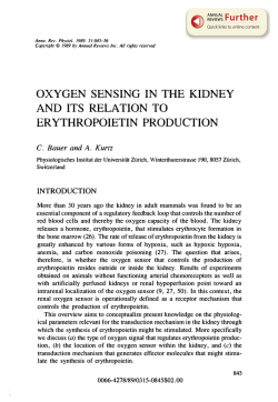
© Copyright 2026