
THE PATHOLOGY OF HYDROCEPHALUS
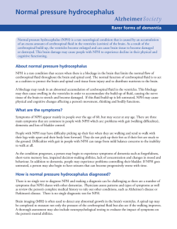
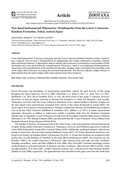
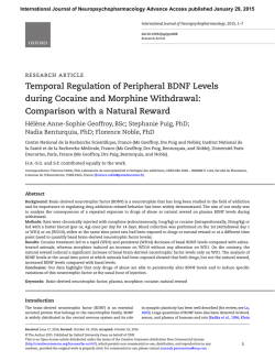
THE PATHOLOGY OF HYDROCEPHALUS
Erasmus Wilson Demonstration delivered at the Royal College of Surgeons of England
on
23rd October 1958
by
K. M. Laurence, M.A., M.B., Ch.B.
Department of Morbid Anatomy, The Hospital for Sick Children, Great Ormond Street*
ERASMUS WILSON, APART from being an eminent dermatologist of the
latter end of the last century, and a past President of this College, was
also a keen Egyptologist. I am sure that had he been fortunate enough
to discover the hydrocephalic mummy of the Roman period reported
by Douglas Derry in 1913, his interest in hydrocephalus might well
have been aroused.
Today, I would like to discuss the naked eye pathology of hydrocephalus, and shall base this talk on post-mortem material obtained
during my tenure of the Hydrocephalus Research Fellowship at The
Hospital for Sick Children, Great Ormond Street. Most of these cases
were examined using a special technique first described by Martin (1952)
which was later modified (Laurence and Martin, 1959) whereby the brain
is allowed to fix in situ within the skull several weeks before a dissection
is made. This not only permits grossly hydrocephalic brains to be
examined without danger of distortion, but it enables minor degrees of
malformation and abnormality to be recognised. some of which would
undoubtedly have been missed if conventional methods of removing the
brain from the bony cage were employed. Most important of all, this
method allows the careful examination of the extra-cerebral cerebro-spinal
fluid pathways, such as the basal cisterns, almost impossible by any
other method.
Hydrocephalus is best defined as the excessive accumulation of cerebrospinal fluid within the cranial cavity. I wish, however, to exclude hydrocephalus e vacuo where an excessive amount of cerebro-spinal fluid replaces
volume lost through primary atrophy of the brain.
Theoretically, hydrocephalus may be brought about in three separate
ways (see Table I).
TABLE I
CAUSES OF HYDROCEPHALUS
Choroid plexus papilloma and villous hypertrophy.
Over-production of C.S.F.
Defective absorption of C.S.F. .. ? ? Venous sinus thrombus.
Obstruction to the C.S.F. pathway
Within ventricular system:
Foramina of Monro
Aqueduct
Exit foramina
Subarachnoid space:
Arnold-Chiari malformation
Basal cistern block
Outside C.N.S.:
Skull abnormalities
Over-production of cerebro-spin al fluid
That over-production of cerebro-spinal fluid occurs is now conceded by
most authorities (Russell, 1954). The choroid plexus papillomata, in which
* Present address: Department of Paediatric Pathology, Department of Child
Health, Welsh National School of Medicine.
388
THE PATHOLOGY OF HYDROCEPHALUS
I
CMS
I
I
I
2
3
4
$
Fig. 1. A parasagittal section through a brain to show a large choroid plexus
papilloma situated in the posterior portion of the markedly dilated lateral ventricle.
The tumour is attached to the tela choroidea anteriorly, as well as to the ventricular
wall by a secondary glial stalk posteriorly. The subependymal haemorrhages are
a terminal event.
this is seen, form about 3 per cent. of childhood intra-cranial tumours.
The cauliflower-like tumour, friable and vascular, is attached to the
choroid plexus and is usually situated in the posterior portion of a lateral
ventricle and the whole ventricular system is symmetrically and often
markedly dilated (Fig. 1). Microscopically most of these tumours,
although more vascular, have the normal structure. One would therefore
expect such a tumour to be functional and by virtue of its bulk to secrete
excessive amounts of cerebro-spinal fluid. Such functional activity is
well known in a number of types of tumour. The absence of obstruction
to the flow of cerebro-spinal fluid has been demonstrated in a number
of cases, including one of this series. not only by encephalography when
air is found to pass from the basal cisterns into the Sylvian fissures,
but also at autopsy when even in the presence of considerable hydrocephalus, a comparatively wide and almost normal cerebro-spinal fluidfilled subarachnoid space is sometimes seen. However, in an appreciable
number of cases, although no obstruction is found, the meninges are
opaque and considerably thickened. Whether this is due to the effect of
a high protein in the cerebro-spinal fluid, which is usually associated with
such a tumour, or due to a compensatory hypertrophy from a high
cerebro-spinal fluid pressure, analogous to the hypertrophy of blood vessels
389
K. M. LAURENCE
in hypertension, is not easy to decide. A more likely explanation for this
meningeal thickening might be, that repeated minor haemorrhages,
known to occur from these vascular papillomas, set up a reactive fibrosis.
This would be supported by the finding of basal cistern block in an
appreciable proportion of such cases.
Under-absorption
Theoretically, under-absorption of cerebro-spinal fluid would be brought
about by obstruction of the venous return as in venous sinus thrombosis.
This, however, is not well authenticated, and in every case of thrombosis
that I have had the opportunity of examining, I was able to find another
cause for the hydrocephalus.
Obstruction of the cerebro-spinal fluid pathway
This group is numerically and pathologically the most important.
Before considering it in detail I would like to restate the general principle
that proximal to the site of obstruction the cerebro-spinal fluid pathway
becomes dilated. Thus, if the lateral and third ventricles are dilated
and the fourth ventricle is of normal dimensions, the obstruction is
likely to be situated within the aqueduct of Sylvius; when the whole
of the ventricular system is dilated but the basal cisterns are of normal
size, then the block will be found at the exit foramina of the fourthventricle.
If, on the other hand, the whole ventricular system and the basal cisterns
are dilated, then a basal cistern block is likely to be the cause of the
hydrocephalus. As there are two sets of choroid plexi, one set within
the lateral and third ventricles, and another choroid plexus within the
fourth ventricle, dilatation may take place below an aqueduct block
if an additional block in the fourth ventricle or the basal cisterns is present.
MALDEVELOPMENTS
Malformation of the aqueduct of Sylvius
Before considering the malformation of the aqueduct a brief consideration of its normal structure will be of help. The aqueduct is a channel
linking the third and fourth ventricles, normally about 11mm. in length
and 0.9sq.mm. in average minimum cross-sectional area (Woollam and
Milleni, 1953). It is completely lined by ependyma and is surrounded by
normal brain tissue without gliosis.
Two malformations have to be considered, namely stenosis and forking.
The former, stenosis, is an uncommon condition which may be associated
with other cerebral malformations. Here the normal shape of the aqueduct
is sometimes preserved but the aqueduct is of markedly reduced diameter,
but often it has a crenated and irregular outline. The ependymal lining,
however, remains intact and the surrounding brain tissue is normal
and free from excessive gliosis.
Forking of the aqueduct on the other hand is a fairly common anomaly
frequently associated with the Arnold-Chiari malformation. Here,
the aqueduct usually divides into a large dorsal and a number of smaller
390
THE PATHOLOGY OF HYDROCEPHALUS
ventral channels, each generally irregular in outline but with an intact
ependymal lining. These channels have a greatly reduced combined
cross-sectional area. Normal brain tissue, free from gliosis, intervenes
between them.
Spina bifida with hydrocephalus
This is the largest and most important group of malformations associated
with hydrocephalus and, before dealing with the hydrocephalus itself, I
must briefly review the anatomy of spina bifida.
In spina bifida occulta, the mildest abnormality of the group, the
spines have failed to fuse. The spinal cord, with its meninges, remains
within the spinal canal. Frequently, overlying skin shows abnormalities.
The spinal cord may show abnormalities which lead to the associated
weaknesses of the legs and urinary troubles sometimes found.
A meningocoele is a lesion where accompanying the spina bifida the
meninges have herniated out of the spinal canal, and are to be found directly
on the surface or covered with abnormal or normal skin. The cord remains
within the spinal canal, but may show a variety of malformations
(Cameron, 1956). Myelocoeles and myelomeningocoeles are essentially
the same lesion. Here, the neural tube has failed to close early in interuterine life and the neural plate remains on the surface. At birth the
neural plate is seen as a raw, apparently ulcerated area surrounded by a
blue, transparent membrane, representing the meninges. In the fortunate
cases the myelocoele will epithelialise over from the surrounding skin in
seven to ten days. More frequently, however, the neural plate becomes
infected and healing takes place after much fibrosis and scarring, and
finally the whole lesion may become well epithelialised. Thus, the typical
spina bifida cystica of bizarre shape develops.
Meningocoeles are comparatively rare-only about 5 per cent. of the
cases seen at The Hospital for Sick Children prove to be true meningocoeles
and I have not, so far, had the opportunity of examining such a case in
the post-mortem room, for they carry a very hopeful prognosis regarding
life. The majority of cases are myelocoeles, and nearly all of these are
associated with some form of malformation of the hind-brain; a small
proportion of those in whom the myelocoele is situated very low down the
cerebral spinal axis forming the exception. The hind-brain abnormality
generally takes the form of an Arnold-Chiari malformation (Fig. 2).
This consists of the Arnold malformation, where the cerebellum is often
small and poorly lobated, with a midline tongue consisting of vermis
which extends for a variable distance down the spinal canal, and the
Chiari malformation, where an elongated medulla extends for a variable
distance beyond the level of the foramen magnum. In addition the
medulla is frequently kinked backwards upon the cord. Lying between the
cerebellum and the pons and medulla is a long and often narrow fourth
ventricle, the choroid plexus of which generally forms a compact mass
near the tip of the cerebellar prolongation. Sometimes there is an
391
K. M. LAURENCE
Fig. 2. A hemisected skull and spinal cord showing the Arnold-Chiari malformation. The elongated medulla (Chiari malformation) is not only largely intraspinal,
but is a!so kinked upon the cord. A well developed cerebellar tongue (Arnold
malformation) is shown. The myelocoele in the lumbo-sacral region is tvpical,
with nerve roots leaving the neural plate to gain the root canals. The cord shows
slight hydromyelia.
392
THE PATHOLOGY OF HYDROCEPHALUS
extension of the fourth ventricle within the medulla beyond the exit
foramina. In all but the mildest cases the upper spinal nerve routes take
a cranial course. The whole is generally covered by congested meninges.
This congestion in time will lead to thickening and fibrosis. The malformation may be only very slight, measuring 3 or 4mm., and thus easily
missed if the brain is removed and examined by the conventional methods,
while in other cases the Chiari malformation alone may be present. Here,
a lengthened medulla extends for a variable distance down the spinal
canal, but the cerebellum is relatively normal.
The Arnold-Chiari malformation must be differentiated from a tonsillar
herniation. In the latter the tonsils descend down the spinal canal laterally,
while in the Arnold-Chiari malformation it is the vermis which extends in
the midline, posteriorly. Moreover, in the tonsillar herniation, there is no
medullary component.
Associated with the malformation of the hind-brain, other congenital
deformities, especially of the central nervous system, are frequently found.
Aqueduct forking has already been mentioned. The thalamic bodies may
be fused to a variable degree, leading to a large inter-thalamic bar or
massa intermedia. Micro-gyrations are frequently present, and a hypoplastic falx and great longitudinal fissure are also common, together
with inter-digitation between the two cerebral hemispheres. Ectopic
subependymal grey matter is sometimes seen, while in many cases there
is upward herniation of the cerebellum through the tentorial opening
and hydromyelia and diastomatomyelia of the cord. Frequently renal
and skeletal malformations are also found. These are only some of the
anomalies that may be present.
Space will not permit a discussion of the aetiology of these malformations, which are adequately discussed by Cameron (1957), but the aetiology
of the associated hydrocephalus is of prime importance here. Aqueduct
abnormalities will, in themselves, lead to hydrocephalus; so will the
Arnold-Chiari malformation by virtue of the crowding of structures
in the often small posterior fossa and in the upper spinal canal. The
hyperaemia and eventual fibrosis and thickening of the meninges surrounding the Arnold-Chiari malformation may further interfere with an
already embarrassed cerebro-spinal fluid circulation. Most important of
all, however, is the effect of an ascending infection from the raw spina
bifida cystica leading to post-inflammatory obstructions. Thus, a block
of the foramina of Monro (Fig. 3), aqueduct block, blocks of the exit
foramina or basal cistern block may be superimposed upon any congenital malformations that may be present.
Septum formation in the aqueduct, foramina of Monro and exit
foramina of the fourth ventricle is exceedingly rare, and I have not had
the opportunity to dissect a case. Nor have I had the opportunity to
examine skeletal abnormalities such as an achondroplasia leading to
hydrocephalus.
393
K. M. LAURENCE
I
CMSI
I
I
I
I
I
2
3
4
s
Fig. 3. A coronal sc_tion o thte cereoral nemispneres snowing a posL-intlammatory
block of the foramina of Monro following an ascending infection from a myelocoele.
A large interthalamic connexus is also present.
GLIOSIS OF THE AQUEDUCT
Gliosis is an interesting and controversial group as there has been
much discussion about the aetiology of this condition. Here, the aqueduct
is narrowed, subdivided or completely occluded by an overgrowth of
.ubependymal fibrillary neuroglia (Fig. 4). The outline of the original
aqueduct is marked out by disorderly islets of ependymal cells left behind
by the gliosis (Fig. 5). If a central channel remains this rarely has any
ependymal lining. Clinically, hydrocephalus begins insidiously, often
somewhat later in childhood or even in early adult life, and there is never
any history suggestive of an antecedent infection.
394
THE PATHOLOGY OF HYDROCEPHALUS
Fig. 4. x A midline section showing the aqueduct completely occluded by gliosis
in a-case associated with neurofibromatosis. The lateral and third ventricles
are grossly dilated.
Fig. 5. Transverse section through an aqueduct showing gliosis.
phosphotungstic acid haematoxylin. x 18)
395
(Mallory's
K. M. LAURENCE
Spiller (1916) suggested that the condition might be due to an involutional hyperplasia of the subependymal glia, analagous to the closing off of
the central canal of the spinal cord that occurs in the second decade while
others have favoured an inflammatory origin on its similarity to postinflammatory gliosis (Russell, 1949). I have had the opportunity of
examining four cases of gliosis in which the condition was associated with
neurofibromatosis. The latter is well known, being a hereditary and familial
condition which would appear to be a system defect of the neuroectodermal
elements of the central and peripheral nervous system, regarded by Willis
(1948) as hamartomatous. Optic nerve " gliomas " also regarded as
hamartomatous (Crome, 1954) are a common complication in such cases,
while blastoma-like microscopic foci have been found scattered in the glial
tissue (Scharenberg, 1953). On the other hand tumour involvement of the
brain (Turner and Gardner, 1938) (which may have arisen in such a
hamartomatous area) is well known. The aqueduct gliosis in these four
examples of neurofibromatosis may also be of hamartomatous origin, a
view supported by finding nests of bizarre glial cells in the peri-aqueductal
tissues. I would like to suggest that aqueduct gliosis unassociated with
neurofibromatosis may also have the same origin.
Fig. 6. Photomicrograph of the peri-aqueductal glial tissue to show bizarre
cells, including multinucleate forms. (Haematoxylin and Van Gieson. x 180)
INFLAMMATIONS
This is a large and important group, and one of the commonest causes
of post-inflammatory hydrocephalus is blood in the cerebro-spinal fluid
pathway. This generally results from birth trauma, especially in premature
and first-born infants. It has recently been made abundantly clear that
396
THE PATHOLOGY OF HYDROCEPHALUS
to produce such trauma the delivery need not have been at all remarkable
and that the infant need show no signs specifically associated with intracranial mischief (Bound, Butler and Spector, 1956). It is worth remembering that when a subdural haematoma is found the injury responsible
for this has very likely produced bleeding into the cerebro-spinal fluid
as well. Also, the same birth injury may well have produced parenchymal
brain damage in addition. However, bleeding into the cerebro-spinal fluid
pathway resulting from haemorrhage from berry aneurysms, following
operations or head injuries in childhood, may also give rise to hydrocephalus (Foltz and Ward, 1956).
Infections form the other important inflammatory group. These infections may be either acute or chronic, of bacteria], and less frequently
of viral or toxoplasmal origin. Often the mild and missed infections
produce the worst effects, as such cases receive only late treatment or may
receive none at all. On the other hand an E. coli meningitis in the neo-
pi
-_mI
cms
Fig. 7. Horizontal section through the cerebrum of a case of hydrocephalus
following B. coli meningitis. The lateral ventricles have been converted into a
series of intercommunicating cysts.
397
K. M. LAURENCE
natal period may also be completely silent. With all these infections
parenchymal brain damage is common.
A variety of anatomical lesions may follow these inflammations. The
most severe effects, often resulting from an E. coli meningitis, may lead to
an organising pyocephalus. Here, the whole ventricular system may be
converted into a series of intercommunicating cysts, and the foramina of
Monro may also become occluded (Fig. 7). In the less severe inflammations, the aqueduct, always a vulnerable structure in view of its position
and narrowness, may become blocked. This block may result from its
lumen being occluded by pus or a small blood clot. On the other hand the
block may follow from damage to its ependymal lining; the ependyma is
very delicate and once it is damaged or denuded it will not regenerate, the
subependymal glia then proliferates and may finally completely occlude
the channel. In the majority of cases, however, an inflammatory reaction
occurs in the meninges, leading to occlusive fibrosis there. This process may
I I
I
I I
Fig. 8. Midline section through the skull showing a hugely dilated ventricular
system due to a block at the exit foramina of the fourth ventricle. Note the
inconspicuous basal cisterns and the ballooned floor of the third ventricle.
398
THE PATHOLOGY OF HYDROCEPHALUS
rrI cms.
Fig. 9. Midline section through a skull showing a basal cistern block. The third
and fourth ventricles are enormously dilated. Fine fibrous strands traverse the
cisterns. The interpeduncular cistern has so greatly expanded as to elevate the
floor of the third ventricle, elongate the optic nerve and the pituitary stalk and
flatten the pituitary gland.
cause a block of the exit foramina of the fourth ventricle, for the arachnoid
may become bound down in their immediate vicinity (Fig. 8). In other
cases the foramina remain clear, but the arachnoid becomes bound down
on the edge of the basal cisterns and thus gives rise to a basal cistern block.
The cisterns then tend to dilate, compressing the cerebellum, elevating the
floor of the third ventricle, or stretching the optic nerve and the pituitary
stalk (Fig. 9).
Any combination of all these inflammatory lesions may be present in a
particular case. Thus, a case of inflammatory aqueduct stenosis may well
be associated with a block at the exit foramina or the basal cisterns as well.
Tumours
Tumours in any position may give rise to obstructive hydrocephalus.
A six week old infant, for example, who had apparently a congenital
aqueduct block, was found subsequently at post-mortem to have
cerebellar medulloblastoma compressing that structure.
In conclusion I shall consider the aetiology of the hydrocephalus in the
hundred consecutive post-mortems carried out at The Hospital for Sick
Children and at the Westminster Children's Hospital on which this paper
is based. (The numbers shown in Figures 3 and 4 are, however, only
provisional, as the data on all these cases is not yet entirely complete). It
399
32
K. M. LAURENCE
TABLE II
AETIOLOGY OF HYDROCEPHALUS
(100 Post-mortem examinations)
..
..
..
..
..
Malformation:
..
..
..
..
..
..
Alone
..
..
..
..
..
With infection
..
..
..
..
..
With trauma
..
..
..
..
..
Inflammation:
..
..
..
..
Infection alone ..
..
..
Trauma alone
..
Unknown (probably infection or trauma)
..
..
..
..
..
..
Tumours
46
14
30
2
50
17
22
11
4
is seen from Table IL that in almost half the cases a malformation was the
basis, though in the majority of these the malformation was associated
with a super-added inflammatory lesion. In the case where the hydrocephalus was of purely inflammatory origin, trauma, and in nearly all
instances birth trauma, accounted for a large proportion. This is in
agreement with results obtained in another investigation where almost
one-third of all the cases in the series had this aetiology (Laurence, 1958).
In eleven cases it was impossible to decide on the evidence available which
of the two main inflammatory aetiologies was responsible. In four cases a
tumour was found to be the underlying cause of the disease. In all these
four the tumour was an unexpected finding.
TABLE III
SITE OF BLOCK CAUSING HYDROCEPHALUS
(100 Post-mortem examinations)
..
..
..
..
Isolated sites:
..
..
..
..
..
Aqueduct
..
..
..
IV Ventricle foramina
..
..
Basal cistern block
..
..
Arnold-Chiari malformation ..
..
..
..
..
..
Multiple sites:
..
..
..
Basal cistern block
With block at foramina of Monro, aqueduct
or IV ventricle.
..
..
..
Arnold-Chiari malformation
With blocks at foramina of Monro, aqueduct,
IV ventricle or basal cistern.
..
..
..
No block (over-secretion): ..
10
50
7
21
12
20
48
28
2
Table III shows that an anatomical cause for the hydrocephalus was
found in every case examined, though two children had hydrocephalus
due to over-secretion of fluid. It is interesting to note that about half the
cases had more than one site of block.
ACKNOWLEDGMENTS
I am indebted to Mr. D. Martin and the staff of the Department of
Medical Illustration for the illustrations and preparations of the specimens, diagrams and charts shown in the demonstration, which accompanied this lecture. Thanks are due to the Research Committee of The
Hospital for Sick Children, Great Ormond Street, and especially Dr. M.
Bodian and Mr. G. H. Macnab for their help, encouragement and support,
and to Professor G. J. Cunningham for his part in enabling me to give this
lecture-demonstration.
400
THE PATHOLOGY OF HYDROCEPHALUS
REFERENCES
BOUND, J. P., BUTLER, N. R., and SPECTOR, W. G. (1956) Brit. med. J. 2, 1191 and 1260.
CAMERON, A. H. (1956) Lancet 2, 171.
(1957) J. Path. Bact. 73, 195 and 213.
CROME, L. (1954) J. Path. Bact. 67, 407.
CROWE, W. F., SCHULL, W. F., and NEEL, J. V. (1956) Multiple neurofibroma. Springfield,
Ill., U.S.A.
DERRY, D. E. (1912-13) J. Anat. (Lond.) 47, 436.
FOLTZ, E. L., and WARD, A. W. (1956) J. Neurosurg. 13, 546.
LAURENCE, K. M. (1958) Lancet 2, 1152.
and MARTIN, D. (1959) J. clin. Path. 12, 188.
MARTIN, D. (1952) Med. biol. Ill. 2, 260.
RUSSELL, D. S. (1949) M.R.C. Spec. Rep. Ser., No. 265. London.
(1954) Ass. Res. nerv. Dis. Proc. 34, 161.
SPILLER, W. G. (1916) J. nerv. ment. Dis. 44, 395.
TURNER, C. A., and GARDNER, W. D. (1938) Amer. J. Cancer 32, 339.
WILLIS, R. A. (1948) Pathology of tumours. London, Butterworth, p. 838.
WOOLLAM, D. H. M., and MILLEN, J. W. (1953) Brain 76, 104.
PROCEEDINGS OF THE COUNCIL IN MAY
AT A MEETING of the Council on the 14th May, with Professor Sir James
Paterson Ross, President, in the Chair, Mr. E. D. Ahern, Brisbane, and Dr.
John H. Gibbon, Philadelphia, were admitted to the Hon. Fellowship.
The Lady Cade Medal was awarded to Squadron Leader T. C. D.
Whiteside for his research on the effect, on visual performance, of dazzle
from sources of very high brightness.
The Begley Prize was awarded to Dr. Geoffrey S. Makin of Liverpool
University.
Diplomas of Membership were granted to 147 candidates and one
diploma of Fellowship was granted.
Diplomas were granted, jointly with the Royal College of Physicians,
as follows: Physical Medicine (1); Tropical Medicine and Hygiene (2).
It was reported that the College of General Practitioners, which had
hoped to erect a building in Lincoln's Inn Fields, had now been prevented
from doing so by a series of setbacks, the most significant of which were
the town planning conditions and difficulties over security of tenure.
The following hospitals were recognised under paragraph 23 of the
F.R.C.S. regulations:
POSTS RECOGNISED
General
(6 mths. unless
otherwise stated)
HOSPITALS
Hospital
H.S. (S.H.O.)
BATH - Royal United Hospital
(additional)
H.S. (S.H.O.)
BATH - St. Martin's
(additional)
Unspecified
(all 6 mths.)
Under para 23 (c)
Temporary recognition
for one year
S.H.O. (E.N.T.)
LANCASTER-Beaumont Hospital
and Lancaster Royal Infirmary
(additional)
Change of recognition of
Casualty Registrar and Deputy Resident
Surgical Officer from 6mths. unspecified
to 6mths. Casualty.
LONDON - Connaught Hospital,
Walthamstow
401
32-2
Casualty
(all 6 mths.)
© Copyright 2026