
! ! ! DIABETES!MELLITUS!Y!BARRERA!HEMATORRETINIANA
!
!
!
!
!
!
!
!
!
!
!
!
!
DIABETES!MELLITUS!Y!BARRERA!HEMATORRETINIANA.!
ANÁLISIS!IN#VITRO!DE!LA!EXPRESIÓN!DE!PROTEÍNAS!DE!
TIGHT!JUNCTION!Y!SU!TRADUCCIÓN!FUNCIONAL.!
IMPLICACIONES!TERAPÉUTICAS!
!
!
!
%
%
%
%
%
Marta!Villarroel!Fandos!
!
TESIS%DOCTORAL%
%
Barcelona,%2015%
Laboratori%de%Diabetis%i%Metabolisme%
Vall%d’Hebron%Institut%de%Recerca%
%
Departament%de%Bioquímica%i%de%Biologia%Molecular%
Universitat%Autònoma%de%Barcelona%
!
!
%
%
%
%
TESIS%DOCTORAL%
%
Programa%de%Doctorat%en%Bioquímica,%Biologia%Molecular%i%Biomedicina%
Departament%de%Bioquímica%i%de%Biologia%Molecular%
%
!
DIABETES!MELLITUS!Y!BARRERA!HEMATORRETINIANA.!
ANÁLISIS!IN#VITRO!DE!LA!EXPRESIÓN!DE!PROTEÍNAS!DE!
TIGHT!JUNCTION!Y!SU!TRADUCCIÓN!FUNCIONAL.!
IMPLICACIONES!TERAPÉUTICAS!
%
Memoria%presentada%por%%
%
Marta!Villarroel!Fandos!!
%
para%optar%al%grado%de%Doctor%en%Bioquímica,%Biología%Molecular%y%Biomedicina%%
por%la%Universitat%Autònoma%de%Barcelona%
%
Barcelona,%2015%
%
%
%
%
%
%
Dr.%Rafael%Simó%Canonge%%%%%%%Dra.%Cristina%Hernández%Pascual%%%%%%%Dra.%Marta%GarciaSRamírez%
%%%Director(
(
(
((((((Director(
(
(
((((((Director(
%
%
%
%
%
Dr.%Joan%Xavier%Comella%Carnicé%%
Tutor% %
%
%
%
Marta%Villarroel%Fandos%
%%%%%%%%%%%%%%Doctorando%%%
A"mi"padre,"a"mi"abuelo,"
a"toda"mi"familia"y"a"mi"amor
!
ÍNDICE
1
2
ÍNDICE'
!
ÍNDICE'
'
'
'
'
'
'
'
''''
'
'
'1
ABREVIATURAS' '
'
'
'
'
'
'''''
'
'
'7'
INTRODUCCIÓN''
'
'
'
'
'
'''
'
'
15'
1.#LA#RETINOPATÍA#DIABÉTICA##
#
#
#
##
#
#
17#
1.1.'EPIDEMIOLOGÍA'
'
'
'
'
'''
'''
'
'
17'
1.2.'PATOFISIOLOGÍA'
'
'
'
'
'
'''
'
'
18'
1.2.1.'Bases'bioquímicas' '
'
'
'
'
'''
'
18'
1.2.2.'Neurodegeneración''
'
'
'
'
'
'
20'
1.2.3.'Alteraciones'de'la'microcirculación''
'
'
'
'
22'
1.2.4.'Edema'macular'diabético' '
'
'
'
'
'
24'
'
'
'
'
'
27'
1.3.1.'Control'de'los'factores'de'riesgo'
'
'
'
'
'
27'
1.3.2.'Fotocoagulación'con'láser' '
'
'
'
'
'
27'
1.3.3.'Vitrectomía' '
'
'
'
'
'
'
28'
'
'
'
'
'
'
28'
1.3.5.'Nuevas'perspectivas'terapéuticas' '
'
'
'
'
30'
#
#
32#
1.3.'TRATAMIENTO''
'
'
'
1.3.4.'Terapias'farmacológicas'
'
'
2.#FISIOPATOLOGÍA#DE#LA#BARRERA#HEMATORRETINIANA#
2.1.'COMPOSICIÓN''
'
'
'
'
'
'
'
'
32'
'
2.1.1.'Retina'
'
'
'
'
'
'
'
'
32'
'
2.1.2.'Barrera'hematorretiniana' '
'
'
'
'
'
34'
'
2.1.3.'Epitelio'pigmentario'de'la'retina'
'
'
'
'
'
38'
'
'
'
'
'
45'
'
'
'
48'
'
'
'
48'
'
'
52'
2.2.'LÍNEA''CELULAR'ARPEW19'
'
'
2.3.'TIGHT'JUNCTIONS'(UNIONES'CELULARES'ESTRECHAS)'
2.3.1.'Función'y'estructura''
'
'
'
2.3.2.'Resistencia'eléctrica'transepitelial'y'permeabilididad'
'
'
3
ÍNDICE'
!
2.3.3.'Componentes'
'
'
'
'
'
'
'
53'
2.3.3.1.'Ocludina' '
'
'
'
'
'
'
54'
2.3.3.2.'Claudinas' '
'
'
'
'
'
'
56'
2.3.3.3.'Zonula'Occludens' '
'
'
'
'
'
59'
'
3.#AMPK# # #
#
#
#
#
#
#
#
#
#
63#
3.1.'ESTRUCTURA' '
'
'
'
'
'
'
'
'
63'
3.2.'REGULACIÓN'DE'LA'ACTIVIDAD'
'
'
'
'
'
'
64'
3.3.'FUNCIONES'
'
'
'
'
'
'
'
66'
#
#
#
#
#
#
#
68#
'
'
'
4.#MATRIZ#EXTRACELULAR#
4.1.'ESTRUCTURA' '
'
'
'
'
'
'
'
'
68'
4.2.'COMPOSICIÓN''
'
'
'
'
'
'
'
'
69'
4.2.1.'Colágeno'IV' '
'
'
'
'
'
'
'
70'
4.2.2.'Fibronectina''
'
'
'
'
'
'
'
70'
4.2.3.'Laminina'
'
'
'
'
'
'
'
71'
'
'
'
'
'
'
'
71'
'
'
'
'
'
'
'
72'
4.4.'MATRIZ'EXTRACELULAR'Y'RETINOPATÍA'DIABÉTICA' '
'
'
'
73'
5.#EFECTO#DEL#FENOFIBRATO#EN#LA#RETINOPATÍA#DIABÉTICA# #
#
76#
'
4.2.4.'Heparán'sulfato'
4.3.'FUNCIONES'
'
'
'
5.1.'FARMACOCINÉTICA' '
'
'
'
'
'
'
'
76'
5.2.'FARMACODINÁMICA' '
'
'
'
'
'
'
'
77'
5.2.1.'PPARs'
'
'
'
'
'
'
'
77'
'
'
'
'
'
'
79'
'
5.2.2.'Mecanismos'de'acción'
5.3.'ESTUDIOS'CLÍNICOS' '
'
'
'
'
'
'
'
82'
5.3.1.'Estudio'FIELD'
'
'
'
'
'
'
'
82'
5.3.2.'Estudio'ACCORD'
'
'
'
'
'
'
'
84'
'
4
ÍNDICE'
!
HIPÓTESIS'Y'OBJETIVOS'
'
'
'
'
'
'
'
87'
RESULTADOS'
'
'
'
'
'
'
'
91'
'
93'
'
'
CAPÍTULO#I:#Efecto'de'la'hiperglicemia'sobre'la'funcionalidad'de'la'barrera'
hematorretiniana'externa'y'la'expresión'de'las'proteínas'de'tight'junction'en'
células'de'epitelio'pigmentario'de'la'retina'humana'(ARPE?19).'
'
'
'
CAPÍTULO#II:#Efecto'protector'del'ácido'fenofíbrico'sobre'la'disrupción'del''
epitelio'pigmentario'de'la'retina'inducida'por'la'IL?1b'a'través'de'la'supresión''
de'la'activación'de'la'vía'de'la'AMPK.'
'
'
'
'
'
'''''''''''105'
DISCUSIÓN'
'
'
'
'
'
'
'
'
''''''''123'
CONCLUSIONES' '
'
'
'
'
'
'
'
''''''''141'
BIBLIOGRAFÍA' '
'
'
'
'
'
'
'
''''''''145'
ANEXO'
'
'
'
'
'
'
'
''''''''171
'
'
5
!
6
!
ABREVIATURAS
7
!
8
ABREVIATURAS'
!
Abreviaturas
ACCORD'
Action'to'Control'Cardiovascular'Risk'in'Diabetes'Study'
ADN' '
Ácido'desoxirribonucleico'
ARN'
'
Ácido'ribonucleico'
ADP'
'
Adenosín'difosfato'
AGEs' '
Productos'avanzados'de'la'glicación'
AICAR' '
Ribósido'de'5WaminoimidazolW4Wcarboxamida'
AMD' '
Degeneración'macular'asociada'a'la'edad'
AMP' '
Adenosín'monofosfato'
AMPK' '
Quinasa'activa'por'monofosfato'de'adenina'
Apo'
'
Apolipoproteína'
ATP'
'
Adenosín'trifosfato'
BDNF' '
Factor'de'crecimiento'derivado'del'cerebro'
BHA'
'
Barrera'hematoacuosa'
BHE'
'
Barrera'hematoencefálica'
BHR'
'
Barrera'hematorretiniana'
BM'
'
Membrana'de'Bruch'
BREC' '
Células'endoteliales'de'retina'bovina'
CaMKKβ
Proteína'quinasa'dependiente'de'calcioWcalmodulina'β'
CBS'
Dominio'identificado'en'la'enzima'cistationinaWβWsintasa'
CK'
'
Caseína'quinasa'
Cmax'
'
Concentración'máxima'
CNTF' '
Factor'neurotrófico'ciliar'
COX'
Ciclooxigenasa''
'
CRALBP'
Proteína'celular'de'unión'al'11WcisWretinaldehído'
CSF'
Factores'estimulantes'de'colonias'
'
CTGF' '
Factor'de'crecimiento'de'tejido'conectivo'
DAG' '
Diacilglicerol'
DHA' '
Ácido'docosahexaenoico'
DM'
Diabetes'mellitus'
'
9
ABREVIATURAS'
!
DME' '
Edema'macular'diabético'
DR'
'
Retinopatía'diabética'
EL'
'
Capa'de'elastina'
eNOS' '
Óxido'nítrico'sintasa'endotelial'
EPCs' '
Células'endoteliales'progenitoras'
Epo''
Eritropoyetina'
'
EpoWR' '
Receptor'de'eritropoyetina'
ETDRS' '
Early'Treatment'Diabetic'Retinopathy'Study'
FDA'
'
Food'and'Drug'Administration'
FGF'
'
Factores'de'crecimiento'de'fibroblastos'
FHHNC''
Hipomagnesemia'familiar'con'hipercalciuria'y'nefrocalcinosis'
FIELD' '
Fenofibrate'Intervention'and'Event'Lowering'in'Diabetes'Study'
GADPH''
Gliceraldehído'3Wfosfato'deshidrogenasa'
GCL'
Capa'de'células'ganglionares'
'
GBD' '
Dominio'de'unión'a'glucógeno'
GFAP' '
Proteína'ácida'fibrilar'de'la'glía'
GLAST' '
Transportador'de'glutamato/aspartato'
GMP' '
Guanosín'monofosfato'
GTPasa''
Guanosina'trifosfatasa'
GuK'
'
Guanilato'quinasa'
HDL'
'
Lipoproteínas'de'alta'densidad'
HGF'
'
Factor'de'crecimiento'de'hepatocitos'
HGMEC'
Células'endoteliales'humanas'de'la'microvasculatura'glomerular''
HIFW1' '
Factor'inducible'por'la'hipoxia'1'
HREC' '
Células'endoteliales'humanas'de'retina''
HUVEC''
Células'endoteliales'humanas'de'vena'de'cordón'umbilical'
ICL'
Capa'de'colágeno'interna'
'
IFNWγ' '
Interferón'γ'
IGFWI' '
Factor'de'crecimiento'insulínico'tipo'I'
IL'
Interleuquina'
10
'
ABREVIATURAS'
!
INL'
'
Capa'nuclear'interna'
IPL'
'
Capa'plexiforme'interna'
IRBP' '
Proteína'de'unión'a'interfotorreceptores'retinoides'
IRMAs' '
Anomalías'microvasculares'intrarretinianas'
JAM'
'
Molécula'de'adhesión'de'la'unión'
kDa'
'
kiloDalton'
KO'
'
Knockout'
LDL'
'
Lipoproteínas'de'baja'densidad''
LEDGF' '
Factor'de'crecimiento'derivado'el'epitelio'de'la'lente'
LPL'
Lipoproteína'lipasa'
'
LpWPLA2'
Fosfolipasa'A2'asociada'a'lipoproteína'
MAGI' '
Guanilato'quinasa'invertida'asociada'a'la'membrana'
MAGUK'
Guanilato'quinasas'asociadas'a'la'membrana'
MAPK' '
Proteína'quinasa'activada'por'mitógenos'
MCPW1''
Proteína'quimioatrayente'de'monocitos'1'
MDCK' '
Células'de'epitelio'de'riñón'canino'MadinWDarby'
MHC' '
Complejo'mayor'de'histocompatiblidad'
MMP' '
Metaloproteinasa'
MUPP1'
Proteína'1'con'múltiples'dominios'PDZ'
NACos' '
Proteínas'asociadas'con'el'núcleo'y'complejos'de'adhesión''
NFWkB'
Factor'nuclear'potenciador'de'las'cadenas'ligeras'kappa'de'las'células'B'
activadas'
NGF'
'
Factor'de'crecimiento'neuronal'
NMDA''
NWmetilWDWaspartato'
NO'
Óxido'nítrico'
'
NPD1' '
Neuroprotectina'D1'
NPDR' '
Retinopatía'diabética'no'proliferativa'
NTW3' '
Neurotropina'3'
OCL'
'
Capa'de'colágeno'externa'
OCT'
'
Tomografía'óptica'de'coherencia'
11
ABREVIATURAS'
!
ONL'
'
Capa'nuclear'externa'
OPL'
'
Capa'plexiforme'externa'
PDGF' '
Factor'de'crecimiento'derivado'de'las'plaquetas'
PDR'
Retinopatía'diabética'proliferativa'
'
PDZ'
Acrónimo'derivado'de'las'3'primeras'proteínas'en'las'que'se'descubrió'el'
dominio:'PSDW95,'DiscsWlarge'A'y'ZOW1'
PEDF' '
Factor'derivado'del'epitelio'pigmentario'
PKA'
'
Proteína'quinasa'A'
PKC'' '
Proteína'quinasa'C'
PLGF' '
Factor'de'crecimiento'placentario'
PPARs' '
Receptores'activadores'de'la'proliferación'de'peroxisomas'
PPREs' '
Elementos'de'respuesta'a'PPARs'
PP2C' '
Proteína'fosfatasa'2C'
RAS'
'
Sistema'Renina'Angiotensina'
ROS'
'
Especies'reactivas'de'oxígeno'
RPE'
'
Epitelio'pigmentario'de'la'retina'
RPE65' '
Proteína'específica'del'epitelio'pigmentario'de'la'retina'65'kDa'
RXR'
Receptor'x'retinoide'
'
SDSWPAGE'
Electroforesis'en'gel'de'poliacrilamida'con'dodecilsulfato'sódico'
SH3'
Dominio'de'homología'al'dominio'3'de'la'proteína'Src'
'
shRNA''
ARN'de'horquilla'pequeña'
siRNA' '
ARN'pequeño'de'interferencia'
SST'
Somatostatina'
'
SSTR' '
Receptor'de'somatostatina'
TAK1' '
Proteína'quinasa'1'activada'por'el'factor'de'crecimiento'transformante'β
TEER' '
Resistencia'eléctrica'transendotelial'
TER'
Resistencia'eléctrica'transepitelial'
'
TGFWβ
Thr'
Factor'de'crecimiento'transformante'β
'
TIMP' '
12
Treonina'
Inhibidores'tisulares'de'metaloproteinasas'de'matriz'
ABREVIATURAS'
!
TJ'
'
Tight'juncions'(uniones'celulares'estrechas)'
TNFWα
Factor'de'necrosis'tumoral'α'
VEGF' '
Factor'de'crecimiento'endotelial'vascular'
VLDL' '
Lipoproteínas'de'muy'baja'densidad'
ZAK'
Quinasa'asociada'a'ZOW1'
'
ZOW1' '
Zonula'occludens'1'
ZONAB''
Factor'de'transcripción'asociado'a'ZOW1'
13
!
14
!
INTRODUCCIÓN
15
!
16
INTRODUCCIÓN'
!
1.#LA#RETINOPATÍA#DIABÉTICA#
1.1.#EPIDEMIOLOGÍA#
La'diabetes'mellitus'(DM)'es'una'enfermedad'crónica'muy'prevalente'y'se'estima'que'
la'cifra'de'pacientes'diabéticos'crecerá'de'forma'exponencial'en'los'próximos'años'(pasará'
de'366'millones'de'personas'en'2011'a'552'millones'en'2030).'Según'el'estudio'Di@betes'la'
prevalencia'de'la'DM'en'España'es'del'13,8%1.'
La'retinopatía'diabética'(DR)'es'la'complicación'microangiopática'más'frecuente'de'la'
diabetes' y' la' principal' causa' de' ceguera' en' la' población' en' edad' laboral' en' los' países'
industrializados.' El' estudio' epidemiológico' de' Wisconsin' reveló' que' a' los' 15' años' del'
diagnóstico,' el' 98%' de' los' pacientes' afectos' de' diabetes' mellitus' tipo' 1' y' el' 78%' de' los'
pacientes'con'diabetes'mellitus'tipo'2'presentaban'DR.''Durante'el'mismo'periodo'el'33%'de'
los'pacientes'con'diabetes'tipo'1'y'el'17%'de'los'pacientes'con'diabetes'tipo'2'presentarán'
retinopatía'diabética'proliferativa'(PDR),'la'principal'causa'de'ceguera'en'los'pacientes'con'
diabetes'tipo'1.'La'gran'mayoría'de'pacientes'con'diabetes'tipo'2'no'van'a'desarrollar'PDR,'
sino' que' es' mucho' más' frecuente' la' evolución' hacia' el' edema' macular' (DME).' Se' ha'
comunicado' que' la' incidencia' de' DME' en' los' pacientes' con' diabetes' tipo' 2' puede' llegar' a'
casi'el'40%'en'un'periodo'de'seguimiento'de'10'años.'Por'el'contrario,'en'los'pacientes'con'
diabetes' tipo' 1,' la' incidencia' de' DME' durante' el' mismo' periodo' se' ha' cifrado' en' un' 20%.'
Dada'la'mayor'prevalencia'de'la'diabetes'tipo'2'(>90%),'el'DME'representa'la'principal'causa'
de'disminución'de'la'agudeza'visual'y'ceguera'no'sólo'en'los'pacientes'con'diabetes'tipo'2,'
sino'en'la'diabetes'en'general2,3.'
El' buen' control' de' la' glucemia' y' de' la' presión' arterial' es' esencial' para' prevenir' o'
retardar' la' progresión' de' la' DR.' Sin' embargo,' los' objetivos' terapéuticos' son' difíciles' de'
alcanzar'y,'en'consecuencia,'la'DR'va'a'presentarse'en'una'elevada'proporción'de'pacientes.'
Los' tratamientos' actuales' para' la' DR' están' indicados' en' fases' avanzadas,' tiene' una'
17
INTRODUCCIÓN'
!
efectividad'limitada'y'se'asocian'a'importantes'efectos'secundarios.'Por'ello,'es'necesario'el'
desarrollo' de' nuevos' tratamientos' basados' en' el' conocimiento' de' los' mecanismos'
fisiopatológico'que'causan'esta'complicación.''
A' continuación' se' revisan' los' mecanismos' patogénicos' que' ocasionan' la'
microangiopatía'diabética,'con'especial'énfasis'en'las'alteraciones'que'ocurren'en'la'retina.''''
1.2.#PATOFISIOLOGÍA#
1.2.1.#Bases#bioquímicas##
A' nivel' fisiopatológico,' la' hiperglicemia' mantenida' induce' una' serie' de' cambios'
bioquímicos'en'el'metabolismo'glucídico,'reológicos'en'el'flujo'sanguíneo'y'anatómicos'en'la'
pared'vascular'que'son'los'responsables'de'la'aparición'de'una'microangiopatía'a'nivel'de'las'
arteriolas,' capilares' y' vénulas4.' Existen' cuatro' vías' metabólicas' que' se' activan' en'
condiciones' de' hiperglicemia' y' que' contribuyen' al' daño' celular' observado' en' la' retina' de'
pacientes'diabéticos:'la'vía'de'los'polioles'(o'de'la'aldosa'reductasa),'la'vía'de'la'hexosamina,'
la'síntesis'de'novo'de'diacilglicerol'(DAG)'y'la'activación'de'la'proteína'quinasa'C'(PKC)'y'la'
formación'de'productos'avanzados'de'la'glicación'(AGEs).'El'nexo'común'entre'la'activación'
de' estos' cuatro' mecanismos' es' el' estrés' oxidativo' inducido' por' la' hiperglicemia.' La'
hiperglicemia'provoca'un'aumento'de'la'producción'de'superóxido'a'nivel'mitocondrial'que'
reduce'en'un'66%'la'actividad'de'la'gliceraldehído'3Wfosfato'deshidrogenasa5.'La'inhibición'
parcial'de'esta'enzima'provoca'un'aumento'en'la'concentración'de'algunos'metabolitos'de'
la'vía'glicolítica'y'su'utilización'en'otras'vías'metabólicas'como'las'detalladas'anteriormente,'
las'cuales'están'implicadas'en'el'desarrollo'de'la'DR'(Fig'1).''
18
INTRODUCCIÓN'
!
#
Figura# 1.# Mecanismos' moleculares' implicados' en' el' daño' celular' inducido' por' hiperglicemia.' El' estrés'
oxidativo'provoca'el'aumento'de'concentración'y'la'entrada'de'metabolitos'de'la'vía'glicolítica'en'otras'
5
vías'metabólicas'que'contribuyen'al'desarrollo'de'la'DR.'DR:.'Retinopatía'diabética.'Extraído'de'Brownlee'M .'
'
En'el'caso'de'la'vía'de'los'polioles'la'activación'de'la'aldosa'reductasa'cataliza'el'paso'
de' glucosa' a' sorbitol' que' posteriormente' es' oxidado' a' fructosa.' El' acúmulo' de' sorbitol'
origina' un' estrés' osmótico' debido' a' su' capacidad' limitada' para' difundir' a' través' de' las'
membranas.' Sin' embargo,' es' el' aumento' del' cociente' NADH/NAD' y' la' disminución' del'
NADPH' quienes' tienen' los' efectos' lesivos' más' importantes,' generando' una' situación' de'
pseudoisquemia'y'disminuyendo'la'producción'de'glutatión'reducido'respectivamente,'que'
es'uno'de'los'principales'mecanismos'de'eliminación'de'los'radicales'libres.'En'la'vía'de'las'
hexosaminas' la' fructosa' 6Wfosfato' actúa' como' sustrato' para' la' formación' de' UDPWNW
acetilglucosamina' y' la' posterior' modificación' de' factores' de' transcripción' y' de' proteínas'
implicadas' en' la' patogénesis' de' la' DR.' La' vía' de' la' PKC' está' relacionada' con' la' vía' de' los'
polioles'ya'que,'en'los'dos'casos,'el'aumento'del'cociente'NADH/NAD'favorece'la'síntesis'de'
novo'de'DAG,'que'a'su'vez'es'el'principal'estímulo'regulador'de'la'PKC.'La'activación'de'la'
PKC' tiene' importantes' efectos' sobre' la' expresión' de' moléculas' implicadas' en' mecanismos'
de'vital'importancia'en'la'etiopatogenia'de'la'DR'como'la'permeabilidad'y'el'flujo'vascular,'la'
angiogénesis,'la'matriz'extracelular'y'la'inflamación.'En'la'última'de'las'vías'la'glucosa'puede'
19
INTRODUCCIÓN'
!
unirse'a'los'grupos'amino'de'las'proteínas'mediante'reacciones'no'enzimáticas'dando'lugar'
a'los'productos'de'Amadori.'A'partir'de'estos'productos,'y'con'la'exposición'continuada'a'la'
hiperglicemia,'se'generan'otros'productos'más'complejos'conocidos'como'AGEs.' Los'AGEs'
intracelulares' pueden' causar' daño' celular' por' tres' mecanismos' principales:' reaccionando'
con'proteínas'intracelulares'y'alterando'su'función,'modificando'las'proteínas'de'la'matriz'
extracelular'provocando'interacciones'anormales'con'otros'componentes'de'dicha'matriz'y'
alterando' las' proteínas' del' plasma' que' se' unirán' a' receptores' de' AGEs' induciendo' la'
producción'de'ROS.'
1.2.2.#Neurodegeneración#
Como' consecuencia' de' la' activación' de' las' vías' metabólicas' mencionadas'
anteriormente'se'producen'una'serie'de'cambios'bioquímicos'que'provocan'alteraciones'en'
la' retina' neural' (neurodegeneración)' y' en' los' capilares' de' la' parte' interna' de' la' retina'
(microangiopatía)6.' El' proceso' de' neurodegeneración' de' la' retina' ocurre' en' las' primeras'
etapas' de' la' DR,' estando' presente' antes' de' que' se' puedan' observar' alteraciones' en' la'
microcirculación7.' El' concepto' de' que' la' neurodegeneración' de' la' retina' es' un' hecho'
temprano' en' la' patogenia' de' la' DR' fue' descrito' por' primera' vez' por' Barber' et' al.' y'
actualmente' existen' evidencias' de' que' participa' en' el' desarrollo' de' las' alteraciones'
microvasculares8.''
En'el'proceso'de'neurodegeneración'retiniana'se'observa'un'aumento'de'la'apoptosis,'
pérdida' gradual' de' neuronas,' expresión' alterada' de' la' GFAP' en' las' células' de' Müller,'
activación'de'la'microglía'y'una'alteración'del'metabolismo'del'glutamato.'La'acumulación'
de' glutamato' y' la' disminución' de' factores' neuroprotectores' como' la' SST,' IRBP' y' PEDF,'
inducen'el'aumento'de'VEGF'participando'en'la'disrupción'de'la'BHR.'Asimismo,'la'pérdida'
de' neuronas' y' la' disfunción' glial' contribuyen,' tanto' a' la' disrupción' de' la' BHR' como' a' la'
pérdida'de'pericitos'y'a'la'formación'de'capilares'acelulares,'afectando'a'la'funcionalidad'de'
la' microvasculatura.' Finalmente' la' disminución' de' las' células' endoteliales' progenitoras'
(EPCs)' observada' en' pacientes' diabéticos,' afecta' la' remodelación' vascular' y' favorece' la'
microangiopatía'y'la'neurodegeneración'(Fig'2)9.'Estos'cambios'explican'parte'del'déficit'de'
20
INTRODUCCIÓN'
!
visión' observado' en' pacientes' diabéticos' antes' de' que' las' alteraciones' vasculares' sean'
detectables' y' muestran' la' interconexión' entre' el' proceso' de' neurodegeneración' y' la'
microangiopatía'en'la'DR10.'''
#
#
Figura#2.#Diagrama'donde'se'muestra'la'interconexión'entre'los'principales'mecanismos'implicados'en'la'
neurodegeneración'y'la'microangiopatía'en'el'desarrollo'de'la'DR.' AGE:'Advanced'glycation'endWproducts;'DAGW
PKC:' DiacylglycerolWprotein' kinase' C;' NMDA:' NWmethylWDWaspartate;' RAS:' ReninWangiotensin' system.' Extraído' de' Simó' R' y'
9
Hernández'C .'
#
21
INTRODUCCIÓN'
!
1.2.3.#Alteraciones#de#la#microcirculación#
Como' se' ha' explicado' en' los' puntos' anteriores,' durante' los' primeros' años' de'
evolución' de' la' DR' se' activan' una' serie' de' vías' metabólicas' y' se' produce' un' proceso'
neurodegenerativo' en' la' retina' que' contribuyen' de' manera' importante' al' desarrollo' de' la'
enfermedad.' Es' necesario' un' periodo' de' al' menos' 5' años' para' que' las' primeras' lesiones'
vasculares'sean'detectables'en'un'examen'oftalmoscópico11.'''
Las'primeras'lesiones'vasculares'que'ocurren'en'la'retina'son'el'engrosamiento'de'la'
membrana' basal,' el' daño' endotelial,' la' disrupción' de' las' uniones' celulares' estrechas'
(también'llamadas'tight'junctions)'y'la'pérdida'de'los'pericitos12.'Estas'lesiones'se'localizan'
de'manera'característica'en'los'pequeños'vasos'sanguíneos'del'polo'posterior'de'la'retina,'
en'concreto'en'la'zona'macular13.'La'pérdida'de'los'pericitos'tiene'una'gran'repercusión'y'es'
el'principal'factor'responsable'de'las'primeras'anormalidades'oftalmoscópicas.'Estas'células'
regulan'el'tono'vascular'de'los'capilares'gracias'a'los'filamentos'de'actina'que'contienen'e'
inhiben'la'proliferación'de'las'células'endoteliales'mediante'la'producción'de'TGFWβ14,15.'El'
engrosamiento' de' la' membrana' basal' impide' el' contacto' entre' los' pericitos' y' las' células'
endoteliales,' dificultando' así' la' nutrición' de' estos' y' contribuyendo' a' su' muerte' por'
apoptosis.'Como'consecuencia'de'la'pérdida'de'pericitos'hay'una'pérdida'del'tono'vascular'y'
un'déficit'de'TGFWβ'que'favorece'la'proliferación'de'las'células'endoteliales.'Estos'cambios'
son'cruciales'para'el'desarrollo'de'microaneurismas'y'hemorragias'intrarretinianas''que'son'
unas' de' las' primeras' alteraciones' que' se' pueden' observar' en' la' retinopatía' diabética' no'
proliferativa' (NPDR).' Además' aparecen' exudados' duros' que' son' el' resultado' del' paso' de'
componentes' del' plasma' al' espacio' intersticial,' especialmente' lípidos' y' proteínas,' como'
consecuencia' del' aumento' de' la' permeabilidad' causado' por' la' alteración' de' la' membrana'
basal'y'la'disrupción'de'las'uniones'estrechas'de'las'células'endoteliales'de'los'capilares'(Fig'
3'B).''
En' estadios' avanzados' de' la' retinopatía' diabética' no' proliferativa' se' observa' muerte'
de' las' células' endoteliales' que' hasta' ahora' estaban' dañadas,' quedando' los' capilares'
recubiertos' solamente' por' una' gruesa' membrana' basal.' Estos' capilares' son' muy'
trombogénicos'y'pueden'obstruirse'debido'a'la'agregación'plaquetaria'o'por'la'adhesión'de'
22
INTRODUCCIÓN'
!
leucocitos'a'las'paredes'de'los'vasos'(leucostasis).'Este'hecho'se'traduce'en'la'observación'
en' el' examen' fundoscópico' de' exudados' blandos' o' algodonosos,' que' son' engrosamientos'
isquémicos'de'la'capa'de'fibras'nerviosas'e'indican'áreas'de'importante'isquemia.'También'
se' observan' dilataciones' venosas' y' vasos' finos' de' calibre' irregular' y' trayecto' tortuoso'
diferentes' a' la' arquitectura' vascular' retiniana,' denominados' anomalías' microvasculares'
intrarretinianas'(IRMAs).'Este'estadio'se'conoce'como'retinopatía'diabética'preproliferativa'
(Fig'3'C).'
Con' el' empeoramiento' de' la' enfermedad' se' alcanza' el' estadio' más' severo' de' la'
retinopatía' diabética,' conocido' como' retinopatía' diabética' proliferativa' (PDR)' y'
caracterizado' por' la' neovascularización.' La' hipoxia' producida' por' la' obstrucción' de' los'
capilares'estimula'la'producción'de'factores'angiogénicos'a'través'del'factor'inducible'por'la'
hipoxia' (HIFW1)16.' Estos' factores' promueven' la' proliferación' de' las' células' endoteliales' y'
aumentan' la' expresión' de' proteasas' e' integrinas,' las' cuales' son' importantes' para' la'
migración' celular.' Entre' todos' los' factores' angiogénicos,' el' VEGF' es' el' más' crítico' en' la'
patogénesis' de' la' DR' y' el' DME17.' Además' de' estimular' la' producción' de' estos' factores,' la'
hipoxia' también' disminuye' la' síntesis' de' factores' antiangiogénicos' como' el' PEDF18,19,'
rompiendo'el'balance'existente'en'condiciones'normales'entre'estos'dos'tipos'de'factores'y'
que'mantiene'el'vítreo'como'una'zona'avascular.'De'este'modo'se'favorece'la'formación'de'
nuevos' vasos' sanguíneos' para' solucionar' la' situación' de' isquemia,' pero' estos' vasos' son'
frágiles' y' tienden' a' sangrar.' Crecen' hacia' el' vítreo' y' están' anclados' a' tejido' fibrótico' que'
puede'contraerse'y'provocar'un'desprendimiento'de'retina.'En'el'examen'fundoscópico'se'
observan'neovasos'así'como'grandes'hemorragias'y'membranas'epirretinianas'formadas'por'
dicho'tejido'fibrótico'(Fig'3'D).'''
!
!
!
23
INTRODUCCIÓN'
!
'
#
Figura# 3.# Imágenes' de' fondo' de' ojo' correspondientes' a' diferentes' etapas' en' la' evolución' de' la'
retinopatía' diabética.' (A)' Fondo' de' ojo' de' paciente' normal.' (B)' NPDR' moderada,' donde' aparecen'
microaneurismas,'microhemorragias'y'exudados.'(C)'NPDR'grave,'donde'se'observan'microhemorragias'
intrarretinianas' en' los' cuatro' cuadrantes' y' en' número' superior' a' 20,' así' como' exudados' duros' y'
microaneurismas.'(D)'PDR,'existe'proliferación'fibrovascular'con'tracción'retiniana'y'neovascularización.'
NPDR:'Retinopatía'diabética'no'proliferativa;'PDR:'Retinopatía'diabética'proliferativa.''
1.2.4.#Edema#macular#diabético#
Como'se'ha'explicado'en'el'punto'anterior,'la'primera'de'las'etapas'en'la'evolución'de'
la'retinopatía'diabética'es'la'retinopatía'diabética'no'proliferativa'(NPDR).'A'partir'de'aquí'la'
historia'natural'de'la'enfermedad'puede'seguir'dos'caminos'diferentes'sin'ser'excluyentes'el'
uno' del' otro' (Fig' 4).' Uno' de' ellos' es' evolucionar' hacia' PDR,' donde' existe' un' desequilibrio'
entre' factores' angiogénicos' y' antiangiogénicos' producido' por' la' hipoxia' que' favorece' la'
neovascularización.' Este' proceso' es' más' común' en' pacientes' diabéticos' tipo' 1.' El' otro'
camino'es'la'evolución'hacia'el'edema'macular'diabético'(DME).'En'este'caso'se'observa'una'
rotura' de' la' barrera' hematorretiniana' (BHR)' que' provoca' la' acumulación' de' líquido'
24
INTRODUCCIÓN'
!
extracelular' en' la' mácula' y' la' consiguiente' pérdida' de' la' agudeza' visual.' El' DME' puede'
desarrollarse'asociado'a'diferentes'grados'de'DR'y'es'más'frecuentes'en'personas'de'edad'
avanzada' y' con' DM' tipo' 2.' El' 10%' de' pacientes' con' NPDR' moderada' presentan' DME,'
aumentando'hasta'un'70%'en'casos'de'PDR'severa20.''
'
#
Figura#4.#Diagrama'donde'se'muestra'la'evolución'de'la'DR'hacia'PDR'o'DME.' DME:'Edema'macular'diabético;'
DR:'Retinopatía'diabética;'NPDR:'Retinopatía'diabética'no'proliferativa;'PDR:'Retinopatía'diabética'proliferativa.'Adaptado'de'
11
Simó'R'y'Hernández'C .'
'
El' DME' es' la' principal' causa' de' disminución' de' la' agudeza' visual' en' pacientes'
diabéticos.' La' manifestación' clínica' más' relevante' es' una' disminución' de' la' visión' central,'
asociada'a'la'deformación'de'las'imágenes'y'visión'borrosa'(Fig'5).'El'líquido'acumulado'en'
la'mácula,'que'es'la'parte'del'ojo'que'provee'la'visión'central'clara,'hace'que'ésta'se'inflame'
nublando'la'visión.''
25
INTRODUCCIÓN'
!
#
Figura# 5.# Síntomas' del' DME:' disminución' de' la' agudeza' visual,' deformación' de' imágenes' y' visión'
borrosa.' (A)' Visión' normal.' (B)' Visión' paciente' con' DME.' DME:' Edema' macular' diabético.' Adaptado' de'
www.gene.com#
'
El''DME'se'produce'por'el'aumento'de'la'permeabilidad'vascular'debido'a'la'rotura'de'
BHR.'El'incremento'de'mediadores'inflamatorios'como'citoquinas,'quimiocinas,'angiotensina'
II,' prostaglandinas,' metaloproteinasas' de' matriz,' selectinas,' moléculas' de' adhesión' celular'
(VCAMW1,' ICAMW1)' y' células' inflamatorias' (macrógafos,' neutrófilos' y' leucocitos)' son'
elementos'cruciales'en'el'desarrollo'del'DME21.'
'Como' se' explica' más' adelante,' la' BHR' se' divide' en' dos' partes,' una' interna' formada'
por' el' endotelio' vascular' de' la' retina' y' otra' externa' constituida' por' las' uniones' celulares'
estrechas'del'epitelio'pigmentario'de'la'retina'(RPE).'En'pacientes'diabéticos'la'hiperglicemia'
mantenida' y' la' hipoxia' estimulan' la' producción' de' VEGF' que,' además' de' ser' un' factor'
angiogénico,' tiene' una' importante' actividad' permeabilizante.' Los' cambios' en' las' uniones'
celulares' estrechas,' la' pérdida' de' pericitos' y' de' células' endoteliales,' el' incremento' de'
permeabilidad' del' endotelio' y' el' RPE' así' como' la' alteración' de' la' matriz' extracelular' y' el'
engrosamiento'de'la'membrana'basal,'son'factores'que'contribuyen'en'la'disrupción'de'la'
BHR20,22,23.'Además,'otros'factores'sistémicos'como'la'hipertensión'provocan'un'incremento'
en' la' presión' hidrostática' de' los' capilares' y' una' disminución' de' la' presión' oncótica'
respectivamente24.'Todo'ello'favorece'el'aumento'de'permeabilidad'de'la'BHR'interna'y'la'
extravasación' de' líquido' del' compartimento' intravascular' al' espacio' extracelular,'
provocando'así'un'engrosamiento'de'la'retina'en'el'área'macular.'El'líquido'extravasado'se'
26
INTRODUCCIÓN'
!
acumula'en'la'retina'neurosensorial'debido'a'que'la'membrana'limitante'externa'dificulta'el'
paso' hacia' el' RPE' que' es' el' encargado' de' eliminarlo' por' transporte' activo' hacia' los'
coriocapilares25.' Alrededor' del' área' de' engrosamiento' pueden' aparecer' exudados' duros,'
que' están' formados' por' material' lipídico' y' proteináceo' extravasado' de' los' vasos' y'
depositado'en'las'capas'externas'de'la'retina.''
1.3.#TRATAMIENTO#
1.3.1.#Control#de#los#factores#de#riesgo#
El' buen' control' de' la' glucemia' y' de' la' presión' arterial' es' esencial' para' prevenir' o'
retardar'la'progresión'de'la'DR26,27.'La'dislipemia,'aunque'se'ha'asociado'con'la'presencia'de'
exudados'duros'en'la'retina28,29,'no'parece'jugar'un'papel'fundamental'en'el'desarrollo'de'la'
DR.'Existen'varios'estudios'clínicos'realizados'en'pacientes'diabéticos'tipo'2'que'han'evaluado'
el' efecto' del' tratamiento' con' terapias' hipolipemiantes' sobre' la' DR.' En' el' estudio' CARDS,' el'
tratamiento'con'atorvastatina'no'demostró'ninguna'reducción'en'la'progresión'de'la'DR30.'En'el'
estudio' FIELD' el' tratamiento' con' fenofibrato' redujo' significativamente' la' necesidad' de'
tratamiento'con'láser'en'estos'pacientes.'Sin'embargo,'este'efecto'protector'del'fenofibrato'no'
estaba' asociado' con' la' reducción' de' los' niveles' plasmáticos' de' lípidos,' cosa' que' sugirió' que'
este' fármaco' tiene' otros' efectos' sobre' la' DR' que' van' más' allá' de' sus' propiedades'
hipolipemiantes31.' Por' último,' en' el' estudio' ACCORDWEye' se' observó' que' el' tratamiento' con'
simvastatina'más'fenofibrato'también'supuso'una'reducción'en'la'progresión'de'la'DR.32'
1.3.2.#Fotocoagulación#con#láser#
El' objetivo' de' este' tratamiento' no' es' mejorar' ni' recuperar' la' visión' perdida,' sino'
estabilizar'la'DR'para'evitar'una'pérdida'de'visión'mayor.'Se'utilizan'láseres'de'efecto'térmico'
para' conseguir' una' vaporización' del' tejido' con' necrosis' celular,' una' desnaturalización' de' las'
proteínas' y' una' coagulación' intravascular,' con' lo' que' la' zona' tratada' adquiere' un' aspecto'
27
INTRODUCCIÓN'
!
blancoWamarillento.' Existen' dos' tipos' de' tratamiento' con' láser:' la' fotocoagulación' focal' y' la'
panfotocoagulación.' La' primera' está' indicada' en' casos' de' edema' macular' clínicamente'
significativo'y'consiste'en'fotocoagular'específicamente'la'zona'de'la'mácula'para'mantener'la'
visión' y' prevenir' la' pérdida' visual' progresiva.' Se' intenta' reducir' la' permeabilidad' vascular'
fotocoagulando'los'microaneurismas'que'presentan'fugas'y'las'zonas'de'rotura'de'la'barrera'
hematorretiniana.'Según'los'resultados'del'ETDRS'la'fotocoagulación'focal'con'láser'reduce'en'
un'50%'el'riesgo'de'pérdida'de'la'agudeza'visual'en'pacientes'con'DME33,34.'En'el'caso'de'la'
panfotocoagulación'se'fotocoagula'toda'la'retina'con'el'objetivo'de'destruir'zonas'isquémicas'
de'la'retina'periférica'para'reducir'la'inducción'de'factores'angiogénicos.'Está'indicada'en'casos'
severos'de'NPDR'y'PDR,'reduciendo'en'un'60%'el'riesgo'de'ceguera35,36.'
1.3.3.#Vitrectomía#
La' vitrectomía' está' indicada' en' casos' severos' de' PDR,' especialmente' cuando' presenta'
hemorragia'vítrea'reciente'o'proliferación'fibrovascular'que'traccione'la'retina'y/o'la'mácula.'Es'
un'procedimiento'quirúrgico'que'consiste'en'la'realización'de'dos'incisiones'en'la'pars'plana'de'
la'esclera'para'acceder'a'la'cavidad'vítrea'con'el'fin'de'retirar'la'totalidad'o'parte'del'humor'
vítreo.' Permite' realizar' una' limpieza' de' las' hemorragias' vítreas' y' la' eliminación' de' las'
membranas'de'tejido'fibrótico'causantes'de'desprendimientos'de'retina'por'tracción.'También'
es'posible'aplicar'la'panfotocoagulación'con'endoláser'durante'la'vitrectomía.'
1.3.4.#Terapias#farmacológicas#
Recientemente' se' han' desarrollado' nuevas' las' terapias' farmacológicas' dirigidas' a'
bloquear' la' angiogénesis' y' que' tienen' como' diana' diferentes' moléculas' implicadas' en' los'
mecanismos' bioquímicos' de' la' DR.' La' administración' sistémica' de' estos' fármacos' tiene' el'
inconveniente'de'que'debido'a'la'existencia'de'la'BHR'se'requieren'dosis'altas'para'alcanzar'
dosis'efectivas'en'el'vítreo'y'en'la'retina,'además'de'los'posibles'efectos'adversos'sistémicos.'
Por' este' motivo' la' vía' de' administración' suele' ser' local,' en' forma' de' gotas' o' inyecciones'
intravítreas.''
28
INTRODUCCIÓN'
!
Uno'de'los'tratamientos'más'utilizados'son'las'inyecciones'intravítreas'de'antagonistas'
del'VEGF'(Pegaptanib37W39,'Ranibizumab40W43','Bevacizumab44W50),'que'es'un'potente'promotor'de'
la' angiogénesis' y' de' la' permeabilidad' vascular.' Ranibizumab' y' bevacizumab' son' anticuerpos'
que' se' unen' e' inhiben' la' actividad' biológica' de' todas' las' isoformas' de' VEGFWA' circulante'
(VEGF165,' VEGF121,' VEGF110).' Sin' embargo' Pegaptanib' es' un' aptámero' que' se' une'
específicamente' al' VEGF165,' que' es' la' principal' isoforma' de' VEGF' responsable' de' la'
neovascularización'patológica'pero'no'de'la'fisiológica.'Debido'a'la'gran'afinidad'y'especificidad'
del'pegaptanib'con'el'VEGF165'se'puede'considerar'la'mejor'opción'de'tratamiento'para'evitar'
los'efectos'sistémicos'adversos'de'la'inhibición'de'la'angiogénesis'en'pacientes'diabéticos.'La'
FDA'ha'aprobado'el'uso'de'pegaptanib'y'ranibizumab'para'el'tratamiento'de'la'AMD'húmeda'
en'el'2004'y'en'el'2006'respectivamente.'Posteriormente,'la'FDA'también'ha'autorizado'el'uso'
de'ranibizumab'para'el'tratamiento'del'edema'macular'con'oclusión'de'las'venas'retinianas'en'
el'2010'y'para'el'DME'en'el'2012.'En'el'caso'de'bevacizumab'fue'desarrollado'y'aprobado'por'la'
FDA' en' el' 2004' para' el' tratamiento' de' cáncer' de' colon' metastásico' y' es' de' aplicación'
intravenosa.'Aunque'el'uso'intraocular'no'está'indicado,'también'se'está'ha'utilizando'en'casos'
de'AMD,'DME'y'como'tratamiento'previo'a'la'vitrectomía'en'pacientes'con'PDR'severa'por'ser'
un' fármaco' tan' similar' (derivan' del' mismo' anticuerpo' monoclonal)' y' efectivo' como' el'
ranibizumab' pero' mucho' más' barato51,52.' En' el' 2011' la' FDA' ha' aprobado' la' utilización' de'
Aflibercept'(VEGF'TrapWEye),'un'fármaco'de'última'generación'para'el'tratamiento'de'la'AMD'
húmeda.' A' diferencia' de' ranibizumab' y' bevacizumab' que' son' anticuerpos' antiWVEGF,'
aflibercept'es'una'proteína'de'fusión'que'incorpora'el'segundo'dominio'de'unión'del'receptor'
VEGFRW1'y'el'tercer'dominio'del'VEGFRW2'a'la'región'Fc'de'la'inmunoglubulina'G'humana.'Se'
une'a'todas'las'isoformas'de'VEGFWA'circulantes,'igual'que'ranibizumab'y'bevacizumab.'En'el'
2014'la'FDA'ha'aprobado'el'uso'de'aflibercept'para'el'tratamiento'del'DME53,54.''
Otro'de'los'tratamientos'actuales'de'la'DR'y'el'DME'son'los'fármacos'antiinflamatorios'
como' los' costicosteroides' (Triamcinolona,' Fluocinolona,' Dexametasona).' Las' inyecciones'
intravítreas' de' triamcinolona' acetónido' son' una' alternativa' para' pacientes' que' no' han'
respondido'al'tratamiento'con'láser55.'Tienen'la'capacidad'de'estabilizar'la'BHR'gracias'a'sus'
propiedades'antiinflamatorias,'antiapoptóticas,'antiedematosas'y'antiangiogénicas,'reduciendo'
los' niveles' de' VEGF56W58.' Existen' ensayos' clínicos' en' los' que' se' ha' demostrado' el' efecto'
29
INTRODUCCIÓN'
!
beneficioso'de'las'inyecciones'de'triamcinolona'en'el'tratamiento'del'DME59.'Una'limitación'de'
este'tipo'de'tratamiento'es'la'necesidad'de'repetidas'inyecciones'de'triamcinolona'y'el'riesgo'
asociado'de'endoftalmitis,'hemorragia'vítrea'y'desprendimiento'de'retina.'Como'alternativa'se'
han'desarrollado'los'implantes'intravítreos'que'se'colocan'en'el'segmento'posterior'del'ojo'y'
liberan'corticosteroides'a'nivel'local'durante'periodos'prolongados'de'tiempo.'Estos'implantes'
pueden' ser' de' dos' tipos,' biodegradables' como' los' de' dexametasona' (Ozurdex)' o' no'
biodegradables'como'los'de'fluocinolona'(Retisert,'Iluvien).'En'los'ensayos'clínicos'realizados'se'
ha' observado' una' mejora' del' DME' en' pacientes' diabéticos' con' implante' Retisert,' pero' la'
necesidad'de'colocar'y'sustituir'el'implante'de'manera'quirúrgica'ha'limitado'el'uso'de'este'tipo'
de' tratamiento60.' Iluvien,' es' un' nuevo' tipo' de' implante' de' fluocinolona' acetónido,' cuya'
principal' mejora' respecto' Retisert' es' que' se' coloca' mediante' inyección' intravítrea' sin'
necesidad'de'cirugía.'Los'resultados'de'los'ensayos'clínicos'demuestran'una'reducción'del'DME'
en' pacientes' diabéticos' con' Iluvien' y' la' FDA' ha' autorizado' en' el' 2014' su' utilización' en' el'
tratamiento' del' DME61,62.' Ozurdex' es' un' implante' biodegradable' de' dexametasona' que'
también'se'aplica'mediante'inyección'intravítrea.'Según'el'resultado'de'los'ensayos'clínicos'el'
tratamiento'con'Ozurdex'produce'una'mejora'del'DME'en'pacientes'diabéticos63,64.'En'el'2014'
la'FDA'ha'autorizado'su'aplicación'para'el'tratamiento'del'DME.'
1.3.5.#Nuevas#perspectivas#terapéuticas#
Los'inhibidores'selectivos'de'la'PKCWβ'(Ruboxistaurina)'son'otra'de'las'terapias'en'estudio.''
La'hipertensión,'hiperglicemia,'VEGF,'estrés'oxidativo,'AGEs'y'la'activación'del'sistema'reninaW
antigotensia' estimulan' esta' vía' de' señalización' celular,' aumentando' la' actividad' de' la' PCK.'
Entre'las'diferentes'isoformas,'la'PKCWβ2'es'la'que'se'activa'por'la'hiperglicemia'y'la'implicada'
en' la' patogénesis' de' la' DR65.' Además' de' participar' en' la' transducción' de' señales' de' los'
receptores' de' VEGF,' regula' la' expresión' del' mRNA' de' VEGF' y' su' activación' provoca' un'
aumento'de'permeabilidad'debido'a'su'efecto'sobre'las'uniones'celulares'estrechas66W68.'El'uso'
de'inhibidores'como'la'ruboxistaurina'previene'y'revierte'las'complicaciones'microvasculares'
en' modelos' animales' de' diabetes,' reduce' la' neovascularización' e' inhibe' el' efecto'
permeabilizante'del'VEGF69.'Según'los'ensayos'clínicos'la'administración'oral'de'ruboxistaurina'
30
INTRODUCCIÓN'
!
reduce'el'riesgo'de'pérdida'de'visión'así'como'la'necesidad'de'tratamiento'con'láser'en'casos'
de' DR' probablemente' debido' a' su' efecto' sobre' el' edema' macular70.' Por' último' hay' que'
mencionar'la'somatostatina'(SST)'y'sus'análogos'(octeótrido)'como'tratamiento'potencial'de'la'
DR.'La'SST'es'una'molécula'que'está'presente'de'manera'natural'en'el'vítreo'y'tiene'un'efecto'
antiangiogénico'directo'sobre'la'retina'debido'a'la'existencia'de'receptores'específicos17.'Se'ha'
observado' un' déficit' de' SST' en' el' vítreo' de' pacientes' con' PDR' y' DME' que' podría' estar'
implicado' en' la' neovascularización' retiniana71,72.' Según' los' estudios' en' pacientes' con' NPDR'
severa'y'PDR'temprana,'la'administración'de'octreótrido'reduce'la'incidencia'y'la'progresión'de'
la'enfermedad'hacia'PDR73,74.'Se'ha'demostrado'que'la'administración'tópica'de'SST'previene'la'
neurodegeneración' de' la' retina' en' ratas' a' las' que' se' la' inducido' la' diabetes' con'
estreptozotocina,'reduciendo'la'activación'glial,'apoptosis''y'la'excitotoxicidad'por'glutamato75.'
En'estos'momentos'se'está'realizando'el'estudio'clínico'EUROCONDOR'(European'Consortium'
for'the'Early'Treatment'of'Diabetic'Retinopathy)'financiado'por'la'EC'(European'Comission)'para'
evaluar' si' la' administración' tópica' (colirio)' de' SST' en' humanos' es' efectiva' para' prevenir' la'
neurodegeneración'así'como'la'aparición'y'desarrollo'de'la'DR76.'
'
31
INTRODUCCIÓN'
!
2.#FISIOPATOLOGÍA#DE#LA#BARRERA#HEMATORRETINIANA#
2.1.#COMPOSICIÓN##
2.1.1.#Retina#
La' retina' es' una' túnica' semitransparente,' delgada,' de' tejido' nervioso' que' recubre' los'
dos' tercios' posteriores' de' la' pared' del' globo' ocular.' Es' un' órgano' complejo,' diseñado' para'
captar'la'luz'y'convertirla'en'impulsos'eléctricos'que'serán'transmitidos'al'cerebro'a'través'del'
nervio'óptico'para'la'interpretación'de'las'imágenes.'Está'formada'por'diez'capas,'nueve'de'las'
cuales'constituyen'la'neuroretina'o'retina'sensorial'y'la'décima'corresponde'al'RPE'que'es'una'
monocapa' de' células' epiteliales' polarizadas.' La' parte' más' interna' de' la' neuroretina' está' en'
contacto'con'el'humor'vítreo'y'la'parte'más'externa'con'el'RPE.'A'continuación,'entre'el'RPE'y'
la'coroides'se'encuentra'la'membrana'de'Bruch'que'permite'la'adhesión'y'la''alineación'del'
RPE77.' Su' estructura' es' compleja' y' está' formada' por' diferentes' tipos' celulares' (Fig' 6):'
Neuronas' (fotorreceptores,' células' ganglionares,' células' bipolares,' células' horizontales' y'
células' amacrinas),' macroglía' (células' de' Müller' y' astrocitos),' microglía' (macrófagos'
residentes),'RPE'y'células'de'la'microvasculatura'(pericitos'y'células'endoteliales)78.''
'
#
#
Figura#
6.#
Representación'
esquemática' de' la' estructura' y'
componentes'de'la'retina'donde'
se' muestran' las' células'
neuronales,'macroglía,'microglía,'
RPE' y' las' células' de' la'
microvasculatura.' Adaptado' de'
79
Antonetti'et'al .''
32
INTRODUCCIÓN'
!
Las' neuronas' están' organizadas' en' capas' alternas,' tres' capas' formadas' por' cuerpos'
celulares'y'dos'capas'formadas'por'sinapsis.'Empezando'por'el'lado'más'externo'(próximo'al'
RPE)'se'encuentra'la'capa'nuclear'externa'(ONL)'que'contiene'los'cuerpos'celulares'de'los'
fotorreceptores'(conos'y'bastones).'A'continuación,'en'la'capa'plexiforme'externa'(OPL),'los'
fotorreceptores' establecen' sinapsis' con' las' células' bipolares' y' horizontales.' A' su' vez' los'
bastones' también' interaccionan' con' el' RPE' como' parte' del' ciclo' de' la' visión' para' la'
regeneración'de'la'rodopsina'después'de'la'fototransducción.'La'capa'nuclear'interna'(INL)'
contiene' los' cuerpos' celulares' de' las' células' bipolares,' horizontales' y' amacrinas.' La' región'
de' sinapsis' entre' dichas' células' amacrinas' y' bipolares' con' las' células' ganglionares' está'
localizada' en' la' capa' plexiforme' interna' (IPL).' Los' cuerpos' de' las' células' ganglionares' se'
encuentran'en'la'capa'de'células'ganglionares'(GCL)'(Fig'7).'Cuando'la'señal'luminosa'llega'a'
los'fotorreceptores'(neuronas'de'primer'orden)'es'transformada'en'impulsos'eléctricos'que'
serán' transmitidos' a' las' células' bipolares' (neuronas' de' segundo' orden)' y' de' aquí' a' las'
células' ganglionares' (neuronas' de' tercer' orden).' Los' axones' de' las' células' ganglionares'
forman' la' capa' de' fibras' nerviosas' y' el' nervio' óptico,' a' través' del' cual' transmiten' la'
información'a'la'corteza'visual'del'cerebro.''
'
!
!
#
Figura# 7.# Sección' de' retina'
humana' normal' teñida' con'
hematoxilinaWeosina.'
Se'
muestran'las'nueve'capas'que'
componen'la'neuroretina'y'el'
RPE.' Adaptado' de' Simó' et'
80
al .#
33
INTRODUCCIÓN'
!
La' macroglía,' formada' por' las' células' de' Müller' y' los' astrocitos,' aporta' soporte'
nutricional'a'las'neuronas'y'realiza'funciones'de'sostén.'Controla'el'microambiente'celular'
regulando'las'concentraciones'extracelulares'de'iones.'Es'importante'para'el'desarrollo'y'el'
mantenimiento'de'la'integridad'de'la'pared'vascular81'y'puede'volverse'“reactiva”'en'cuanto'
se' produce' un' daño' en' la' retina' como' en' el' caso' de' la' diabetes.' La' reactividad' glial' tiene'
como' objetivo' reparar' el' daño' producido' y' normalizar' los' niveles' de' nutrientes' y'
neurotransmisores.'Suele'preceder'a'la'activación'de'la'microglía'e'implica'un'aumento'de'la'
proliferación'celular.'La'glía'reactiva'presenta'células'de'mayor'tamaño'que'cuando'están'en'
reposo'y'una'sobreexpresión'de'proteínas'del'citoesqueleto'como'la'proteína'ácida'fibrilar'
de'la'glía'(GFAP)'y'la'tubulina.'La'microglía'está'constituída'por'macrófagos'residentes'en'la'
retina.' Se' activan' ante' un' estímulo' inflamatorio' o' un' daño' en' la' retina,' modificando' su'
funcionalidad'y'comportamiento'para'reducir'la'inflamación'y'fagocitar'células'muertas'por'
apoptosis.' El' último' componente' de' la' retina' son' las' células' de' la' microvasculatura,'
formadas'por'las'células'endoteliales'y'los'pericitos.'Las'células'endoteliales'constituyen'las'
paredes' de' los' capilares,' regulando' el' flujo' sanguíneo' y' la' homeostasis' de' la' retina.' Los'
pericitos'son'células'modificadas'de'la'musculatura'lisa'que'rodean'las'células'endoteliales'y'
ayudan'a'la'contracción'de'los'vasos'sanguíneos82.''
La'retina'recibe'su'aporte'sanguíneo'de'dos'orígenes:'los'coriocapilares'y'las'ramas'de'
la' arteria' central' de' la' retina.' Los' coriocapilares' abastecen' el' tercio' externo' de' la'
neuroretina' y' el' RPE.' La' coroides' recibe' el' mayor' flujo' sanguíneo' (65W85%)' y' es'
imprescindible'para'el'mantenimiento'del'tercio'externo'de'la'retina,'especialmente'para'los'
fotorreceptores.'El'resto'del'flujo'sanguíneo'(20W30%)'llega'a'la'retina'a'través'de'las'ramas'
de'la'arteria'central'de'la'retina'para'irrigar'los'dos'tercios'internos83.'
2.1.2.#Barrera#hematorretiniana#
El'concepto'de'barrera'hematoencefálica'(BHE)'fue'descrito'por'primera'vez'en'1885'por'
Ehrlich.'En'1913'Goldman'demostró'en'sus'experimentos'con'el'colorante'azul'de'tripano'que'
existe'una'barrera'que'separa'y'protege'el'cerebro'de'la'circulación'sistémica84.'En'1953'varios'
trabajos'apuntaban''la'existencia'en'el'segmento'anterior'del'ojo'de'algún'tipo'de'barrera'
34
INTRODUCCIÓN'
!
similar'a'la'BHE,'pero'en'el'caso'del'segmento'posterior'la'información'era'escasa85.'En'1965,'
Ashton' y' CunhaWVaz' describieron' por' primera' vez' la' existencia' de' la' barrera'
hematorretiniana' (BHR)' en' el' segmento' posterior' del' ojo86.' En' sus' experimentos' sobre' el'
efecto'de'la'histamina'en'la'permeabilidad'de'los'vasos'sanguíneos'oculares'observaron'que'
los'capilares'de'la'retina'mostraban'un'comportamiento'similar'a'los'capilares'de'la'BHE87.'
Los' estudios' de' microscopía' electrónica' revelaron' la' presencia' de' “zonulae' occludente”'
entre'las'células'endoteliales'de'los'capilares'de'la'retina.'Este'tipo'de'unión'celular,'también'
observado'en'los'epitelios,'explicaba'la'reducida'permeabilidad'de'estos'capilares88.'En'base'
a'los'estudios'morfológicos'y'de'permeabilidad'se'propuso'una'estructura'de'BHR'formada'
por' dos' componentes' principales:' las' células' endoteliales' de' los' vasos' sanguíneos' de' la'
retina'(BHR'interna)'y'el'epitelio'pigmentario'de'la'retina'(BHR'externa)'(Fig'8)89.''
'
#
Figura# 8.# Esquema' de' la' barrera' hematorretiniana' (BHR).' La' BHR' interna' está' formada' por' las' células'
endoteliales'de'los'capilares'de'la'retina,'las'cuales'están'rodeadas'de'pericitos'y'células'de'Müller.'La'
BHR'externa'está'formada'por'el'epitelio'pigmentario'de'la'retina'(RPE).'El'control'de'fluidos'y'solutos'
que' atraviesan' la' BHR' viene' dado' por' uniones' celulares' estrechas' (tight' junctions).' AC:' Amacrine'cell;'BC:'
Bipolar'cell;'CC:'Photoreceptor'cell'(cone'cell);'GC:'Ganglion'cell;'HC:'Horizontal'cell;'MC:'Müller'cell;'RC:'Photoreceptor'cell'(rod'
90
cell).'Adaptado'de'Hosoya'et'al .#
35
INTRODUCCIÓN'
!
La' BHR' controla' el' microambiente' de' la' retina' a' través' de' procesos' de' secreción,'
absorción' y' transporte91.' Es' una' barrera' selectiva' que' regula' el' balance' osmótico,' la'
concentración'iónica'y'el'transporte'de'nutrientes'(azúcares,'lípidos'y'aminoácidos).'Hace'de'
la'retina'un'lugar'inmunológicamente'privilegiado'ya'que'limita'el'paso'de'inmunoglobulinas'
y' de' células' inmunes' circulantes.' Como' se' ha' mencionado' anteriormente,' son' las' uniones'
celulares' estrechas' las' responsables' del' control' de' fluidos' y' solutos' que' atraviesan' la'
BHR78,92.'El'correcto'funcionamiento'de'la'BHR'es'muy'importante'para'la'retina'neural,'ya'
que'es'un'tejido'muy'vulnerable'y'cualquier'alteración'vascular'que'provoque'una'reducción'
de'las'propiedades'de'barrera'puede'afectar'la'función'visual.''
La'rotura'de'la'BHR'es'la'principal'causa'implicada'en'la'patogénesis'del'DME.'En'las'
primeras'etapas'del'DME'se'produce'una'alteración'en'los'capilares'de'la'retina'que'forman'
la'BHR'interna.'La'disrupción'de'uniones'celulares'estrechas'de'las'células'endoteliales'de'la'
microvasculatura' provoca' la' rotura' de' la' BHR' interna,' permitiendo' el' paso' de' fluidos' y'
proteínas'desde'la'circulación'hacia'la'neuroretina'y'aumentando'la'presión'oncótica'en'este'
tejido.' La' membrana' limitante' externa,' anterior' al' RPE,' actúa' como' una' barrera' e' impide'
que'éste'pueda'eliminar'el'exceso'de'líquido'que'se'está'acumulando'en'la'retina'sensorial,'
desencadenando'la'formación'del'DME.'La'BHR'externa,'formada'por'el'RPE,'también'juega'
un' papel' importante' en' la' patogénesis' del' DME.' Las' células' del' RPE' constituyen' una'
importante' barrera' semipermeable' localizada' entre' la' retina' sensorial' y' los' coriocapilares.'
Contribuye'al'mantenimiento'del'microambiente'de'estas'dos'estructuras'para'garantizar'su'
correcto'funcionamiento.'La'alteración'de'las'uniones'celulares'estrechas'de'cualquiera'de'
las' dos' BHR' favorece' el' aumento' de' permeabilidad' y' la' extravasación' del' contenido'
intravascular,'desencadenándose'los'procesos'que'conducirán'a'la'formación'del'DME.'
Barrera'hemorretiniana'interna'
La'BHR'interna'está'formada'por'dos'capas'de'capilares'situadas'en'la'capa'de'células'
ganglionares'y'en'las'capas'plexiformes'interna'y'externa.'Los'capilares'que'forman'la'BHR'
interna'están'formados'por'una'monocapa'de'células'endoteliales'unida'una'la'lámina'basal'
y' rodeada' de' otros' tipos' celulares' como' pericitos,' astrocitos' y' microglía.' Este' conjunto' se'
36
INTRODUCCIÓN'
!
conoce' como' unidad' neurovascular.' Las' células' endoteliales' de' los' capilares' de' la' retina'
presentan' uniones' estrechas,' también' llamadas' tight' junctions' (TJ),' que' los' hacen' muy'
impermeables' y' limitan' la' difusión' de' moléculas' desde' la' sangre' hacia' la' neuroretina.'
Además' de' tener' un' elevado' número' de' TJ,' estas' células' carecen' de' fenestraciones.' Estas'
dos' características,' similares' a' las' del' endotelio' de' la' BHE,' se' traducen' en' una' elevada'
resistencia' eléctrica' transendotelial' y' una' permeabilidad' paracelular' restringida.' Las'
propiedades' de' barrera' del' endotelio' de' la' retina' permiten' el' transporte' selectivo' de'
moléculas' mediante' dos' procesos,' la' ruta' paracelular' y' la' ruta' transcelular.' El' transporte'
paracelular'está'regulado'por'las'uniones'intercelulares'de'las'células'endoteliales'mientras'
que' en' el' transporte' transcelular' intervienen' vesículas' de' transporte' especializadas'
(caveolas)'y'transporte'mediado'por'receptores93.''
Los' pericitos' son' células' de' la' musculatura' lisa' modificadas' que' rodean' las' células'
endoteliales.'En'la'retina'el'ratio'pericito/célula'endotelial'es'alto'(1:1)'en'comparación'con'
la' BHE' (1:3)' u' otros' capilares' (1:10),' sugiriendo' una' importante' función' en' la' BHR.'
Proporcionan'soporte'físico,'intervienen'en'la'contracción'de'los'capilares'de'la'retina'y'se'
comunican' con' las' células' endoteliales' adyacentes,' astrocitos,' microglía' y' neuronas,'
formando' la' unidad' neurovascular.' En' los' capilares' las' células' endoteliales' y' los' pericitos'
están' separados' por' la' lámina' basal' pero' ésta' contiene' agujeros' a' través' de' los' cuales'
pueden' establecer' contactos' directos.' Las' interacciones' entre' los' pericitos' y' las' células'
endoteliales' son' importantes' para' la' maduración,' remodelación' y' mantenimiento' del'
sistema'vascular94.''
Las'células'de'las'glía'también'ejercen'un'papel'importante'en'el'mantenimiento'de'la'
BHR'interna.'Los'dos'tipos'principales'de'células'de'la'macroglía'son'las'células'de'Müller'y'
los'astrocitos.'Mientras'que'los'núcleos'de'las'células'de'Müller'se'localizan'en'la'INL'y'sus'
prolongaciones' se' extienden' a' través' de' todas' las' capas' de' la' retina' desde' la' membrana'
limitante'externa'a'la'interna,'los'astrocitos'se'encuentran'en'la'capa'de'fibras'nerviosas.'Las'
células'gliales'dan'soporte'a'la'BHR,'no'solamente'de'un'modo'estructural'si'no'que'facilitan'
la'comunicación'entre'las'células'neurales'y'la'vasculatura.'Participan'en'la'formación'y'en'el'
mantenimiento'de'la'BHR,'aportan'nutrientes'a'las'neuronas,'realizan'funciones'de'sostén,'
37
INTRODUCCIÓN'
!
recaptan' los' neurotransmisores' de' los' terminales' nerviosos' y' eliminan' productos' de'
desecho95.''
Los'modelos'experimentales'de'BHR'interna'más'utilizados'in'vitro'son'los'cultivos'de'
células'endoteliales'de'retina'bovina'(BREC).'Pueden'ser'en'forma'de'una'única'monocapa'
de'células'BREC'cultivadas'sobre'soportes'permeables'(transwells)'o'en'forma'de'cocultivos'
para'generar'modelos'experimentales'más'complejos.'En'este'último'caso'las'células'BREC'
crecen'en'una'cara'del'filtro'y'los'astrocitos'en'la'cara'opuesta,'mientras'que'los'pericitos'se'
cultivan' en' el' fondo' del' pocillo.' Todos' ellos' permiten' realizar' estudios' de' resistencia'
eléctrica'transendotelial'(TEER)'y'de'permeabilidad'96.''
Barrera'hematorretiniana'externa'
La'BHR'externa'está'formada'por'el'RPE'que'se'encuentra'entre'la'superficie'externa'
de' los' fotorreceptores' y' la' coroides.' El' RPE' está' constituido' por' una' monocapa' de' células'
epiteliales' polarizadas' que' presentan' uniones' estrechas' (TJ)' y' restringen' el' transporte'
paracelular'de'moléculas97.'A'diferencia'de'los'vasos'sanguíneos'que'nutren'la'neuroretina,'
las' paredes' de' los' coriocapilares' son' finas' y' tienen' múltiples' fenestraciones.' Esta'
característica' hace' que' sean' más' permeables' y' que' el' plasma' se' escape' al' espacio'
extravascular.' Por' este' motivo' el' RPE' juega' un' papel' importante' limitando' el' paso' de' las'
moléculas' que' provienen' de' la' circulación' hacia' la' neuroretina.' Además' de' esta' función'
protectora,' el' RPE' está' implicado' en' otros' procesos' que' contribuyen' al' correcto'
funcionamiento'de'la'retina'y'que'se'detallan'a'continuación.''
2.1.3.#Epitelio#pigmentario#de#la#retina#
El'epitelio'pigmentario'de'la'retina'(RPE)'está'formado'por'una'monocapa'de'células'
epiteliales' polarizadas' y' constituye' la' BHR' externa.' Situado' entre' la' neuroretina' y' la'
coroides,'tiene'un'origen'neuroectodérmico'y'por'tanto'se'considera'parte'de'la'retina.'La'
membrana' apical' del' RPE' está' en' contacto' con' los' segmentos' externos' de' los'
fotorreceptores'y'la'parte'basolateral'con'la'membrana'de'Bruch,'la'cual'separa'el'RPE'del'
38
INTRODUCCIÓN'
!
endotelio' fenestrado' de' la' coroides.' Las' células' del' RPE' están' conectadas' entre' ellas' por'
uniones'celulares'estrechas'(TJ)'que'lo'hace'impermeable'al'paso'de'macromoléculas'y'evita'
la' entrada' de' componentes' del' plasma' en' la' retina.' La' función' oclusiva' de' estas' uniones'
celulares'es'esencial'para'el'mantenimiento'de'la'integridad'de'la'retina98.''
Las'células'del'RPE'son'de'vital'importancia'para'el'mantenimiento'de'la'homeostasis'
de' la' retina.' Las' principales' funciones' del' RPE' son' las' siguientes:' (1)' transporte' de'
nutrientes,' iones' y' agua;' (2)' absorción' de' la' luz' y' protección' contra' la' fotooxidación;' (3)'
reisomerización' del' todoWtransWretinal' en' 11WcisWretinal,' elemento' clave' para' el' ciclo' de' la'
visión;' (4)' fagocitosis' de' los' discos' membranosos' de' los' segmentos' externos' de' los'
fotorreceptores;'(5)'secreción'de'factores'esenciales'para'el'mantenimiento'de'la'integridad'
estructural'de'la'retina'(Fig'9).'Además'de'estas'funciones,'el'RPE'estabiliza'la'concentración'
de' iones' en' el' espacio' subretiniano,' lo' cual' es' crucial' para' el' mantenimiento' de' la'
excitabilidad'de'los'fotorreceptores99.'Como'parte'de'la'BHR,'el'RPE'está'involucrado'en'el'
privilegio'inmune'del'ojo'y'también'a'través'de'la'secreción'de'factores'inmunosupresores'
en' el' interior'de'dicha'estructura80.' Un'fallo'en'cualquiera'de'estas'funciones'puede'tener'
graves'consecuencias'como'la'degeneración'de'la'retina,'la'pérdida'de'visión'y'ceguera.''
#
Figura# 9.# Principales' funciones' del' epitelio' pigmentario' de' la' retina.' PEDF:'Pigment'epitheliumWderived'factor,'
98
.'
VEGF:'Vascular'endothelial'growth'factor.'Extraído'de'Strauss'O
39
INTRODUCCIÓN'
!
Transporte'transepitelial'
El' transporte' a' través' del' RPE' es' bidireccional,' del' espacio' subretiniano' hacia' la'
coroides' transporta' electrolitos' y' agua' y' en' la' otra' dirección,' desde' la' sangre' hacia' los'
fotorreceptores,'transporta'glucosa'y'otros'nutrientes.'
Debido' a' la' elevada' actividad' metabólica' de' las' neuronas' y' los' fotorreceptores' se'
produce'una'gran'cantidad'de'agua'en'la'retina.'Por'otra'parte,'la'presión'intraocular'genera'
un' movimiento' de' agua' desde' el' cuerpo' vítreo' hacia' la' retina.' Estos' dos' procesos' hacen'
necesaria' la' eliminación' constante' de' agua' de' la' capa' interna' de' la' retina' hacia' la'
coroides100.' Este' movimiento' de' agua' del' espacio' subretiniano' produce' una' fuerza' de'
adhesión'entre'la'retina'y'el'RPE.'El'transporte'transepitelial'se'debe'a'un'transporte'de'ClW'y'
K+'y'utiliza'la'energía'generada'por'la'bomba'Na+K+WATPasa,'localizada'en'la'membrana'apical'
del' RPE101,102.' Las' uniones' celulares' estrechas' que' existen' entre' las' células' del' RPE' hacen'
que' la' resistencia' paracelular' de' esta' barrera' sea' 10' veces' mayor' que' la' resistencia'
transcelular103,104.'Por'esta'razón'el'agua'no'puede'atravesar'el'RPE'por'la'vía'paracelular'y'lo'
hace'por'la'vía'transcelular'a'través'de'la'AquaporinaW1105W107.'
En' sentido' contrario,' desde' la' sangre' hacia' los' fotorreceptores,' el' RPE' absorbe'
nutrientes'como'la'glucosa,'retinol,'ácido'ascórbico'y'ácidos'grasos.'En'las'membranas'apical'
y' basolateral' del' RPE' existen' grandes' cantidades' de' transportadores' de' glucosa' (GLUT)'
siendo' GLUT1' y' GLUT3' los' más' expresados108W110.' GLUT3' media' el' transporte' basal' de'
glucosa,'mientras'que'GLUT1'se'encarga'del'transporte'inducido'de'glucosa'en'respuesta'a'
diferentes' demandas' metabólicas.' Otra' de' las' funciones' importantes' del' RPE' es' el'
transporte'de'retinol'para'garantizar'el'subministro'de'retinal'a'los'fotorreceptores.'El'todoW
transWretinol'formado'en'los'fotorreceptores'durante'el'ciclo'de'la'visión'es'transportado'al'
RPE' donde' se' isomeriza' a' 11WcisWretinal' para' ser' entregado' nuevamente' a' los'
fotorreceptores111.'El'transporte'de'ácidos'grasos'como'el'ácido'docosahexaenoico'(DHA)'a'
los' fotorreceptores' ' es' muy' importante' para' la' función' visual' ya' que' es' un' ácido' graso'
esencial' del' tipo' omegaW3,' que' no' puede' ser' sintetizado' en' el' tejido' nervioso' y' es'
indispensable' para' la' estructura' de' las' membranas' de' las' neuronas' y' de' los'
40
INTRODUCCIÓN'
!
fotorreceptores112.' Además,' el' DHA' es' el' precursor' de' la' neuroprotectina' D1' (NPD1),' un'
docosatrieno'que'protege'el'RPE'contra'el'estrés'oxidativo113.''
Absorción'de'luz'y'protección'contra'la'fotooxidación'
La'retina'es'el'único'tejido'neural'que'está'expuesto'a'la'luz'directamente'y'de'manera'
continua,' hecho' que' favorece' la' fotooxidación' de' lípidos' que' se' vuelven' extremadamente'
tóxicos'para'las'células114.'Además,'es'el'tejido'del'cuerpo'que'proporcionalmente'consume'
más'oxígeno,'generando'así'una'elevada'producción'de'especies'reactivas'de'oxígeno'(ROS).'
Por'este'motivo'el'RPE'tiene'un'papel'muy'importante'contrarrestando'el'estrés'oxidativo'
que' se' produce' en' la' retina.' Para' llevar' a' cabo' está' función' contiene' varios' tipos' de'
pigmentos' como' melanina' y' lipofucsina,' especializados' en' diferentes' longitudes' de' onda,'
que' le' permiten' absorber' y' filtrar' la' luz115.' En' una' segunda' línea' de' defensa' contiene'
antioxidantes'enzimáticos'(superóxido'dismutasa,'catalasa)'y'no'enzimáticos'(carotenoides,'
ascorbato)116W118.' El' glutatión' y' la' melanina' también' contribuyen' como' protectores' ante' el'
estrés'oxidativo.'
Ciclo'de'la'visión'
El' ciclo' de' la' visión' es' una' cascada' de' reacciones' enzimáticas' de' fotólisis' y'
regeneración'de'los'pigmentos'sensibles'a'la'luz,'presentes'en'los'discos'membranosos'de'
los' segmentos' externos' de' los' fotorreceptores.' Es' un' proceso' cíclico' que' depende' del'
intercambio'de'retinoides'entre'los'fotorreceptores'y'el'RPE.'
Se' inicia' con' la' absorción' de' la' luz' por' la' rodopsina,' compuesta' por' una' proteína'
receptora' acoplada'a'una'proteína'G,'llamada'opsina,'y'por'el'cromóforo'11WcisWretinal.' La'
absorción'de'la'luz'provoca'la'isomerización'del'11WcisWretinal'en'todoWtransWretinal.'Debido'a'
que' los' fotorreceptores' no' tienen' la' isomerasa' cis?trans,' la' regeneración' del' 11WcisWretinal'
debe' hacerse' en' el' RPE' donde' sí' existe' esta' enzima.' Para' ello,' es' necesario' que' el' todoW
transWretinal'sea'metabolizado'a'todoWtransWretinol'en'los'fotorreceptores'y'transportado'al'
RPE'unido'a'la'proteína'de'unión'a'interfotorreceptores'retinoides'(IRBP).'En'el'RPE,'el'todoW
41
INTRODUCCIÓN'
!
transWretinol' es' esterificado' y' sometido' a' trans?isomerización' a' 11WcisWretinal' gracias' a' la'
acción'de'dos'enzimas,'la'proteína'específica'del'epitelio'pigmentario'de'la'retina'de'65'kDa'
(RPE65)' y' la' 11WcisWretinol' deshidrogenasa.' El' 11WcisWretinal' reisomerizado' es' transportado'
desde'el'RPE'a'los'fotorreceptores'unido'a'la'IRBP119'(Fig'10).'La'IRBP'es'una'glicoproteína'
que'se'sintetiza'en'los'fotorreceptores'y'se'extruye'a'la'matriz'interfotorreceptora.'Solubiliza'
los' retinoides' hidrofóbicos' insolubles' en' agua' y' dirige' su' transporte' entre' los' diferentes'
compartimentos'celulares120W122.''
!
Figura# 10.# El'ciclo'de'la'visión.'Cascada'de'reacciones'enzimáticas'para'la'regeneración'de'los'retinoides'
utilizados' durante' la' detección' de' la' luz' en' los' fotorreceptores.' PR:' Photoreceptor;' RHO:' Rhodopsin;' RHO*:'
RhodopsinWactivated;'IRBP:'Insterstitial'retinolWbinding'protein;'RPE:'Retinal'pigment'epithelium;'FA:'Fatty'acyl'group.'Extraído'de'
119
Wright'et'al '
Fagocitosis'
Otra' de' las' funciones' del' RPE' es' el' mantenimiento' de' la' excitabilidad' de' los'
fotorreceptores'a'través'de'renovación'de'sus'segmentos'externos.'La'exposición'constante'
a' niveles' intensos' de' luz' produce' la' acumulación' de' proteínas' y' lípidos' oxidados' en' el'
interior'de'los'fotorreceptores'que'pueden'interferir'en'el'proceso'de'transducción'de'la'luz.'
Con' el' fin' de' mantener' su' correcto' funcionamiento' y' eliminar' las' sustancias' tóxicas'
acumuladas,' los' segmentos' externos' de' los' fotorreceptores' se' renuevan' constantemente'
reconstruyéndose' desde' su' base123,124.' Las' extremidades' de' los' segmentos' externos' que'
42
INTRODUCCIÓN'
!
contienen' mayor' concentración' de' radicales' libres,' proteínas' y' lípidos' fotooxidados' se'
desprenden' de' manera' coordinada' y' se' forman' nuevas' extremidades,' manteniendo' una'
longitud' constante.' Las' extremidades' desprendidas' de' los' segmentos' externos' son'
fagocitadas' por' el' RPE,' el' cual' las' digiere' y' entrega' a' los' fotorreceptores' moléculas'
esenciales' como' DHA' y' retinal,' para' volver' a' reconstruir' nuevos' segmentos' externos'
sensibles'a'la'luz125,126.''
Secreción'
Además'de'las'funciones'descritas'anteriormente,'el'RPE'produce'y'secreta'diferentes'
factores' que' son' esenciales' para' el' mantenimiento' de' la' estructura' y' la' integridad' de' la'
retina' y' los' coriocapilares17,127.' Produce' moléculas' que' favorecen' la' supervivencia' de' los'
fotorreceptores'y'aseguran'una'estructura'básica'para'la'correcta'circulación'y'suministro'de'
nutrientes.'También'secreta'factores'inmunosupresores'que'contribuyen'al'mantenimiento'
del' privilegio' inmune' del' ojo.' Entre' todos' estos' factores' destacan' el' factor' derivado' del'
epitelio'pigmentario'(PEDF)17,128,129,'el'factor'de'crecimiento'endotelial'vascular'(VEGF)17,130W
133
,' los' factores' de' crecimiento' de' fibroblastos' (FGFW1,' FGFW2' y' FGFW5)17,134W137,' el' factor' de'
crecimiento'transformante'β'(TGFWβ)17,138,139,'el'factor'de'crecimiento'insulínico'tipo'I'(IGFW
I)140,141,' el' factor' de' crecimiento' neuronal' (NGF),' el' factor' de' crecimiento' derivado' del'
cerebro'(BDNF),'la'neurotropinaW3'(NTW3),'el'factor'neurotrófico'ciliar'(CNTF)142,143,'el'factor'
de'crecimiento'derivado'de'las'plaquetas'(PDGF)17,144,145,'el'factor'de'crecimiento'derivado'el'
epitelio' de' la' lente' (LEDGF)146,' varios' miembros' de' la' familia' de' las' interleucinas147,148,'
quimiocinas,' el' factor' de' necrosis' tumoral' α' (TNFWα)147,' factores' estimulantes' de' colonias'
(CSF),'diferentes'tipos'de'inhibidores'tisulares'de'metaloproteinasas'de'matriz'(TIMP)149W151.'
El' RPE' es' muy' sensible' a' muchas' citoquinas' inflamatorias' las' cuales' provocan' respuestas'
como' la' expresión' del' complejo' mayor' de' histocompatibilidad' de' clase' II' (MHC)' en'
superficie,' expresión' de' moléculas' de' adhesión,' alteración' de' la' función' de' barrera' y' la'
secreción'de'otras'citoquinas'tanto'proinflamatorias'como'antiinflamatorias147.''
Entre'todos'los'factores'sintetizados'y'secretados'por'el'RPE,'se'consideran'el'PEDF'y'el'
VEGF' como' los' más' significativos.' El' PEDF' actúa' de' dos' maneras' en' el' RPE:' como' factor'
43
INTRODUCCIÓN'
!
neuroprotector' ante' la' apoptosis' inducida' por' glutamato' o' hipoxia152W154' y' como' factor'
antiangiogénico' inhibiendo' la' proliferación' de' las' células' endoteliales17,128.' El' VEGF,' sin'
embargo,' es' un' factor' proangiogénico' pero' en' condiciones' fisiológicas' es' secretado' por' el'
RPE'a'bajas'concentraciones17.'Previene'la'apoptosis'de'las'células'endoteliales'y'es'esencial'
para' el' mantenimiento' del' endotelio' y' de' los' coriocapilares.' Además,' el' VEGF' regula' la'
permeabilidad'vascular'y'la'estabilización'de'las'fenestraciones'del'endotelio155.'El'PEDF'y'el'
VEGF' son' secretados' en' lados' opuestos' del' RPE.' El' PEDF' se' secreta' en' el' extremo' apical,'
actuando'así'sobre'las'neuronas'y'los'fotorreceptores,'mientras'que'la'secreción'del'VEGF'es'
basolateral' para' actuar' sobre' el' endotelio' de' la' coroides156,157.' El' mantenimiento' del'
equilibrio' entre' los' niveles' de' factores' proangiogénicos' (ej.' VEGF)' y' antiangiogénicos' (ej.'
PEDF)' es' muy' importante' en' la' prevención' del' desarrollo' de' alteraciones' retinianas'
asociadas'a'la'diabetes'como'la'retinopatía'diabética.''
Nuestro'grupo'de'investigación'ha'identificado'que'el'RPE'también'sintetiza'SST,'Epo'y'
Apo'A1.'La'somatostatina'(SST)'es'fundamental'en'el''mantenimiento'de'la'homeostasis'de'
la'retina.'La'concentración'intravítrea'de'SST'es'mucho'mayor'que'la'plasmática,'cosa'que'
sugiere' una' importante' producción' intraocular,' si' se' tiene' en' cuenta' que' los' niveles' de'
proteína'total'en'el'humor'vítreo'son'20'veces'menores'que'en'suero158,159.'Aunque'también'
se'sintetiza'en'la'neuroretina,'la'mayor'fuente'de'SST'en'el'ojo'es'el'RPE160.'También'se'han'
identificado'en'la'retina'los'cinco'subtipos'de'receptores'para'la'somatostatina'(SSTRs'1W5),'
siendo'el'SSTR1'y'SSTR2'los'más'expresados161W164.'La'presencia'simultánea'de'SST'y'de'SSTRs'
sugiere'una'acción'autocrina'en'la'retina.'La'SST'es'un'factor'angiostático,'ya'que'reduce'la'
proliferación' de' las' células' endoteliales' y' la' neovascularización165W167.' Está' implicada' en'
transporte' de' iones' y' de' agua' en' el' RPE,' evitando' así' la' acumulación' de' fluidos' en' la'
retina168.' Actúa' como' neuromodulador' sobre' diferentes' vías' como' la' señalización'
intracelular' mediada' por' calcio169,' óxido' nítrico170' y' la' liberación' de' glutamato' por' los'
fotorreceptores171.'Es'uno'de'los'factores'neuroprotectores'y'neurotróficos'más'relevantes,'
cuyo'déficit'se'ha'relacionado'con'la'neurodegeneración'en'las'primeras'etapas'de'la'DR172.''
El' RPE' también' expresa' eritropoyetina' (Epo)' y' su' receptor' (EpoWR)' a' unos' niveles'
mayores'que'la'neuroretina173,174.'Como'en'el'caso'de'la'SST,'los'niveles'intravítreos'de'Epo'
44
INTRODUCCIÓN'
!
son' superiores' a' los' plasmáticos173.' La' Epo' tiene' función' neuroprotectora175,' además' de'
estimular' la' movilización' de' las' células' endoteliales' progenitoras' hacia' zonas' de' la' retina'
donde'se'ha'producido'un'daño176.'Sin'embargo,'presenta'un'potencial'angiogénico'similar'
al'VEGF177.'Por'este'motivo,'en'el'caso'de'enfermedades'como'la'DR,'su'efecto'puede'variar'
según'el'grado'de'evolución'de'la'enfermedad.'En'estadios'iniciales'tiene'un'papel'protector,'
mientras' que' etapas' más' avanzadas' de' la' enfermedad' su' efecto' angiogénico' puede'
potenciar'el'efecto'del'VEGF'y'favorecer'la'neovascularización178,179.'
Otro' de' los' factores' secretados' por' el' RPE' es' la' apolipoproteína' A1' (apoA1),' siendo'
éste'el'mayor'productor'de'apoA1'en'la'retina'humana80.'El'RPE'es'un'importante'regulador'
del' transporte' de' lípidos' en' la' retina' debido' a' su' gran' capacidad' de' internalización' y'
extrusión'de'lípidos.'La'apoA1'participa'en'el'transporte'reverso'de'estos'lípidos'para'evitar'
su'acumulación180,'la'fotooxidación'y'la'consecuente'lipotoxicidad,'además'de'contribuir'a'la'
eliminación'de'ROS181,182.''
2.2.#LÍNEA#CELULAR#ARPE_19#
Existen'líneas'celulares'de'RPE'de'humanos'y'de'otras'especies'que'han'sido'creadas'
por' transformación' con' oncogenes' o' proteínas' virales.' Estas' líneas' se' utilizan' en'
investigación' y' son' una' buena' alternativa' al' uso' de' cultivos' primarios,' para' evitar'
dificultades'en'la'obtención'y'purificación'así'como'variabilidad'entre'donantes.''
La'línea'celular'ARPEW19'es'una'línea'de'células'de'RPE'humano,'obtenidas'de'manera'
espontánea'en'1986'por'AotakiWKeen'a'partir'de'un'cultivo'primario'de'RPE'de'un'donante'
masculino' de' 19' años' muerto' en' un' accidente' de' tráfico183.' A' diferencia' de' la' mayoría' de'
líneas' celulares' obtenidas' espontáneamente' que' suelen' ser' aneuploides,' las' ARPEW19' son'
células' diploides' con' un' cariotipo' normal.' Estas' células' forman' monocapas' estables' que'
mantienen'in'vitro''las'características'morfológicas'y'fisiológicas'del'RPE'nativo.'Su'aspecto'
adoquinado' y' su' rápida' tasa' de' proliferación' las' distinguen' de' otros' cultivos' primarios' de'
45
INTRODUCCIÓN'
!
RPE' (Fig' 11).' Una' de' las' características' que' aseguran' la' integridad' funcional' de' las' células'
ARPEW19'es'la'presencia'de'monocapas'polarizadas'(Fig'12).''
'
#
Figura#11.#Monocapa'de'la'línea'celular'ARPEW19'de'epitelio'pigmentario'de'retina'humana.'Escala=200'µm.'
#
#
#
#
Figura# 12.# Sección'de'una'monocapa'de'la'línea'celular'ARPEW19.'Las'células'han'sido'cultivadas'sobre'un'
soporte'permeable'(Transwell)'con'un'recubrimiento'de'matrigel.' A:'Parte'apical'de'las'células;'M:'Matrigel;'F:'Filtro.'
183
Escala=50'µm.'Extraído'de'Dunn'et'al .'
46
INTRODUCCIÓN'
!
La'expresión'y'la'localización'de'proteínas'que'forman'parte'de'las'uniones'celulares'
estrechas'(ocludina,'claudinaW1,'zonula'occludensW1)'y'de'la'bomba'ATPasa'Na+/K+,'las'cuales'
presentan'una'localización'apical'en'el'RPE,'se'utiliza'para'determinar'la'polarización'de'la'
monocapa'y'son'indicativas'de'unas'buenas'propiedades'de'barrera'(Fig'13).'La'medida'de'la'
resistencia'transepitelial'(TER)'de'los'cultivos'se'utiliza'para'evaluar'la'funcionalidad'de'las'
uniones'celulares.'Las'células'ARPEW19'presentan'un'TER'de'50W100'Ω-cm2'pero'puede'variar'
según'las'condiciones'de'cultivo183W185.'Los'estudios'de'permeabilidad'a'diferentes'tipos'de'
moléculas' marcadas,' como' el' dextrano' o' la' inulina,' son' otro' método' muy' utilizado' para'
evaluar'las'propiedades'de'barrera'de'los'epitelios.'Respecto'a'los'marcadores'bioquímicos'
de'diferenciación,'las'células'ARPEW19'expresan'la'proteína'de'unión'al'11WcisWretinaldehído'
(CRALBP)' y' RPE65183.' Aunque' este' tipo' de' células' pueden' crecer' sobre' diferentes' tipos' de'
matrices,' las' células' ARPEW19' que' crecen' directamente' sobre' plástico' son' las' que' mejor'
mantienen'las'características'del'RPE'nativo186,187.'
#
#
Figura# 13.# Inmunohistoquímica' de' una' monocapa' de' células' ARPEW19' obtenida' por' microscopía'
confocal.'En'la'parte'inferior'de'las'imágenes'se'muestran'las'proyeccionesWZ'donde'puede'observarse'la'
+ +
localización'apical'de'las'uniones'celulares'estrechas'(tight'junctions)'y'de'la'ATPasa'Na /K .'(A)'ZOW1:'color'
+
+
rojo;' ClaudinaW1:' color' verde.' (B)' ATPasa' Na /K :' color' rojo;' Ocludina:' color' verde.' Escala=20' µm.' Extraído' de' GarcíaW
187
Ramírez'et'al .''
'
'
47
INTRODUCCIÓN'
!
2.3.#TIGHT#JUNCTIONS#(UNIONES#CELULARES#ESTRECHAS)#
2.3.1.#Función#y#estructura#
Las'células'epiteliales'y'endoteliales'forman'barreras'celulares'que'separan'diferentes'
tejidos' y' compartimentos' de' nuestro' organismo.' Para' poder' llevar' a' cabo' esta' función' es'
necesario'que'se'polaricen,'es'decir,'que'presenten'un'dominio'apical'y'otro'basolateral'de'
diferente'composición'proteica'y'lipídica'y'que'estén'unidas'entre'ellas'a'través'de'uniones'
celulares.'Las'uniones'celulares'son'puntos'de'contacto'entre'las'membranas'plasmáticas'de'
las'células'o'entre'las'células'y'la'matriz'extracelular.''
En' los' vertebrados' las' células' epiteliales' están' unidas' por' cuatro' tipos' de' uniones'
intercelulares:'tight'junctions'(uniones'celulares'estrechas),'adherens'junctions'(uniones'de'
adherencia),'desmosomas'y'gap'junctions'(uniones'de'hendidura)'(Fig'14).'Las'tight'junctions'
(TJ)' se' encuentran' en' el' extremo' más' apical' de' la' membrana' lateral' y' están' unidas' al'
citoesqueleto' de' actina.' Forman' una' barrera' semipermeable' que' limita' la' difusión'
paracelular'de'fluidos'y'solutos,'además'de'limitar'la'difusión'lateral'de'lípidos'y'proteínas'de'
membrana'para'mantener'la'diferente'composición'entre'los'dominios'apical'y'basolateral.'
Las'adherens'junctions'están'formadas'por'placas'de'cadherina'unida'a'los'microfilamentos'
de'actina'y'pueden'encontrarse'cercanas'a'las'TJ'o'distribuidas'a'lo'largo'de'la'membrana'
lateral,' según' el' tipo' de' epitelio.' Ayuda' a' las' superficies' epiteliales' a' resistir' la' separación'
durante'las'actividades'contráctiles.'Los'desmosomas'se'localizan'a'lo'largo'de'la'membrana'
lateral' y' están' formados' por' placas' de' cadherina,' como' las' adherens' junctions,' pero'
asociada' a' los' filamentos' intermedios.' Contribuyen' al' mantenimiento' de' la' estabilidad'
cuando'están'bajo'presión'y'ante'la'tracción'mecánica.'El'último'tipo'de'uniones'celulares'
son'las'gap'junctions,'que'forman'poros'intercelulares'que'permiten'el'intercambio'de'iones'
y'pequeñas'moléculas'hidrofílicas'entre'las'células'vecinas.'Estos'poros'están'formados'por'
proteínas' llamadas' conexinas' que' se' unen' para' formar' complejos' llamados' conexones,'
distribuidos'a'lo'largo'de'la'membrana'lateral'o'en'ocasiones'intercalados'con'las'TJ188,189.''
#
48
INTRODUCCIÓN'
!
#
#
Figura# 14.# Tipos' de'
uniones'
intercelulares.'
Esquema' de' una' célula'
epitelial' polarizada' donde'
pueden' observarse' los'
diferentes'tipos'de'uniones'
celulares'y'sus'anclajes'con'
el' citoesqueleto.' Extraído'
188
de'Matter'K'y'Balda'MS .'#
'
'
'
'
Las' uniones' estrechas' o' TJ' están' localizadas' en' la' zona' más' apical' de' las' células'
polarizadas,'especialmente'en'las'células'epiteliales'y'endoteliales'de'los'vertebrados.'Tienen'
dos'funciones'principales,'por'un'lado'actúan'como'una'barrera'para'evitar'el'paso'o'la'libre'
difusión'de'moléculas'a'través'de'la'vía'paracelular.'Esta'barrera'es'semipermeable'y'permite'el'
paso' selectivo' de' ciertos' solutos190.' En' segundo' lugar' evitan' la' difusión' lateral' de' lípidos' y'
proteínas' de' membrana,' manteniendo' así' la' diferente' composición' lipídica' y' proteica' en' las'
regiones' apical' y' basolateral' para' formar' diferentes' dominios' de' membrana191.' Además' de'
estas' funciones,' las' TJ' son' muy' importantes' para' biogénesis,' el' mantenimiento' y' la'
funcionalidad' de' los' epitelios.' Intervienen' en' la' adhesión,' aportan' resistencia' mecánica' y'
regulan'vías'de'señalización'reclutando'moléculas'que'controlan'la'proliferación,'diferenciación'
y'expresión'génica188.''
En' el' microscopio' electrónico' de' transferencia' se' observan' puntos' donde' las'
hemimembranas'externas'de'las'células'adyacentes'parece'que'se'fusionen'(kissing'points).'La'
técnica' de' criofractura' da' una' idea' tridimensional' de' las' uniones' estrechas' y' muestra' en' las'
zonas' de' contacto' partículas' de' unos' 10' nm' organizadas' en' redes' o' filas.' En' estas' zonas' el'
49
INTRODUCCIÓN'
!
espacio'intermembranoso'queda'obstruido,'con'una'profundidad'de'0,2W0,5'µm192.'En'general'
la'disposición'de'las'filas'es'rectilínea'o'anastomosada'y'el'número'de'filas'es'proporcional'a'la'
permeabilidad'y'a'la'resistencia'eléctrica'de'la'unión'(Fig'15).''
'
'
'
'
'
#
Figura# 15.# Fotografía' de' microscopía' electrónica'
de' las' uniones' celulares' entre' dos' células'
epiteliales.' TJ:' ' Tight' junction;' ZA:' Zonula' adherens;' D:'
Desmosoma;'IF:'Filamento'intermedio.' Extraído'de'Young'
193
B'and'Heath'JW .'
'
'
'
'
'
La'estructura'básica'de'las'TJ'consiste'en'varias'proteínas'transmembrana'unidas'a'una'
placa'citoplasmática'formada'por'una'red'de'proteínas'adaptadoras'que'conectan'las'uniones'
celulares' con' el' citoesqueleto' (Fig' 16).' En' esta' placa' citoplasmática' es' donde' se' reclutan' las'
diferentes'proteínas'de'señalización.'Las'proteínas'transmembrana'son'las'constituyentes'de'la'
barrera'paracelular'y'las'mediadoras'de'la'adhesión'celular'y'pueden'ser'de'dos'tipos:'con'un'
único' dominio' transmemebrana' (JAMs)' o' con' cuatro' dominios' transmembrana' (ocludina,'
claudinas,'tricelulina).'Las'proteínas'de'la'placa'citoplasmática'actúan'como'conectores'con'el'
50
INTRODUCCIÓN'
!
citoesqueleto' y' como' reguladores' del' ensamblaje' y' de' la' funcionalidad' de' las' TJ.' Muchas' de'
estas'proteínas'de'la'placa'(ZOW1,'ZOW2,'ZOW3,'MUPP1,'MAGI)'interaccionan'con'las'proteínas'
transmembrana'a'través'de'dominios'de'uniónWPDZ.'Los'dominios'de'uniónWPDZ'son'dominios'
de' 80W90' aminoácidos' que' ayudan' a' anclar' proteínas' transmembrana' al' citoesqueleto' y' a'
mantener'unidos'los'complejos'de'señalización.'Son'frecuentes'en'proteínas'estructurales'y'de'
señalización.'PDZ'es'un'acrónimo'cuyas'letras'corresponden'a'las'tres'proteínas'en'las'que'se'
identificó'el'dominio'por'primera'vez:'PSDW95,'DiscsWlarge'A'y'ZOW1.'Las'proteínas'de'la'placa'
también' pueden' interaccionar' a' través' de' otros' dominios' con' otras' proteínas' reguladoras'
como'las'GTPasas,'PKC'o'proteínas'asociadas'con'el'núcleo'y'complejos'de'adhesión'(NACos)194.''
#
#
#
Figura#
16.#
Representación'
esquemática' de' la' estructura'
básica' de' los' componentes' de' las'
uniones' celulares' estrechas' (TJ).'''
JAMW1:' ' Molécula' de' adhesión' de' la' unión'
1;' MAGI:' Proteína' guanilato' quinasa'
invertida'asociada'a'la'membrana;'MUPP1:'
Proteína'1'con'múltiples'dominios'PDZ;'ZOW
1/2:' Zonula' occludens' 1/2.' Extraído' de'
Niessen'CM
195
.#
'
'
'
La' alteración' de' las' TJ' provocada' por' la' inflamación' es' una' causa' importante' de'
enfermedades' como' la' DR,' enfermedad' de' Crohn,' esclerosis' múltiple,' fibrosis' quística' y'
algunos'tipos'de'cáncer'como'el'cáncer'de'mama'y'de'próstata.'Existen'numerosos'estudios'
sobre' el' efecto' de' las' citoquinas' y' los' factores' de' crecimiento' en' la' funcionalidad' y'
estructura'de'las'TJ'en'este'tipo'de'patologías.'Las'principales'citoquinas'que'regulan'las'TJ'
son'la'interleuquinaW1 β'(ILW1β),'el'factor'de'necrosis'tumoral'α'(TNFWα),'el'interferón'γ'(IFNW
γ),'el'factor'de'crecimiento'transformante'β (TGFWβ)'y'el'factor'de'crecimiento'derivado'de'
51
INTRODUCCIÓN'
!
las'plaquetas'(PDGF).'Estas'moléculas,'además'del'factor'de'crecimiento'endotelial'vascular'
(VEGF)' y' el' factor' de' crecimiento' de' hepatocitos' (HGF),' producen' un' aumento' de'
permeabilidad' y' una' disminución' de' la' expresión' de' ZOW1' o' de' ocludina' en' células'
endoteliales'y'epiteliales196W198.'
2.3.2.#Resistencia#eléctrica#transepitelial#y#permeabilidad#
La'resistencia'de'un'epitelio'está'directamente'determinada'por'las'propiedades'de'las'TJ,'
que'regulan'el'paso'de'fluidos'y'solutos'entre'las'células'que'componen'el'epitelio'a'través'de'la'
ruta' de' transporte' paracelular.' Existen' diferentes' modelos' experimentales' y' estrategias' para'
estudiar'la'formación'de'las'TJ'y'su'regulación.'La'mayoría'de'ellos'consisten'en'el'cultivo'de'
una'línea'celular'epitelial'sobre'un'soporte'permeable.'Esta'metodología'permite'la'medida'de'
parámetros'característicos'de'la'integridad'y'funcionalidad'de'las'TJ'que'forman'la'barrera'de'
difusión'paracelular.'Los'dos'parámetros'que'se'miden'más'frecuentemente'son'la'resistencia'
eléctrica'transepitelial'(TER)'y'la'permeabilidad'paracelular187.''Normalmente'una'reducción'en'
la'resistencia'eléctrica'transepitelial'va'acompañada'de'un'aumento'de'permeabilidad.''
La'resistencia'eléctrica'transepitelial'de'una'monocapa'de'células'consiste'en'la'medida'
instantánea'de'la'conductividad'iónica'con'el'objetivo'de'determinar'la'integridad,'así'como'la'
selectividad' iónica' de' las' TJ.' Se' utilizan' voltímetros' como' el' EVOM' (World' Precision'
Instruments)' con' un' par' de' electrodos' que' se' colocan' a' ambos' lados' de' la' monocapa' y' se'
genera'el'paso'de'corriente'a'través'de'ellos199.'Para'ello'es'necesario'que''las'células'epiteliales'
se' cultiven' sobre' soportes' permeables' (transwells)' (Fig' 17).' La' resistencia' eléctrica'
transepitelial' de' una' monocapa' de' células' representa' la' suma' de' la' resistencia' paracelular'
(resistencia'de'la'unión'y'del'espacio'intercelular)'y'de'la'resistencia'transcelular'(resistencia''de'
la'parte'apical'y'basolateral'de'la'membrana'celular)200.'
'
52
INTRODUCCIÓN'
!
#
#
Figura# 17.# Cultivo' de' células' RPE' sobre' soportes' permeables' (transwells)' y' medida' de' la' resistencia'
eléctrica'transepitelial'(TER).'En'verde'se'observan'los'dos'electrodos,'colocados'en'el'compartimento'
apical'y'en'el'basal'respectivamente,'con'el'fin'de'cuantificar'la'resistencia'de'la'monocapa'al'paso'de'
201
corriente.'Extraído'de'Rizzolo'et'al .'
'
Otra'de'las'determinaciones'es'la'medida'de'la'permeabilidad'paracelular.'Se'utiliza'para'
cuantificar' el' paso' de' moléculas' hidrofílicas' a' través' de' la' monocapa' de' células' durante' un'
periodo'de'tiempo'de'varias'horas.'Permite'la'evaluación'de'la'difusión'lenta'a'través'de'las'TJ'y'
la'determinación'de'la'selectividad'por'tamaño'de'la'barrera'de'difusión'paracelular.'Para'ello'
se' utilizan' moléculas' de' diferente' peso' molecular,' como' el' dextrano' conjugado' con'
fluoresceína' (FITCWdextran),' o' conjugadas' con' radiactividad,' como' el' manitol' tritiado' o' la'
inulina.' La' molécula' marcada' se' añade' al' compartimento' apical' del' transwell' y' se' incuba'
durante'varias'horas'a'37'grados'para'permitir'su'difusión'hacia'el'compartimento'basolateral'a'
través'de'las'TJ'de'la'monocapa'celular199.''
2.3.3.#Componentes#
Se' han' identificado' más' de' 40' proteínas' que' están' asociadas' con' las' TJ,' incluyendo'
proteínas'transmembrana,'adaptadoras'y'proteínas'de'señalización202.'Las'más'estudiadas,'
en'lo'referente'a'la'BHR,'son'la'ocludina,'claudinas'y'la'zonula'occludens.''
53
INTRODUCCIÓN'
!
2.3.3.1.'Ocludina'
La'ocludina'fue'la'primera'proteína'integral'de'membrana'de'la'familia'de'las'TJ'que'se'
identificó.' Fue' aislada' en' 1993' a' partir' de' hígado' de' pollo' por' Furuse' et' al203' y'
posteriormente' en' mamíferos' por' AndoWAkatsuka' et' al204.' Su' nombre' deriva' del' latín'
“occludere”'que'significa'cerrar.'Igual'que'las'claudinas'son'proteínas'integrales'con'cuatro'
regiones'transmembrana,'dos'dominios'extracelulares'y'con'los'extremos'carboxi'terminal'y'
amino' terminal' orientados' hacia' el' citoplasma' (Fig' 18).' En' el' caso' de' la' ocludina' los' dos'
dominios' extracelulares' son' aproximadamente' del' mismo' tamaño,' no' presentan'
aminoácidos'con'carga'y'son'muy'ricos'en'tirosina.'En'el'primer'dominio'extracelular'más'de'
la'mitad'de'los'residuos'son'tirosinas'y'glicinas'(60%).'El'hecho'de'no'presentar'aminoácidos'
con' carga' en' los' dominios' extracelulares' hace' pensar' que' la' ocludina' no' contribuye'
directamente'a'la'selectividad'de'moléculas'por'carga'en'los'poros'de'las'TJ.'Sin'embargo,'
puede'aumentar'la'resistencia'eléctrica'transepitelial'a'través'de'la'interacción'con'residuos'
cargados' de' los' dominios' extracelulares' de' las' diferentes' claudinas205.' El' extremo' carboxi'
terminal'se'une'con'la'proteína'adaptadora'ZOW1,'así'como'con'la'ZOW2'y'ZOW3,'para'unir'la'
ocludina' con' el' citoesqueleto' de' actina206.' Además,' tanto' por' el' extremo' amino' terminal'
como'carboxi'terminal'interacciona'con'factores'que'determinan'su'localización207.'
'
'
'
Figura# 18.# Representación'
esquemática' de' las' principales'
proteínas'
integrales'
de'
membrana' de' las' TJ,' ocludina' y'
claudina.' Extraído' de' GonzálezW
202
Mariscal'et'al .'
'
'
54
INTRODUCCIÓN'
!
La'ocludina'está'formada'por'504'aminoácidos'y'presenta'un'peso'molecular'de'55,9'
kDa.' En' la' electroforesis' en' gel' de' poliacrilamida' con' dodecilsulfato' sódico' (SDSWPAGE)' se'
detectan' múltiples' bandas' de' ocludina' de' diferentes' pesos' moleculares,' una' de' bajo' peso'
molecular'(62W68'kDa)'y'otra'de'mayor'peso'molecular'(70W82'kDa).'La'diferencia'de'tamaño'
de'las'bandas'corresponde'a'diferentes'grados'de'fosforilación'de'la'ocludina'en'residuos'de'
serinas,'treoninas'y'tirosinas.'Este'tipo'de'modificación'postraduccional'se'lleva'a'cabo'por'
proteínas' quinasas' como' la' PKC208,' caseína' quinasa' 1' y' 2' (CK1' y' 2)209,' p34cdc2/complejo'
ciclinaWB210'y'la'tirosina'quinasa'cWyes211.'De'este'modo'se'regula'la'distribución'celular'de'la'
ocludina,'su'señalización'y'sus'interacciones'con'las'TJ.'En'las'células'epiteliales'las'ocludinas'
fosforiladas'se'localizan'en'las'TJ,'mientras'que'las'poco'o'no'fosforiladas'se'encuentran'en'
el' citoplasma212.' En' el' caso' de' las' células' endoteliales' el' efecto' es' opuesto' ya' que' los'
tratamientos'con'VEGF'o'citoquinas'provocan'un'aumento'de'la'fosforilación'de'la'ocludina,'
cosa' que' se' traduce' en' un' aumento' de' permeabilidad68.' La' desregulación' de' la' PKC' en' la'
diabetes' juega' un' papel' importante' en' el' desarrollo' de' la' retinopatía' diabética.' Factores'
como' el' VEGF' estimulan' la' actividad' de' esta' enzima,' cosa' que' provoca' un' aumento' de' la'
fosforilación'de'la'ocludina'y'un'incremento'de'la'permeabilidad'en'las'células'endoteliales.'''
La' ocludina' es' una' molécula' sensible' a' cambios' de' oxidaxiónWreducción.' En'
condiciones'reductoras'como'en'la'hipoxia'o'ante'el'estrés'oxidativo'producido'durante'la'
inflamación,' los' oligómeros' de' ocludina' tienden' a' disociarse,' produciéndose' un'
desensamblaje'de'las'TJ.'En'cambio'la'oxidación'favorece'la'oligomerización'y'el'ensamblaje'
de'las'TJ213.'
Las' ocludinas' están' implicadas' en' la' función' oclusiva' de' las' TJ' pero' por' sí' solas' no'
forman' uniones' estrechas,' es' necesaria' la' interacción' con' las' claudinas' ya' sea' directa' o'
indirectamente214.' Se' ha' observado' que' la' expresión' de' ocludina' se' correlaciona' con' las'
propiedades'de'barrera'de'algunos'tejidos,'como'en'el'caso'del'endotelio'arterial'y'cerebral.'
Estos' tejidos' presentan' una' elevada' expresión' de' ocludina' y' forman' una' barrera' muy'
impermeable'para'limitar'el'paso'de'solutos196.'En'la'retina'se'han'realizado'experimentos'
con'RNA'de'interferencia'(siRNA)'en'los'que'se'demuestra'que'la'ocludina'contribuye''a'la'
función'de'barrera'de'las'TJ'y'a'la'regulación'de'la'permeabilidad.'Phillips'et'al.'observó'que'
55
INTRODUCCIÓN'
!
el' tratamiento' de' una' línea' celular' humana' de' RPE' (ARPEW19)' con' siRNA' para' la' ocludina,'
además'de'reducir'el'contenido'de'esta'proteína'en'un'65%,'provocó'una'disminución'del'
TER'del'25%'y'aumentó'la'permeabilidad'un'15%215.'Se'han'realizado'estudios'en'retinas'de'
ratas' diabéticas' por' estreptozotocina' en' los' que' se' demuestra' una' disminución' en' el'
contenido'de'ocludina'y'un'cambio'en'su'distribución'inducido'por'el'tratamiento'con'VEGF.'
Antonetti' et' al.' observó' el' mismo' efecto,' tanto' en' ratas' diabéticas' como' en' células'
endoteliales' de' retina' bovina' (BREC)' tratadas' con' VEGF,' con' el' consiguiente' aumento' de'
permeabilidad216.'En'cultivos'de'células'de'RPE'el'tratamiento'con'HGF'produce'cambios'en'
la' distribución' y' en' el' contenido' de' las' TJ,' así' como' un' aumento' de' permeabilidad.' Este'
efecto'se'debe'a'que'el'HGF'estimula'la'fosforilación'de'la'ocludina'y'de'la'ZOW1,'induciendo'
su' migración' desde' la' membrana' hacia' el' citoplasma,' y' provoca' una' reducción' en' el'
contenido' neto' de' éstas198.' De' todos' estos' estudios' se' concluye' que' los' cambios' en' el'
contenido'de'la'ocludina'están'asociados'con'una'alteración'de'la'permeabilidad'en'la'retina'
y'sugieren'un'posible'papel'en'la'regulación'del'flujo'paracelular'de'iones'y'otras'moléculas.'
2.3.3.2.'Claudinas'
Las' claudinas' fueron' descubiertas' por' Furuse' y' Tsukita' en' 1998' a' partir' de' la' misma'
fracción'de'hígado'de'pollo'donde'previamente'habían'identificado'la'ocludina.'Después'de'los'
resultados' obtenidos' en' los' experimentos' con' ratones' KO' para' la' ocludina,' Tsukita' y' sus'
colaboradores' continuaron' buscando' otras' proteínas' de' TJ' y' fue' así' como' descubrieron' las'
claudinas,'en'concreto'la'claudinaW1'y'la'claudinaW2217.'Su'nombre'deriva'del'latín'“claudere”'
que'significa'cerrar.''
Son' una' familia' multigénica' compuesta' por' 24' tipos' diferentes' de' proteínas'
transmembrana' con' un' peso' molecular' de' 20W27' kDa.' Son' proteínas' integrales' con' unos'
dominios' estructurales' similares' a' los' de' la' ocludina.' Presentan' cuatro' regiones'
transmembrana,'dos'dominios'extracelulares'y'los'extremos'carboxi'terminal'y'amino'terminal'
orientados' hacia' el' citoplasma' (Fig' 18).' A' diferencia' de' la' ocludina' los' dos' dominios'
extracelulares' son' de' diferente' tamaño,' siendo' el' primer' dominio' mucho' mayor' que' el'
segundo'y'presentan'gran'cantidad'de'residuos'cargados'(+,'W)'que'influyen'en'el'paso'de'iones''
56
INTRODUCCIÓN'
!
a'través'del'espacio'extracelular202.'La'secuencia'de'aminoácidos'del'primer'dominio'varía'entre'
los' diferentes' tipos' de' claudinas.' Está' implicado' en' las' interacciones' homofílicas' (entre' el'
mismo'tipo'de'claudinas)'y'heterofílicas'(entre'diferentes'tipos'de'claudinas)'y'es'el'responsable'
de' la' selectiva' permeabilidad' paracelular' de' las' TJ218,219.' El' extremo' carboxi' terminal' de' la'
claudina'es'más'corto'que'el'de'la'ocludina.'Presenta'sitios'de'fosforilación'y'motivos'de'unión'
PDZ,'a'través'de'los'cuales'se'une'a'proteínas'adaptadoras'con'dominios'PDZ'como'la'ZOW1,'2'y'
3'para'anclarse'al'citoesqueleto'de'actina220.''
Las' claudinas' son' los' componentes' mayoritarios' de' las' uniones' celulares' estrechas.'
Son' las' responsables' de' la' formación' de' las' fibrillas' características' de' las' TJ' y' son' un'
elemento'fundamental'en'la'regulación'de'la'permeabilidad'paracelular'y'en'la'formación'de'
poros'selectivos'de'iones.'Cuando'se'transfectan'fibroblastos,'que'normalmente'no'forman'
TJ,' y' se' sobreexpresa' la' claudina' se' observan' filas' de' partículas' de' 10' nm' que' forman' las'
uniones' celulares' estrechas214.' La' claudinaW1' es' un' componente' estructural' de' las' TJ' muy'
estable,'con'una'fracción'móvil'del'25%.'Cuando'se'elimina'el'extremo'carboxi'terminal'que'
contiene'el'dominio'de'unión'PDZ'y'se'impide'la'interacción'con'la'ZOW1'y'2,'no'se'observan'
cambios'en'la'estabilidad'de'la'claudinaW1.'Este'hecho'sugiere'que'la'interacción'con'la'ZO'no'
es' necesaria' para' la' estabilización' de' la' claudinaW1,' una' vez' ensamblada' en' las' TJ.' A'
diferencia'de'la'claudina,'la'ocludina'presenta'una'fracción'móvil'del'80%'y'es'mucho'más'
dinámica.'Tiene'una'función'más'importante'como'copolimerizadora'y'ayudando'a'regular'la'
formación' de' las' TJ,' que' ' como' componente' estructural.' Mientras' que' la' ocludina' es' una'
proteína'sensible'a'cambios'de'oxidaciónWreducción,'las'claudinas'se'ven'poco'afectadas'por'
el'estrés'oxidativo221.''
Los'diferentes'tipos'de'claudinas'se'pueden'clasificar'en'dos'categorías'funcionales,'las'
que' aumentan' la' permeabilidad' paracelular' a' través' de' la' formación' de' poros' como' la'
claudinaW2,'7,'10,'15'y'16,'y'las'claudinas'que'reducen'la'permeabilidad'paracelular'porque'
tienen'una'función'de'sellado'como'la'claudinaW1,'3,'5,'11'y'19221.'Presentan'una'distribución'
variable'según'el'tejido'donde'se'expresen'y'son'las'responsables'de'la'variedad'de'resistencias'
eléctricas' y' selectividad' iónica' paracelular' de' los' epitelios' y' endotelios202.' La' claudinaW1' se'
expresa'en'muchos'tejidos'del'cuerpo'y'tiene'una'función'muy'importante'actuando'como'
57
INTRODUCCIÓN'
!
barrera'para'aumentar'la'resistencia'epitelial.'Los'ratones'KO'para'claudinaW1'presentan'un'
fenotipo'embrionario'letal'debido'a'un'aumento'en'la'permeabilidad'de'la'epidermis'que'les'
provoca' una' grave' deshidratación' y' la' muerte' al' primer' día' de' vida.' En' estos' ratones,' las'
células'que'expresan'ocludina'pero'no'claudinaW1'permiten'el'paso'de'moléculas'marcadas,'
demostrándose' así' que' la' combinación' de' claudinaW1' y' ocludina' es' necesaria' para' la'
formación' de' una' barrera' paracelular' efectiva222.' Algunas' claudinas' son' características' de'
ciertos' tipos' celulares,' como' la' claudinaW5' en' el' caso' de' células' endoteliales' o' la' claudinaW11'
que' se' expresa' en' los' oligodendrocitos' y' las' células' de' Sertoli.' Otras' se' expresan' durante' el'
desarrollo'embrionario'como'la'claudinaW6.'En'el'caso'de'la'retina'las'claudinas'mayoritarias'son'
la'claudinaW5'en'las'células'endoteliales'y'la'claudinaW1,'3'y'19'en'el'RPE.'Otros'tejidos'con'gran'
expresión' de' diferentes' tipos' de' claudinas' son' el' riñón,' el' tracto' gastrointestinal' y' el' tracto'
respiratorio223.'
La'regulación'de'las'claudinas'y'por'consiguiente'de'las'propiedades'de'las'TJ'ocurre'en'
varios'niveles,'como'regulación'transcripcional,'modificaciones'postraduccionales,'interacción'
con'proteínas'adaptadoras,'interacción'con'claudinas'de'la'misma'membrana'(interacciónWcis)'o'
interacción' con' claudinas' de' células' vecinas' (interacciónWtrans).' En' conjunto,' todos' estos'
procesos' determinan' el' ensamblaje' de' las' TJ,' la' remodelación' y' su' degradación.' ' A' nivel'
transcripccional' los' mayores' reguladores' de' las' claudinas' son' el' TNFWα,' el' factor' nuclear'
potenciador' de' las' cadenas' ligeras' kappa' de' las' células' B' activadas' (NFWkB)' y' el' TGFWβ.' En'
condiciones'experimentales'el'tratamiento'con'citoquinas'como'el'TNFWα,'IFNWγ'e'ILW13,'las'
cuales' se' encuentran' elevadas' en' la' enfermedad' inflamatoria' intestinal,' provoca' una'
disminución'de'la'expresión'de'las'claudinasW1,'3,'4,'5,'7,'8'y'un'incremento'de'la'claudinaW2'
(claudina' formadora' de' poro)' similar' al' observado' en' estos' pacientes223.' Amasheh' et' al.'
demostró'en'la'línea'celular'intestinal'HTW29/B6'que'el'tratamiento'con'TNFWα'disminuía'la'
expresión'de'claudinaW1'y'aumentaba'la'expresión'de'claudinaW2'actuando'a'través'de'la'vía'
del' NFWkB224.' Resultados' similares' se' han' observado' en' el' caso' de' la' BHE,' donde' el' TNFWα'
provoca'una'disminución'de'la'expresión'de'claudinaW5'a'través'de'la'vía'de'NFWkB,'afectando'
a' la' funcionalidad' de' dicha' barrera225.' El' TGFWβ' también' tiene' un' papel' importante' en' la'
transición' epitelio' mesénquima' en' algunos' tipos' de' tumores,' así' como' en' el' desarrollo'
vascular' y' en' el' mantenimiento' de' la' funcionalidad' de' la' barrera' intestinal.' En' células'
58
INTRODUCCIÓN'
!
endoteliales' el' TGFWβ,' a' través' de' SMAD' 2/3' produce' una' disminución' de' la' expresión' de'
claudinaW5226.' En' tumores' de' cáncer' de' mama' invasivos' y' en' adenocarcinomas' de' colon,'
alteraciones' en' la' vía' del' TGFWβ/SMAD' provocan' diferencias' de' expresión' en' varios' tipos'
claudinas' que' están' relacionadas' con' riesgo' de' metástasis227,228.' La' localización' de' las'
claudinas' y' su' inserción' en' las' TJ' se' regula' por' diferentes' mecanismos' de' modificación'
postraduccional' siendo' el' más' importante' la' fosforilación.' El' caso' de' la' claudinaW1' la'
fosforilación'por'enzimas'como'la'proteína'quinasa'activada'por'mitógenos'(MAPK),'PKC'o'
PKA' promueve' su' inserción' en' la' TJ229,230.' Otros' mecanismos' de' modificación'
postraduccional'que'determinan'la'localización'de'las'claudinas'son'la'palmitoilación'y'la'OW
glicosilación' en' residuos' del' extremo' carboxi' terminal,' y' la' NWglicosilación' en' residuos' del'
primer'dominio'extracelular223.''
2.3.3.3.'Zonula'Occludens'
En' 1986,' Stevenson' et' al.' identificaron' a' partir' de' células' epiteliales' de' riñón' canino'
MadinWDarby' (MDCK)' la' primera' proteína' asociada' de' TJ,' a' la' que' llamaron' zonula'
occludensW1' (ZOW1)231.' A' principios' de' los' 90' se' secuenció' su' cDNA' y' se' descubrió' su'
homología' con' la' proteína' supresora' de' tumores' Dlg' de' Drosophila' y' con' la' proteína' de'
unión'sináptica'PSD95/SAP90232.'Posteriormente'ZOW2233'y'ZOW3234'fueron'identificadas'como'
proteínas' que' coinmunoprecipitaban' con' la' ZOW1.' La' ZOW1' es' una' proteína' de' 220' kDa,'
mientras'que'la'ZOW2'y'la'ZOW3'tienen'un'peso'molecular'de'160'y'130'kDa'respectivamente.'
Todas' ellas' pertenecen' a' la' familia' de' las' guanilato' quinasas' asociadas' a' la' membrana'
(MAGUK),'las'cuales'se'caracterizan'por'tener'tres'dominios'estructuralmente'conservados:'
PDZ,' de' homología' al' dominio' 3' de' la' proteína' Src' (SH3)' y' guanilato' quinasa' (GuK).' Los'
dominios' PDZ' son' muy' importantes' para' el' agrupamiento' y' el' anclaje' de' proteínas'
transmembrana.' Las' proteínas' que' contienen' múltiples' dominios' PDZ,' como' por' ejemplo'
PSD95,' Dlg' y' ZOW1,' funcionan' como' adaptadores' para' reclutar' proteínas' integrales,' de'
señalización'o'del'citoesqueleto'en'regiones'específicas'de'la'membrana'citoplasmática.'En'
las'proteínas'MAGUK,'el'dominio'GuK'no'es'enzimáticamente'activo'debido'a'la'ausencia'de'
ciertos' aminoácidos'críticos'para'la'unión'del'guanosín'monofosfato'(GMP)'y'del'adenosín'
59
INTRODUCCIÓN'
!
trifosfato' (ATP).' En' su' lugar,' el' dominio' GuK' actúa' mediando' las' interacciones' entre'
proteínas' y' la' asociación' intramolecular' con' el' dominio' SH3.' La' ZOW1' contiene' múltiples'
dominios' de' unión' que' le' permiten' organizar' la' estructura' de' las' TJ' (Fig' 19).' A' través' del'
dominio'PDZW1'interacciona'con'la'claudina220,'el'dominio'PDZW2'facilita'la'dimerización'de'la'
ZOW1'mediante'la'interacción'con'la'ZOW2235'y'a'la'región'SH3WGuK'se'unen'diversas'proteínas'
como'la'ocludina236'y'dos'proteínas'de'las'uniones'de'adherencia,'afadina237'y'cadherina'vía'
αWcatenina238.'Finalmente'el'extremo'carboxi'terminal,'rico'en'prolinas,'interacciona'con'la'
FWactina'para'unir'las'TJ'a'los'microfilamentos'del'citoesqueleto239.''
#
Figura#19.#Representación'esquemática'de'la'proteína'adaptadora'de'tight'junction'Zonula'occludens'1'
(ZOW1).'Se'muestran'los'diferentes'dominios'de'unión,'así'como'las'proteínas'con'las'que'interacciona.'
92
Extraído'de'Erickson'et'al .'
'
Las'tres'ZO'presentan'diferente'expresión'según'el'tejido.'La'ZOW1'y'ZOW2'se'expresan'
tanto' en' células' epiteliales' como' endoteliales,' mientras' que' la' ZOW3' se' expresa'
exclusivamente'en'los'epitelios.'La'expresión'de'la'ZOW1'se'regula'a'nivel'postranscripcional'
por'splicing'alternativo.'Esta'proteína'tiene'en'su'extremo'carboxi'terminal'un'dominio'de'
splicing' alternativo' de' 80' aminoácidos' llamado' motivo' α.' La' isoforma' α+' es'
cuantitativamente' más' abundante' en' células' epiteliales' y' la' αW' es' mayoritaria' en' células'
endoteliales,'aunque'las'dos'se'expresan'en'los'dos'tipos'celulares.'Estas'isoformas'tienen'
diferentes'funciones,'la'α+'está'relacionada'con'la'formación'de'TJ'funcionales'mientras'que'
la'αW'se'relaciona'con'TJ'dinámicas'a'lo'largo'de'la'membrana'lateral'de'la'célula'como'en'el'
caso'de'las'células'de'Sertoli'o'células'que'no'presentan'TER'como'los'podocitos240,241.'
60
INTRODUCCIÓN'
!
''Las'proteínas'asociadas'a'TJ,'como'la'ZO,'tienen'funciones'muy'diversas'debido'a'las'
múltiples'interacciones'con'otras'moléculas.'Su'función'principal'es'regular'la'permeabilidad'
paracelular' y' actuar' como' barrera.' Permiten' la' polimerización' de' las' claudinas' en' la' parte'
apical' de' la' membrana' lateral' y' actúan' como' un' nexo' de' unión' entre' las' proteínas'
transmembrana'de'las'TJ'y'el'citoesqueleto'de'actina'y'miosina.'Además,'reclutan'moléculas'
como' las' quinasas' y' las' fosfatasas' que' regulan' la' estabilidad' de' las' TJ.' A' parte' de' estas'
funciones' juegan' un' papel' muy' importante' en' la' organización' de' procesos' como' la'
morfogénesis,' el' establecimiento' de' la' polaridad,' la' proliferación' celular' y' la'
diferenciación242.' Por' todo' ello' se' cree' que' la' placa' citoplasmática' de' las' TJ' es' una' de' las'
regiones'donde'se'coordinan'más'vías'de'señalización.'En'dicha'placa'se'pueden'encontrar'
dos'tipos'de'proteínas,'las'asociadas'a'las'TJ'y'las'proteínas'de'señalización.'Las'primeras'son'
proteínas'asociadas'a'la'ZOW1,'como'la'ZOW2,'ZOW3,'AF6'y'cingulina,'cuya'función'es'organizar'
las'proteínas'transmembrana'y'anclarlas'a'otras'proteínas'citoplasmáticas'y'a'los'filamentos'
de' actina.' Las' proteínas' de' señalización,' como' el' factor' de' transcripción' asociado' a' ZOW1'
(ZONAB),'RhoA,'RalA'y'RafW1,'intervienen'en'el'ensamblaje'de'las'TJ,'en'la'regulación'de'la'
permeabilidad'y'en'la'transcripción'génica205.'Un'ejemplo'es'el'caso'de'la'ZOW1'que'funciona'
como'un'inhibidor'de'la'proliferación'y'lo'hace'a'través'del'factor'de'transcripción'ZONAB.'
En' células' que' están' proliferando,' la' ZOW1' se' localiza' en' el' núcleo' a' unos' niveles' bajos'
mientras' que' los' niveles' de' ZONAB' en' el' núcleo' son' elevados,' estimulando' así' la'
transcripción'de'genes'reguladores'del'ciclo'celular.'Sin'embargo,'en'células'confluentes'se'
observa'un'aumento'en'los'niveles'de'ZOW1'en'las'TJ'y'una'redistribución'de'ZONAB'que'pasa'
del'núcleo'al'citoplasma,'para'ser'reclutado'posteriormente'en'las'TJ189.''
El' grado' de' fosforilación' de' la' ZOW1' tiene' un' papel' esencial' en' la' permeabilidad' y' la'
remodelación'de'las'TJ.'Antonetti'et'al.'Observaron'que'el'tratamiento'con'VEGF'en'células'
endoteliales' de' retina' de' rata' producía' un' aumento' de' la' fosforilación' de' la' ZOW1' en' los'
residuos' de' tirosina,' así' como' un' aumento' de' la' fosforilación' de' la' ocludina.' En' los' dos'
casos,' este' aumento' de' la' fosforilación' estaba' relacionado' con' un' incremento' de' la'
permeabilidad'paracelular243.'En'los'experimentos'realizados'por'Stevenson'et'al.'en'células'
MDCK,' se' observó' que' las' células' con' menor' TER' presentaban' mayores' niveles' de' ZOW1'
fosforilada' en' comparación' con' las' monocapas' que' tenían' TER' elevado244.' ' La' ZOW1' puede'
61
INTRODUCCIÓN'
!
ser'regulada'por'fosforilación'en'residuos'de'tirosina,'mediada'por'la'vía'de'señalización'de'
MAPK,'o'por'fosforilación'en'residuos'de'serinas'y'treoninas'por'parte'de'kinasas'como'la'
PKC'o'la'quinasa'asociada'a'la'ZOW1'(ZAK)245,246.'
La'inhibición'de'la'expresión'de'los'tres'tipos'de'ZO'demostró'que'tanto'la'ZOW1'como'
la'ZOW2'son'imprescindibles'para'la'formación'de'las'TJ'y'para'su'función'de'barrera.'Umeda'
et'al.'utilizaron'en'sus'experimentos'una'línea'de'células'epiteliales'(Eph4)'que'no'expresaba'
la' ZOW1' ni' la' ZOW3' y' bloquearon' la' expresión' de' la' ZOW2' con' ARN' de' horquilla' pequeña'
(shRNA).'Como'consecuencia'de'esta'triple'inhibición,'las'proteínas'transmembrana'de'las'TJ'
como'la'ocludina,'claudina'y'JAM,'estaban'desorganizadas'y'se'redujo'la'función'de'barrera'
del' epitelio,' observándose' un' aumento' de' permeabilidad' y' una' disminución' del' TER.' Sin'
embargo,' la' expresión' exógena' de' ZOW1,' ZOW2' o' de' las' dos' proteínas' permitió' la'
polimerización'de'la'claudina'y'la'formación'de'las'TJ247.'
Se' han' realizado' estudios' con' citoquinas,' hormonas,' y' factores' de' crecimiento' que'
relacionan'la'abundancia'de'ZOW1'con'el'grado'de'oclusión'de'las'TJ.'En'el'caso'de'la'BHE,'
citoquinas' proinflamatorias' como' el' TNFWα' y' la' ILW1β' favorecen' la' ruptura' de' esta' barrera'
debido'a'la'degradación'y'disminución'de'la'síntesis'de'las'proteínas'de'TJ,'especialmente'de'
ocludina'y'ZOW1.'El'aumento'de'las'metaloproteinasas'(MMP)'de'la'matriz'extracelular'y'la'
reducción'de'los'inhibidores'de'metaloproteinasas'provocado'por'la'inflamación,'es'una'de'
las' causas' de' la' disrupción' de' la' BHE205.' En' la' retinopatía' diabética' también' se' observan'
alteraciones' de' la' BHR' debido' a' la' degradación' poteolítica' de' las' TJ' por' parte' de' la'
metaloproteinasas.' Se' han' encontrado' niveles' elevados' de' MMPW9' en' células' endoteliales'
de' retina' cultivadas' en' medio' con' elevada' concentración' de' glucosa.' Estas' células'
presentaban'un'aumento'de'la'degradación'de'ocludina'y'una'alteración'generalizada'de'las'
TJ248.'Además'de'la'glucosa'y'las'citoquinas'proinflamatorias,'la'hipoxia'también'contribuye'a'
la'producción'de'MMP'en'la'retina.'Lo'hace'estimulando'la'secreción'de'TGFWβ'por'parte'de'
las'células'de'Müller,'el'cual'favorece'la'síntesis'endotelial'de'MMP249.'Otro'de'los'factores'
implicado'en'la'disrupción'de'las'TJ'de'las'células'endoteliales'en'la'retina'es'el'VEGF.'Este'
factor' produce' una' activación' de' la' PKC,' la' cual' fosforila' a' la' ZOW1' y' a' la' ocludina,'
provocando'así'un'aumento'de'la'permeabilidad'vascular68.''
'
62
INTRODUCCIÓN'
!
3.#AMPK#
3.1.#ESTRUCTURA#
La' quinasa' activa' por' monofosfato' de' adenina' (AMPK)' es' un' sensor' de' energía'
evolutivamente' conservado' en' eucariotas.' Se' activa' cuando' aumenta' la' relación' AMP/ATP'
en' la' célula,' con' el' objetivo' de' estimular' vías' metabólicas' que' generen' ATP' e' inhibir' vías'
anabólicas'que'lo'consuman'para'mantener'el'balance'energético'de'la'célula.''
La' AMPK' es' un' complejo' enzimático' heterotrimérico' formado' por' una' subunidad'
catalítica'(α)'y'dos' subunidades'reguladoras'(β' y'γ)'(Fig'20).'Cada'una'de'ellas'tiene'dos'o'
más' isoformas' que' se' expresan' de' diferente' manera' según' el' tejido250.' La' subunidad'
catalítica'AMPKα'tiene'dos'isoformas'(α1'y'α2)'que'en'mamíferos'están'codificadas'por'dos'
genes'(PRKAA1'y'PRKAA2)'respectivamente.'En'el'caso'de'la'isoforma'a1'es'principalmente'
citoplasmática,' mientras' que' la' isoforma' α2' es' nuclear' y' juega' un' papel' importante' en' la'
regulación'transcripcional.'Las'dos'isoformas'contienen'un'dominio'serina/treonina'quinasa'
en'la'región'NWterminal,'con'un'residuo'conservado'de'treonina'(Thr)'en'la'posición'172'cuya'
fosforilación'es'imprescindible'para'la'correcta'activación'y'funcionamiento'de'la'AMPK251.'
La' región' CWterminal' es' necesaria' para' la' interacción' de' la' subunidad' α' con' la' β.' La'
subunidad' reguladora' AMPKβ' también' tiene' dos' isoformas' (β1' y' β2)' codificadas' por' los'
genes' PRKAB1' y' PRKAB2' respectivamente.' La' región' C' terminal' de' esta' subunidad' actúa'
como'un'puente'donde'se'unen'la'subunidad'α'y'la'γ'y'permite'el'correcto'ensamblaje'de'
del'complejo'enzimático.''También'contiene'un'dominio'central'conocido'como'dominio'de'
unión'a'glucógeno'(GBD),'a'través'del'cual'interacciona'con'moléculas'de'este'carbohidrato.'
Se' cree' que' es' a' través' de' este' domino' GBD' como' la' AMPK' es' sensible' a' las' reservas'
celulares' de' energía' en' forma' de' glucógeno252.' Finalmente' la' subunidad' AMPKγ' tiene' tres'
isoformas'(γ1,'γ2'y'γ3)'codificadas'por'tres'genes'diferentes'(PRKAG1,'PRKAG2'y'PRKAG3).'
Esta'subunidad'reguladora'contiene'los'sitios'de'unión'de'nucleótidos'de'adenina,'formados'
por' 4' repeticiones' en' tándem' de' una' secuencia' conocida' como' motivo' CBS,' debido' a' su'
identificación'por'primera'vez'en'la'enzima'cistationinaWβWsintasa.'En'una'de'las'repeticiones'
63
INTRODUCCIÓN'
!
se'une'el'adenosín'monofosfato'(AMP)'específicamente'y'de'una'manera'tan'fuerte'que'no'
se' intercambia' con' adenosín' difosfato' (ADP)' o' ATP.' Esta' unión' tiene' un' papel' estructural'
porque' causa' un' cambio' conformacional' en' la' AMPK' que' impide' la' desfosforilación' de' la'
Thr172.'Sin'embargo'en'las'otras'repeticiones'el'AMP,'ADP'y'ATP'compiten'por'unirse,'cosa'
que'permite'a'la'célula'detectar'su'estado'energético253.''
#
#
Figura# 20.' Estructura' de' las' tres' subunidades' que' componen' la' AMPK.' Se' muestran' los' diferentes'
dominios'de'cada'una'de'las'subunidades.'Debido'a'que'las'isoformas'α1/α2'y'β1/β2'son'muy'similares'
se' muestra' un' ejemplo' de' cada' una' de' ellas.' AIS:' Autoinhibitory' sequence;' CBS:' Cystathionine' βWsynthase;' CTD:' CW
250
terminal'domain;'GBD:'GlycogenWbinding'domain;'NTD:'NWterminal'domain.'Extraído'de'Hardie'DG .''
3.2.#REGULACIÓN#DE#LA#ACTIVIDAD#
La'activación'de'la'AMPK'se'produce'debido'a'un'aumento'en'la'concentración'de'AMP'
provocado'por'cambios'metabólicos'o'estímulos'ambientales'que'consumen'ATP.'La'unión'
del' AMP' a' la' subunidad' γ' provoca' un' cambio' conformacional' que' expone' el' residuo' de'
Thr172'de'la'subunidad'α'a'la'acción'de'las'quinasas'para'su'fosforilación,'además'de'activar'
64
INTRODUCCIÓN'
!
alostéricamente' el' complejo' AMPK' ya' fosforilado.' Este' cambio' conformacional' reduce' la'
afinidad'de'la'AMPK'por'las'fofastasas,'como'la'proteína'fosfatasa'2C'(PP2C),'evitando'así'la'
desfosforilación.'Todos'estos'mecanismos'en'conjunto'producen'un'aumento'de'la'actividad'
de'la'AMPK'superior'a'2000'veces254.'Como'la'AMPK'es'sensible'a'los'cambios'de'AMP/ATP,'
cuando'los'niveles'de'ATP'aumentan'se'produce'una'inactivación'de'esta'enzima'debido'a'la'
desfosforilación'del'residuo'Thr172'por'acción'de'las'fosfatasas.''
Existen' tres' tipos' diferentes' de' quinasas' que' pueden' fosforilar' la' AMPK.' La' principal'
activadora' es' la' quinasa' LKB1' y' sus' subunidades' accesorias' STRAD' y' MO25.' LKB1' fue'
descubierta'originariamente'como'una'proteína'supresora'de'tumores'y'está'mutada'en'el'
síndrome' de' PeutzWJeghers' de' susceptibilidad' a' cáncer' humano255.' En' segundo' lugar' se'
encuentra' la' proteína' quinasa' dependiente' de' calcioWcalmodulina' (CaMKKβ), que' activa' la'
AMPK'estimulada'por'señales'que'provocan'un'aumento'del'calcio'en'el'citoplasma'en'vez'
de'un'aumento'de'AMP.'En'este'caso'la'activación'de'la'AMPK'puede'entenderse'como'un'
mecanismo'para'anticipar'grandes'demandas'de'ATP,'las'cuales'suelen'ir'acompañadas'de'
un' aumento' de' calcio' citosólico.' Mientras' que' LKB1' se' encuentra' en' todos' los' tipos'
celulares,' la' expresión' de' CaMKKβ' es' más' restringida' expresándose' preferentemente' en'
tejido' neural,' células' endoteliales' y' células' hematopoyéticas256W258.' Finalmente,' la' proteína'
quinasa' 1' activada' por' el' factor' de' crecimiento' transformanteWβ (TAK1)' es' otro' de' los'
activadores' del' dominio' catalítico' de' la' AMPK.' TAK1' se' fosforila' en' respuesta' a' los'
receptores' de' citoquinas' y' participa' en' la' vía' de' señalización' de' las' MAPK' (JNK)' y' de' NFW
kβ259.'
La'AMPK'puede'ser'activada'farmacológicamente'in'vitro'o'in'vivo''por'diferentes'tipos'
de'compuestos.'El'ribósido'de'5WaminoimidazolW4Wcarboxamida'(AICAR)'es'un'análogo'de'la'
adenosina' que' se' utiliza' frecuentemente' como' activador' farmacológico' de' la' AMPK' en'
estudios' experimentales.' El' AICAR' entra' en' las' células' mediante' transportadores' de'
adenosina' y' es' convertido' por' la' adenosina' quinasa' en' un' nucleótido' monofosforilado'
llamado'ZMP.'En'la'célula'el'ZMP'se'une'a'la'subunidad'γ'de'la'AMPK'simulando'todos'los'
efectos'del'AMP,'tanto'en'la'activación'alostérica'de'la'quinasa'como'en'la'inhibición'de'la'
desfosforilación,' aunque' es' un' activador' menos' potente260.' La' AMPK' también' puede' ser'
65
INTRODUCCIÓN'
!
activada' por' dos' tipos' de' fármacos' para' el' tratamiento' de' la' diabetes' de' tipo' 2,' las'
biguanidas'como'la'metformina'y'las'tiazolidinedionas'como'la'pioglitazona.'Estos'fármacos'
activan' la' AMPK' de' manera' indirecta' mediante' la' inhibición' del' complejo' I' de' la' cadena'
respiratoria' de' la' mitocondria,' lo' cual' provoca' una' disminución' en' la' síntesis' de' ATP' y' un'
aumento' de' la' ratio' AMP/ATP' dentro' de' la' célula261.' Sustancias' naturales' como' el'
resveratrol'y'la'berberina'también'producen'una'activación'indirecta'de'la'AMPK'mediante'
la'inhibición'mitocondrial'de'la'producción'de'ATP262.'
3.3.#FUNCIONES#
La'función'principal'de'la'AMPK'es'actuar'como'un'sensor'de'los'niveles'de'energía'en'
la'célula.'Esta'enzima'se'activa'cuando'aumenta'la'ratio'AMP/ATP' debido'a'estímulos'que'
inhiben'la'producción'de'ATP,'como'la'hipoxia'y'la'deprivación'de'glucosa,''o'procesos'que'
favorecen'el'consumo'de'ATP,'como'la'activación'de'proteínas'motoras,'la'división'celular'y'
las'vías'biosintéticas.''Una'vez'fosforilada,'la'AMPK'activa'vías'catabólicas'que'generan'ATP'
como'la'glucólisis'y'la'oxidación'de'ácidos'grasos'y'también'inhibe'vías'anabólicas'como'la'
síntesis'de'glucógeno,'proteínas,'colesterol'y'ácidos'grasos,'así'como'el'crecimiento'celular.'
Todo'ello'se'consigue'a'corto'plazo'mediante'la'fosforilación'de'enzimas'metabólicos'clave'y'
a' largo' plazo' mediante' la' regulación' de' la' transcripción' de' genes' implicados' en' estos'
procesos250.''
Además'de'sus'efectos'sobre'el'metabolismo,'la'AMPK'juega'un'papel'importante'en'
establecimiento' y' el' mantenimiento' de' la' polaridad' celular,' especialmente' en' células'
epiteliales.' Estudios' en' Drosophila' melanogaster' demostraron' que' la' quinasa' LKB1' es'
necesaria' para' la' polarización' de' las' células' epiteliales' y' que' mutaciones' en' este' gen' son'
letales' durante' el' desarrollo' de' los' embriones263.' Baas' et' al.' observó' en' sus' experimentos'
con' líneas' celulares' de' epitelio' intestinal' (LS174T)' que' al' inducir' la' expresión' de' la' unidad'
reguladora'STRAD'y'activar'LKB1,'se'producía'una'remodelación'del'citoesqueleto'de'actina,'
la' formación' de' las' TJ' y' la' completa' polarización' de' dichas' células264.' En' la' misma' línea'
celular'se'observó'una'respuesta'muy'similar'cuando'se'activó'la'AMPK'al'reducir'los'niveles'
66
INTRODUCCIÓN'
!
de' ATP' utilizando' 2Wdesoxiglucosa,' un' inhibidor' de' la' glucólisis265.' ' En' experimentos' con'
células'epiteliales'MDCK'se'ha'observado'que'la'activación'de'la'AMPK'es'necesaria'para'la'
repolarización' de' la' monocapa,' después' de' haberse' producido' cambios' en' las'
concentraciones'de'calcio'del'medio.'Esta'eliminación'de'calcio'del'medio'de'cultivo'provoca'
la'disrupción'de'las'TJ'y'la'pérdida'de'polaridad.'Una'vez'reintroducido'el'calcio,'la'activación'
de' la' AMPK' juega' un' papel' importante' porque' facilita' el' ensamblaje' de' las' TJ' y' la'
repolarización'de'la'monocapa266,267.''
Los' efectos' de' la' activación' de' la' AMPK' pueden' se' diferentes' según' el' tipo' celular.'
Scharl' et' al.' observó' en' células' epiteliales' de' intestino' (T84)' que' el' tratamiento' con' IFNW
γ estimula'la'activación'de'la'AMPK,'independientemente'de'los'niveles'celulares'de'energía.'
Como' consecuencia' se' produce' una' reducción' del' TER' y' un' aumento' de' la' permeabilidad'
celular,' así' como' una' disminución' de' la' expresión' de' las' proteínas' de' TJ' ocludina' y' ZOW1,'
alterando'las'propiedades'de'barrera'del'epitelio'intestinal268.''
67
INTRODUCCIÓN'
!
4.#MATRIZ#EXTRACELULAR#
4.1.#ESTRUCTURA#
La'membrana'de'Bruch'(BM)'es'una'estructura'pentalaminar,'situada'entre'el'RPE'y'los'
capilares' fenestrados' de' la' coroides.' Esta' localización' estratégica' entre' la' retina' y' la'
circulación'sistémica'hace'que'juegue'un'importante'papel'en'la'funcionalidad'de'la'retina'
regulando'procesos'como'el'intercambio'de'nutrientes,'oxígeno'y'eliminación'de'deshechos'
metabólicos'así'como'la'comunicación'celular'y'la'proliferación.''
Según'la'clasificación'de'Hogan'en'1960'la'BM'está'formada'por'cinco'capas'(Fig'21)269.'
La'primera'de'ellas,'desde'la'retina'hacia'la'coroides,'es'la'membrana'basal'del'RPE.'Es'una'
membrana' basal' continua' de' unas' 0,14W0,15' µm' de' grosor' y' con' una' composición' muy'
similar' a' la' membrana' basal' de' los' coriocapilares' (colágeno' de' tipo' IV,' laminina,'
fibronectina,'heparán'sulfato'y'condroitín/dermatán'sulfato).'A'diferencia'de'la'membrana'
basal'de'la'coroides,'la'membrana'basal'del'RPE'no'presenta'colágeno'VI.'En'segundo'lugar'
se'encuentra'la'capa'de'colágeno'interna'(ICL).'Está'formada'por'fibras'de'colágeno'I,'III'y'V'
organizadas'en'una'estructura'de'red,'entre'las'cuales'se'encuentran'glicosaminoglicanos'y'
componentes'del'sistema'de'coagulación'y'del'complemento.'La'tercera'capa'es'la'capa'de'
elastina'(EL),'formada'principalmente'por'fibras'de'elastina'y'por'algunas'fibras'de'colágeno'
VI'y'fibronectina.'Frecuentemente'las'fibras'de'la'capa'de'colágeno'interna'y'externa'cruzan'
esta' capa' de' elastina.' A' continuación' se' encuentra' la' capa' de' colágeno' externa' (ECL),' de'
menor' grosor' que' la' ICL' pero' de' idéntica' composición.' La' última' capa' corresponde' a' la'
membrana' basal' de' los' coriocapilares.' A' diferencia' de' la' membrana' basal' del' RPE' es'
discontinua' y' presenta' colágeno' VI' como' componente' mayoritario.' Este' tipo' de' colágeno'
está' implicado' en' la' adhesión' de' la' BM' a' las' células' endoteliales' de' los' capilares' de' la'
coroides.'También'contiene'laminina,'heparán'sulfato'y'colágeno'de'tipo'IV'y'V270.''
68
INTRODUCCIÓN'
!
#
#
Figura#21.'Fotografía'de'la'membrana'de'Bruch'obtenida'con'un'microscopio'electrónico'de'transmisión'
y' representación' esquemática' donde' se' muestran' las' cinco' capas' que' la' componen.' Extraído' de'
http://kepler.uag.mx'
'
En'1949'Ashton'describió'por'primera'vez'una'alteración'estructural'en'la'membrana'
basal' de' pacientes' con' DR' al' observar' un' aumento' de' grosor' y' de' intensidad' en' las'
tinciones271.' Estudios' posteriores' en' pacientes' diabéticos' tipo' I' y' tipo' II' han' establecido' el'
engrosamiento' de' la' membrana' basal' como' una' de' las' primeras' y' principales' alteraciones'
estructurales' de' la' DR' debido' a' la' acumulación' excesiva' de' componentes' de' la' matriz'
extracelular.' El' engrosamiento' de' la' membrana' basal' afecta' a' la' integridad' y' a' la'
funcionalidad' de' la' BHR.' Provoca' cambios' estructurales' en' los' capilares' de' la' retina' que'
resultan'en'una'pérdida'de'células'endoteliales,'un'incremento'de'la'permeabilidad'vascular'
y'a'largo'plazo'en'una'pérdida'de'visión'asociada'a'DR272.''
4.2.#COMPOSICIÓN#
Como' se' ha' mencionado' en' el' punto' anterior,' la' BM' está' compuesta' por' varias'
proteínas' estructuradas' de' una' manera' muy' organizada.' Por' su' naturaleza' acelular,' la'
síntesis' de' las' proteínas' de' la' matriz' extracelular' depende' principalmente' del' RPE' y' de' la'
coroides.' El' RPE' también' produce' metaloproteinasas' (MMP)' e' inhibidores' tisulares' de' las'
metaloproteinasas'(TIMP),'cuyo'balance'determina'la'remodelación'de'la'BM.''A'lo'largo'de'
la'vida'se'duplica'su'grosor'debido'a'la'reducción'de'la'solubilidad'de'las'fibras'de'colágeno'
69
INTRODUCCIÓN'
!
al' aumentar' el' número' de' entrecruzamientos' y' al' incremento' en' la' deposición' de'
moléculas273.'Los'componentes'principales'de'la'BM'son'colágeno'IV,'fibronectina,'laminina'
y'proteoglicanos'como'el'heparán'sulfato'(Fig'22).''
4.2.1.#Colágeno#IV#
El' colágeno' IV' es' un' tipo' de' colágeno' que' se' encuentra' exclusivamente' en' la'
membrana'basal'y'tiene'un'peso'molecular'de'500'kDa.'Tiene'una'estructura'de'triple'hélice,'
formada' por' dos' cadenas' α1' idénticas' y' una' cadena' α2.' Las' moléculas' de' colágeno' IV'
interaccionan' entre' ellas' para' formar' dímeros' por' el' sitio' de' unión' NC1' localizado' en' el'
extremo'CWterminal.'Estos'dímeros'se'entrecruzan'con'otros'por'el'dominio'7S'situado'en'el'
extremo' NWterminal' para' crear' una' organización' en' forma' de' red.' El' colágeno' IV' también'
presenta' sitios'de'unión,'como'el'dominio'CD3,'a'través'de'los'cuales'interacciona'con'las'
integrinas' α1β1' y' α2β1' permitiendo' la' adhesión' de' las' células' a' la' membrana' basal' y' el'
inicio'de'la'señalización'celular274.'Diferentes'estudios'han'demostrado'que'tanto'la'diabetes'
como' la' hiperglicemia' estimulan' la' síntesis' de' colágeno' IV' en' las' células' vasculares' de' la'
retina'contribuyendo'al'engrosamiento'de'la'membrana'basal272,275.''
4.2.2.#Fibronectina#
La' fibronectina' es' un' componente' muy' importante' de' la' membrana' basal' porque'
facilita' el' mantenimiento' de' la' organización' y' de' la' estructura.' Interviene' en' la' adhesión,'
migración,'crecimiento'celular'y'diferenciación'debido'a'interacciones'específicas'con'otros'
componentes'de'la'matriz'extracelular.'Es'un'dímero'formado'por'dos'grandes'subunidades'
idénticas' de' 250' kDa' unidas' por' el' extremo' CWterminal' a' través' de' puentes' disulfuro276.'
Como' en' el' caso' del' colágeno' IV,' existen' estudios' in' vivo' e' in' vitro' donde' se' observa' un'
aumento'de'la'síntesis'de'fibronectina'y'una'acumulación'de'esta'proteína'en'la'membrana'
basal'de'las'células'vasculares'de'la'retina272,277.''
70
INTRODUCCIÓN'
!
4.2.3.#Laminina#
Esta'glicoproteína'es'el'componente'mayoritario'de'la'membrana'basal.'Con'un'peso'
molecular'de'820'kDa'está'formada'por'tres'cadenas'polipeptídicas:'α,'β'y'γ'que'presentan'
sitios'de'unión'a'integrinas'y'a'heparán'sulfato.'Como'otras'proteínas'de'matriz'interviene'
en'la'adhesión'celular,'proliferación,'diferenciación'y'movilidad.''
4.2.4.#Heparán#sulfato#
Este' proteoglicano' es' otro' de' los' componentes' mayoritarios' de' la' membrana' basal.'
Está' formado' por' cadenas' largas' de' glicosaminoglicanos' de' 65' kDa' cada' una,' unidas'
covalentemente' a' una' única' cadena' polipeptídica' de' 400' kDa.' Su' función' principal' es'
contribuir' a' la' adhesión' celular' mediante' la' interacción' con' otros' componentes' de' la'
membrana'basal.'
#
Figura# 22.' Composición' de' la' membrana' basal' e' interacciones' entre' sus' componentes.' En' amarillo' se'
muestran' los' dímeros' de' colágeno' IV' unidos' entre' sí' por' el' sitió' de' unión' NC1' e' interaccionando' con'
otros' dímeros' por' el' dominio' 7S.' También' pueden' unirse' a' integrinas' (verde)' por' el' dominio' CD3.' El'
resto' de' componentes' como' la' fibronectina' (rojo)' y' laminina' (azul)' pueden' interaccionar' tanto' con' el'
278
colágeno'IV'como'con'las'integrinas'a'través'de'sitios'específicos'e'unión.'Extraído'de'Roy'et'al .#
71
INTRODUCCIÓN'
!
4.3.#FUNCIONES#
Debido' a' su' localización' estratégica' entre' la' retina' y' la' circulación' sistémica,' la' BM'
ejerce' un' papel' muy' importante' en' la' funcionalidad' retiniana' y' en' la' patología' ocular.'
Además' de' regular' el' intercambio' de' nutrientes' también' participa' en' la' diferenciación'
celular,'proliferación,'migración'y'remodelación'de'tejidos'durante'los'procesos'patológicos.'
Las'principales'funciones'de'la'membrana'basal'y'por'consiguiente'de'la'membrana'de'Bruch'
de'la'cual'forma'parte'son'tres:'regula'la'difusión'de'moléculas'entre'la'coroides'y'el'RPE,'
aporta'un'soporte'físico'para'la'adhesión'del'RPE'a'la'membrana'y'actúa'como'una'barrera'
física'para'prevenir'la'migración'celular'a'través'de'la'membrana.'
Difusión'de'moléculas'
La'BM'actúa'como'una'barrera'semipermeable'regulando'el'intercambio'de'moléculas'
entre'la'retina'y'la'coroides.'Debido'a'la'ausencia'de'componente'celular'en'esta'membrana'
el'transporte'de'moléculas'es'pasivo'y'está'regulado'por'procesos'de'difusión.'La'difusión'a'
través' de' la' BM' depende' principalmente' de' su' composición' molecular,' que' puede' variar'
según' la' edad' y' la' localización' en' la' retina,' y' también' de' otros' factores' como' la' presión'
hidrostática'a'los'dos'lados'de'la'membrana'y'de'la'concentración'de'las'sustancias'que'se'
intercambian.' Las' que' atraviesan' la' BM' desde' la' coroides' hacia' el' RPE' son' nutrientes,'
lípidos,'precursores'de'pigmentos,'vitaminas,'oxígeno,'minerales'y'antioxidantes,'todas'ellas'
moléculas' necesarias' para' el' correcto' funcionamiento' de' los' fotorreceptores' y' de' la'
neuroretina.' En' sentido' contrario,' desde' el' RPE' hacia' la' coroides,' se' transportan'
principalmente'productos'de'deshecho'como'CO2,'agua,'iones,'lípidos'y'colesterol'oxidados'
y'otros'metabolitos'resultantes'del'ciclo'de'la'visión'así'como'fragmentos'de'los'segmentos'
externos'de'los'fotorreceptores.'
Adhesión'y'diferenciación'
Otra' de' las' funciones' de' la' BM' es' proporcionar' soporte' para' facilitar' la' adhesión'
celular'del'RPE.'En'la'membrana'basal'del'RPE'se'expresan'integrinas'que'interaccionan'con'
72
INTRODUCCIÓN'
!
componentes' de' la' matriz' extracelular' como' laminina,' fibronectina' y' colágeno' IV' para'
asegurar' la' adhesión' del' RPE' a' la' BM.' La' especificidad' de' las' integrinas' por' estos'
componentes'viene'determinada'por'las'diferentes'combinaciones'de'las'subunidades'α'y'β.'
Además'de'participar'en'la'adhesión'también'están'implicadas'en'el'inicio'de'la'señalización'
celular'de'procesos'como'la'supervivencia'y'la'diferenciación'del'RPE279,280.''
Barrera'para'la'migración'celular'
Las' células' del' RPE' que' forman' la' BHR' externa' actúan' como' una' barrera'
semipermeable' previniendo' el' paso' de' moléculas' mayores' de' 300' kDa.' La' BM' aporta' un'
soporte'físico'a'estas'células'para'favorecer'la'correcta'función'de'barrera.'En'el'caso'de'la'
BHR' interna,' la' membrana' basal' de' los' capilares' de' la' retina' evita' la' infiltración' de' los'
leucocitos'y'del'componente'inflamatorio281.'
4.4.#MATRIZ#EXTRACELULAR#Y#RETINOPATIA#DIABÉTICA#
En'pacientes'diabéticos'se'observa'un'engrosamiento'de'la'BM'debido'a'un'aumento'
en'la'síntesis'de'los'componentes'de'la'matriz'extracelular'estimulada'por'la'hiperglicemia.'
Estas' alteraciones' biosintéticas' ocurren' en' las' primeras' etapas' de' la' diabetes' y' son'
detectables'antes'de'que'se'observen'otras'lesiones'morfológicas'propias'de'la'DR'.'A'parte'
del' incremento' de' la' síntesis' de' componentes' de' la' matriz' extracelular,' la' hiperglicemia'
afecta' otros' mecanismos' que' en' conjunto' favorecen' el' engrosamiento' de' la' membrana'
basal'(Fig'23).'
73
INTRODUCCIÓN'
!
'
#
Figura#23.#Efecto'de'la'hiperglicemia'sobre'el'engrosamiento'de'la'membrana'basal'y'su'influencia'en'el'
278
desarrollo'de'la'retinopatía'diabética.'Extraído'de'Roy'et'al .'
'
La'principal'causa'del'engrosamiento'de'la'BM'es'la'ruptura'del'balance'que'existe'en'
condiciones' normales' entre' la' síntesis' y' la' degradación' de' los' componentes' de' la' matriz'
extracelular.'Hay'que'mencionar'que'la'actividad'de'las'MMP'y'de'la'uroquinasa,'encargadas'
74
INTRODUCCIÓN'
!
de' la' degradación' de' la' matriz' extracelular,' están' elevadas' en' pacientes' con' DR' como'
mecanismo' compensatorio.' Mientras' que' la' síntesis' de' colágeno' IV' y' de' fibronectina'
aumenta'en'condiciones'de'hiperglicemia,'la'tasa'de'degradación'por'parte'de'las'MMP'es'
insuficiente,' produciéndose' una' acumulación' de' estas' proteínas' y' un' engrosamiento' de' la'
BM248.''
Otro'de'los'factores'que'contribuye'al'engrosamiento'de'la'BM'es'la'activación'de'la'
PKC.' Existen' estudios' donde' se' ha' demostrado' un' aumento' en' la' síntesis' de' colágeno' IV,'
fibronectina' y' laminina' en' respuesta' a' la' activación' de' la' PKC282.' La' acumulación' de' AGEs'
también'estimula'la'producción'de'estos'componentes'de'la'matriz'extracelular'y'disminuye'
su' degradación,' alterando' el' balance' entre' los' dos' procesos' y' favoreciendo' el'
engrosamiento' de' la' BM283,284.' Factores' de' crecimiento' como' el' TGFWβ' y' el' factor' de'
crecimiento'de'tejido'conectivo'(CTGF)'se'sintetizan'en'la'retina'estimulados'por'las'elevadas'
concentraciones' de' VEGF' existentes' en' condiciones' de' hiperglicemia.' Estos' factores' de'
crecimiento' son' potentes' activadores' de' la' expresión' de' colágeno' IV,' fibronectina' y'
laminina285,286.'Las'endotelinas'también'estimulan'la'síntesis'de'los'componentes'de'la'BM,'
en' concreto' la' isoforma' endotelinaW1.' En' estudios' en' ratas' diabéticas' el' tratamiento' con''
inhibidores'de'los'receptores'de'endotelina,'como'el'Bosentan,'redujo'la'sobreexpresión'de'
colágeno'IV'y'fibronectina'provocada'por'la'hiperglicemia'y'previno'el'engrosamiento'de'la'
BM'de'los'capilares'de'la'retina287.'Por'último,'la'inflamación'así'como'el'aumento'de'flujo'
de'la'vía'de'los'polioles'también'se'han'asociado'a'un'engrosamiento'de'la'BM288,289.''
#
75
INTRODUCCIÓN'
!
5.# EFECTO# DEL# FENOFIBRATO# EN# LA# RETINOPATÍA#
DIABÉTICA#
El' uso' de' los' fibratos' empezó' en' 1962' con' la' descripción' del' clofibrato' por' Thorp' y'
Waring290.' Posteriormente' se' desarrollaron' otros' fármacos' como' el' gemfibrozilo,'
fenofibrato,'bezafibrato'y'ciprofibrato.'Todos'ellos'son'derivados'del'ácido'fíbrico'y'se'usan'
en'clínica'para'el'tratamiento'de'las'dislipemias.''
5.1.#FARMACOCINÉTICA##
El'fenofibrato,'es'un'fibrato'de'tercera'generación'que'fue'introducido'en'la'práctica'
clínica' en' 1975.' Actualmente' es' uno' de' los' fibratos' más' prescritos' a' nivel' mundial' para' el'
tratamiento' de' la' ' dislipemia,' en' especial' de' la' hipertrigliceridemia.' Químicamente' se'
conoce' como' 1Wmetiletil' éster' del' ácido' 2W[4W(4Wclorobenzoil)fenoxi]W2Wmetilpropanoico' (Fig'
24).''
'
Figura# 24.# Estructura'química'del'fenofibrato.'Las'líneas'indican'enlaces'de'carbono.' O:'Oxígeno;'Cl:'Cloro.'
291
Extraído'de'Noonan'et'al .'
'
El'fenofibrato'se'administra'de'forma'oral'como'un'profármaco'que'posteriormente'es'
hidrolizado'por'las'esterasas'en'el'intestino'y'en'el'hígado'para'convertirse'en'su'metabolito'
activo,'el'ácido'fenofíbrico.'Después'de'la'administración'oral'de'una'dosis'de'200'mg/día'de'
fenofibrato' micronizado' y' tras' una' rápida' absorción,' se' alcanza' una' concentración'
76
INTRODUCCIÓN'
!
plasmática' media' de' 15' µg/mL' de' ácido' fenofíbrico.' La' concentración' máxima' en' plasma'
(Cmax)'se'observa'a'las'5'horas'después'de'su'administración.'Aproximadamente'el'99%'del'
ácido'fenofíbrico'se'encuentra'unido'a'proteínas'plasmáticas,'concretamente'a'la'albúmina.'
En'pacientes'con'una'función'renal'normal'no'se'ha'observado'acumulación'del'fármaco'tras'
un' tratamiento' continuado.' Tiene' una' vida' media' de' aproximadamente' 20' horas,'
permitiendo' así' la' administración' de' una' sola' dosis' diaria.' Tras' ser' metabolizado' por' el'
citocromo' hepático' P450' (CYP3A4)' se' excreta' principalmente' por' la' orina' conjugado' con'
ácido'glucorónico'(60%)'y'por'las'heces'(25%)292.''
5.2.#FARMACODINÁMICA#
5.2.1.#PPARs#
El' fenofibrato' es' un' agonista' de' una' superfamilia' de' receptores' nucleares,' los'
receptores' activadores' de' la' proliferación' de' peroxisomas' (PPARs),' en' concreto' de' la'
isoforma'α.'Esta'superfamilia'está'formada'por'tres'miembros'(PPARWα,'PPARWγ,'PPARWβ/δ)'
codificados'por'diferentes'genes'y'con'un'patrón'de'distribución'variable'según'el'tejido.'En'
el' núcleo' actúan' como' factores' de' transcripción' modulando' la' expresión' de' genes'
implicados' en' el' metabolismo' de' la' glucosa' y' de' los' lípidos,' adipogénesis,' inflamación' y'
estrés' oxidativo.' Los' PPARs' también' regulan' su' propia' expresión' mediante'
retroalimentación'positiva'o'interaccionando'con'otros'factores'de'transcripción.'Pueden'ser'
activados'por'ligandos'endógenos'como'los'ácidos'grasos'o'sus'derivados'(prostaglandinas'y'
leucotrienos)' o' por' agonistas' sintéticos' como' el' fenofibrato,' en' el' caso' del' PPARWα,' o' las'
tiazolidinedionas,'en'el'caso'del'PPARWγ293,294.'Los'PPARWα'presentan'una'expresión'elevada'
en' tejidos' que' tienen' una' alta' tasa' de' βWoxidación' mitocondrial' o' peroxisomal' de' ácidos'
grasos,'como'el'hígado,'el'corazón,'el'riñón,'el'músculo'esquelético,'el'tejido'adiposo'marrón'
y' la' retina.' Los' PPARWα' también' están' presentes' en' monocitos,' macrófagos' y' células'
endoteliales.'Los'PPARWβ/δ'tienen'una'expresión'ubicua'en'casi'todos'los'tejidos'y'participan'
en' la' proliferación,' angiogénesis' e' inflamación.' Finalmente' los' PPARWγ' juegan' un' papel'
77
INTRODUCCIÓN'
!
importante' en' el' metabolismo' de' la' glucosa' y' sensibilidad' a' la' insulina,' regulan' el'
metabolismo' de' lípidos' aumentando' su' absorción' y' almacenamiento' y' participan' en' la'
diferenciación'y'funcionalidad'de'adipocitos295.'
El' ácido' fenofíbrico' producido' por' la' acción' de' las' esterasas' se' une' al' PPARWα' del'
citoplasma'y'lo'activa'provocando'su'migración'hacia'el'núcleo'de'la'célula.'Una'vez'allí,'el'
PPARWα' activado' heterodimeriza' con' el' receptor' nuclear' X' retinoide' (RXR)' que' a' su' vez' se'
encuentra' unido' a' su' ligando,' el' ácido' 9WcisWretinoico.' Estos' dímeros' se' unen' a' secuencias'
específicas'de'ADN'llamadas'elementos'de'respuesta'a'PPARs'(PPREs)'para'activar'o'inhibir'
la'expresión'de'genes'implicados'en'el'metabolismo'lipídico291'(Fig'25).''
'
#
#
Figura#25.#Mecanismo'de'acción'del'ácido'fenofíbrico'en'la'célula.' FA:'Ácido'fenofíbrico;'RA:'ácido'9WcisWretinoico;'
PPARα:'receptor'activador'de'la'proliferación'de'peroxisomas;'RXR:'receptor'X'retinoide;'PPRE:'elemento'de'respuesta'a'PPARs.'
Extraído'de'Noonan'et'al
'
#
78
291
.'
INTRODUCCIÓN'
!
5.2.2.#Mecanismos#de#acción#
Mecanismos'lipídicos'
El'fenofibrato'está'indicado'en'el'tratamiento'de'la'hipertrigliceridemia'y'la'dislipemia'
mixta.'Los'efectos'que'ejercen'los'derivados'del'ácido'fíbrico'sobre'el'perfil'lipídico'a'través'
de'los'PPARWα'se'caracterizan'por'una'reducción'importante'de'los'niveles'de'triglicéridos'en'
plasma' (20W50%)' y' un' aumento' de' la' concentración' de' colesterol' HDL' (10W50%).' También'
producen'una'disminución'moderada'de'las'concentraciones'de'colesterol'total,'colesterol'
unido'a'proteínas'de'baja'densidad'(LDL)'(5W20%)'y'de'colesterol'VLDL.''
La'activación'de'los'PPARs'provoca'un'aumento'de'la'lipólisis'gracias'al'aumento'en'la'
expresión' de' la' lipoproteína' lipasa' (LPL)' y' a' una' reducción' del' inhibidor' de' la' lipoproteína'
lipasa' apoC3.' Además' de' disminuir' la' expresión' de' apoB,' el' tratamiento' con' fenofibrato'
también'estimula'la'síntesis'de'la'apoA1'y'apoA2,'principales'proteínas'de'las'HDL296.'
A'pesar'de'los'importancia'del'fenofibrato'reduciendo'los'niveles'de'lípidos'circulantes,'
parece' que' estas' acciones' no' están' relacionadas' con' sus' efectos' beneficiosos' sobre' la' DR''
observados' en' los' estudios' clínicos.' Es' importante' destacar' que' en' la' retina' de' pacientes'
diabéticos' se' ha' observado' una' sobreexpresión' de' la' apoA1297,298.' La' apoA1' es' un' factor'
clave' para' el' transporte' intrarretiniano' de' lípidos,' evitando' así' su' deposición' y' por'
consiguiente' la' lipotoxicidad.' ' También' elimina' los' ROS' y' protege' a' la' retina' del' estrés'
oxidativo.' Por' estos' motivos' el' alto' contenido' en' apoA1' que' presentan' los' pacientes'
diabéticos'se'considera'un'mecanismo'protector'contra'la'deposición'de'lípidos'(exudados'
duros)'y'contra'el'estrés'oxidativo298,299.''
Mecanismos'no'lipídicos'
Además' de' su' efecto' hipolipemiante' el' fenofibrato,' o' su' metabolito' activo' el' ácido'
fenofíbrico,' participa' en' otros' mecanismos' moleculares' no' lipídicos' a' través' de' los' cuales'
podría'ejercer'sus'efectos'beneficiosos'sobre'la'DR.'
79
INTRODUCCIÓN'
!
Efecto#neuroprotector:'Como'se'ha'mencionado'anteriormente,'la'neurodegeneración'
juega'un'papel'importante'en'la'patogénesis'de'la'DR.'Ocurre'en'etapas'muy'iniciales'de'la'
RD,' incluso' antes' de' que' ésta' sea' detectable.' En' modelos' experimentales' de' isquemia'
cerebral' y' de' neurodegeneración' se' ha' demostrado' que' la' activación' de' los' PPARWα''
produce' un' efecto' neuroprotector' independiente' de' los' cambios' en' la' concentración' de'
lípidos'del'suero.'Sus'propiedades'antiinflamatorias,'antioxidantes'y'antiapoptóticas'se'han'
relacionado' con' este' efecto300.' En' ratones' diabéticos' db/db,' se' ha' observado' como' el'
tratamiento'con'ácido'fenofíbrico'produce'un'efecto'protector'sobre'la'neurodegeneración'
retiniana.'Los'ratones'db/db'se'consideran'un'buen'modelo'animal'de'diabetes'tipo'2'para'el'
estudio' de' la' neurodegeneración' porque' desarrollan' unas' alteraciones' similares' a' las'
observadas' en' pacientes' diabéticos' en' las' primeras' etapas' de' la' DR301.' El' tratamiento' de'
estos' ratones' con' ácido' fenofíbrico' a' corto' plazo' (1' semana)' reduce' la' activación' glial' y' la'
apoptosis' en' la' GCL,' produce' una' mejora' en' los' electroretinogramas' y' previene' la'
disminución' de' la' expresión' del' transportador' de' glutamato/aspartato' (GLAST).' Este'
transportador' facilita' la' eliminación' del' glutamato' por' parte' de' las' células' de' Müller,'
evitando' la' acumulación' en' el' espacio' extracelular.' Debido' a' la' importancia' de' la'
excitotoxicidad' en' el' proceso' de' neurodegeneración' inducida' por' la' acumulación' de'
glutamato,' es' posible' que' los' efectos' del' ácido' fenofíbrico' sobre' la' expresión' de' GLAST'
estén' relacionados' con' su' acción' neuroprotectora302.' En' otros' trabajos' se' ha' demostrado'
que'el'tratamiento'con'fenofibrato'provoca'una'disminución'de'la'fosfolipasa'A2'asociada'a'
lipoproteína'(LpWPLA2)'que,'igual'que'ocurre'con'el'glutamato,'produce'muerte'celular'en'el'
cerebro'y'podría'causar'efectos'similares'en'la'retina.'En'este'último'caso'se'necesitan'más'
estudios' para' confirmar' si' la' capacidad' reductora' del' fenofibrato' sobre' los' niveles' de' LpW
PLA2'puede'estar'implicada'en'sus'efectos'neuroprotectores'en'la'retina303.'
Mejora#de#la#función#endotelial#y#de#la#actividad#antiapoptótica:'El'ácido'fenofíbrico'
ejerce' un' efecto' protector' sobre' la' microvasculatura,' suprimiendo' la' apoptosis' y'
estimulando'la'fosforilación'de'la'óxido'nítrico'sintasa'endotelial'(eNOS)'y'por'consiguiente'
la' producción' de' óxido' nítrico' (NO).' Estos' efectos' protectores' no' son' dependientes' de' la'
activación'de'los'PPARWα,'sino'que'son'mediados'por'la'AMPK'tal'y'como'se'ha'demostrado'
en'diferentes'modelos'celulares,'incluyendo'las'células'endoteliales'de'la'retina'humana304W
80
INTRODUCCIÓN'
!
307
.' Además,' en' células' de' RPE' se' ha' observado' que' el' ácido' fenofíbrico' ejerce' un' efecto'
dual,'inhibiendo'las'vías'de'señalización'activadas'por'el'estrés'celular'y'activando'las'vías'de'
supervivencia'y'autofagia308.'''
Actividad#antioxidante#y#antiinflamatoria:'El'fenofibrato,'a'través'de'la'activación'de'
los' PPARWα,' estimula' la' expresión' y' la' activación' de' enzimas' antioxidantes' como' la'
superóxido' dismutasa' y' la' glutatión' peroxidasa300.' Esta' activación' de' los' PPARWα' también'
induce'la'apoptosis'de'los'macrófagos'derivados'de'monocitos'e'inhibe'la'expresión'de'las'
moléculas' de' adhesión' endoteliales,' dos' efectos' importantes' en' la' prevención' de' la'
leucostasis309,310.' Ademas,' el' fenofibrato' reduce' la' inflamación' sistémica' y' aumenta' los'
niveles' plasmáticos' de' adiponectina,' la' cual' ejerce' un' efecto' protector' sobre' los' vasos'
sanguíneos'de'la'retina'modulando'la'vía'de'del'TNFWα311,312.'El'efecto'antiinflamatorio'del'
fenofibrato' se' lleva' a' cabo' mediante' la' inhibición' de' la' actividad' de' NFWkB313' y' evita' el'
aumento' en' la' expresión' de' ILW6' y' la' ciclooxigenasa' 2' (COXW2)' inducido' por' la' ILW1314,315.'
Otros'estudios'han'demostrado'como'el'fenofibrato'es'capaz'de'inhibir'la'vía'de'señalización'
celular'de'Wnt'evitando'la'fosforilación'del'coreceptor'LRP6'y'la'acumulación'de'βWcatenina.'
En'la'DR'la'hiperglicemia'y'el'estrés'oxidativo'activan'la'vía'de'Wnt,'provocando'un'aumento'
en' la' generación' de' ROS' y' estimulando' la' transcripción' de' genes' proangiogénicos.' Los'
efectos' beneficiosos' del' fenofibrato' sobre' la' inhibición' de' la' vía' de' Wnt' son' debidos' a' un'
mecanismo'dependiente'de'los'PPARWα291,316.'Todos'estos'experimentos'demuestran'como'
el'tratamiento'con'fenofibrato'es'capaz'de'mejorar'el'estrés'oxidativo'y'la'inflamación,'que'
son'factores'muy'importantes'en'el'desarrollo'de'la'DR.'
Actividad#antiangiogénica:'En'células'endoteliales'humanas'de'cordón'umbilical,'se'ha'
observado' que' la' activación' de' los' PPARWα' provoca' una' inhibición' de' la' expresión' del'
receptor'2'del'VEGF'y'de'la'neovascularización317.'Existen'otros'estudios'como'el'de'Varet'et'
al.' en' los' que'se'demuestra'como' el'tratamiento'con'fenofibrato'inhibe'la'angiogénesis' in'
vitro' e' in' vivo318.' En' otros' experimentos' con' modelos' murinos' de' DM' tipo' 1,' tras' la'
administración'oral'e'intravítrea'de'fenofibrato'se'observa'una'mejora'de'la'leucostasis'y'de'
la' permeabilidad' vascular,' así' como' una' reducción' de' la' sobreexpresión' de' moléculas' de'
adhesión'y'de'VEGF319.'Estos'efectos'beneficiosos'del'fenofibrato'pueden'bloquearse'con'el'
81
INTRODUCCIÓN'
!
uso'de'antagonistas'de'los'PPARWα,'hecho'que'nos'sugiere'que'este'fármaco'actúa'en'estos'
casos'a'través'de'un'mecanismo'dependiente'de'los'PPARWα.''
5.3.#ESTUDIOS#CLÍNICOS#
5.3.1.#Estudio#FIELD#
El' estudio' FIELD' (Fenofibrate' Intervention' and' Event' Lowering' in' Diabetes)' fue' un'
estudio' clínico' específicamente' diseñado' para' evaluar' el' efecto' del' fenofibrato' sobre' los'
accidentes' cardiovasculares' en' pacientes' con' DM' tipo' 2.' Se' llevó' a' cabo' en' 63' centros' de'
Australia,' Nueva' Zelanda' y' Finlandia' y' se' reclutaron' 9795' pacientes' diabéticos' tipo' 2' con'
edades'comprendidas'entre'50'y'75'años'y'sin'tratamiento'con'estatinas'de'base.'La'mitad'
de' los' pacientes' (n=4895)' recibieron' de' manera' randomizada' tratamiento' con' fenofibrato'
micronizado'(200'mg/día)'y'el'resto'de'pacientes'(n=4900)'recibió'placebo.'El'estudio'clínico'
duró'5'años'y'los'pacientes'fueron'visitados'a'intervalos'de'4W6'meses.'Una'vez'finalizado,'no'
se'observó'un'efecto'significativo'del'fenofibrato'sobre'el'objetivo'primario' del'estudio,'la'
reducción' de' la' muerte' por' enfermedad' cardiovascular' e' infarto' de' miocardio' (11%' de'
reducción' vs.' placebo;' p=0.16).' Sin' embargo' el' tratamiento' con' fenofibrato' sí' redujo'
significativamente' la' incidencia' general' de' accidentes' cardiovasculares' (disminución' del'
13,9%' al' 12,5%' placebo' vs.' fenofibrato;' p=0.035).' El' 8%' del' total' de' pacientes' del' estudio'
FIELD'presentaba'DR'(retinopatía'proliferativa'o'DME)'al'inicio'del'estudio'y'la'evaluación'del'
efecto' del' tratamiento' con' fenofibrato' sobre' la' progresión' de' la' DR' y' la' necesidad' de'
tratamiento' con' fotocoagulación' con' láser' se' incluyeron' como' un' objetivo' terciario.' En' el'
grupo'de'pacientes'tratados'con'fenofibrato'se'observó'una'reducción'significativa'del'30%'
en' la' necesidad' de' tratamiento' con' láser' (5.2%' vs.' 3.6%;' p=0.0003)' en' casos' de' DR.' En'
pacientes'con'DME'esta'reducción'fue'del'31%'(3.4%'vs.'2.4%;'p=0.002)'y'en'pacientes'con'
PDR' la' reducción' fue' del' 30%' (2.2%' vs.' 1.5%;' p=0.015).' No' se' observaron' diferencias'
significativas' en' la' concentración' de' lípidos' entre' los' pacientes' que' requirieron' láser' y' los'
que' no' lo' necesitaron,' hecho' que' sugiere' que' los' efectos' beneficiosos' del' fenofibrato,' ya'
82
INTRODUCCIÓN'
!
evidentes' a' los' 8' meses' desde' el' inicio' del' tratamiento,' son' independientes' de' su' acción'
hipolipemiante.'Sólo'se'observaron'diferencia'significativas'en'la'reducción'de'la'necesidad'
de'tratamiento'con'láser'en'pacientes'sin'DR'al'inicio'del'estudio320,321.'
El' estudio' FIELD' incorporó' un' subestudio' oftalmológico' en' el' cual' se' tomaron'
fotografías' de' fondo' de' ojo' de' manera' sistemática.' En' el' subestudio' se' incluyeron' 1012'
pacientes' sin' evidencia' de' retinopatía' clínicamente' significativa' (proliferativa' o' no'
proliferativa' severa),' DME' o' historia' de' tratamiento' con' láser' al' inicio' del' estudio.' A'
diferencia' del' estudio' principal,' en' el' subestudio' oftalmoscópico' el' tratamiento' con'
fenofibrato' sí' redujo' de' manera' significativa' la' progresión' de' la' DR' en' el' subgrupo' de'
pacientes' con' DR' preexistente' (14.6%' vs.' 3.1%;' p=0.0004)' pero' no' en' aquellos' sin' DR.'
Además' el' fenofibrato' redujo' la' progresión' de' la' DR' equivalente' a' dos' niveles' en' la'
clasificación' ETDRS,' el' desarrollo' de' DME' clínicamente' significativo' y' la' reducción' de' la'
necesidad'de'tratamiento'con'láser'en'un'34%'(p=0.022)'(Fig'26)31.'
'
Figura# 26.# Porcentage' de' pacientes' que' necesitaron' tratamiento' con' láser' en' el' estudio' FIELD' y' en' el'
322
subestudio'oftalmoscópico.'Extraído'de'Ansquer'et'al .''
83
INTRODUCCIÓN'
!
Existen'una'serie'de'puntos'débiles'en'el'estudio'FIELD'que'hay'que'tener'en'cuenta'
para' interpretar' sus' resultados.' Primeramente,' en' el' estudio' principal' la' DR' se' valoró' por'
datos' de' la' historia' clínica' y' no' se' realizaron' retinografías' de' forma' sistemática.' Esto' es'
importante'ya'que'el'mayor'determinante'de'la'progresión'de'la'DR'es'la'propia'situación'
basal' de' ésta.' En' segundo' lugar,' los' criterios' que' debían' seguir' los' centros' participantes'
respecto' a' la' indicación' de' la' fotocoagulación' no' se' definieron' al' inicio' del' estudio' y,' en'
consecuencia,'fueron'heterogéneos.'En'tercer'lugar,'el'número'de'eventos'en'el'subestudio'
oftalmológico' fue' muy' pequeño.' Únicamente' 28' pacientes' requirieron' tratamiento' con'
láser,' de' los' cuales' 23' recibieron' placebo' y' 5' fenofibrato.' En' cuarto' lugar,' existe' una'
discrepancia' entre' el' estudio' principal' y' el' estudio' oftalmológico:' en' el' primero' el'
fenofibrato' sólo' fue' efectivo' en' los' pacientes' sin' historia' de' DR,' mientras' que' en' el'
subestudio'oftalmológico'lo'fue'sólo'en'los'pacientes'que'ya'la'presentaban.'La'posible'razón'
es'que'los'pacientes'del'estudio'principal'catalogados'como'“sin'retinopatía”'en'realidad'sí'la'
tuvieran,' cosa' que' se' habría' objetivado' si' se' hubieran' realizado' retinografías' al' inicio' del'
estudio323.''
5.3.2.#Estudio#ACCORD#
El'estudio'ACCORD'(Action'to'Control'Cardiovascular'Risk'in'Diabetes)'fue'un'estudio'
clínico' diseñado' para' evaluar' el' efecto' de' diferentes' estrategias' de' control' intensivo'
(glucemia,' concentraciones' séricas' de' lípidos' y' presión' sanguínea)' sobre' los' accidentes'
cardiovasculares'en'pacientes'con'DM'tipo'2'con'riesgo'cardiovascular.'Este'estudio'duró'4'
años,' se' llevó' a' cabo' en' 77' centros' de' Estados' Unidos' y' Canadá' y' se' reclutaron' 10251'
pacientes'diabéticos'tipo'2.'Se'les'asignó'de'manera'randomizada'un'control'intensivo'de'la'
glucemia'(HbA1c'<'6.0%)'o'un'control'estándar'(7.0%'<'HbA1c'<'7.9%).'Del'total'de'pacientes,'a'
5518'que'presentaban'dislipemia'se'les'asignó'aleatoriamente'un'tratamiento'combinado'de'
fenofibrato'en'cápsulas'(160'mg/dia)'más'simvastatina'o'placebo'más'simvastatina'Esta'dosis'
de'fenofibrato'administrada'junto'a'una'estatina'(simvastatina)'es'bioequivalente'a'los'200'mg'
de'fenofibrato'micronizado'administrado'en'el'estudio'FIELD324.'Los'4733'pacientes'restantes'
fueron' sometidos' de' manera' randomizada' a' un' control' intensivo' de' la' presión' sanguínea'
84
INTRODUCCIÓN'
!
(presión'sistólica'<'120'mm'Hg)'o'un'control'estándar'(presión'sistólica'<'140'mm'Hg).'Respecto'
al' objetivo' primario' del' estudio,' el' tratamiento' con' fenofibrato' y' simvastatina' no' redujo'
significativamente'la'tasa'de'accidentes'cardiovasculares'en'comparación'con'la'administración'
de'simvastatina'sola32,325.''
El'estudio'ACCORD'incorporó'un'subestudio'oftalmológico'(ACCORDWEye)'con'el'objetivo'
de' determinar' si' alguna' de' las' tres' intervenciones' evaluadas' en' el' estudio' ACCORD' (control'
glicémico'intensivo,'combinación'de'fenofibrato'más'estatina'y'control'intensivo'de'la'presión'
sanguínea)'producía'una'reducción'del'riesgo'de'aparición'o'progresión'de'DR,'en'comparación'
con'los'tratamientos'estándares.'En'este'estudio'la'progresión'de'la'DR'se'definió'como'tres'o'
más' niveles' en' la' clasificación' ETDRS' o' PDR' con' necesidad' de' tratamiento' con' láser' o'
vitrectomía.' 1593' pacientes' con' DM' tipo' 2' de' un' total' de' 2856' incluidos' en' el' subestudio'
ACCORDWEye'fueron'tratados'con'fenofibrato'(n=806)'o'con'placebo'(n=787).'A'diferencia'del'
estudio'FIELD,'la'duración'de'la'diabetes'en'los'pacientes'del'estudio'ACCORD'fue'mayor'(10.0'
años'vs.'5.1'años)'y'presentaban'una'prevalencia'más'elevada'de'DR'preexistente'(50%'vs.'8%)'
al' inicio' del' estudio.' Sin' embargo,' los' resultados' generales' del' estudio' ACCORDWEye' fueron'
consistentes'con'los'obtenidos'en'el'estudio'FIELD,'observándose'un'40%'de'reducción'en'la'
progresión'de'la'retinopatía'tras'el'tratamiento'con'fenofibrato'(10.2%'vs.'6.5%;'p=0.006)'y'un'
mayor' beneficio' en' pacientes' con' evidencia' de' DR' al' inicio' del' estudio.' Respecto' al' resto' de'
intervenciones' se' observó' una' reducción' de' la' progresión' de' la' DR' con' el' control' glicémico'
intensivo'(7.3%'vs.'10.4%;'p=0.003)'y'también'con'el'control'intensivo'de'la'presión'sanguínea'
(10.4%' vs.' 8.8%;' p=0.29).' De' las' tres' estrategias' evaluadas,' sólo' el' control' intensivo' de' la'
glicemia'y'el'tratamiento'combinado'de'la'dislipemia'redujeron'de'una'manera'significativa'la'
progresión'de'la'DR32,326.''
En' resumen,' los' resultados' del' estudio' FIELD' y' del' estudio' ACCORDWEye' nos'
demuestran'que'los'efectos'beneficiosos'del'fenofibrato'en'la'progresión'de'la'DR'y'del'DME'
van'más'allá'de'su'acción'hipolipemiante.'Probablemente'sus'propiedades'antiapoptóticas,'
antiinflamatorias'y'antioxidantes'mejoran'la'vasculatura,'atenuando'la'progresión'de'la'DR'y'
la' necesidad' de' tratamiento' con' láser' pero' se' necesitan' más' estudios' para' determinar' los'
mecanismos'exactos'a'través'de'los'cuales'actúa'el'fenofibrato'sobre'la'DR.'
85
!
'
86
!
HIPÓTESIS Y OBJETIVOS
87
!
88
HIPÓTESIS'Y'OBJETIVOS'
!
La'etiopatogenia'del'DME'ha'sido'menos'estudiada'que'la'de'la'DR'pero'se'sabe'que'para'
su' desarrollo' es' necesario' que' se' produzca' una' disrupción' de' la' BHR.' Como' se' ha' explicado'
anteriormente'existen'dos'barreras'hematorretinianas:'la'BHR'interna,'formada'por'las'uniones'
celulares'estrechas'o'TJ'de'las'células'endoteliales'de'los'vasos'sanguíneos'de'la'retina'y'la'BHR'
externa,'formada'por'el'RPE'que'también'presenta'uniones'celulares'de'tipo'TJ.'La'alteración'
de'cualquiera'de'estos'dos'sistemas'debido'a'la'disrupción'de'las'TJ'provoca'un'aumento'de'
permeabilidad'y'la'extravasación'del'contenido'intravascular,' iniciándose' diferentes' procesos'
que'conducen'al'desarrollo'del'DME.
El'estudio'de'los'factores'que'modulan'la'permeabilidad'de'la'BHR'es'fundamental,'no'
sólo'para'el'mejor'conocimiento'de'la'etiopatogenia'del'DME,'sino'para'establecer'las'bases'
que' permitan' el' diseño' de' nuevas' estrategias' terapéuticas.' Mientras' que' la' alteración' de' las'
proteínas' implicadas' en' la' disrupción' de' las' TJ' de' la' BHR' interna' ha' sido' ampliamente'
estudiada,'existe'poca'información'sobre'este'proceso'en'el'RPE'que'forma'la'BHR'externa.'Por'
este' motivo' el' primer' objetivo' de' esta' tesis' doctoral' ha' sido' evaluar' el' efecto' del' medio'
diabético'sobre'la'permeabilidad'celular'y'la'expresión'de'moléculas'de'TJ'que'determinan'el'
funcionamiento' de' la' BHR' externa' (ocludina,' zonula' occludensW1' y' claudinaW1)' en' cultivos' de'
células'de'RPE.''
'Como'segundo'objetivo'se'ha'estudiado'el'efecto'del'fenofibrato'en'cultivos'de'RPE,'un'
fármaco'que'ha'resultado'eficaz'para'reducir'la'progresión'del'DME'en'ensayos'clínicos.'Según'
los'resultados'observados'en'el'estudio'FIELD,'el'tratamiento'con'fenofibrato'redujo'en'un'30%'
la'necesidad'de'tratamiento'con'láser'en'pacientes'diabéticos'de'tipo'2'con'DME'y'DR.'En'el'
estudio'ACCORDWEye'se'demostró'una'reducción'del'40%'en'la'progresión'de'la'DR.'Los'efectos'
beneficiosos'del'fenofbrato'sobre'la'progresión'de'la'DR'observados'en'estos'ensayos'clínicos'
no' están' relacionados' con' su' efecto' hipolipemiante,' pero' no' se' conocen' los' mecanismos'
específicos' a' través' de' los' cuales' actúa' en' la' retina.' Por' este' motivo,' además' de' evaluar' el'
efecto' protector' del' tratamiento' con' fenofibrato,' se' han' estudiado' diferentes' vías' de'
señalización'para'determinar'el'mecanismo'de'acción'de'este'fármaco'en'la'BHR'externa.''
En'base'a'lo'explicado'anteriormente,'los'objetivos'del'presente'estudio'han'sido:'
89
HIPÓTESIS'Y'OBJETIVOS'
!
Capítulo# I:' Efecto' de' la' hiperglicemia' sobre' la' funcionalidad' de' la' barrera'
hematorretiniana' externa' y' la' expresión' de' las' proteínas' de' tight' junction' en' células' de'
epitelio'pigmentario'de'la'retina'humana'(ARPE?19).''
1.
Estudio' del' efecto' de' dos' concentraciones' de' glucosa,' 5.5' mM'
(normoglicemia)' y' 25' mM' (hiperglicemia)' sobre' la' permeabilidad' y' la'
resistencia'transepitelial'(TER)'de'una'monocapa'de'células'ARPEW19.'
2.
Estudio' del' efecto' de' dos' concentraciones' de' glucosa,' 5.5' mM'
(normoglicemia)' y' 25' mM' (hiperglicemia)' sobre' la' expresión' de' las' tres'
principales'proteínas'de'tight'junction'en'el'RPE:'ocludina,'ZOW1'y'claudinaW1.'
Capítulo# II:' Efecto' protector' del' ácido' fenofíbrico' sobre' la' disrupción' del' epitelio'
pigmentario'de'la'retina'inducida'por'la'IL?1β'a'través'de'la'supresión'de'la'activación'de'la'
vía'de'la'AMPK.'
1.
Determinación'de'las'condiciones'de'cultivo'de'las'células'ARPEW19'que'mejor'
simulan' la' alteración' de' la' barrera' hematorretiniana' externa' observada' en'
pacientes'diabéticos.''
2.
Evaluación'del'efecto'protector'de'dos'concentraciones'de'ácido'fenofíbrico,'
el' metabolito' activo' del' fenofibrato,' sobre' el' aumento' de' permeabilidad'
inducido' por' la' ILW1β y' la' expresión' de' las' proteínas' de' tight' junction'
(ocludina,'ZOW1'y'claudinaW1).'
3.
Estudio' de' la' implicación' de' la' vía' de' la' AMPK' en' la' hiperpermeabilidad'
provocada'el'medio'diabético'y'como'posible'mecanismo'de'acción'a'través'
del'cual'el'ácido'fenofíbrico'ejerce'su'efecto'protector'en'el'RPE.''
90
!
RESULTADOS
91
!
92
!
CAPÍTULO#I#
#
Efecto' de' la' hiperglicemia' sobre' la' funcionalidad' de' la' barrera'
hematorretiniana' externa' y' la' expresión' de' las' proteínas' de' tight'
junction' en' células' de' epitelio' pigmentario' de' la' retina' humana'
(ARPE?19).
93
!
94
RESULTADOS'
!
No'existen'estudios'sobre'el'efecto'directo'de'la'concentración'de'glucosa'en'el'RPE.'
Por' este' motivo' el' objetivo' de' nuestro' trabajo' fue' estudiar' el' efecto' de' la' elevada'
concentración' de' glucosa' sobre' la' permeabilidad' y' la' expresión' de' las' proteínas' de' TJ'
(ocludina,'ZOW1'y'claudinaW1)'en'una'línea'celular'humana'de'RPE'(ARPEW19).'
Las' células' ARPEW19' se' mantuvieron' en' cultivo' durante' 3' semanas' a' 5.5' mM' de' DW
Glucosa,' simulando' condiciones' fisiológicas' de' normoglicemia,' y' a' 25' mM' de' DWGlucosa,'
simulando'la'hiperglicemia'existente'en'pacientes'diabéticos.'Para'evaluar'la'funcionalidad'
de' la' monocapa' realizamos' medidas' de' la' resistencia' transepitelial' (TER)' en' células'
cultivadas' sobre' transwells' así' como' ensayos' de' permeabilidad' con' dextrano' marcado' de'
varios' pesos' moleculares.' Las' células' crecidas' en' condiciones' de' hiperglicemia' (25' mM' DW
Glucosa)'presentaron'valores'de'TER'significativamente'más'elevados'que'las'cultivadas'en'
condiciones' de' normoglicemia' (5.5' mM' DWGlucosa).' Las' medidas' de' permeabilidad' de' los'
cultivos' mantenidos' a' 25' mM' de' DWGlucosa' fueron' significativamente' menores' en'
comparación'con'los'cultivos'mantenidos'a'5.5'mM'de'DWGlucosa,'tanto'para'dextrano'de'40'
kDa'como'de'70'kDa.'''
La'expresión'de'las'proteínas'de'TJ'se'evaluó'por'PCR'a'tiempo'real'y'por'Western'Blot.'
No'se'observaron'diferencias'significativas'en'los'niveles'de'mRNA'y'de'proteína'de'ocludina'
y'de'ZOW1'entre'los'cultivos'mantenidos'a'5.5'y'a'25'mM'de'DWGlucosa.'Sin'embargo,'en'el'
caso' de' la' claudinaW1' se' observaron' niveles' significativamente' mayores' de' mRNA' y' de'
proteína' en' las' células' cultivadas' en' condiciones' de' hiperglicemia.' Para' determinar' si' este'
aumento' de' claudinaW1' estaba' relacionado' con' una' mejora' de' la' permeabilidad' y' de' la'
funcionalidad' del' RPE' transfectamos' las' células' con' siRNA' para' bloquear' la' expresión'
claudinaW1.'No'observamos'diferencias'significativas'en'las'medidas'de'TER'ni'en'los'ensayos'
de' permeabilidad' en' condiciones' de' hiperglicemia' en' las' células' transfectadas.' Mediante'
inmunohistoquímica,' confirmamos' que' las' células' crecían' formando' una' monocapa' y' que'
estaban' correctamente' polarizadas.' Para' ello' utilizamos' anticuerpos' para' detectar' las' tres'
proteínas'de'TJ'(ocludina,'ZOW1'y'claudinaW1)'y'para'la'Na+/K+WATPasa'que'es'un'marcador'de'
polarización' que' presenta' una' localización' apical' en' el' RPE.' Los' resultados' de' la'
inmunohistoquímica' corroboraron' que' a' 25' mM' de' DWGlucosa' se' produce' un' aumento' de'
95
RESULTADOS'
!
expresión'de'claudinaW1'en'comparación'con'las'células'mantenidas'a'5.5'mM'de'DWGlucosa.'
En' las' células' ARPEW19' en' las' que' se' había' silenciado' la' expresión' de' claudinaW1' mediante'
siRNA'no'observamos'diferencias'significativas'en'la'disposición'de'las'otras'proteínas'de'TJ,'
ZOW1'y'ocludina,'manteniéndose'la'integridad'y'la'funcionalidad'de'la'monocapa'celular.''
De' los' experimentos' realizados' podemos' concluir' que' la' hiperglicemia' produce' una'
disminución' de' la' permeabilidad' en' las' células' ARPEW19' y' un' aumento' de' los' niveles' de'
claudinaW1.' Sin' embargo,' la' sobreexpresión' de' claudinaW1' inducida' por' la' hiperglicemia' no'
está' relacionada' con' los' mecanismos' a' través' de' los' cuales' la' glucosa' aumenta' la' función'
oclusiva'de'las'TJ.'!
!
!
!
!
!
96
Experimental Eye Research 89 (2009) 913–920
Contents lists available at ScienceDirect
Experimental Eye Research
journal homepage: www.elsevier.com/locate/yexer
Effects of high glucose concentration on the barrier function and the expression
of tight junction proteins in human retinal pigment epithelial cells
Marta Villarroel a, Marta Garcı́a-Ramı́rez a, b, Lidia Corraliza a, b, Cristina Hernández a, b, Rafael Simó a, b, *
a
b
Diabetes and Metabolism Research Unit, Institut de Recerca Hospital Vall d’Hebron, Universitat Autònoma de Barcelona (UAB), Pg. Vall d’Hebron 119-129, 08035 Barcelona, Spain
CIBER for Diabetes and Associated Metabolic Diseases (CIBERDEM), Barcelona, Spain
a r t i c l e i n f o
a b s t r a c t
Article history:
Received 20 February 2009
Accepted in revised form 29 July 2009
Available online 4 August 2009
There is no information on the direct effect of high glucose concentrations on the barrier function of
retinal pigment epithelium (RPE). The aim of this study was to explore the effect of high glucose
concentrations on the permeability and the expression of tight junction proteins (occludin, zonula
occludens-1 (ZO-1) and claudin-1) in a human RPE line (ARPE-19). For this purpose, ARPE-19 cells were
cultured for 3 weeks in a medium containing 5.5 mM D-glucose (mimicking physiological conditions) and
25 mM D-glucose (mimicking hyperglycemia that occurs in diabetic patients). The permeability was
evaluated by measuring transepithelial electrical resistance (TER) and apical–basolateral movements of
dextran. The expression of tight junction proteins was evaluated by real-time PCR (RT-PCR) and Western
blot. Cells grown at 25 mM of D-glucose showed a significant higher TER and a significant lower dextran
diffusion than the ones maintained at 5.5 mM of D-glucose. Occludin and ZO-1 mRNA levels and protein
content were similar in cultures maintained in 5.5 mM and 25 mM D-glucose. By contrast, high glucose
concentrations induced a significant overexpression of claudin-1 (mRNA: 1.03 ! 0.48 vs 2.29 ! 0.7 RQ;
p ¼ 0.039, at 21 days. Protein levels: 0.92 ! 0.12 vs 1.14 ! 0.28 arbitrary units; p ¼ 0.03, at 21 days).
However, after blocking claudin-1 expression using siRNA no changes in TER and permeability were
observed. We conclude that high glucose concentration results in a reduction of permeability in ARPE-19
cells. In addition, our results suggest that the overexpression of claudin-1 induced by high glucose
concentrations is not involved in the mechanisms by which glucose increases the tight junction sealing
function. Further studies addressed to unravel the complexity of permeability regulation in RPE are
needed.
! 2009 Elsevier Ltd. All rights reserved.
Keywords:
blood–retinal barrier
cell culture
retinal pigment epithelium
tight junction
1. Introduction
The retinal pigment epithelium (RPE) is a highly specialized
epithelium that serves as a multifunctional and indispensable
component of the vertebrate eye (Strauss, 2005). RPE forms the
outer blood retinal barrier (BRB), thus controlling the flow of solutes
and fluid from the choroidal vasculature into the outer retina
(Erickson et al., 2007; Strauss, 2005). The inner BRB is constituted by
the blood vessels of the retina and directly controls the flux into the
inner retina (Erickson et al., 2007; Strauss, 2005). The strict control
of fluid and solutes that cross the BRB is achieved through welldeveloped tight junctions. Over 40 proteins have been found to be
associated with tight junctions (Gonzalez-Mariscal et al., 2003).
* Corresponding author at: Diabetes and Metabolism Research Unit, Institut de
Recerca Hospital Vall d’Hebron, Universitat Autònoma de Barcelona (UAB), Pg.
Vall d’Hebron 119-129, 08035 Barcelona, Spain. Tel.: þ34 934894172; fax: þ34
934894015.
E-mail address: [email protected] (R. Simó).
Zonula occludens-1 (ZO-1), claudins and occludin are the most
studied of these proteins, especially regarding how they are related
to the BRB.
Diabetic macular edema is one of the primary causes of poor
visual acuity in patients with diabetic retinopathy (Congdon et al.,
2003; Lightman and Towler, 2003). The breakdown of the BRB due
to the disruption of the tight junctions is the main factor accounting
for diabetic macular edema (Joussen et al., 2007). While extensive
work has been carried out to identify the factors involved in the
disruption of the tight junctions of the inner BRB, the mechanisms
implicated in the outer BRB regulation have been poorly explored.
Treatment of RPE cells with either serum, interferon-g, tumor
necrosis factor-a, hepatocyte growth factor (HGF), interleukin (IL)1b or placental growth factor-1 (PLGF-1) decreased transepithelial
electrical resistance (TER), increased permeability and altered the
expression or content of tight junction molecules (Abe et al., 2003;
Chang et al., 1997; Jin et al., 2002; Miyamoto et al., 2007; Zech et al.,
1998). However, to the best of our knowledge the direct effect of
high glucose concentrations has never been reported.
0014-4835/$ – see front matter ! 2009 Elsevier Ltd. All rights reserved.
doi:10.1016/j.exer.2009.07.017
97
914
M. Villarroel et al. / Experimental Eye Research 89 (2009) 913–920
The aim of the study was to explore the effect of 5.5 mM
and 25 mM D-glucose on the permeability and the
expression of tight junction proteins (occludin, ZO-1 and claudin-1)
in a human RPE line (ARPE-19).
2.3. Permeability assay
D-glucose
2. Methods
2.1. Human RPE cell cultures
ARPE-19 was obtained from American Type Culture Collection
(Manassas, VA, USA). The cells used in these experiments were
between passages 16 and 19. Initially, the culture was started at
a concentration of 80,000 RPE cells/well (800,000 cells/mL) in
a medium with 10% FBS and 5.5 mM D-glucose (0.2 mL on the apical
side and 0.6 mL on the basolateral side). Seven days after the
culture was started, half of the transwells were maintained with
D-glucose 5.5 mM and the other half were switched to D-glucose
25 mM. Cells grew in these conditions for 21 days (total time in
culture: 28 days). The media were changed every 3–4 days. In order
to rule out a potential bias by an osmotic effect the experiment was
also performed using mannitol (5.5 mM D-glucose þ 19.5 mM
mannitol vs 25 mM D-glucose) as an osmotic control agent.
Cultures were grown directly on plastic (polyester filters; HTSTranswell; Costar, Corning Inc, NY, USA) instead of matrices. This is
because RPE cells grown on plastic have the closest gene expression
profile to native RPE (Tian et al., 2004), and synthesize a matrix that is
similar to its in vivo basement membrane (Campochiaro et al., 1986).
2.2. Measurement of TER
TER was measured using an epithelial voltmeter (MILLICELLERS; Millipore, Billerica, MA, USA) with STX100C (for 24-well
format) electrode (World Precision Instruments, Sarasota, FL, USA)
according to the manufacturer’s instructions. Net TER measurements were calculated by subtracting the resistance of the filter
alone (background) from the values obtained with the filters with
RPE cells. Measurements were performed every 3 days on 4
different wells (twice each).
The permeability of the RPE cells was determined at 21 days by
measuring the apical-to-basolateral movements of fluorescein
isothiocyanate (FICT) dextran (40 and 70 kDa) (Sigma, Saint Louis,
Missouri, USA). The test molecule was added to the apical
compartment of the cells in a concentration of 100 mg/mL 200 mL
samples were collected from the basolateral side at 3, 30, 60, 90,
120, 195 and 270 min after adding the molecules. A minimum of
three cultures were used for each time measurement. The absorbance was measured at 485 nm of excitation and 528 nm of
emission with a microplate reader (SpectraMax Gemini; Molecular
Devices, Sunnyvale, CA, USA).
2.4. Real-time PCR
RNA was extracted with the Rneasy Mini kit with DNAase
digestion. RT-PCR specific primers were used (TaqMan assays):
OCLN Hs00170162_m1; TJP1 (ZO-1) Hs00268480_m1; CLN1
Hs00221623_m1. Automatic relative quantification data was
obtained with ABI Prism 7000 SDS software (Applied Biosystems,
Foster City, CA, USA) using b-actin as endogenous control gene
(ACTB Hs99999903_m1). The measurements were performed at
14 and 21 days. The DDCt method was applied to estimate
relative transcript levels. Levels of b-actin amplification were
used for endogenous reference to normalize each sample Ct
(threshold cycle) value. D-glucose (5.5 mM) medium at 14 days
was used as a calibrator. Units are expressed as relative quantification (RQ).
2.5. Western blot analysis
Protein was extracted and a total of 20 mg protein was resolved
by 10% SDS-PAGE (for claudin-1 and occludin) and 7.5% SDS-PAGE
(for ZO-1) and transferred to a polyvinylidene fluoride membrane
(Millipore, Billerica, MA, USA). Incubation with rabbit anti-claudin-1,
rabbit anti-occludin and mouse anti-ZO-1, all diluted 1:1000,
(Zymed Lab Gibco; Invitrogen, San Diego, CA, USA), was performed at
Fig. 1. (A) Results of TER. The vertical axis represents the TER, expressed in Ohm $ cm2, and the horizontal axis the time. (B) Results of 70 kDa dextran permeability. (C) Results of
40 kDa dextran permeability. The vertical axis is the concentration of dextran and the horizontal axis is the time after addition of the molecule. ( ) 25 mM D-glucose; ( ) 5.5 mM
D-glucose. (D) Results of 40 kDa dextran permeability controlling by osmotic effect with mannitol. (
) 25 mM D-Glu; ( ) 5.5 mM D-Glucose þ 19.5 mM Mannitol. Dextran
permeability was measured at 3, 30, 60, 90, 120, 195 and 270 min. Results are expressed as the mean ! SD. *p < 0.05.
98
M. Villarroel et al. / Experimental Eye Research 89 (2009) 913–920
room temperature (RT) for 1 h. After washing, goat anti-rabbit or
mouse horseradish peroxidase-conjugated secondary antibody
(Pierce; Thermo Scientific, Rockford, IL, USA) was applied and
proteins were visualized using the enhanced chemiluminescence
detection system (Supersignal CL-HRP Substrate System; Pierce;
Thermo Scientific, Rockford, IL, USA). The same blot was stripped and
reblotted with a mouse primary antibody specific to b-actin
(Calbiochem; Merck, Nottingham, UK) to normalize the protein
levels. Densitometric analysis of the autoradiographs was performed
with a GS-800 calibrated densitometer (Bio-Rad Laboratories,
Hercules, CA, USA) and analyzed with Quantity One 4.6.2 software
(Bio-Rad Laboratories, Hercules, CA, USA). The measurements were
performed at 14 and 21 days. Results are presented as densitometry
arbitrary units.
2.6. siRNA experiments
To determine whether claudin-1 protein expression contributes
to maintenance of barrier function in the RPE, we used small
interfering RNA (siRNA) to inhibit transiently the expression of
claudin-1 in the ARPE-19 cells.
A siRNA probe targeted to Claudin-1 was purchased from Dharmacon (Dharmacon, Inc., Lafayette, CO, USA). The target sequences
for the human-specific CLDN1 Accell SMARTpool siRNA mixture
were as follows: UCAUGAUGUGUGAGUGUAA (A-017369-17),
CUUUGAACAUGAACUAUGC (A-017369-18), CCGUUGGCAUGAAGU
GUAU (A-017369-19), GUGUGAAUAUUAAUUAGUU (A-017369-20).
A control Accell siRNA pool of cyclophilin B (CYP B) (D-001970-01)
was used in the experiments. ARPE-19 were transfected with
Accell siRNAs in Accell delivery media (B-005000) according to the
manufacturer’s instructions. Cell monolayers grown for 3 weeks in
euglycemic or hyperglycemic conditions in 24-well transwells were
treated with Accell siRNA probes for 72 h (from day 25 to day 28) and
then the medium was replaced by standard conditions for an
additional 24 h (from day 28 to day 29). TER and permeability
measurements were performed as described above.
915
2.9. Cytotoxicity
Lactate dehydrogenase (LDH) was measured as an indicator of
cell death by using a cytotoxicity detection kit (Roche; Applied
Science, Barcelona, Spain). LDH activity was measured in a 96-well
plate with two replicates for each condition at an absorbance of
490 nm. Results are expressed as percentage of cells showing
cytotoxicity ! SD. Percent cytotoxicity ¼ (Exp Value & Low
Control)/(High Control & Low control) $ 100.
2.10. ATP measurements
ARPE-19 cells were plated in 96-well white plates (Costar,
Corning Inc, NY, USA). ATP concentration in cultures maintained in
5.5 mM of D-Glucose and 25 mM of D-Glucose was detected using
the ApoSENSOR" Cell Viability Assay Kit (MBL International,
Woburn, MA, USA) based on the luciferin–luciferase reaction.
Luminescence was measured with a microplate reader (SpectraMax
Gemini; Molecular Devices, Sunnyvale, CA, USA). Results are
expressed as [ATP] in mg/ml.
2.7. Immunohistochemistry
Immunohistochemistry was performed in cells grown for 21
days at confluence in 24-well plates containing one circle cover slip
of glass (12 mm of diameter) (Thermo scientific, Menzel-Gläser;
Braunschweig, GE) inside each well. Cells were washed with PBS
and fixed with methanol for 10 min, washed again with PBS two
%
times and blocked with PBS BSA 2% 0.05% Tween overnight at 4 C.
Rabbit anti-claudin-1 or occludin, mouse anti-ZO-1 (Zymed Lab
Gibco; Invitrogen, San Diego, CA, USA), and mouse anti-Naþ/Kþ
ATPase (Millipore, Billerica, MA, USA) all diluted 1/200 were incubated for 1 h at RT. After washing with PBS, cells were further
incubated with Alexa 488 goat anti-rabbit and Alexa 594 donkey
anti-mouse secondary antibodies (Invitrogen; San Diego, CA, USA)
for 1 h at RT. After washing with PBS the slides were mounted with
Vectashield mounting medium for fluorescence with DAPI (Vector
Laboratories; Burlingame, CA, USA). Images were acquired with
a confocal laser scanning microscope (FV1000; Olympus, Hamburg,
Germany).
2.8. Cell counting
Nuclei from seven fields of each condition were counted to
determine the total number of cells and cells in division per field.
Images, equivalent to an area of 0,57 mm2, were acquired at 20x
with a fluorescence microscope (BX61; Olympus, Hamburg,
Germany).
Fig. 2. Results of Real-Time PCR. Results of mRNA levels of occludin (A), ZO-1 (B) and
claudin-1 (C). The vertical axis is the relative quantification (RQ) level of each gene.
Bars represent the mean ! SD of the values obtained. Levels of statistical significance
were set at p < 0.05.
99
916
M. Villarroel et al. / Experimental Eye Research 89 (2009) 913–920
2.11. Statistical analysis
Student’s t test was used to compare continuous variables that
were expressed as mean ! SD. Levels of statistical significance were
set at p < 0.05.
3. Results
3.1. Measurement of TER
The cells grown at 25 mM of D-glucose showed higher TER
values than the ones maintained at 5.5 mM of D-glucose (Fig. 1A).
These differences were already evident at 7 days of switched on
25 mM (p < 0.001) and continued to be significantly different for at
least two weeks (p ' 0.002 at days 14, 16, 18, 21, 23, 25, 28, 30).
3.2. Permeability assay
Permeability was significantly lower in cultures under 25 mM of
in comparison with 5.5 mM of D-glucose. These results
were similar when using 70 kDa dextran (p < 0.05 at 30, 60, 90, 120,
195 and 270 min) (Fig. 1B), 40 kDa dextran (p < 0.05 at 90, 120, 195
and 270 min) (Fig. 1C) or after controlling by osmotic effect using
mannitol (5.5 mM D-glucose þ 19.5 mM mannitol) (p < 0.05 at 30,
60, 90, 120, 195, 270 min) (Fig. 1D).
D-glucose
3.3. Real-time PCR
Occludin and ZO-1 mRNA levels were similar in cultures maintained in 5.5 mM and 25 mM of D-glucose at 14 and 21 days (Fig. 2A
and B). By contrast, high glucose concentration produced a clear
upregulation of claudin-1 mRNA expression at 21 days (1.03 ! 0.48
vs 2.29 ! 0.7; p ¼ 0.039) (Fig. 2C).
3.4. Western blot analysis
The results of Western blot analysis are displayed in Fig. 3. For
occludin there were no significant differences between both
glucose conditions in the measurements performed at 14 days
(0.52 ! 0.22 vs 0.66 ! 0.28; p ¼ 0.42) and at 21 days (0.44 ! 0.10 vs
0.38 ! 0.19; p ¼ 0.56) (Fig. 3A). For ZO-1, a low protein content was
observed in samples grown at 25 mM of D-glucose at 14 (0.53 ! 0.15
vs 0.27 ! 0.06; p ¼ 0.09) and 21 days (0.28 ! 0.09 vs 0.09 ! 0.07;
p ¼ 0.10), but these differences were not significant (Fig. 3B). By
contrast, we found significant differences in claudin-1 expression
between the two glucose conditions. At 14 days we observed
a significantly higher claudin-1 protein content in cells cultured
under 25 mM of D-glucose (0.14 ! 0.08 vs 0.28 ! 0.11; p ¼ 0.04).
This difference was even more evident at 21 days (0.92 ! 0.12 vs
1.14 ! 0.28; p ¼ 0.03) (Fig. 3C).
3.5. siRNA to claudin-1
siRNA to claudin-1 was able to significantly reduce mRNA levels
of claudin-1 (p ¼ 0.002) as well as the protein content (p ¼ 0.03)
(Fig. 4A and B).
siRNA to claudin-1 in ARPE-19 cells failed to demonstrate any
differences in TER (Fig. 4C) and permeability (Fig. 4D) and,
Fig. 3. Results of Western blot analysis. (A) Results of occludin, (B) ZO-1 and (C)
claudin-1. Protein levels are expressed in arbitrary units after correction for b-actin.
Bars represent the mean ! SD. Levels of statistical significance were set at p < 0.05. NS:
no significant.
100
M. Villarroel et al. / Experimental Eye Research 89 (2009) 913–920
therefore, its role in reducing permeability under high (25 mM)
glucose concentrations was negligible.
3.6. Immunohistochemistry
To demonstrate that the cells were grown forming a monolayer and exhibiting polarity, ARPE-19 cells were stained with
ZO-1, occludin, claudin-1 and with the apical marker enzyme
Naþ/Kþ ATPase (Fig. 5). As expected, the confocal vertical (X–Z)
sections showed a predominant apical Naþ/Kþ ATPase localization (Fig. 5H) and apical staining pattern for the tight junction
proteins ZO-1 (Fig. 5B), occludin (Fig. 5D) and claudin-1
(Fig. 5F).
The results of the immunohistochemistry performed in cells
grown under euglycemic and hyperglycemic conditions are
shown in Fig. 6. When cells were cultured at 25 mM of
D-Glucose, claudin-1 immunostaining (Fig. 6D, green) appeared
to be stronger than when cultured at 5.5 mM (Fig. 6A). Claudin1 was observed to colocalize with ZO-1 in junctional complexes
(Fig. 6C and F, yellow). Claudin-1 immunoreactivity disappeared
to the background level when cells were treated with siRNA to
claudin-1 (Fig. 6G). However, monolayer integrity was maintained (Fig. 6H).
3.7. Cell counting and cytotoxicity detection
In order to rule out a potential bias in the results due to changes
in cell proliferation, the total number of cells and cells in division
were counted. No significant differences were found in the total cell
number between 5.5 mM of D-glucose and 25 mM (146 ! 27.37 vs
141.29 ! 20.01; p ¼ ns). The number of cells in division was similar
between both glucose concentrations (16.14 ! 4.95 vs 19.57 ! 7.00;
p ¼ ns). In addition, we did not observe any significant differences
regarding cytotoxicity as measured by LDH assay (5.43% ! 0.56 vs
6.72% ! 0.46; p ¼ ns).
917
3.8. ATP measurements
ATP concentrations were measured in order to determine
whether 25 mM of D-Glucose maintained ATP better than 5.5 mM of
D-Glucose. ATP concentrations of cells grown under hyperglycemic
conditions were higher than the ATP of cells grown in euglycemic
media but these differences were not significant (2.86 ! 0.86 vs
2.16 ! 0.58 mg/ml; p ¼ 0.17).
4. Discussion
The effect of the RPE on the properties of the neighboring cells is
well documented but the effects of neighboring environments on
RPE are less well studied (King and Suzuma, 2000; Peng et al.,
2003). Intercellular junction integrity of RPE can be impaired by
several proinflammatory cytokines, HGF and PLGF-1 (Abe et al.,
2003; Jin et al., 2002; Miyamoto et al., 2007; Zech et al., 1998).
However, the specific effects of high glucose concentrations on the
function and molecular constituents of RPE cell tight junctions have
never been reported. In the present study, we have found that
glucose at a concentration mimicking severe hyperglycemia
significantly increases TER and decreases permeability. In addition,
this reduction in permeability was associated with a significant
increase of expression and content of claudin-1. Therefore, it seems
that high glucose concentrations strengthen rather than weaken
the tight junction properties of ARPE-19 cells.
Tight junction integrity in cell culture is generally measured
using TER and/or paracellular tracer flux. TER is measuring the
resistance of the paracellular pathway rather than transcellular
permeability and, therefore, the higher the TER the lower the
permeability (Harhaj and Antonetti, 2004). In fact, in the present
study we have observed an inverse relationship between TER and
permeability assessed by dextran diffusion (data not shown).
We did not find any significant difference in the cell count and
cytotoxicity assay between both glucose conditions. In addition, we
Fig. 4. Results of siRNA to claudin-1 experiments. (A) Results of Real-Time PCR. The vertical axis is the relative expression level of claudin-1. Gene expression levels were calculated
after normalizing with b-actin. (B) Results of Western blot analysis of claudin-1. Protein levels are expressed in arbitrary units after correction for b-actin. Bars represent the
mean ! SD. (C) Results of TER. The vertical axis represents the TER, expressed in Ohm $ cm2, and the horizontal axis the time. (D) Results of 40 kDa dextran permeability. The
vertical axis is the concentration of dextran and the horizontal axis is the time after addition of the molecule. ( ) 25 mM D-glucose; ( ) 25 mM D-glucose þ CLDN1 siRNA; ( )
25 mM D-Glucose þ CYP B siRNA (positive control). Dextran permeability was measured at 5, 35, 67, 97, 135, 170, 228 and 285 min. Results are expressed as the mean ! SD. Levels of
statistical significance were set at p < 0.05. TER and permeability values were not significant (p > 0.05).
101
918
M. Villarroel et al. / Experimental Eye Research 89 (2009) 913–920
Fig. 5. Evidence for tight junctions and polarity in ARPE-19 monolayers. (A) Expression of ZO-1, (C) occludin, (E) claudin-1 and (G) Naþ/Kþ ATPase. Confocal vertical (X–Z) sections
showing polarization of ARPE-19 cells. (H) Immunofluorescence of the apical marker enzyme Naþ/Kþ ATPase and (B) ZO-1, (D) occludin and (F) claudin-1 staining showing apical
localization of tight junctions. Bar: 10 mm.
monitored by means of confocal microscopy that the growing cells
were forming a monolayer. These results suggest that the significant differences observed in TER and permeability detected in the
medium with high glucose concentration were not due to changes
in cell proliferation, damage or cell growth in multilayers. In
addition, the difference between 5.5 mM and 25 mM of glucose on
ARPE-19 permeability cannot be attributed to osmotic effects
because similar results were obtained after controlling the osmotic
effect using mannitol.
There is little information regarding the effect of glucose and
cytokines on ARPE-19 tight junction proteins. Abe et al. reported
that IL-1b impaired the barrier function in ARPE-19 cells and was
accompanied by an aberrant expression of the tight junction
molecules (Abe et al., 2003). Ghassemifar et al. (2006) demonstrated that VEGF significantly upregulates ZO-1aþ and ZO-1a&
transcripts and proteins resulting in a significant increase in their
TER. Miyamoto et al. (2007) reported that PLGF-1 increases ARPE-19
permeability and that injection of PLGF-1 into the vitreous of
Lewis rats induced an opening of the RPE tight junctions with
subsequent sub-retinal fluid accumulation and retinal edema. In
102
the present study we have found that high glucose concentrations
lead to a decrease of permeability and a differential expression of
tight junction proteins in ARPE-19 cells. Whereas occludin
expression was unaffected, a low but not significant protein
content of ZO-1 was detected. The significant upregulation of
claudin-1 expression, observed in cultures treated with glucose
25 mM, might suggest that glucose exerts its effects on the barrier
function by a process involving a specific increase in this tight
junction protein. However, after blocking claudin-1 expression by
using siRNA there were no effects on measurements of TER and
permeability, thus arguing against claudin-1 as a significant
contributor to the increase of sealing function associated with high
glucose concentrations. The complexity of the tight junction
complex is just beginning to be understood in epithelial model
systems and the relative contribution of the various junctional
proteins to BRB properties and the changes in permeability in
disease states will be critical areas for future studies. However,
our observations suggest that occludin, ZO-1 and claudin-1 are
unrelated to the functional strengthening in RPE that occurs in
hyperglycemic conditions. Therefore, further studies focused on
M. Villarroel et al. / Experimental Eye Research 89 (2009) 913–920
919
Fig. 6. Immunohistochemistry performed in cells grown under 5.5 mM and 25 mM of D-Glucose and cells treated with siRNA to claudin-1. (A, D) Expression of claudin-1 and (B, E)
ZO-1 in ARPE-19 cultured cells. (C, F) Merged image showing colocalization of claudin-1 and ZO-1 (yellow). (G) Claudin-1 staining in siRNA treated cells. Note that the immunostaining disappears giving way to the background level. Bar: 25 mm. (For interpretation of the references to colour in this figure legend, the reader is referred to the web version of
this article.)
other tight junction proteins, as well as other systems involved in
RPE permeability are needed.
Our results cannot be easily transferred to clinical practice
because the diabetic milieu is something more than high blood
glucose levels, and other elements such as cytokines, growth
factors, reactive oxygen species and advanced glycation endproducts could be involved in tight junction dysfunction. However,
our findings strongly suggest that hyperglycemia per se is not an
important factor accounting for the impairment of the outer BRB in
diabetic retinopathy. It is worth noting that Busik et al. (2008) have
recently reported that in vivo diabetes-related endothelial injury in
the retina may be due primarily to the release of cytokines induced
by glucose but not a direct effect of high glucose. Another potential
weakness of our study is that cultured cells do not perfectly fit in as
a model of the tissue from which they were derived, simply because
cells need to interact with their environment to maintain a native
phenotype. One of the most difficulty properties to retain in
epithelial cell culture is precisely the barrier function performed by
tight junctions. However, ARPE-19 cell line is a line of human RPE
that retains barrier function and, therefore, is a good model for
studying RPE tight junctions (Luo et al., 2006).
In conclusion, high glucose concentration results in a reduction
of permeability in ARPE-19 cells. This finding has important
implications in both the design and the interpretation of the results
of in vitro experimental studies using ARPE-19 cultured cells. In
addition, our results suggest that the overexpression of claudin-1
induced by high glucose concentrations is not involved in the
mechanisms by which glucose increases the tight junction sealing
function.
Competing interests
None.
Acknowledgments
This study was supported by grants from Novo Nordisk Pharma
S.A, Fundación para la Diabetes, the Generalitat de Catalunya
(2005SGR0030), and Ministerio de Ciencia y Tecnologı́a (SAF200605284). CIBER for Diabetes and Associated Metabolic Diseases is an
initiative of the Instituto de Salud Carlos III.
References
Abe, T., Sugano, E., Saigo, Y., Tamai, M., 2003. Interleukin-1b and barrier function of
retinal pigment epithelial cells (ARPE-19): aberrant expression of junctional
complex molecules. Invest. Ophthalmol. Vis. Sci. 44, 4097–4104.
Busik, J.V., Mohr, S., Grant, M.B., 2008. Hyperglycemia-induced reactive oxygen
species toxicity to endothelial cells is dependent on paracrine mediators.
Diabetes 57, 1952–1965.
Campochiaro, P.A., Jerdon, J.A., Glaser, B.M., 1986. The extracellular matrix of
human retinal pigment epithelial cells in vivo and its synthesis in vitro. Invest.
Ophthalmol. Vis. Sci. 27, 1615–1621.
103
920
M. Villarroel et al. / Experimental Eye Research 89 (2009) 913–920
Chang, C.W., Ye, L., Defoe, D.M., Caldwell, R.B., 1997. Serum inhibits tight junction
formation in cultured pigment epithelial cells. Invest. Ophthalmol. Vis. Sci. 38,
1082–1093.
Congdon, N.G., Friedman, D.S., Lietman, T., 2003. Important causes of visual
impairment in the world today. JAMA 290, 2057–2060.
Erickson, K.K., Sundstrom, J.M., Antonetti, D.A., 2007. Vascular permeability in
ocular disease and the role of tight junctions. Angiogenesis 10, 103–117.
Ghassemifar, R., Lai, C.M., Rakoczy, P.E., 2006. VEGF differentially regulates
transcription and translation of ZO-1alphaþ and ZO-1alpha& and mediates
trans-epithelial resistance in cultured endothelial and epithelial cells. Cell
Tissue Res. 323, 117–125.
Gonzalez-Mariscal, L., Betanzos, A., Nava, P., Jaramillo, B.E., 2003. Tight junction
proteins. Prog. Biophys. Mol. Biol. 81, 1–44.
Harhaj, N.S., Antonetti, D.A., 2004. Regulation of tight junctions and loss of barrier
function in pathophysiology. Int. J. Biochem. Cell Biol. 36, 1206–1237.
Jin, M., Barron, E., He, S., Ryan, S.J., Hinton, D.R., 2002. Regulation of RPE intercellular
junction integrity and function by hepatocyte growth factor. Invest.
Ophthalmol. Vis. Sci. 43, 2782–2790.
Joussen, A., Smyth, N., Niessen, C., 2007. Pathophysiology of diabetic macular
edema. Dev. Ophthalmol. 39, 1–12.
104
King, G.L., Suzuma, K., 2000. Pigment-epithelium-derived factor: a key coordinator
of retinal neuronal and vascular functions. N. Engl. J. Med. 342, 349–351.
Lightman, S., Towler, H.M., 2003. Diabetic retinopathy. Clin. Cornerstone 5, 12–21.
Luo, Y., Zhuo, Y., Fukuhara, M., Rizzolo, L.J., 2006. Effects of culture conditions on
heterogeneity and the apical junctional complex of the ARPE-19 cell line. Invest.
Ophthalmol. Vis. Sci. 47, 3644–3655.
Miyamoto, N., de Kozak, Y., Jeanny, J.C., Glotin, A., Mascarelli, F., Massin, P.,
BenEzra, D., Behar-Cohen, F., 2007. Placental growth factor-1 and epithelial
haemato–retinal barrier breakdown: potential implications in the pathogenesis
of diabetic retinopathy. Diabetologia 50, 461–470.
Peng, S., Rahner, C., Rizzolo, L.J., 2003. Apical and basal regulation of the
permeability of the retinal pigment epithelium. Invest. Ophthalmol. Vis. Sci.
44, 808–817.
Strauss, O., 2005. The retinal pigment epithelium in visual function. Physiol. Rev. 85,
845–881.
Tian, J., Ishibashi, K., Handa, J.T., 2004. The expression of native and cultured RPE
grown on different matrices. Physiol. Genomics 17, 170–182.
Zech, J.C., Pouverau, I., Cotinet, A., Goureau, O., Le Varlet, B., deKozak, Y., 1998. Effect
of cytokines and nitric oxide on tight junctions in cultured rat retinal pigment
epithelium. Invest. Ophthalmol. Vis. Sci. 39, 1600–1608.
!
CAPÍTULO#II#
#
Efecto'protector'del'ácido'fenofíbrico'sobre'la'disrupción'del'epitelio'
pigmentario'de'la'retina'inducida'por'la'IL?1β'a'través'de'la'supresión'
de'la'activación'de'la'vía'de'la'AMPK.'
105
!
106
RESULTADOS'
!
El' objetivo' de' este' estudio' fue' evaluar' el' efecto' protector' del' ácido' fenofíbrico' (el'
metabolito'activo'del'fenofibrato)'sobre'la'funcionalidad'de'la'BHR''externa'y'los'niveles'de'
expresión' de' las' proteínas' de' TJ' (ocludina,' ZOW1' y' claudinaW1)' en' células' de' epitelio'
pigmentario'de'la'retina.'
Para'llevar'a'cabo'este'estudio'utilizamos'una'línea'celular'humana'de'RPE'(ARPEW19)'
la'cual'se'mantuvo'en'cultivo'durante'18'días'en'condiciones'de'normoglicemia'(5.5'mM'de'
DWGlucosa)'e'hiperglicemia'(25'mM'de'DWGlucosa).'En'cada'uno'de'estos'cultivos'se'probaron'
4'condiciones'diferentes:'(1)'células'control,'las'cuales'no'recibieron'ningún'tratamiento,'(2)'
células'tratadas'con'ILW1β'(10'ng/mL)'durante'48h'('días'16'y'17;'1'aplicación'diaria)'con'la'
finalidad' de' provocar' la' disrupción' de' la' monocapa,' (3)' células' tratadas' con' dos'
concentraciones' de' ácido' fenofíbrico,' 25' µM' y' 100' µM' durante' 72h' (' días' 15,' 16' y' 17;' 1'
aplicación'diaria)'para'evaluar'los'efectos'citotóxicos'de'dicho'fármaco'y'(4)'células'tratadas'
con'dos'concentraciones'de'ácido'fenofíbrico,'25'µM'y'100'µM'durante'72h'('días'15,'16'y'
17;'1'aplicación'diaria)'y'con'ILW1β'(10'ng/mL)'durante'48h'('días'16'y'17;'1'aplicación'diaria)'
para'evaluar'el'efecto'protector'del'ácido'fenofíbrico'sobre'el'daño'celular'provocado'por'la'
ILW1β.'
Los'experimentos'de'permeabilidad'realizados'sobre'células'ARPEW19'cultivadas'sobre'
transwells,'revelaron'que'el'tratamiento'con'ácido'fenofíbrico'reducía'significativamente'el'
incremento'de'permeabilidad'provocado'por'la'ILW1β'de'un'modo'dosisWdependiente.'En'los'
ensayos'de'inmunohistoquímica'pudimos'observar'como'la'monocapa'de'células'cultivadas'
a'25'mM'de'DWGlucosa'y'tratadas'con'ILW1β'presentaba'una'alteración'en'la'estructura'y'en'
la'distribución'de'las'TJ.'El'tratamiento'con'una'concentración'de'25'µM'de'ácido'fenofíbrico'
redujo' significativamente' la' desorganización' de' las' TJ,' mientras' que' concentraciones'
mayores' de' este' fármaco' (100' µM)' potenciaron' su' efecto' protector' manteniendo' la'
integridad' de' la' monocapa' celular' totalmente' preservada.' En' relación' a' los' niveles' de'
expresión' de' las' proteínas' de' TJ' no' se' observaron' diferencias' significativas' entre' las'
diferentes' condiciones' en' el' caso' de' la' ocludina' y' de' la' ZOW1.' Sin' embargo,' el' tratamiento'
107
RESULTADOS'
!
con'ILW1β'provocó'un'aumento'de'expresión'de'claudinaW1'que'se'redujo'de'manera'dosisW
dependiente'cuando'las'células'fueron'tratadas'previamente'con'ácido'fenofíbrico.'
Con'la'finalidad'de'determinar'si'la'vía'de'señalización'de'la'AMPK'estaba'implicada'en'
los' efectos' protectores' del' ácido' fenofíbrico' sobre' la' funcionalidad' del' RPE' estudiamos' la'
activación' de' esta' enzima' en' las' diferentes' condiciones' de' cultivo.' El' tratamiento' con' ILW
1β produjo' una' activación' máxima' de' la' AMPK' por' fosforilación' de' la' Thr172' de' la'
subunidad'catalítica'α.'El'tratamiento'con'25'µM'de'ácido'fenofíbrico'redujo'parcialmente'la'
activación' de' la' AMPK' mientras' que' una' concentración' superior' (100' µM),' previa' a' la'
suplementación' con' ILW1β,' previno' la' fosforilación' de' la' AMPK' y' la' mantuvo' a' niveles'
similares'a'las'células'control.'Para'evaluar'la'contribución'de'la'activación'de'la'AMPK'sobre'
la' permeabilidad' epitelial' y' la' organización' de' las' TJ' se' trataron' las' células' ARPEW19' con'
AICAR,'un'precursor'del'AMP'que'provoca'la'activación'de'dicha'enzima.'Los'experimentos'
de' permeabilidad' e' inmunohistoquímica' demostraron' como' el' tratamiento' con' AICAR'
producía'un'aumento'de'permeabilidad'y'una'disrupción'de'la'monocapa'celular'similar'a'la'
producida'por'la'ILW1β.'El'tratamiento'con'100'µM'de'ácido'fenofíbrico,'previo'a'la'adición'
de' AICAR,' previno' de' manera' significativa' la' desorganización' de' las' TJ' preservando' la'
integridad' de' la' monocapa' de' células' ARPEW19.' Realizamos' experimentos' con' RNA' de'
transferencia'para'confirmar'que'la'AMPKα'jugaba'un'papel'importante'en'el'aumento'de'
permeabilidad'inducido'por'la'ILW1β.'Para'ello'transfectamos'las'células'ARPEW19'con'siRNA'
para' silenciar' las' dos' isoformas' de' la' AMPK,' la' α1' y' la' α2.' Los' resultados' de' estos'
experimentos'demostraron'que'en'las'células'en'las'que'se'había'bloqueado'la'expresión'de'
la'AMPKα'se'redujo'significativamente'el'aumento'de'permeabilidad'inducido'por'la'ILW1β.'
Además,' en' los' ensayos' de' inmunohistoquímica' se' pudo' observar' como' en' las' células'
transfectadas' se' mantuvo' parcialmente' la' estructura' de' la' monocapa' después' del'
tratamiento'con'ILW1β.'Finalmente'quisimos'evaluar'los'niveles'de'fosforilación'de'la'AMPK'
en'el'RPE' de'donantes'diabéticos'con'NPDR'y'donantes'no'diabéticos.'De'acuerdo'con'los'
resultados'obtenidos'en'los'cultivos'celulares,'observamos'como'los'niveles'de'fosforilación'
de' la' AMPK' eran' superiores' en' los' pacientes' diabéticos' en' comparación' con' los' no'
diabéticos.'A'su'vez,'estos'niveles'de'fosforilación'eran'similares'a'los'obtenidos'en'células'
ARPEW19'cultivadas'a'25'mM'de'DWGlucosa'y'tratadas'con'ILW1β,'condición'escogida'en'este'
108
RESULTADOS'
!
estudio' para' simular' in' vitro' la' lesión' que' se' produce' en' el' RPE' de' pacientes' diabéticos'
después'de'años'de'evolución'de'la'enfermedad.''
Los'resultados'de'este'estudio'demuestran'como'el'tratamiento'de'las'células'ARPEW19'
con'ácido'fenofíbrico'reduce'significativamente,'y'de'manera'dosisWdependiente,'el'aumento'
de'permeabilidad'y'la'disrupción'de'la'monocapa'celular'provocada'por'la'ILW1β.'Este'efecto'
obedece'a'que'el'ácido'fenofíbrico'suprime'la'activación'de'la'AMPK'inducida'por'el'medio'
diabético.' Estos' hallazgos' contribuyen' a' aumentar' nuestro' conocimiento' sobre' los' efectos'
beneficiosos'del'fenofibrato'en'el'tratamiento'del'DME'y'sobre'su'mecanismo'de'acción'en'
el'RPE.'
109
!
110
Diabetologia (2011) 54:1543–1553
DOI 10.1007/s00125-011-2089-5
ARTICLE
Fenofibric acid prevents retinal pigment epithelium
disruption induced by interleukin-1β by suppressing
AMP-activated protein kinase (AMPK) activation
M. Villarroel & M. Garcia-Ramírez & L. Corraliza &
C. Hernández & R. Simó
Received: 3 November 2010 / Accepted: 18 January 2011 / Published online: 3 March 2011
# Springer-Verlag 2011
Abstract
Aims/hypothesis The mechanisms involved in the beneficial effects of fenofibrate on the development and progression of diabetic macular oedema (DMO) remain to be
elucidated. To shed light on this issue we have explored the
effect of fenofibric acid on the barrier function of human
retinal pigment epithelium (RPE) cells.
Methods ARPE-19 cells (a human RPE line) were cultured
for 18 days under standard conditions and under conditions
leading to the disruption of the monolayer (D-glucose,
25 mmol/l, with IL-1β, 10 ng/ml, added at days 16 and 17).
Fenofibric acid, 25 μmol/l and 100 μmol/l, was added on
the last 3 days of the experiment (one application/day).
RPE cell permeability was evaluated by measuring apical-
Electronic supplementary material The online version of this article
(doi:10.1007/s00125-011-2089-5) contains supplementary material,
which is available to authorised users.
M. Villarroel : M. Garcia-Ramírez : L. Corraliza : C. Hernández :
R. Simó (*)
Centro de Investigación Biomédica en Red de Diabetes y
Enfermedades Metabólicas Asociadas (CIBERDEM), Spain
e-mail: [email protected]
URL: www.ciberdem.org
M. Villarroel : M. Garcia-Ramírez : L. Corraliza : C. Hernández :
R. Simó
Diabetes and Metabolism Research Unit,
Vall d’Hebron Institut de Recerca (VHIR),
Pg Vall d’Hebron 119–129,
08035 Barcelona, Spain
M. Villarroel : M. Garcia-Ramírez : L. Corraliza : C. Hernández :
R. Simó
Universitat Autònoma de Barcelona,
Barcelona, Spain
basolateral movements of FITC-dextran (40 kDa). The
production of tight junction proteins and AMP-activated
protein kinase (AMPK) phosphorylation was assessed by
western blot. Immunohistochemical studies of tight junction
proteins and small interfering RNA transfection to AMPK
were also performed in ARPE-19 monolayers.
Results Treatment of ARPE-19 cells with fenofibric acid
significantly reduced the increment of permeability and the
breakdown of the ARPE-19 cell monolayer induced by Dglucose, 25 mmol/l, and IL-1β, 10 ng/ml, in a dose-dependent
manner. This effect was unrelated to changes in the content of
tight junction proteins. Fenofibric acid prevented the activation of AMPK induced by IL-1β and the hyperpermeability
induced by IL-1β was blocked by silencing AMPK.
Conclusions/interpretation Disruption of RPE induced by
IL-1β is prevented by fenofibric acid through its ability to
suppress AMPK activation. This mechanism could be involved
in the beneficial effects of fenofibrate on DMO development.
Keywords AMPK . Blood–retinal barrier . Diabetic
macular oedema . Diabetic retinopathy . Fenofibric acid .
IL-1β . Permeability . Retinal pigment epithelium
Abbreviations
AICAR 5-Aminoimidazole-4-carboxamide riboside
AMPK AMP-activated protein kinase
BRB
Blood–retinal barrier
DAPI
4′-6-Diamidino-2-phenylindole
DMO
Diabetic macular oedema
DMSO Dimethylsulphoxide
DR
Diabetic retinopathy
LDH
Lactate dehydrogenase
NPDR
Non-proliferative diabetic retinopathy
PDR
Proliferative diabetic retinopathy
PPAR
Peroxisome proliferator-activated receptor
111
1544
Diabetologia (2011) 54:1543–1553
RPE
ZO-1
Retinal pigment epithelium
Zonula occludens-1
Introduction
Proliferative diabetic retinopathy (PDR) remains the leading
cause of blindness and vision loss in adults under 40 years in
the developed world [1]. Diabetic macular oedema (DMO),
another important event that occurs in diabetic retinopathy
(DR), is more frequent in type 2 than in type 1 diabetes, and
it is the primary cause of poor visual acuity in type 2
diabetes. Because of the high prevalence of type 2 diabetes,
DMO is the main cause of visual impairment for diabetic
patients [2]. Vascular leakage caused by the breakdown of
the blood–retinal barrier (BRB) is the main event involved in
the pathogenesis of DMO [3, 4]. In the Fenofibrate
Intervention and Event Lowering in Diabetes (FIELD) study
on DR, treatment with fenofibrate (a peroxisome proliferatoractivated receptor [PPAR]-α agonist) reduced the need for
laser treatment for DMO and PDR by 30% [5]. In addition,
the Action to Control Cardiovascular Risk in Diabetes
(ACCORD) Eye Study has recently shown 40% reduction
in the odds of having progression of DR in the group of
patients receiving fenofibrate plus simvastatin compared
with those patients treated with placebo plus simvastatin
[6]. However, the mechanisms by which fenofibrate exert its
beneficial effects in DR remain to be elucidated [7, 8].
Because of the notable effect of fenofibrate in preventing
DMO progression, it could be hypothesised that fenofibrate
exerts an important effect in preventing and/or restoring the
sealing function of the BRB. In fact, it has been recently
reported that PPAR-α WY 14643 reduces inflammation and
vascular leakage in a murine model of lung injury [9].
The BRB is composed of two elements: (1) the inner
BRB, which is constituted by the blood vessels of the retina
and directly controls the flux into the inner retina; and (2)
the outer BRB formed by the retinal pigment epithelium
(RPE), which controls the flow of solutes and fluid from the
choroidal vasculature into the outer retina [10, 11]. The
strict control of fluid and solutes that cross the inner and the
outer BRB is achieved through well-developed tight
junctions, zonula occludens-1 (ZO-1), claudins and occludin being the most studied of these proteins. While
extensive work has been carried out to identify the factors
involved in the disruption of the tight junctions of the inner
BRB, the mechanisms implicated in the regulation of the
outer BRB have been poorly explored.
The increase in pro-inflammatory cytokines plays a key role
in the pathogenesis of DMO [4, 12, 13]. In fact, treatment of
RPE cells with either serum, interferon-γ, tumour necrosis
factor-α, hepatocyte growth factor (HGF), IL-1β or placental
112
growth factor-1 (PLGF-1) increases permeability and alters
the levels or content of tight junction molecules [14–18]. As
IL-1β plays an essential role in the development of DR [19–
22], we decided to use this cytokine to provoke the
breakdown of the RPE cell monolayer and to test the
potential preventive effects of fenofibrate.
On these bases, the aim of the present study was to explore
the effect of fenofibric acid (the active metabolite of
fenofibrate) on the barrier function and the levels of tight
junction proteins (occludin, ZO-1 and claudin-1) in a human
RPE cell line cultured under different glucose concentrations
with and without IL-1β. In addition, the role of AMPactivated protein kinase (AMPK; a cellular energy sensor) in
mediating the hyperpermeability induced by IL-1β and the
effect of fenofibrate on AMPK activation was also evaluated.
Methods
Human RPE cell cultures ARPE-19, a spontaneously
immortalised human RPE cell line, was obtained from
American Type Culture Collection (Manassas, VA, USA).
Cells were cultured in euglycaemic conditions (D-glucose,
5.5 mmol/l) and hyperglycaemic conditions (D-glucose,
25 mmol/l) for 18 days at 37°C under 5% (vol./vol.) CO2 in
medium (DMEM/F12) supplemented with 10% (vol./vol.)
FBS (Hyclone; Thermo Fisher Scientific, UT, USA) and
1% (vol./vol.) penicillin/streptomycin (Hyclone; Thermo
Fisher Scientific). ARPE-19 cells from passage 20 were
used and the medium was changed every 3–4 days. For
permeability studies, ARPE-19 cells were seeded at
400,000 cells/ml (80,000 RPE cells/well) in 0.33 cm2
HTS-Transwells (Costar; Corning, NY, USA). For real-time
PCR, western blot analysis and immunofluorescence cells
were seeded at 20,000 cells/ml.
Four different conditions were tested in cells cultured under
either 5 or 25 mmol/l D-glucose: (1) control cells that did not
receive any treatment—in order to rule out a potential bias
by an osmotic effect in cells cultured under D-glucose,
25 mmol/l, permeability was also measured using mannitol
(D-glucose, 5.5 mmol/l, and mannitol, 19.5 mmol/l) as an
osmotic control agent; (2) cells treated with IL-1β (Preprotech; Rock Hill, NJ, USA), 10 ng/ml, for 48 h (days 16 and
17 at one application/day) in order to provoke the disruption
of the monolayer; (3) cells treated with two concentrations of
fenofibric acid, 25 μmol/l and 100 μmol/l, for 72 h (days 15,
16 and 17 at one application/day) to evaluate the potential
cytotoxic effects of fenofibric acid; (4) cells treated with two
concentrations of fenofibric acid, 25 and 100 μmol/l, for 72 h
(days 15, 16 and 17 at one application/day) and with IL-1β,
10 ng/ml, for 48 h (days 16 and 17 at one application/day) in
order to evaluate the effect of fenofibric acid in preventing the
cell damage provoked by IL-1β.
Diabetologia (2011) 54:1543–1553
In addition, some cells were treated with 5-aminoimidazole4-carboxamide riboside (AICAR; Santa Cruz Biotechnology;
Santa Cruz, CA, USA), 2 mmol/l, for 48 h (days 16 and 17 at
one application/day) to induce AMPK activation.
The cells were subjected to serum starvation (1% [vol./vol.]
FBS) during the treatments. Fenofibric acid was dissolved in
dimethylsulphoxide (DMSO) but the final concentration of
DMSO in the medium did not exceed 0.03% (vol./vol.).
DMSO was added to the control culture medium at the same
concentration.
Small interfering RNA transfection A small interfering
RNA (siRNA) probe targeted to AMPKα1 (also known as
PRKAA1) and AMPKα2 (also known as PRKAA2) was
purchased from Dharmacon (Lafayette, CO, USA). The
target sequences for the human-specific PRKAA1 Accell
SMARTpool siRNA mixture are detailed in the electronic
supplementary material (ESM). A control Accell siRNA
pool of cyclophilin B (CYPB [also known as PPIB]; D001970-01) was used in the experiments. ARPE-19 cells
were transfected with 1 μmol/l of Accell siRNAs in Accell
delivery media (B-005000) according to the manufacturer’s
instructions. Cell monolayers were treated with Accell siRNA
probes for 72 h and then the medium was replaced by standard
conditions and the cells were treated with IL-1β (10 ng/ml)
and fenofibric acid (100 μmol/l) as described above.
Permeability assay The permeability of RPE cells was
determined at 18 days by measuring the apical-tobasolateral movements of FITC-dextran (40 kDa) (Sigma,
St Louis, MI, USA) following a procedure previously
reported by this group [23].
Real-time PCR RNA was extracted with the RNeasy Micro
kit (Qiagen Sciences, Germantown, MD, USA). RT-PCR
specific primers were used (Thermo Scientific Solaris qPCR
Gene Expression Assays; Thermo Fisher Scientific):
PRKAA1 (AX-005027-00-0100) and PRKAA2 (AX005361-00-0100). Thermo Scientific Solaris qPCR Gene
Expression ROX Master Mix was used. Automatic relative
quantification data was obtained with ABI Prism 7000 SDS
software (Applied Biosystems, Foster City, CA, USA)
using RPS18 as endogenous control gene (AX-011890-000100). The ΔΔCt method was applied to estimate relative
transcript levels. Levels of 18S amplification were used for
endogenous reference to normalise each sample threshold
cycle value. Units are expressed as relative quantification.
Western blot analysis After treatment, cells were washed
with ice-cold PBS and lysed with 200 μl of lysis buffer
(RIPA buffer: PMSF, 1 mmol/l; Na3VO4, 2 mmol/l; NaF,
100 mmol/l; and containing 1× protease inhibitor cocktail
[Sigma]). Protein was extracted and a total of 20 μg protein was
1545
resolved by 10% (vol./vol.) SDS-PAGE (for claudin-1 and
occludin) and 7.5% (vol./vol.) SDS-PAGE (for ZO-1,
p-AMPK-α-Thr172, AMPK) and transferred to a nitrocellulose
membrane (GE Healthcare, Waukesha, WI, USA). The blots
were probed with rabbit anti-claudin-1, rabbit anti-occludin
and mouse anti-ZO-1, all diluted 1:1000, (Zymed Lab Gibco;
Invitrogen, San Diego, CA, USA) and with rabbit anti-pAMPK-α-Thr172 (1:1,000) and rabbit anti-AMPK (1:1,000;
Cell Signaling Technology, Danvers, MA, USA). After
washing, goat anti-rabbit or -mouse horseradish peroxidase
(HRP)-conjugated secondary antibody (Pierce; Thermo Scientific, Rockford, IL, USA) was applied and proteins were
visualised using the chemiluminescent HRP substrate Immobilon Western (Millipore, Billerica, MA, USA). The same blot
was stripped and reblotted with a mouse primary antibody
specific to β-actin (Calbiochem; Merck, Nottingham, UK) to
normalise the protein levels. Densitometric analysis of the
autoradiographs was performed with a GS-800 calibrated
densitometer (Bio-Rad Laboratories, Hercules, CA, USA) and
analysed with Quantity One 4.6.2 software (Bio-Rad Laboratories). The measurements were performed at 18 days. Results
are presented as densitometry arbitrary units.
Immunohistochemistry Immunohistochemistry was performed in cells grown for 18 days at confluence in 24 well
plates containing one circular cover slip of glass (12 mm
diameter) (Thermo Scientific, Menzel-Gläser; Braunschweig,
Germany) inside each well. The details of the method have
been reported elsewhere [23].
Cell counting and cytotoxicity Details of cell counting and
the methods used to measure cytotoxicity are specified in
the ESM.
Human retinas Eight human postmortem eyes were
obtained from diabetic donors with non-proliferative diabetic retinopathy (NPDR) in ophthalmological examinations performed during the preceding 2 years. Eight eye
cups obtained from non-diabetic donors closely matched by
age (68±8 vs 69±7 years) were selected from our eye bank
as the control group. The time elapsed from death to eye
enucleation was less than 4 h. After enucleation, eyes were
snap frozen at −80°C and stored until assayed. Neuroretina
and RPE were harvested under the microscopic dissection
of isolated eye cups from donors following the protocol
described by Sonoda et al. [24].
All ocular tissues were used in accordance with
applicable laws and with the Declaration of Helsinki for
research involving human tissue. In addition this study was
approved by the ethics committee of our hospital.
Statistical analysis Data obtained were evaluated statistically
using one-way ANOVA for the comparisons performed
113
1546
Diabetologia (2011) 54:1543–1553
among more than two groups. Unpaired Student’s t test was
used to determine the significance of the difference between
two different groups. Results were expressed as mean ± SD.
Levels of statistical significance were set at p<0.05.
Results
Effect of high glucose and IL-1β on RPE cell permeability
As previously reported by this group [23] permeability was
significantly lower in ARPE-19 cells cultured under
25 mmol/l D-glucose compared with 5.5 mmol/l D-glucose
and this could not be attributed to an osmotic effect
(Fig. 1a). The increase in permeability after IL-1β treatment
was similar in cells cultured in 5.5 mmol/l D-glucose to that
in cells cultured in 25 mmol/l D-glucose (Fig. 1b).
Therefore, IL-1β is the main factor accounting for the
breakdown of the ARPE-19 cell monolayer.
Fenofibric acid prevents the hyperpermeability induced by
IL-1β Treatment of ARPE-19 cells with fenofibric acid
significantly reduced the increment of permeability induced
by IL-1β. This protective effect on monolayer permeability
was more evident in cultures treated with fenofibric acid,
100 μmol/l, (p=0.02 at 40 min) than in cultures treated
with fenofibric acid, 25 μmol/l, (p=0.04 at 40 min; Fig. 2).
Fenofibric acid prevents the disorganisation of tight
junction proteins Immunofluorescence analysis showed a
change in cell shape and tight junction disruption in ARPE19 cells cultured under D-glucose, 25 mmol/l, and IL-1β.
By contrast, treatment with 25 μmol/l fenofibric acid prior
to IL-1β supplementation partially preserved monolayer
integrity. This protective effect of fenofibric acid was more
evident when using a higher concentration (100 μmol/l),
which resulted in monolayer integrity being totally preserved (Fig. 3). Claudin-1 immunostaining in IL-1βsupplemented cell cultures appeared to be stronger than in
the untreated cells, but no significant differences were
observed in ZO-1 and occludin staining (Fig. 3).
We did not observe any significant differences for
occludin and ZO-1 under different conditions in western
blot analyses (data not shown). By contrast, IL-1β-treated
cultures showed higher levels of claudin-1 than the
untreated cells. This increase in claudin-1 after IL-1β
supplementation was reduced in a dose-dependent manner
when the cells were previously treated with fenofibric acid,
25 or 100 μmol/l (Fig. 4).
Fenofibric acid prevents the activation of AMPK induced
by IL-1β AMPK activation was examined in order to study
whether this cellular energy sensor participates in the
fenofibric-acid-induced effects on epithelial barrier function. We did not find any difference in AMPK activation
Fig. 1 Results of 40 kDa dextran permeability. The vertical axis is the
concentration of dextran and the horizontal axis is the time after the
addition of the molecule. a ARPE-19 permeability after treatment
with: D-glucose, 5.5 mmol/l (dotted bars); D-glucose, 5.5 mmol/l, and
mannitol, 19.5 mmol/l (striped bars); and D-glucose, 25 mmol/l (white
bars). Results are expressed as the mean ± SD, n=6. †p=0.04
compared with the other conditions at 40 min. Dextran permeability
was measured at 3 and 40 min. b ARPE-19 permeability after
treatment with: D-glucose, 5.5 mmol/l, and IL-1β, 10 ng/ml, for 48 h
(dark grey bars); and D-glucose, 25 mmol/l, and IL-1β, 10 ng/ml, for
48 h (black bars). Results are expressed as the mean ± SD, n=6.
Dextran permeability was measured at 3 and 40 min
114
Fig. 2 ARPE-19 permeability after treatment with: D-glucose,
5.5 mmol/l (dotted bars); D-glucose, 25 mmol/l, and IL-1β, 10 ng/ml,
for 48 h (black bars); D-glucose, 25 mmol/l, fenofibric acid, 25 μmol/l,
for 72 h, and IL-1β, 10 ng/ml, for 48 h (light grey bars); and D-glucose,
25 mmol/l, fenofibric acid, 100 μmol/l, for 72 h and IL-1β, 10 ng/ml,
for 48 h (striped bars). Results are expressed as the mean ± SD, n=4.
ANOVA: p<0.001; Student’s t test: *p<0.05 compared with the other
conditions at 40 min. Dextran permeability was measured at 3 and
40 min
Diabetologia (2011) 54:1543–1553
1547
Fig. 3 Immunohistochemistry of ARPE-19 cells showing the disruption of the monolayer induced by IL-1β and the beneficial effects of
fenofibric acid, 25 and 100 μmol/l, in preventing the disorganisation
of tight junction proteins and in maintaining the integrity of the
monolayer. (a–d) Occludin and (i–l) claudin-1 staining appears in
green and (e–h) ZO-1 staining appears in red. m–p Merged images
show colocalisation of claudin-1 and ZO-1 (yellow). The nuclei were
stained with 4′-6-diamidino-2-phenylindole (DAPI; blue). Scale bar,
20 μm. Glu, glucose; Claud-1, claudin-1
between 5 and 25 mmol/l D-glucose. IL-1β treatment
caused maximal activation of AMPK in ARPE-19 cells as
assessed by phosphorylation of Thr172 of the AMPK
catalytic α-subunit, which is a well-established marker of
AMPK activation. Treatment with 25 μmol/l of fenofibric
acid prior to IL-1β supplementation partially prevented IL1β-induced activation of AMPK. A higher concentration of
fenofibric acid (100 μmol/l), prior to the addition of IL-1β,
strongly reduced the phosphorylation of AMPK, almost to
levels similar to those of the control cells (Fig. 5).
Furthermore, in an additional experiment, cells were
treated with 100 μmol/l fenofibric acid for 1 h before
adding IL-1β, 10 ng/ml. AMPK and AMPK activation
were assessed at 0, 0.25, 1, 6, and 24 h after incubation. As
115
1548
Diabetologia (2011) 54:1543–1553
Fig. 4 Western blot showing the increase of claudin-1 after treatment
with D-glucose, 25 mmol/l, and IL-1β, 10 ng/ml, for 48 h and the
preventive effect of fenofibric acid, 25 and 100 μmol/l. Protein levels
are expressed in arbitrary units after correction for β-actin. Bars
represent the mean ± SD, n=4. **p<0.01 vs control (IL-1β−,
fenofibric acid−); †p=0.04 vs control. AU, arbitrary unit
shown in Fig. 6, the protective effect of fenofibric acid in
preventing AMPK phosphorylation induced by IL-1β was
lost after 24 h incubation.
AMPK activation mediates the hyperpermeability induced
by IL-1β and it is prevented by fenofibric acid To evaluate
the role of AMPK activation in epithelial permeability and
tight junction disruption, cells were treated with AICAR, a
precursor of AMP that enters cells and causes activation of
AMPK. Treatment of ARPE-19 cells with AICAR, 2 mmol/l,
caused significant AMPK activation, as assessed by phos-
Fig. 5 Western blot showing the increase of AMPK phosphorylation
after treatment with D-glucose, 25 mmol/l, and IL-1β, 10 ng/ml, and
the preventive effect of fenofibric acid, 25 and 100 μmol/l. AMPK
activity is expressed as the ratio of the phosphorylated form of the protein
to total protein. Protein levels are expressed in arbitrary units after
correction for β-actin. Bars represent the mean ± SD, n=4. ***p<0.001
vs control (IL-1β−, fenofibric acid−); †p=0.007 vs control; ‡p=0.003 vs
IL-1β+, fenofibric acid−
116
Fig. 6 Western blot of phosphorylated and total AMPKα after treatment
with fenofibric acid, 100 μmol/l, for 1 h prior to the addition of IL-1β,
10 ng/ml. AMPK activity is expressed as the ratio of the phosphorylated
form of the protein to total protein. Protein levels are expressed in
arbitrary units after correction for β-actin. Bars represent the mean ± SD,
n=4. **p<0.01 compared with the other conditions
phorylation of Thr172 of the AMPK catalytic α-subunit
(p=0.04). Treatment with 100 μmol/l fenofibric acid prior
to AICAR supplementation prevented AICAR-induced
activation of AMPK (Fig. 7a). To determine whether
AMPK activation mediates IL-1β-induced alterations in
RPE permeability we measured FITC-dextran flux in cells
treated with IL-1β and in cells treated with AICAR. Under
both conditions a significant increase in epithelial permeability was observed compared with cells cultured with
25 mmol/l D-glucose (p=0.01 and p=0.02, respectively;
Fig. 7b). Notably, the increase in permeability detected
under both conditions was very similar. In addition,
treatment with fenofibric acid, 100 μmol/l, prior to the
addition of AICAR, was able to prevent the increment of
permeability induced by AICAR supplementation (p=0.04;
Fig. 7b). According to these results, immunofluorescence
images showed that exposure to fenofibric acid treatment
prior to the addition of IL-1β or AICAR prevented the
disruption of tight junction proteins and preserved monolayer integrity (Fig. 7c).
In order to confirm whether AMPKα was relevant in
accounting for the hyperpermeability induced by IL-1β,
ARPE-19 cells were transfected with siRNA oligonucleotides targeting AMPKα1 and AMPKα2 isoforms. siRNA to
AMPKα was able to significantly reduce mRNA levels of
both AMPKα1 and AMPKα2 (by 91.2% [p=0.03; Fig. 8a]
and 60% [p=0.004; Fig. 8b], respectively). AMPKα protein
content was measured by western blot and a 56% of reduction
was observed in cells treated with siRNA to AMPKα1 and
AMPKα2 (p=0.04; Fig. 8c). To examine the functional effects
of these findings we measured the flux of FITC-dextran
(40 kDa) across ARPE-19 monolayers. As shown before in
Diabetologia (2011) 54:1543–1553
1549
Fig. 7 Results of pharmacological activation of AMPK by AICAR
and its effect on human RPE cell permeability. a Western blot of
phosphorylated and total AMPKα showing the increase of AMPK
phosphorylation induced by AICAR, 2 mmol/l, for 48 h, and the
preventive effects of fenofibric acid, 100 μmol/l, for 72 h. AMPK
activity is expressed as the ratio of the phosphorylated form of the
protein to total protein. Protein levels are expressed in arbitrary units
after correction for β-actin. Bars represent the mean ± SD, n=4. *p<
0.05 compared with the other conditions. b Results of 40 kDa dextran
permeability. D-Glucose, 25 mmol/l, white bars; D-glucose, 25 mmol/l,
and IL-1β, 10 ng/ml, for 48 h, black bars; D-glucose, 25 mmol/l, and
AICAR, 2 mmol/l, for 48 h, grey bars; D-glucose, 25 mmol/l,
fenofibric acid, 100 μmol/l, for 72 h and AICAR, 2 mmol/l, for
48 h, striped bars. Results are expressed as the mean ± SD, n=4. **p=
0.01, †p=0.02 and ‡p=0.04 compared with D-glucose, 25 mmol/l.
Dextran permeability was measured at 3 and 40 min. c Immunohistochemistry of ARPE-19 monolayers showing either the disruption of
tight junction due to AMPK activation induced by IL-1β, 10 ng/ml,
for 48 h or by AICAR, 2 mmol/l, for 48 h, and the beneficial effects of
previous treatment with fenofibric acid, 100 μmol/l, for 72 h on the
maintenance of monolayer integrity. Merged images show colocalisation of claudin-1 and ZO-1 (yellow). Scale bar, 20 μm. Fe, fenofibric
acid; Glu, glucose
Fig. 1, IL-1β produced an increment of permeability that was
almost prevented in AMPKα-transfected cells (p=0.03;
Fig. 8d). Finally, the results of the immunohistochemistry
showed that the monolayer integrity in AMPKα-knockdown
cells treated with IL-1β was partially preserved compared
with those cells treated with IL-1β (Fig. 8e).
Discussion
Cell counting and cytotoxicity Results relating to cell
counting and cytotoxicity are shown in the ESM.
AMPK activation in human RPE from diabetic and nondiabetic donors AMPK phosphorylation was significantly
higher in RPE from diabetic donors with NPDR than in
RPE from non-diabetic donors (Fig. 9). In addition, the
levels detected in the RPE from diabetic patients were very
similar to those obtained in ARPE-19 cells cultured with
D-glucose, 25 mmol/l, and IL-1β.
It has recently been shown that fenofibrate, a PPAR-α agonist
indicated for the treatment of hypertriacylglycerolaemia and
mixed dyslipidaemia, reduces the progression of existing DR,
thus lessening the need for laser treatment in both DMO and
PDR [5]. This beneficial effect is unrelated to quantitative
changes in serum lipids but other potential mechanisms,
including its potential effect on the BRB, have recently been
proposed [8]. In the present study we provide evidence that
fenofibrate is able to prevent in a dose-dependent manner the
breakdown of the RPE cell monolayer induced by the
diabetic milieu, and that this effect is mainly mediated by its
ability to lower AMPK activation.
The RPE is a specialised epithelium lying in the interface
between the neural retina and the choriocapillaris, where it
forms the outer BRB. Tight junctions between neighbouring
117
1550
Diabetologia (2011) 54:1543–1553
Fig. 8 Results of AMPKα knockdown using siRNA oligonucleotides.
a, b Results of real-time PCR. The vertical axis is the relative
expression level of (a) the AMPKα1 isoform or (b) the relative
expression level of the AMPKα2 isoform. As can be seen both
isoforms were significantly silenced by siRNA probes. Gene expression levels were calculated after normalising with S18. Bars represent
the mean ± SD, n=3. *p<0.05. c Results of western blot analysis
showing the effectiveness of siRNA oligonucleotides in reducing the
content of AMPKα. AMPKα protein levels are expressed in arbitrary
units after correction for β-actin. Bars represent the mean ± SD, n=3.
*p=0.04. d Results of 40 kDa dextran permeability showing that
AMPK-induced hyperpermeability is prevented by siRNA. D-glucose,
25 mmol/l, white bars; D-glucose, 25 mmol/l, and IL-1β, 10 ng/ml, for
48 h, black bars; D-glucose, 25 mmol/l, and siRNA targeting AMPKα1
and AMPKα2 and IL-1β, 10 ng/ml, for 48 h, grey bars. Results are
expressed as the mean ± SD, n=4. *p<0.05 compared with the other
conditions at 40 min. Dextran permeability was measured at 3 and
40 min. e Immunohistochemistry of ARPE-19 cells treated with IL1β, 10 ng/ml, for 48 h and ARPE-19 cells transfected with siRNA
targeting the AMPKα1 and AMPKα2 isoforms. Merged images show
colocalisation of claudin-1 and ZO-1 (yellow). Scale bar, 20 μm. AU,
arbitrary unit; Glu, glucose
Fig. 9 Western blot showing the increase of AMPK phosphorylation in
RPE from diabetic patients (†p=0.04 vs non-diabetic patients), and in
ARPE-19 cells after treatment with D-glucose, 25 mmol/l, and IL-1β,
10 ng/ml, for 48 h (‡p=0.03 vs cells treated with D-glucose, 5.5 mmol/l).
AMPK activity is expressed as the ratio of the phosphorylated form of
the protein to total protein. Protein levels are expressed in arbitrary units
after correction for β-actin. Bars represent mean ± SD
RPE cells and neighbouring endothelial cells are essential in
the strict control of fluids and solutes that cross the BRB, as
well as in preventing the entrance of toxic molecules and
plasma components into the retina. Apart from this sealing
function, RPE cells have other essential functions for the
integrity of the retina [25]. Most of the research on the
pathophysiology of diabetic retinopathy has been focused
on the impairment of the neuroretina and the breakdown of
the inner BRB. By contrast, the effects of diabetes on the
RPE have received less attention.
Pro-inflammatory cytokines such as IL-1β play a crucial
role in the pathogenesis of both PDR and DMO [19–22].
Apart from its intrinsic deleterious effect, IL-1β has been
shown to stimulate several pro-inflammatory cytokines
such as IL-6, IL-8 and monocyte chemotactic protein 1
(MCP-1) [26, 27], which, in turn, have also been involved
in both PDR and DMO [28–30]. It is well known that IL1β participates in the breakdown of the inner BRB, which
is constituted by retinal capillaries [31–33]. In addition, it
has been also demonstrated that IL-1β induces the
disruption of the barrier function of RPE cells, thus
118
Diabetologia (2011) 54:1543–1553
resulting in an increased permeability [17]. Although this
effect was associated with the aberrant production of tight
junctions (downregulation of occludin and upregulation of
claudin-1), the intracellular signalling pathways that mediate these effects remain to be elucidated. In the present
study we have confirmed that exposure to IL-1β is a good
method for inducing the breakdown of the RPE cells and
that this is associated with an upregulation of claudin-1.
However, we have not found any differences in the levels
of occludin and ZO-1. The upregulation of claudin-1
induced by IL-1β was prevented by fenofibric acid in a
dose-dependent manner. It could be expected that claudin-1
enhancement should be associated with a decrease rather
than an increase in permeability, but this was not the case.
In addition, we have recently shown that the upregulation
of claudin-1 in ARPE-19 cells cultured under high glucose
conditions (D-glucose, 25 mmol/l) was not related to
changes in permeability [23]. Taken together, these findings
suggest that an ordered distribution, rather than a crude
assortment, of tight junction proteins is essential for the
efficient functioning of RPE barrier. Fenofibric acid was
able to reduce (at 25 μmol/l) or prevent (at 100 μmol/l) the
disorganisation of tight junction proteins and it was
associated with the preservation of the sealing function of
RPE cells, which was also dose dependent. It is reasonable
to deduce that the effect of fenofibric acid in preventing the
breakdown of the RPE monolayer is mediated by its effect
in maintaining the structural disposition of tight junction
proteins. However, the complexity of the tight junction
complex is just beginning to be understood in epithelial model
systems and the relative contribution of the various functional
proteins to BRB properties and the changes in permeability in
disease states will be critical areas for future study. Therefore,
apart from preventing the abnormal distribution of the tight
junction proteins herein determined, fenofibric acid might also
modulate other tight junction proteins, as well as other systems
involved in RPE permeability.
AMPK is an evolutionarily conserved energy sensor in
eukaryotic cells. It is activated by allosteric binding of
AMP and through phosphorylation of its Thr172 residue in
the activation loop by upstream kinases [34–36]. AMPK
functions as a metabolic switch, thereby coordinating the
cellular enzymes involved in carbohydrate and fat metabolism to enable ATP conservation and synthesis. When
AMPK is activated by AMPK kinase, and a conformational
change is induced by combining with AMP, the AMP/ATP
ratio is decreased because ATP-consuming pathways are
switched off and ATP-generating pathways are switched on
[34–36]. AMPK can be triggered by an increased cellular
AMP/ATP ratio under energy stress, such as hypoxia,
ischaemia, glucose deprivation and oxidative stress. AMPK
can also be activated in response to physiological stimuli
such as exercise and contraction in skeletal muscle, and to
1551
the peptide hormones leptin and adiponectin [34–36]. The
induction of AMPK activation by IL-1β detected in the
present study, as well as the effect of fenofibric acid in
preventing this activation, has not been previously reported.
In addition, we have found that AMPK activation induced
by AICAR leads to an increase of permeability due to the
breakdown of the ARPE-19 cell monolayer similar to that
provoked by IL-1β, and it is also prevented by fenofibric
acid. Furthermore, the hyperpermeability induced by IL-1β
can be prevented by silencing AMPKα. These findings
strongly suggest: (1) the disruption of RPE cells provoked
by IL-1β is mediated by AMPK activation rather than as a
direct effect of IL-1β on tight junction protein production;
and (2) the effect of fenofibric acid in preventing the
disruption of human RPE cells is mediated by its ability to
lower AMPK activation induced by IL-1β or, in other
words, fenofibric acid is able to anchor tight junction
proteins and prevent their disorganisation by downregulating AMPK activation. Notably, we found that AMPK
activation in human RPE from diabetic donors was
significantly higher than in RPE from non-diabetic donors,
and very similar to those obtained in ARPE-19 cells
cultured under D-glucose, 25 mmol/l, and IL-1β. These
findings suggest that our results obtained in vitro could be
transferred to the events that are taking place in the human
diabetic retina, and point towards suppression of AMPK
activation as a mechanism by which fenofibrate might
prevent or arrest DMO.
AMPK activation can exert different effects in maintaining tight junction integrity depending on the cell type. In
this regard, whereas AMPK activation has been recently
involved in the disruption of the intestinal epithelial barrier
induced by interferon-γ [37], it can also facilitate the
assembly of tight junctions in certain epithelial cells such as
Madin–Darby canine kidney (MDCK; a canine line of
kidney epithelial cells) [38, 39]. Therefore, the effects of
fenofibric acid in increasing the sealing function of RPE
cells by means of lowering AMPK activation cannot be
extrapolated to other cell types.
Finally, it should be stressed that we found that IL-1β
rather than high glucose level was the main factor
accounting for the breakdown of the ARPE-19 cell
monolayer. In addition, we provide evidence that fenofibric
acid exerts its dose-dependent protective effects by blocking IL-1β-induced AMPK activation independently of
glucose levels. These findings support the concept that
inflammation, and in particular IL-1β, plays a crucial role
in the pathogenesis of DMO. In this regard it is worth
noting that Busik et al. [40] have reported that diabetesrelated endothelial injury in the retina may be due primarily
to glucose-induced cytokine release by neighbouring cells
rather than the direct effect of high glucose on endothelial
cells. However, we have only explored the effect of
119
1552
Diabetologia (2011) 54:1543–1553
fenofibric acid in RPE cells (outer BRB) and, as a
consequence, further studies are needed to elucidate the
effect of fenofibric acid on the sealing function of retinal
endothelial cells (inner BRB).
In summary, treatment of ARPE-19 cells with fenofibric
acid significantly reduced the increment of permeability
and the breakdown of the ARPE cell monolayer induced by
IL-1β in a dose-dependent manner. This effect was mainly
mediated by its ability to lower AMPK activation induced
by IL-1β. These findings contribute significantly to
increasing our knowledge about why fenofibrate has
beneficial effects on DMO development.
Acknowledgements This study was supported by grants from
Ministerio de Ciencia e Innovación (SAF2009-07408) and CIBERDEM. CIBERDEM is an initiative of the Instituto de Salud Carlos III.
We acknowledge the assistance of Abbott Laboratories in providing
fenofibric acid.
Duality of interest R. Simó received grant support from Novo
Nordisk and Abbott Laboratories, and advisory fees from Novo
Nordisk, Elli Lilly, Pfizer and Novartis, as well as travel and
accommodation expenses from all these companies. The remaining
authors declare that there is no duality of interest associated with this
manuscript.
References
1. Congdom N, Friedman DS, Lietman T (2006) Important causes of
visual impairment in the world today. JAMA 290:2057–2060
2. Lightman S, Towler HM (2003) Diabetic retinopathy. Clin
Cornerstone 5:12–21
3. Simó R, Carrasco E, García-Ramírez M, Hernández C (2006)
Angiogenic and antiangiogenic factors in proliferative diabetic
retinopathy. Curr Diabet Rev 2:71–98
4. Joussen A, Smyth N, Niessen C (2007) Pathophysiology of
diabetic macular edema. Dev Ophthalmol 39:1–12
5. Keech A, Mitchell P, Summanen P et al (2007) Effect of fenofibrate
on the need for laser treatment for diabetic retinopathy (FIELD
study): a randomised controlled trial. Lancet 370:1687–1697
6. The ACCORD Study Group, ACCORD Eye Study Group (2010)
Effects of medical therapies on retinopathy progression in type 2
diabetes. N Engl J Med 363:233–244
7. Simó R, Hernández C (2007) Fenofibrate for diabetic retinopathy.
Lancet 370:1667–1668
8. Simó R, Hernández C (2009) Advances in the medical treatment
of diabetic retinopathy. Diab Care 32:1556–1562
9. Schaefer MB, Pose A, Ott J et al (2008) Peroxisome proliferatoractivated receptor-alpha reduces inflammation and vascular
leakage in a murine model of acute lung injury. Eur Respir J
32:1344–1353
10. Strauss O (2005) The pigment epithelium in visual function.
Physiol Rev 85:845–881
11. Erickson KK, Sundstrom JM, Antonetti DA (2007) Vascular
permeability in ocular disease and the role of tight junctions.
Angiogenesis 10:103–117
12. Kern TS (2007) Contributions of inflammatory processes to the
development of the early stages of diabetic retinopathy. Exp
Diabetes Res 2007:95103
120
13. Gardner TW, Antonetti DA (2008) Novel potential mechanisms
for diabetic macular edema: leveraging new investigational
approaches. Curr Diab Rep 8:263–269
14. Chang CW, Ye L, Defoe DM, Caldwell RB (1997) Serum inhibits
tight junction formation in cultured pigment epithelial cells. Invest
Ophthalmol Vis Sci 38:1082–1093
15. Zech JC, Pouverau I, Cotinet A, Goureau O, Le Varlet B, deKozak
Y (1998) Effect of cytokines and nitric oxide on tight junctions in
cultured rat retinal pigment epithelium. Invest Ophthalmol Vis Sci
39:1600–1608
16. Jin M, Barron E, He S, Ryan SJ, Hinton DR (2002) Regulation of
RPE intercellular junction integrity and function by hepatocyte
growth factor. Invest Ophthalmol Vis Sci 43:2782–2790
17. Abe T, Sugano E, Saigo Y, Tamai M (2003) Interleukin-1β and
barrier function of retinal pigment epithelial cells (ARPE-19):
aberrant expression of junctional complex molecules. Invest
Ophthalmol Vis Sci 44:4097–4104
18. Miyamoto N, de Kozak Y, Jeanny JC et al (2007) Placental
growth factor-1 and epithelial haemato-retinal barrier breakdown:
potential implications in the pathogenesis of diabetic retinopathy.
Diabetologia 50:461–470
19. Kowluru RA, Odenbach S (2004) Role of interleukin-1beta in the
pathogenesis of diabetic retinopathy. Br J Ophthalmol 88:1343–1347
20. Gerhardinger C, Costa MB, Coulombe MC, Toth I, Hoehn T,
Grosu P (2005) Expression of acute-phase response proteins in
retinal Müller cells in diabetes. Invest Ophthalmol Vis Sci
46:349–357
21. Demircan N, Safran BG, Soylu M, Ozcan AA, Sizmaz S (2006)
Determination of vitreous interleukin-1 (IL-1) and tumour
necrosis factor (TNF) levels in proliferative diabetic retinopathy.
Eye 20:1366–1369
22. Vincent JA, Mohr S (2007) Inhibition of caspase-1/interleukin-1beta
signaling prevents degeneration of retinal capillaries in diabetes and
galactosemia. Diabetes 56:224–230
23. Villarroel M, Garcia-Ramírez M, Corraliza L, Hernández C, Simó
R (2009) Effects of high glucose concentration on the barrier
function and the expression of tight junction proteins in human
retinal pigment epithelial cells. Exp Eye Res 89:913–920
24. Sonoda S, Spee C, Barron E, Ryan SJ, Kannan R, Hinton DR
(2009) A protocol for the culture and differentiation of highly
polarized human retinal pigment epithelial cells. Nat Protoc
4:662–673
25. Simó R, Villarroel M, Corraliza L, Hernández C, Garcia-Ramírez
M (2010) The retinal pigment epithelium: something more than a
constituent of the blood–retinal barrier—implications for the
pathogenesis of diabetic retinopathy. J Biomed Biotechnol
2010:190724
26. Holtkamp GM, van Rossem M, de Vos AF, Willekens B, Peek R,
Kjilstra A (1998) Polarized secretion of IL-6 and IL-8 by human
retinal pigment epithelial cells. Clin Exp Immunol 112:34–43
27. Holtkamp GM, de Vos AF, Peek R, Kjilstra A (1999) Analysis of
the secretion pattern of monocyte chemotactic protein-1 (MCP-1)
and transforming growth factor-beta 2 (TGF-beta 2) by human
retinal pigment epithelial cells. Clin Exp Immunol 118:35–40
28. Funatsu H, Noma H, Mimura T, Eguchi S, Hori S (2009)
Association of vitreous inflammatory factors with diabetic
macular edema. Ophthalmology 116:73–79
29. Yoshimura T, Sonoda KH, Sugahara M et al (2009) Comprehensive
analysis of inflammatory immune mediators in vitreoretinal
diseases. PLoS ONE 4:e8158
30. Hernández C, Segura RM, Fonollosa A, Carrasco E, Francisco G,
Simó R (2005) Interleukin-8, monocyte chemoattractant protein-1
and IL-10 in the vitreous fluid of patients with proliferative
diabetic retinopathy. Diabet Med 22:719–722
Diabetologia (2011) 54:1543–1553
31. Martiney JA, Lieak M, Berman JW, Arezzo JC, Brosnan CF
(1990) Pathophysiologic effect of interleukin-1β in the rabbit
retina. Am J Pathol 137:1411–1423
32. Luna JD, Chan CC, Derevjanik NL et al (1997) Blood-retinal barrier
(BRB) breakdown in experimental autoimmune uveoretinitis: comparison with vascular endothelial growth factor, tumor necrosis factor
alpha, and interleukin-1beta-mediated breakdown. J Neurosci Res
49:268–280
33. Bamforth SD, Lightman SL, Greenwood J (1997) Interleukin-1
beta-induced disruption of the retinal vascular barrier of the
central nervous system is mediated through leukocyte recruitment
and histamine. Am J Pathol 150:329–340
34. Luo Z, Saha AK, Xiang X, Ruderman NB (2005) AMPK, the
metabolic syndrome and cancer. Trends Pharmacol Sci 26:69–76
35. Carling D (2004) The AMP-activated protein kinase cascade—a
unifying system for energy control. Trends Biochem Sci 29:18–24
1553
36. Kahn BB, Alquier T, Carling D, Hardie DG (2005) AMPactivated protein kinase: ancient energy gauge provides clues to
modern understanding of metabolism. Cell Metab 1:15–25
37. Scharl M, Paul G, Barrett KE, McCole DF (2009) AMP-activated
protein kinase mediates the interferon-gamma-induced decrease in
intestinal epithelial barrier function. J Biol Chem 284:27952–
27963
38. Zhang L, Li J, Young LH, Caplan MJ (2006) AMP-activated
protein kinase regulates the assembly of epithelial tight junctions.
Proc Natl Acad Sci USA 103:17272–17277
39. Zheng B, Cantley LC (2007) Regulation of epithelial tight
junction assembly and disassembly by AMP-activated protein
kinase. Proc Natl Acad Sci USA 104:819–822
40. Busik JV, Mohr S, Grant MB (2008) Hyperglycemia-induced
reactive oxygen species toxicity to endothelial cells is dependent
on paracrine mediators. Diabetes 57:1952–1965
121
122
!
DISCUSIÓN
123
!
124
DISCUSIÓN'
!
DISEÑO# DE# UN# MODELO# QUE# SIMULA# IN# VITRO# LA# DISRUPCIÓN# DE# LA#
BARRERA#HEMATORRETINIANA#EXTERNA#INDUCIDA#POR#LA#DIABETES
En'la'presente'tesis'se'ha'puesto'a'punto'un'modelo'experimental'de'lesión'de'la'BHR'
externa' que' simula' la' producida' por' la' diabetes.' Esta' metodología' se' ha' publicado' en' la'
revista'Methods'in'Molecular'Biology'(ver'PDF'en'anexo)187.'
Se' utilizaron' células' ARPEW19' que' son' una' línea' comercial' de' RPE' de' retina' humana'
que'se'caracteriza'por'conservar'las'propiedades'de'barrera'y'se'considera'un'buen'modelo'
para' el' estudio' de' las' TJ' in' vitro327.' Las' células' ARPEW19' se' cultivaron' sobre' plástico' de'
acuerdo'con'los'resultados'publicados'por'Tian'et'al.'en'los'cuales'se'valoró'el'fenotipo'de'
mRNA'del'RPE'cultivado'sobre'diferentes'tipos'de'matrices.'En'sus'experimentos'demostró'
que'el'RPE'crecido'directamente'sobre'plástico'es'el'que'presenta'un'perfil'de'expresión'más'
parecido' al' RPE' nativo,' debido' a' que' este' material' estimula' al' RPE' a' producir' in' vitro' una'
matriz'mucho'más'similar'a'la'membrana'basal'que'las'matrices'individuales'comerciales.'El'
RPE' cultivado' sobre' colágeno' IV,' laminina' o' fibronectina' presenta' un' fenotipo' similar'
cuando' se' utilizan' cualquiera' de' estas' tres' matrices' pero' difiere' del' RPE' cultivado' sobre'
colágeno' I.' Esto' se' debe' a' que' el' colágeno' IV,' la' laminina' y' la' fibronectina' son' los'
componentes' mayoritarios' de' la' membrana' basal,' sin' embargo' el' colágeno' I' no' es' un'
componente' normal' de' la' membrana' basal' sino' que' se' produce' en' condiciones'
patológicas186.'
Las'células'ARPEW19'se'mantuvieron'en'cultivo'durante'3'semanas'en'condiciones'de'
normoglicemia'(5.5'mM'de'DWGlucosa)'e'hiperglicemia'(25'mM'de'DWGlucosa)'para'simular'
las' condiciones' de' hiperglicemia' crónica' de' los' pacientes' diabéticos.' Con' la' finalidad' de'
valorar'la'funcionalidad'de'las'TJ'de'la'monocapa'de'células'ARPEW19'se'realizaron'medidas'
de' la' resistencia' eléctrica' transepitelial' (TER)' y' de' permeabilidad.' Al' contrario' de' lo'
esperado,' observamos' que' la' hiperglicemia' provocaba' un' aumento' del' TER' y' una'
disminución'de'la'permeabilidad'en'los'cultivos'de'células'ARPEW19.'Respecto'a'la'expresión'
de'las'proteínas'de'TJ,'no'se'observaron'diferencias'significativas'en'el'caso'de'la'ocludina'y'
la' ZOW1' pero' sí' un' aumento' significativo' del' contenido' de' claudinaW1' en' condiciones' de'
hiperglicemia.' Realizamos' estudios' de' citotoxicidad,' proliferación' e' inmunohistoquímica'
125
DISCUSIÓN'
!
para' descartar' otras' posibles' causas' que' pudieran' provocar' una' disminución' de' la'
permeabilidad,'como'el'daño'celular'o'el'crecimiento'de'las'células'en'multicapas'en'lugar'
de' formar' monocapas.' Una' vez' descartados' estos' factores,' se' realizaron' experimentos' de'
transfección' con' RNA' de' interferencia' para' determinar' si' el' aumento' de' expresión' de' la'
claudinaW1' estaba' relacionado' con' la' disminución' de' la' permeabilidad' observada' en'
condiciones' de' hiperglicemia.' Después' de' bloquear' la' expresión' de' la' claudinaW1,' no'
observamos' cambios' significativos' en' las' medidas' del' TER' ni' de' la' permeabilidad.' De' los'
resultados'obtenidos'en'nuestro'primer'experimento'pudimos'concluir'que'ninguna'de'las'3'
proteínas' de' TJ' estudiadas' (ocludina,' ZOW1' y' claudinaW1)' es' la' responsable' directa' de' la'
disminución'de'permeabilidad'observada'en'el'RPE'en'condiciones'de'hiperglicemia'ni'de'la'
mejora'de'la'función'de'sellado'de'las'TJ'observada.''
'
Este'primer'estudio'nos'demuestra'la'importancia'del'diseño'y'de'la'interpretación'de'
los'resultados'al'realizar'estudios'in'vitro'con'células'ARPEW19'como'modelo'de'BHR'externa.'
Los'resultados'obtenidos'no'son'fácilmente'transferibles'a'la'práctica'clínica'debido'a'que'la'
hiperglicemia' no' es' el' único' factor' presente' en' pacientes' diabéticos.' Además' de'
concentraciones' elevadas' de' glucosa,' el' medio' diabético' está' compuesto' por' otros'
elementos' como' citoquinas,' factores' de' crecimiento,' especies' reactivas' de' oxígeno' y'
productos' avanzados' de' la' glicación' que' habría' que' tener' en' cuenta' en' el' diseño' de' los'
experimentos.'Según'los'resultados'expuestos'en'el'capítulo'I,'la'hiperglicemia'por'sí'sola'no'
es' un' factor' importante' para' explicar' la' rotura' de' la' BHR' externa' observada' en' pacientes'
con''DR.'Es'la'combinación'de'los'diferentes'factores'presentes'en'el'medio'diabético'junto'
con'la'hiperglicemia'lo'que'produce'una'alteración'de'la'funcionalidad'de'la'BHR'externa'y'la'
disrupción'de'las'TJ.''El'aumento'de'las'citoquinas'proinflamatorias'observado'en'pacientes'
diabéticos' juega' un' papel' destacado' en' la' patogénesis' de' la' DR328W331.' En' este' sentido,'
nuestro' grupo' demostró' una' gran' elevación' de' la' concentración' de' varias' citoquinas'
proinflamatorias' en' el' humor' vítreo' de' los' pacientes' con' PDR,' en' el' mismo' rango' que' la'
detectada'en'los'derrames'pleurales'metaneumónicos332.'Citoquinas'como'el'TNFWα,'ILW1β'e'
IFNWγ'influyen'en'el'comportamiento'celular'provocando'respuestas'inmunes'e'inflamatorias'
importantes.' Cuando' el' RPE' es' estimulado' por' la' ILW1β' y' el' TNFWα' secreta' ILW6' e' ILW8,' dos'
126
DISCUSIÓN'
!
potentes'mediadores'de'inflamación333.'Los'pacientes'diabéticos'con'PDR'presentan'niveles'
significativamente'superiores'de'ILW1β'y'de'TNFWα'en'comparación'con'los'pacientes'control'
tanto'en'el'vítreo'(ILW1β:'34,18'vs.'5,58'pg/mL'//'TNFWα:'160,77'vs.'12,38'pg/mL)'como'en'el'
suero'(ILW1β:'12,87'vs.'0,42'pg/mL'//'TNFWα:'103,87'vs.'5,97'pg/mL).'En'los'dos'grupos,'los'
niveles'de'estas'dos'citoquinas'proinflamatorias'son'mayores'en'el'vítreo'que'en'el'suero,'
indicando'una'posible'secreción' intraocular'o'un'aumento'de'la'concentración'debido'a'la'
rotura' de' la' BHR334.' Existen' estudios' en' la' bibliografía' realizados' en' cultivos' de' células' de'
RPE,'las'cuales'se'han'tratado'con'diferentes'tipos'de'citoquinas'como'IFNWγ,'TNFWα,'HGF,'ILW
1β,'factor'de'crecimiento'placentarioW1'(PLGFW1)'y'con'suero,'con'el'fin'de'desvelar'el'efecto'
de'estas'moléculas'sobre'la'funcionalidad'del'RPE.'En'todos'ellos,'los'diferentes'tratamientos'
producen' una' alteración' del' RPE,' observándose' una' disminución' de' la' resistencia'
transepitelial,' un' incremento' de' permeabilidad' y' una' disrupción' de' las' proteínas' de' las'
uniones'celulares'estrechas'(TJ)198,335W338.'
'
Tal'y'como'se'ha'comentado'anteriormente,'la'hiperglicemia'y'la'inflamación'son'dos'
factores' importantes' presentes' en' los' pacientes' con' DR' y' DME' que' pueden' tener' graves'
consecuencias' para' el' correcto' funcionamiento' de' la' BHR.' Una' de' las' dificultades' de' los'
estudios'que'se'describen'en'el'capítulo'II'fue'encontrar'las'condiciones'que'mejor'simularan'
el'medio'diabético'in'vitro.'La'limitación'que'presentan'los'cultivos'celulares'es'que'no'son'
un' modelo' exacto' del' tejido' del' cual' derivan,' debido' a' que' necesitan' interactuar' con' su'
entorno'para'mantener'su'fenotipo'original.''
Con' la' finalidad' de' encontrar' las' condiciones' de' cultivo' que' mejor' reprodujeran' la'
alteración' del' RPE' observado' en' pacientes' con' DME' y' PDR' se' probaron' diferentes'
combinaciones'de'citoquinas'proinflamatorias'en'células'ARPEW19.'Tal'y'como'se'explica'en'
el'capítulo'II,'la'combinación'de'hiperglicemia'(25'mM'de'DWGlucosa)'con'ILW1β'(10'ng/mL)'
durante'48'horas'fue'la'que'produjo'un'aumento'de'permeabilidad'mayor'y'una'alteración'
de' las' TJ' que' provocó' la' disrupción' de' la' monocapa' celular.' Se' ha' demostrado' en' cultivos'
primarios' de' RPE' humano' y' en' células' ARPEW19' que' el' tratamiento' con' ILW1β' estimula' la'
producción' de' citoquinas' proinflamatorias' como' la' ILW6,' ILW8' y' la' proteína' quimioatrayente'
de' monocitos' (MCPW1),' las' cuales' también' se' han' relacionado' con' el' desarrollo' de' PDR' y'
127
DISCUSIÓN'
!
DME332,339,340.' La' ILW1β' participa' en' la' rotura' de' la' BHR' interna,' actuando' sobre' las' células'
endoteliales'de'los'vasos'sanguíneos'de'la'retina341.'Según'los'experimentos'realizados'por'
Abe' et' al.' en' cultivos' de' células' ARPEW19,' el' tratamiento' con' ILW1β (10' ng/mL)' produjo'
también' un' aumento' significativo' de' la' permeabilidad' y' una' reducción' de' la' resistencia'
transepitelial' de' la' monocapa.' Asimismo' el' tratamiento' con' esta' citoquina' alteró' la'
expresión'de'las'proteínas'de'TJ,'hecho'que'se'tradujo'en'una'disminución'de'la'expresión'
de'ocludina'y'un'aumento'de'claudinaW1337.''Nuestros'resultados'están'de'acuerdo'con'los'
experimentos'de'Abe'et'al.'y'confirman'que'el'tratamiento'de'las'células'ARPEW19'con'ILW1β'
es'un'buen'método'para'inducir'la'disrupción'de'las'uniones'celulares'in'vitro.'Mientras'que''
observamos'un'aumento'en'la'expresión'de'claudinaW1'en'células'cultivadas'en'condiciones'
de'hiperglicemia'(25'mM'de'DWGlucosa)'y'tratadas'con'ILW1β,'en'el'caso'de'la'ocludina'y'de'la'
ZOW1'no'se'observaron'diferencias'significativas.'Según'Abe'et'al.'los'cambios'en'la'expresión'
de' la' ocludina' y' claudinaW1' inducidos' por' el' tratamiento' con' ILW1β' pueden' alterar' las'
interacciones' homofílicas' y' herofílicas' que' se' establecen' entre' las' proteínas' de' TJ' de' las'
células' adyacentes.' Dichas' interacciones' son' necesarias' para' la' correcta' organización' de'
estas'uniones'celulares'y'para'garantizar'la'selectiva'permeabilidad'paracelular'de'cada'una'
de' ellas337.' Sin' embargo,' según' nuestros' resultados' expuestos' en' el' capítulo' I,' después' de'
bloquear'la'expresión'de'la'claudinaW1'no'se'observan'cambios'significativos'en'las'medidas'
del'TER'ni'de'permeabilidad.'Estas'observaciones'nos'indican'que'para'garantizar'una'buena'
funcionalidad' de' las' uniones' celulares' es' más' importante' la' correcta' distribución' y'
organización'de'las'proteínas'de'TJ'que'los'cambios'en'el'contenido'neto'de'éstas.''
Toda'la'metodología' utilizada'en'los'experimentos'que'hemos'realizado'se' recoje'en'
una'revisión'que'se'adjunta'como'anexo'al'final'de'esta'tesis'(GarciaWRamírez'et'al187).'En'ella'
se'describe'el'protocolo'para'inducir'en'cultivos'de'células'de'RPE'una'lesión'similar'a'la'que'
se'produce'en'la'retina'durante'la'DR.'Además,'se'detallan'diferentes'métodos'para'evaluar'
la'funcionalidad'de'la'monocapa'de'células'de'RPE'y'para'el'estudio'de'las'uniones'celulares'
estrechas'(TJ).''
'
128
DISCUSIÓN'
!
EFECTO# DEL# FENOFIBRATO# SOBRE# LA# DISRRUPCIÓN# DE# LA# BARRERA#
HEMATORRETINIANA#EXTERNA#INDUCIDA#POR#LA#DIABETES#
'
Una' vez' establecidas' las' condiciones' de' cultivo' que' mejor' simulaban' la' lesión'
observada'en'pacientes'diabéticos'quisimos'evaluar'el'efecto'protector'del'ácido'fenofíbrico,'
el' metabolito' activo' del' fenofibrato,' sobre' la' disrupción' del' RPE.' El' fenofibrato' es' un'
agonista' de' los' PPARWα,' indicado' habitualmente' para' el' tratamiento' de' dislipemias,' en'
especial' cuando' existe' hipertrigliceridemia.' Además' de' estas' clásicas' indicaciones,' dos'
importantes'ensayos'clínicos'han'demostrado'el'efecto'del'fármaco'en'la'prevención'de'la'
progresión'del'DME'y'la'DR31,32.'En'el'estudio'FIELD,'publicado'en'el'año'2007,'el'tratamiento'
con'fenofibrato'redujo'en'un'30%'la'necesidad'de'tratamiento'con'láser'en'casos'de'DME'y'
PDR31,320.' Posteriormente' se' publicó' el' estudio' ACCORD,' cuyos' resultados' fueron'
consistentes' con' el' estudio' FIELD,' y' en' el' cual' se' observó' una' reducción' del' 40%' en' la'
probabilidad' de' progresión' de' la' DR' en' el' grupo' de' pacientes' tratados' con' fenofibrato'
combinado' con' simvastatina' en' comparación' con' los' tratados' con' placebo' y'
simvastatina32,325.''
'
No'se'conocen'los'mecanismos'exactos'a'través'de'los'cuales'el'fenofibrato'reduce'la'
progresión'del'DME'y'la'DR'pero'sí'se'ha'demostrado,'según'los'resultados'de'los'estudios'
FIELD' y' ACCORD,' que' son' independientes' de' su' efecto' hipolipemiante.' Según' nuestra'
hipótesis,' el' fenofibrato' podría' ejercer' un' importante' efecto' protector' sobre' la' retina,'
fortaleciendo'las'uniones'celulares'estrechas'y'evitando'la'disrupción'de'la'BHR.'Realizamos'
unas' serie' de' experimentos' en' células' de' RPE' en' condiciones' que' simularan' el' medio'
diabético,' combinando' hiperglicemia' (25' mM' de' DWGlucosa)' e' ILW1β (10' ng/mL),' para'
provocar' la' disrupción' de' la' monocapa' y' poder' evaluar' el' efecto' protector' del' ácido'
fenofíbrico' (el' metabolito' activo' del' fenofibrato)' sobre' las' TJ.' Los' resultados' de' estos'
experimentos' se' recogen' en' el' capítulo' II' de' esta' tesis.' Los' estudios' de' permeabilidad'
revelaron' que' el' tratamiento' con' ILW1β,' y' no' la' hiperglicemia' por' sí' sola,' es' el' principal'
causante' de' la' disrupción' de' la' monocapa' de' células' ARPEW19.' Se' utilizaron' dos'
concentraciones'de'ácido'fenofíbrico,'25'µM'y'100'µM,'para'evaluar'los'efectos'protectores'
129
DISCUSIÓN'
!
de' este' fármaco' y' determinar' si' eran' dosisWdependientes.' Efectivamente,' tanto' los'
experimentos'de'permeabilidad'como'los'de'inmunohistoquímica'demostraron'que'la''dosis'
menor' de' ácido' fenofíbrico' (25' µM)' reducía' significativamente' el' incremento' de'
permeabilidad'y'la'desorganización'de'las'TJ'provocada'por'la'ILW1β'mientras'que'dosis'más'
altas' (100' µM)' provocaban' una' mayor' disminución' de' permeabilidad' y' favorecían' el'
mantenimiento'de'la'estructura'de'las'monocapa'y'la'correcta'distribución'de'las'TJ.'Como'
hemos' mencionado' anteriormente,' no' encontramos' cambios' en' la' expresión' de' las'
proteínas' de' TJ' ocludina' y' ZOW1' en' células' tratadas' con' ILW1β' pero' sí' observamos' un'
incremento'de'claudinaW1,'el'cual'se'redujo'de'manera'dosisWdependiente'cuando'las'células'
fueron' tratadas' con' ácido' fenofíbrico.' Podríamos' pensar' que' el' incremento' de' claudinaW1'
está' relacionado' con' una' disminución' de' la' permeabilidad' más' que' con' aumento' de' ésta,'
pero' tal' y' como' hemos' demostrado' en' el' capítulo' I' los' cambios' en' el' contenido' de' esta'
proteína' de' TJ' no' se' relacionan' con' la' función' de' sellado' de' la' monocapa' de' RPE.' Estas'
observaciones' nos' indican' que' para' el' correcto' funcionamiento' de' la' BHR' externa' es' más'
importante' una' adecuada' distribución' y' estructura' de' las' TJ' que' un' aumento' en' el'
contenido' neto' de' éstas.' Nuestros' experimentos' nos' demuestran' como,' de' acuerdo' con'
esta' idea,' el' ácido'fenofíbrico'ejerce'sus'efectos'beneficiosos'sobre'el'RPE' favoreciendo' el'
mantenimiento'de'la'estructura'de'la'monocapa'de'células'y'la'correcta'organización'de'las'
uniones'celulares'estrechas.'El'efecto'protector'del'ácido'fenofíbrico'es'dosisWdependiente,'
concentraciones' bajas' de' ácido' fenofíbrico' (25' µM)' disminuyen' parcialmente' la'
desorganización'de'las'TJ'mientras'que'concentraciones'más'elevadas'(100' µM)'preservan'
totalmente'la'integridad'y'la'estructura'del'RPE.'Es'importante'mencionar'que'en'esta'tesis'
hemos'estudiado'las'tres'proteínas'de'TJ'más'importantes'pero'existen'más'de'40'proteínas'
que' forman'parte'del'complejo'de'las'uniones'celulares'estrechas.'Por'tanto,'no'podemos'
descartar'que'el'ácido'fenofíbrico'module'otras'proteínas'de'TJ'u'otros'sistemas'implicados'
en' la' permeabilidad' del' RPE.' La' contribución' relativa' de' otras' proteínas' sobre' la'
funcionalidad'de'la'BHR,'así'como'los'efectos'del'ácido'fenofíbrico'sobre'ellas'deberían'ser'
motivo' de' futuros' experimentos' para' desvelar' la' importancia' de' este' fármaco' en' la'
prevención'del'DME'y'de'la'DR.''
'
130
DISCUSIÓN'
!
MECANISMOS# DE# ACCIÓN# DEL# FENOFIBRATO# EN# EL# EPITELIO# PIGMENTARIO#
DE#LA#RETINA#
Las' vías' de' señalización' a' través' de' las' cuales' el' fenofibrato' ejerce' sus' efectos'
beneficiosos'sobre'la'retina'se'conocen'parcialmente.'Según'los'estudios'de'Murakami'et'al.'
realizados' en' células' endoteliales' humanas' de' vena' de' cordón' umbilical' (HUVEC),' el'
tratamiento'con'fenofibrato'estimula'eNOS'y'aumenta'la'producción'de'NO,'ejerciendo'un'
efecto' protector' sobre' la' microvasculatura304.' Otros' experimentos' realizados' en' células'
endoteliales' humanas' de' retina' (HREC)' y' en' células' HUVEC' han' demostrado' que' el'
tratamiento'con'fenofibrato'reduce'la'apoptosis'y'aumenta'la'supervivencia'de'estos'tipos'
celulares305,307.'En'células'endoteliales'humanas'de'la'microvasculatura'glomerular'(HGMEC)'
también' se' ha' observado' que' el' tratamiento' con' fenofibrato' produce' una' activación' de'
eNOS' así' como' una' inhibición' de' la' vía' de' NFWkB' y' de' la' apoptosis306.' Estos' efectos'
protectores'del'fenofibrato'sobre'la'microvasculatura,'aumentando'la'supervivencia'celular'
y'reduciendo'la'inflamación,'son'mediados'a'través'de'la'activación'de'la'vía'de'señalización'
de'la'AMPK.'Estas'observaciones'nos'indican'que'además'de'ser'un'agonista'de'los''PPARWα,'
el' fenofibrato' puede' actuar' a' través' de' otros' mecanismos' independientes' de' dichos'
receptores'nucleares.''
'
Centrando' nuestro' trabajo' en' la' BHR' externa' realizamos' varios' experimentos,'
descritos' en' el' capítulo' II,' para' determinar' si' la' vía' de' señalización' de' la' AMPK' también'
podía' estar' implicada' en' los' efectos' protectores' del' ácido' fenofíbrico' sobre' el' RPE' que'
estábamos' observando.' La' AMPK' es' un' sensor' de' energía,' cuya' función' es' coordinar'
enzimas' celulares' implicadas' en' el' metabolismo' de' carbohidratos' y' grasas' con' el' fin' de'
mantener'las'reservas'de'ATP'y'promover'su'síntesis'en'casos'de'demanda'energética.'Esta'
enzima'se'activa'cuando'aumenta'la'ratio'AMP/ATP'debido'a'un'estrés'energético'producido'
por' diferentes' causas' como' hipoxia,' isquemia,' deprivación' de' glucosa' y' estrés' oxidativo.'
También' puede' activarse' por' otros' estímulos' fisiológicos' como' el' ejercicio,' la' contracción'
del' músculo' esquelético' y' hormonas' como' la' leptina' y' adiponectina.' En' nuestros'
experimentos' hemos' demostrado' por' primera' vez' que' los' efectos' beneficiosos' del'
tratamiento' con' ácido' fenofíbrico' en' el' RPE' se' deben' a' su' capacidad' para' prevenir' la'
131
DISCUSIÓN'
!
activación'de'la'AMPK'inducida'por'la'ILW1β.'Las'condiciones'de'cultivo'que'simulan'el'medio'
diabético'(hiperglicemia'+'ILW1β)'producen'una'activación'de'la'AMPK'en'las'células'ARPEW19'
que' provocan' un' aumento' de' permeabilidad' y' la' disrupción' de' las' TJ.' Para' evaluar' la'
activación'de'la'AMPK'utilizamos'la'fosforilación'del'residuo'Thr172'de'la'subunidad'catalítica'
de' la' AMPKα como' marcador' de' activación.' El' medio' diabético' (hiperglicemia' +' ILW1β)'
produjo' una' activación' máxima' de' la' AMPK' en' células' ARPEW19,' que' fue' prevenida'
parcialmente'por'el'tratamiento'con'25'µM'de'ácido'fenofíbrico.'Concentraciones'mayores'
de' este' fármaco' (100' µM)' redujeron' significativamente' la' fosforilación' de' la' AMPKα,'
prácticamente'a'niveles'similares'al'control,'hecho'que'se'tradujo'en'una'disminución'de'la'
hiperpermeabilidad' y' una' conservación' de' la' integridad' y' estructura' de' la' monocapa' de'
células'ARPEW19.'Los'experimentos'con'AICAR,'un'activador'de'la'AMPK,'confirmaron'como'
la'fosforilación'de'esta'enzima'inducida'por'dicho'compuesto,'producía'un'incremento'de'la'
permeabilidad' debido' a' la' disrupción' de' las' TJ' similar' al' provocado' por' la' ILW1β' y' como' el'
tratamiento' con' ácido' fenofíbrico' era' capaz' de' prevenirlo.' De' manera' complementaria,' se'
transfectaron' las' células' con' RNA' de' interferencia' para' silenciar' las' dos' isoformas' de' la'
AMPKα'(AMPKα1'y'AMPKα2)'y'corroborar'que'la'ILW1β'estaba'actuando'a'través'de'esta'vía'
de'señalización.'Efectivamente'tras'el'silenciamiento'de'las'dos'isoformas'de'la'AMPKα'no'
se'observó'ningún'aumento'de'permeabilidad'ni'alteración'significativa'de'la'estructura'de'
la' monocapa' de' células' ARPEW19.' Para' determinar' si' los' resultados' obtenidos' in' vitro' eran'
transferibles' in' vivo,' comparamos' los' niveles' de' fosforilación' de' la' AMPK' entre' RPE' de'
pacientes'donantes'control'y'RPE'de'pacientes'donantes'diabéticos'con'NPDR,'observándose'
un'aumento'significativo'de'la'activación'de'la'AMPK'en'este'último'grupo.'Este'incremento'
de'la'fosforilación'en'pacientes'diabéticos'fue'similar'al'observado'in'vitro'en'células'ARPEW
19'cultivadas'bajo'condiciones'que'simulaban'el'medio'diabético.'Todo'ello'nos'indica'que'
nuestros' resultados'pueden'transferirse'a'la'práctica'clínica'y'presentan'la'supresión'de' la'
activación' de' la' AMPK' como' un' posible' mecanismo' de' actuación' del' fenofibrato' en' la'
prevención' de' la' progresión' del' DME.' Como' resultado' de' los' experimentos' expuestos'
durante' el' capítulo' II' de' esta' tesis' podemos' concluir' que' el' ácido' fenofíbrico' ejerce' un'
efecto' protector' dosisWdependiente' a' través' del' bloqueo' de' la' activación' de' la' AMPK'
inducida' por' la' ILW1β.' El' incremento' de' permeabilidad' observado' en' cultivos' de' células'
132
DISCUSIÓN'
!
ARPEW19'en'presencia'de'ILW1β'es'independiente'de'la'concentración'de'glucosa'en'el'medio,'
hecho'que'nos'indica'que'esta'citoquina'es'el'principal'factor'responsable'de'la'disrupción'
de'la'monocapa'celular.'Estas'observaciones'apoyan'el'concepto'de'que'la'inflamación,'y'en'
particular'la'ILW1β,'tiene'una'importante'contribución'en'la'patogénesis'del'DME.''
'
Cabe'destacar'que'los'efectos'de'la'activación'de'la'AMPK'pueden'ser'diferentes'según'
el' tipo' celular.' Como' hemos' mencionado' anteriormente,' en' algunos' tipos' celulares' la'
activación'de'la'AMPK'produce'efectos'beneficiosos'aumentando'la'supervivencia'mediante'
una'disminución'de'la'apoptosis'y'la'inflamación304W307.'Sin'embargo,'en'otros'tipos'celulares,'
como' ocurre' en' las' células' ARPEW19,' la' activación' de' la' AMPK' tiene' efectos' opuestos'
produciendo'un'aumento'de'la'inflamación'a'través'de'la'activación'de'las'vías''como'NFWkB'
y'p38/MAPK268,342,343.'Según'el'estudio'de'RibouletWChavey'et'al.'el'tratamiento'de'célulasWβ'
pancreáticas'con'una'combinación'de'citoquinas'(TNFWα,'ILW1β'y'IFNWγ)'produjo'un'aumento'
de'la'activación'de'la'AMPK'a'las'48'horas'y'un'aumento'de'la'apoptosis344.''Los'efectos'de'la'
activación' de' la' AMPK' sobre' la' integridad' y' organización' de' las' TJ' también' dependen' del'
tipo' celular.' Mientras' que' en' células' MDCK' se' ha' demostrado' que' la' fosforilación' de' esta'
enzima' favorece' la' polarización' celular' y' el' ensamblaje' de' las' TJ,' en' células' epiteliales' de'
colón' humano' T84' la' activación' de' la' AMPK' produce' el' efecto' contrario,' reduciendo' la'
expresión'de'las'TJ266,267.'Scharl'et'al.'demostraron'en'células'T84'que'el'tratamiento'con'IFNW
γ' (100' ng/mL)' induce' la' activación' de' la' AMPK' a' las' 48' horas,' independientemente' de' la'
concentración'de'ATP'intracelular.'Esta'activación'provoca'una'disminución'de'la'resistencia'
transepitelial' y' un' aumento' de' la' permeabilidad,' así' como' una' disrupción' de' las' TJ' de' la'
barrera'epitelial'intestinal268.'Nuestros'resultados,'junto'con'los'del'experimento'de'Scharl'et'
al.,' sugieren' un' nuevo' papel' para' la' AMPK,' no' sólo' como' sensor' de' energía' celular' sino'
como'transductor'de'señales'de'citoquinas'proinflamatorias.''
'
Como' hemos' mencionado' anteriormente,' la' AMPK' puede' activarse' por' varias' vías,'
siendo' LKB1' y' CaMKKβ' las' más' conocidas.' LKB1' lleva' a' cabo' la' fosforilación' de' la' AMPK'
cuando'aumenta'la'ratio'AMP/ATP'debido'a'una'disminución'en'los'niveles'de'energía'y'la'
CaMKKβ' actúa' en' respuesta' a' un' incremento' en' la' concentración' de' calcio' en' el' citosol.'
133
DISCUSIÓN'
!
Momcilovic'et'al.259'demostraron'que'existe'una'tercera'vía'de'activación'de'la'AMPK'través'
de' la' quinasa' TAK1.' En' sus' experimentos' con' Saccharomyces' cerevisiae' observaron' como'
TAK1' fosforilaba' Snf1' (el' ortólogo' de' la' AMPK' en' levaduras)' mientras' que' en' células' HeLa'
cultivadas' in' vitro' la' quinasa' TAK1' estimulaba' la' fosforilación' de' la' AMPK259.' Inicialmente'
TAK1' fue' identificada' como' mediadora' de' la' señalización' de' TGFWβ' en' células' de'
mamífero345.' Posteriormente' se' ha' descubierto' que' TAK1' también' puede' ser' activada' por'
citoquinas' proinflamatorias' como' TNFWα' e' ILW1β' y' por' el' lipopolisacárido' bacteriano,'
regulando' las' vías' de' señalización' de' NFWkB' y' de' las' MAPK' JNK' y' p38343,346W348.' HerreroW
Martín'et'al.349'demostraron'en'células'MCF10A'procedentes'de'epitelio'de'mama'humano'
que' el' tratamiento' con' ILW1β' (10' ng/mL)' inducía' la' activación' de' la' AMPK' a' través' de' la'
fosforilación'de'TAK1.'En'este'mismo'estudio'se'realizaron'experimentos'en'células'hTERTW
RPEW1'de'epitelio'pigmentario'de'la'retina'en'las'cuales'también'se'observó'una'activación'
de' la' AMPK' por' parte' de' TAK1' independientemente' de' LKB1349.' Todos' estos' trabajos' nos'
presentan'a'TAK1'y'a'AMPK'como'dos'componentes'importantes'en'las'vías'de'señalización'
intracelular' activadas' durante' procesos' inflamatorios.' Según' los' resultados' obtenidos' en'
nuestro' estudio,' la' combinación' de' hiperglicemia' +' ILW1β' (10' ng/mL)' también' estimula' la'
activación'de'la'AMPK'en'células'ARPEW19'produciendo'un'aumento'de'permeabilidad'y'una'
alteración' de' las' uniones' celulares' estrechas.' Asimismo' hemos' demostrado' como' el' ácido'
fenofíbrico'ejerce'un'efecto'protector'sobre'el'RPE,'bloqueando'la'vía'de'señalización'de'la'
AMPK,'mediante'la'inhibición'de'la'fosforilación'de'la'AMPK.'En'un'estudio'que'realizamos'
en' colaboración' con' el' grupo' de' la' Dra.' A.' M.' Valverde' (Instituto' de' Investigaciones'
Biomédicas' Alberto' SolsWCSIC)' se' demostró' que' el' tratamiento' con' ácido' fenofíbrico'
previene'la'activación'de'JNK'y'de'p38'MAPK'inducida'por'la'combinación'de'hiperglicemia'e'
hipoxia' 1%' en' células' ARPEW19308.' Debido' a' que' la' hipoxia' y' el' estrés' oxidativo' son' un'
estímulo' fisiológico' de' la' AMPK,' es' posible' que' los' efectos' observados' se' deban' a' la'
inhibición' de' la' activación' de' esta' enzima' por' parte' del' ácido' fenofíbrico.' Analizando' los'
resultados'de'estos'experimentos,'y'dada'la'importancia'de'TAK1'como'proteína'activadora'
de' la' AMPK' en' procesos' inflamatorios,' es' probable' que' el' ácido' fenofíbrico' esté' actuando'
sobre' esta' quinasa' para' bloquear' la' fosforilación' de' la' AMPK' en' células' ARPEW19.' Será'
134
DISCUSIÓN'
!
necesaria' la' realización' de' nuevos' experimentos' para' determinar' el' papel' ' de' TAK1' en' la'
regulación'de'la'AMPK'en'el'RPE'y'evaluar'su'posible'modulación'por'el''fenofibrato'.''
'
EFECTO# DEL# FENOFIBRATO# SOBRE# LA# SÍNTESIS# DE# COMPONENTES# DE# LA#
MATRIZ#EXTRACELULAR#
'
Otra'de'las'lesiones'características'de'la'DR'es'el'exceso'de'síntesis'de'componentes'de'la'
matriz' extracelular' de' la' retina' y' el' consiguiente' engrosamiento' de' la' membrana' basal.' Este'
tipo'de'alteraciones'ocurren'en'las'primeras'etapas'de'la'diabetes'y'son'detectables'antes'de'
que'se'observen'lesiones'morfológicas'propias'de'la'DR.'La'membrana'basal'es'un'componente'
muy' importante' de' la' BHR' ya' que' participa' en' la' regulación' de' la' permeabilidad' de' la'
microvasculatura' (BHR' interna)' y' del' RPE' (BHR' externa).' Por' este' motivo,' cualquier'
desequilibrio' del' balance' entre' la' síntesis' y' la' degradación' de' componentes' de' la' matriz'
extracelular'puede'traducirse'en'una'alteración'de'la'permeabilidad.'Se'sabe'que'los'cambios'
en' la' composición' de' la' membrana' basal,' ' el' depósito' de' lípidos,' la' formación' de' AGEs' o' el'
estrés' oxidativo,' afectan' al' fenotipo' celular' del' RPE186.' Existen' estudios' previos' realizados' en'
células'endoteliales'de'retina'de'rata,'en'los'que'se'demuestra'que'la'hiperglicemia'induce'la'
sobreexpresión' de' componentes' de' la' membrana' basal' provocando' un' aumento' de' la'
permeabilidad' vascular272.' Cuando' se' normaliza' la' expresión' de' dichos' componentes' se'
observa' una' disminución' de' la' permeabilidad' de' los' vasos' sanguíneos' de' la' retina' y' una'
reducción'de'la'pérdida'de'pericitos,'evitando'así'la'formación'de'capilares'acelulares350W352.'El'
engrosamiento'de'la'membrana'basal,'debido'a'la'sobreexpresión'de'sus'componentes,'se'ha'
relacionado'con'un'aumento'de'permeabilidad'y'con'la'rotura'de'la'BHR'interna'en'pacientes'
diabéticos,'pero'no'existen'estudios'de'este'tipo'realizados'en'la'BHR'externa.'Únicamente'se'
ha' demostrado' en' estudios' previos' que' la' fibronectina' y' el' colágeno' IV' se' localizan' en' la'
membrana' basal' del' RPE' y' que' dicha' membrana' puede' aumentar' su' grosor' debido' al'
envejecimiento'y'a'la'formación'de'productos'avanzados'de'la'glicación353W355.''
135
DISCUSIÓN'
!
También' hemos' realizado' estudios' en' colaboración' con' el' grupo' del' Prof.' Sayon' Roy'
(Departamento' de' Medicina.' Universidad' de' Boston)' para' examinar' el' efecto' del' medio'
diabético' sobre' la' síntesis' de' estos' dos' componentes' de' la' membrana' basal,' fibronectina' y'
colágeno' IV,' y' sus' consecuencias' sobre' la' permeabilidad' del' RPE.' Los' resultados' de' estos'
experimentos' se' detallan' en' el' anexo' de' esta' tesis.' Para' llevar' a' cabo' estos' experimentos'
utilizamos'las'mismas'condiciones'de'cultivo'y'la'misma'composición'del'medio'diabético'que'
en'los'experimentos'anteriores'expuestos'en'el'capítulo'II.'Del'mismo'modo'que'ocurre'en'la'
BHR'interna,'tanto'la'hiperglicemia'por'sí'sola'como'la'combinación'de'hiperglicemia'más'ILW1β'
produjo'una'sobreexpresión'de'fibronectina'y'colágeno'IV'en'los'cultivos'de'células'ARPEW19.'
También'evaluamos'el'efecto'del'tratamiento'con'ácido'fenofíbrico'sobre'el'incremento'de'la'
síntesis' de' fibronectina' y' colágeno' IV,' para' determinar' si' los' efectos' beneficiosos' de' este'
fármaco'en'la'prevención'del'DME'también'podían'estar'relacionados'con'una'prevención'del'
engrosamiento' de' la' membrana' basal' y' una' reducción' de' la' sobreexpresión' de' sus'
componentes.'El'tratamiento'de'las'células'ARPEW19'con'100'µM'de'ácido'fenofíbrico'redujo'
significativamente'la'sobreexpresión'de'fibronectina'y'colágeno'IV'inducida'por'la'hiperglicemia'
y' por' la' combinación' de' ésta' con' ILW1β.' Además' el' tratamiento' con' ácido' fenofíbrico' redujo'
significativamente'y'de'manera'dosisWdependiente'el'aumento'de'permeabilidad'inducido'por'
el' medio' diabético.' Los' estudios' de' inmunohistoquímica' demostraron' que' la' hiperglicemia'
combinada' con' la' ILW1β' producía' una' disrupción' de' la' monocapa' de' células' ARPEW19' y' un'
aumento' de' la' producción' de' fibronectina' y' colágeno' IV' por' parte' de' estas' células.' Sin'
embargo,'después'del'tratamiento'con'ácido'fenofíbrico'se'observó'una'reducción'de'la'síntesis'
de' estos' componentes' de' la' matriz' extracelular' y' un' mantenimiento' de' la' distribución' y'
estructura' de' las' TJ' sin' verse' afectado' el' contenido' de' las' mismas.' Estos' experimentos'
confirman'los'resultados'expuestos'en'el'capítulo'II'de'esta'tesis,'en'los'que'se'demostró'que'el'
principal' factor' implicado' en' la' regulación' de' la' permeabilidad' del' RPE' era' la' correcta'
distribución'de'las'TJ'y'no'el'contenido'neto'de'éstas.'Nuestro'estudio'nos'demuestra'que'el'
ácido' fenofíbrico' ejerce' un' importante' efecto' en' la' regulación' de' la' sobreexpresión' de'
fibronectina' y' colágeno' IV,' normalizando' la' síntesis' de' los' componentes' de' la' matriz'
extracelular.'Este'mecanismo,'junto'a'su'capacidad'para'evitar'la'disrupción'de'las'proteínas'de'
TJ'y'su'contribución'al'mantenimiento'de'la'estructura'y'funcionalidad'de'la'monocapa'del'RPE,'
136
DISCUSIÓN'
!
podrían'explicar'la'eficacia'de'este'fármaco'en'la'prevención'de'la'progresión'del'DME'y'la'DR'
observada'en'los'estudios'FIELD'y'ACCORD.'
Existen' estudios' previos' en' otros' tipos' celulares' en' los' que' se' ha' observado' que' las'
citoquinas'proinflamatorias'estimulan'la'síntesis'de'componentes'de'la'membrana'basal.'En'el'
caso' de' las' células' humanas' de' la' musculatura' lisa' vascular' y' en' células' mesoteliales' del'
peritoneo''se'ha'observado'como'el'tratamiento'con'ILW1β'produce'un'aumento'en'la'expresión'
de' fibronectina' y' colágeno356,357.' En' el' caso' de' la' DR,' la' elevada' concentración' de' citoquinas'
proinflamatorias'estimulan'la'sobreexpresión'de'los'componentes'de'la'membrana'basal'y'su'
engrosamiento,' produciendo' un' aumento' de' la' permeabilidad' y' favoreciendo' la' rotura' de' la'
BHR.'Se'ha'demostrado'experimentalmente'que'la'regulación'de'la'síntesis'de'los'componentes'
de' la' matriz' extracelular' y' la' disminución' de' su' grosor' son' dos' factores' importantes' en' la'
prevención' de' la' apoptosis' y' el' incremento' de' permeabilidad' asociado' con' la' DR350,358.' La'
reducción'de'la'síntesis'de'fibronectina'y'colágeno'IV'tras'el'tratamiento'con'ácido'fenofíbrico'
observada'en'nuestro'estudio,'coincide'con'los'resultados'obtenidos'en'investigaciones'previas'
en'otros'tejidos.'Así,'en'experimentos'realizados'con'ratas'a'las'que'se'les'indujo'la'diabetes'
con' una' inyección' de' estreptozotocina,' se' observó' una' disminución' de' la' acumulación' de'
componentes' de' la' matriz' extracelular' en' el' córtex' renal' después' del' tratamiento' con'
fenofibrato359.' Los' mismos' resultados' se' obtuvieron' en' riñones' de' ratas' hipertensas' tras' el'
tratamiento' con' dicho' fármaco360.' Los' mecanismos' exactos' a' través' de' los' cuales' el' ácido'
fenofíbrico'regula'la'expresión'de'los'componentes'de'la'matriz'extracelular'no'se'conocen.'En'
ratones'se'ha'demostrado'que'el'ácido'fenofíbrico,'mediante'la'activación'de'los'factores'de'
transcripción'PPARWα,'regula'la'remodelación'de'la'matriz'extracelular'a'través'de'la'inhibición'
de' las' MMPs361.' Existen' otros' estudios' en' los' que' se' ha' observado' que' el' tratamiento' con'
fenofibrato' inhibe' el' estrés' oxidativo' y' la' vía' de' señalización' de' las' MAPK,' disminuyendo' los'
niveles' de' TGFWβ' y' evitando' la' acumulación' de' componentes' de' la' matriz' extracelular360.' La'
regulación' de' la' síntesis' de' los' estos' componentes' y' la' reducción' de' su' engrosamiento' son'
factores'muy'importantes'para'el'mantenimiento'de'la'integridad'estructural'de'la'membrana'
basal'de'la'miscrovasculatura'y'del'RPE.'
137
DISCUSIÓN'
!
A' lo' largo' de' esta' tesis' doctoral' hemos' profundizado' en' los' mecanismos' patogénicos'
implicados'en'el'DME'y'la'DR'para'contribuir'al'desarrollo'de'nuevas'estrategias'terapéuticas.'
Los'tratamientos'actuales'para'la'DR'y'el'DME,'como'la'fotocoagulación'con'láser,'la'inyección'
de' corticosteroides' intravítreos' y' de' antagonistas' del' VEGF' y' la' vitrectomía,' sólo' están'
indicados' en' etapas' avanzadas' de' la' enfermedad' y' presentan' numerosos' efectos' adversos.'
Además,'su'efectividad'es'limitada'y'tienen'un'elevado'coste'económico.'Por'este'motivo'es'
necesario'el'desarrollo'de'nuevas'terapias'farmacológicas'que'se'puedan'aplicar'en'las'etapas'
iniciales' de' la' enfermedad,' para' prevenir' su' evolución' y' reducir' la' carga' socioWeconómica' de'
esta' complicación' de' la' diabetes.' Los' resultados' obtenidos' en' los' estudios' clínicos' FIELD' y'
ACCORDWEye,'realizados'en'un'total'de'11,388'pacientes'con'DM'tipo'2,'demuestran'como'el'
tratamiento'con'fenofibrato'reduce'el'riesgo'de'desarrollo'y'progresión'de'la'DR.'Según'estos'
datos' el' tratamiento' con' fenofibrato' podría' ser' una' buena' opción' terapéutica' en' pacientes'
diabéticos'en'las'primeras'etapas'de'la'DR'para'retrasar'su'evolución'y'en'pacientes'sin'DR'para'
prevenir'su'desarrollo.'La'falta'de'conocimiento'de'los'mecanismos'de'acción'específicos'del'
fenofibrato' en' el' contexto' de' la' DR' y' el' DME' es' una' limitación' para' la' indicación' de' este'
fármaco' para' la' RD' en' la' práctica' clínica.' En' esta' tesis' hemos' contribuido' a' ampliar' el'
conocimiento' sobre' los' mecanismos' de' acción' del' fenofibrato' en' la'BHR' externa' para' poder'
explicar' sus' efectos' beneficiosos' observados' en' los' estudios' clínicos' en' pacientes' con' DR' y'
DME.'Hemos'descrito'por'primera'vez'en'células'de'RPE'humano'que'el'tratamiento'con'ácido'
fenofíbrico' ejerce' un' efecto' protector' dosisWdependiente,' previniendo' el' aumento' de'
permeabilidad'y'la'disrupción'de'las'TJ'inducida'por'el'medio'diabético.'Los'efectos'protectores'
del'ácido'fenofíbrico'sobre'el'RPE'son'mediados'a'través'de'la'inhibición'de'la'fosforilación'de'la'
AMPK,' que' se' encuentra' activa' en' pacientes' diabéticos' por' causa' de' la' hiperglicemia' y' la'
inflamación.' Además' de' estos' efectos,' el' ácido' fenofíbrico' previene' la' sobreexpresión' de'
fibronectina' y' colágeno' IV,' dos' componentes' importantes' de' la' membrana' basal' del' RPE,'
evitando' su' engrosamiento' y' por' tanto,' su' contribución' al' aumento' de' permeabilidad' de' la'
BHR'externa.'En'la'bibliografía'se'han'descrito'otros'mecanismos'potenciales'de'acción'para'el'
fenofibrato' en' la' DR,' como' antioxidante,' antiinflamatorio,' antiapoptótico,' antiangiogénico' y'
neuroprotector.'La'amplia'acción'terapéutica'de'este'fármaco'supone'una'ventaja'debido'a'que'
actúa' sobre' diferentes' vías' implicadas' en' la' patogénesis' de' la' DR.' Es' necesario' continuar'
138
DISCUSIÓN'
!
profundizando' en' el' estudio' de' los' mecanismos' de' acción' específicos' del' fenofibrato,' para'
poder'determinar'cuando''utilizar'este'fármaco'en'la'prevención'de'la'aparición'y'la'progresión'
de'la'DR.'
139
!
140
!
CONCLUSIONES
141
!
142
CONCLUSIONES'
!
1.
La' hiperglicemia' por' sí' sola' no' es' el' factor' responsable' de' la' alteración' de' la' BHR'
externa'en'la'DR.'Es'la'combinación'de'los'diferentes'factores'presentes'en'el'medio'
diabético' quien' aumenta' la' permeabilidad' del' RPE' y' altera' su' estructura' y'
organización.'''
2.
La' combinación' de' hiperglicemia' e' ILW1β' produce' en' los' cultivos' de' células' ARPEW19'
una'lesión'similar'a'la'observada'en'el'RPE'de'los'pacientes'diabéticos.'
3.
El' tratamiento' con' ILW1β' estimula' la' activación' de' la' AMPK' en' las' células' ARPEW19'
provocando'un'aumento'de'permeabilidad'y'la'disrupción'de'las'TJ.'
4.
El' aumento' de' permeabilidad' observado' en' el' RPE' se' debe' a' la' alteración' en' la'
distribución'de'las'proteínas'de'TJ''y'no'a'cambios'en'el'contenido'neto'de'éstas.'
5.
El'ácido'fenofíbrico,'el'metabolito'activo'del'fenofibrato,'ejerce'un'importante'efecto'
protector'sobre'el'RPE'evitando'el'aumento'de'permeabilidad'inducido'por'el'medio'
diabético.''
6.
El'ácido'fenofíbrico'previene'la'disrupción'de'las'TJ'provocada'por'el'medio'diabético,'
favoreciendo' el' mantenimiento' de' la' estructura' y' organización' de' las' uniones'
celulares.''
7.
Los'efectos'del'tratamiento'con'ácido'fenofíbrico'sobre'el'RPE'son'dosisWdependientes.'
8.
Los' efectos' beneficiosos' del' ácido' fenofíbrico' sobre' el' RPE' se' deben' a' su' capacidad'
para'bloquear'la'activación'de'la'AMPK'inducida'por'la'ILW1β.'
9.
Los'efectos'beneficiosos'del'tratamiento'con'ácido'fenofíbrico'sobre'el'mantenimiento'de'
la'estructura'y'la'funcionalidad'del'RPE,'corroboran'la'importancia'de'este'fármaco'en'la'
prevención'de'la'progresión'del'DME'y'la'DR'observada'en'los'estudios'FIELD'y'ACCORD.'
143
CONCLUSIONES'
!
10.
El'tratamiento'con'fenofibrato'promete'ser'una'buena'opción'terapéutica'para'prevenir'
la'aparición'de'DR'y'para'retasar'su'evolución'en'pacientes'diabéticos.''
144
!
BIBLIOGRAFÍA
145
!
146
BIBLIOGRAFÍA'
!
1.!
2.!
3.!
4.!
5.!
6.!
7.!
8.!
9.!
10.!
11.!
12.!
13.!
14.!
15.!
16.!
17.!
Soriguer! F,! Goday! A,! Bosch6Comas! A,! et! al.! Prevalence! of! diabetes! mellitus! and!
impaired! glucose! regulation! in! Spain:! the! [email protected]! Study.! Diabetologia.+
2012;55(1):88693.!
Klein! R,! Klein! BE,! Moss! SE,! Davis! MD,! DeMets! DL.! The! Wisconsin! epidemiologic!
study! of! diabetic! retinopathy.! II.! Prevalence! and! risk! of! diabetic! retinopathy!
when!age!at!diagnosis!is!less!than!30!years.!Arch+Ophthalmol.+1984;102(4):5206
526.!
Klein!R,!Klein!BE,!Moss!SE,!Cruickshanks!KJ.!The!Wisconsin!Epidemiologic!Study!
of! Diabetic! Retinopathy.! XV.! The! long6term! incidence! of! macular! edema.!
Ophthalmology.+1995;102(1):7616.!
Aliseda!Pérez!de!Madrid!D,!Berástegui!I.!Diabetic!retinopathy.!An+Sist+Sanit+Navar.+
2008;31!Suppl!3:23634.!
Brownlee! M.! Biochemistry! and! molecular! cell! biology! of! diabetic! complications.!
Nature.+2001;414(6865):8136820.!
Hernández! C,! Simó! R.! Strategies! for! blocking! angiogenesis! in! diabetic!
retinopathy:! from! basic! science! to! clinical! practice.! Expert+ Opin+ Investig+ Drugs.+
2007;16(8):120961226.!
Villarroel!M,!Ciudin!A,!Hernández!C,!Simó!R.!Neurodegeneration:!An!early!event!
of!diabetic!retinopathy.!World+J+Diabetes.+2010;1(2):57664.!
Barber! AJ,! Lieth! E,! Khin! SA,! Antonetti! DA,! Buchanan! AG,! Gardner! TW.! Neural!
apoptosis!in!the!retina!during!experimental!and!human!diabetes.!Early!onset!and!
effect!of!insulin.!J+Clin+Invest.+1998;102(4):7836791.!
Simó! R,! Hernández! C,! (EUROCONDOR)! ECftEToDR.! Neurodegeneration! in! the!
diabetic!eye:!new!insights!and!therapeutic!perspectives.!Trends+Endocrinol+Metab.+
2014;25(1):23633.!
Barber!AJ.!A!new!view!of!diabetic!retinopathy:!a!neurodegenerative!disease!of!the!
eye.!Prog+Neuropsychopharmacol+Biol+Psychiatry.+2003;27(2):2836290.!
Simó!R,!Hernández!C,!(EUROCONDOR)!ECftEToDR.!Neurodegeneration!is!an!early!
event! in! diabetic! retinopathy:! therapeutic! implications.! Br+ J+ Ophthalmol.+
2012;96(10):128561290.!
Giardino! I,! Brownlee! M.! The! biochemical! basis! of! microvascular! disease.! In:!
Pickup! J,! Williams! G,! eds.! Textbook+ of+ Diabetes.! Oxford,! UK:! Blackwell! Science;!
1997:42.41642.16.!
Cunha6Vaz! J,! Bernardes! R,! Lobo! C.! Clinical! Phenotypes! of! Diabetic! Retinopathy.!
In:!Tombran6Tink!J,!Barnstable!C,!Gardner!T,!eds.!Visual+Dysfunction+in+Diabetes.!
London:!Humana!Press;!2012:53668.!
Forrester!JV,!Knott!RM.!The!pathogenesis!of!diabetic!retinopathy!and!cataract.!In:!
Pickup!J,!Williams!G,!eds.! Textbook+of+diabetes.! Vol! 2.! Oxford:! Blackwell! Science;!
1997:45.41645.19.!
Antonelli6Orlidge! A,! Saunders! KB,! Smith! SR,! D'Amore! PA.! An! activated! form! of!
transforming! growth! factor! beta! is! produced! by! cocultures! of! endothelial! cells!
and!pericytes.!Proc+Natl+Acad+Sci+U+S+A.+1989;86(12):454464548.!
Arjamaa! O,! Nikinmaa! M.! Oxygen6dependent! diseases! in! the! retina:! role! of!
hypoxia6inducible!factors.!Exp+Eye+Res.+2006;83(3):4736483.!
Simó! R,! Carrasco! E,! García6Ramírez! M,! Hernández! C.! Angiogenic! and!
antiangiogenic! factors! in! proliferative! diabetic! retinopathy.! Curr+ Diabetes+ Rev.+
2006;2(1):71698.!
147
BIBLIOGRAFÍA'
!
18.!
19.!
20.!
21.!
22.!
23.!
24.!
25.!
26.!
27.!
28.!
29.!
30.!
31.!
32.!
148
Spranger! J,! Osterhoff! M,! Reimann! M,! et! al.! Loss! of! the! antiangiogenic! pigment!
epithelium6derived! factor! in! patients! with! angiogenic! eye! disease.! Diabetes.+
2001;50(12):264162645.!
Ogata!N,!Tombran6Tink!J,!Nishikawa!M,!et!al.!Pigment!epithelium6derived!factor!
in!the!vitreous!is!low!in!diabetic!retinopathy!and!high!in!rhegmatogenous!retinal!
detachment.!Am+J+Ophthalmol.+2001;132(3):3786382.!
Bhagat! N,! Grigorian! RA,! Tutela! A,! Zarbin! MA.! Diabetic! macular! edema:!
pathogenesis!and!treatment.!Surv+Ophthalmol.+2009;54(1):1632.!
Ascaso!FJ,!Huerva!V,!Grzybowski!A.!The!role!of!inflammation!in!the!pathogenesis!
of! macular! edema! secondary! to! retinal! vascular! diseases.! Mediators+ Inflamm.+
2014;2014:432685.!
Ciulla! TA,! Amador! AG,! Zinman! B.! Diabetic! retinopathy! and! diabetic! macular!
edema:! pathophysiology,! screening,! and! novel! therapies.! Diabetes+ Care.+
2003;26(9):265362664.!
Ehrlich! R,! Harris! A,! Ciulla! TA,! Kheradiya! N,! Winston! DM,! Wirostko! B.! Diabetic!
macular!oedema:!physical,!physiological!and!molecular!factors!contribute!to!this!
pathological!process.!Acta+Ophthalmol.+2010;88(3):2796291.!
Andonegui! J,! Jiménez! Lasanta! L.! Diabetic! macular! edema.! An+ Sist+ Sanit+ Navar.+
2008;31(Suppl!3):35644.!
Soliman! W,! Sander! B,! Jørgensen! TM.! Enhanced! optical! coherence! patterns! of!
diabetic! macular! oedema! and! their! correlation! with! the! pathophysiology.! Acta+
Ophthalmol+Scand.+2007;85(6):6136617.!
Diabetes!Control!and!Complications!Trial!Research!Group.!The!effect!of!intensive!
treatment! of! diabetes! on! the! development! and! progression! of! long6term!
complications! in! insulin6dependent! diabetes! mellitus.! N+ Engl+ J+ Med.+
1993;329(14):9776986.!
UK! Prospective! Diabetes! Study! Group.! Intensive! blood6glucose! control! with!
sulphonylureas! or! insulin! compared! with! conventional! treatment! and! risk! of!
complications! in! patients! with! type! 2! diabetes! (UKPDS! 33).! Lancet.+
1998;352(9131):8376853.!
Chew!EY,!Klein!ML,!Ferris!FL,!et!al.!Association!of!elevated!serum!lipid!levels!with!
retinal! hard! exudate! in! diabetic! retinopathy.! Early! Treatment! Diabetic!
Retinopathy! Study! (ETDRS)! Report! 22.! Arch+ Ophthalmol.+ 1996;114(9):10796
1084.!
van!Leiden!HA,!Dekker!JM,!Moll!AC,!et!al.!Blood!pressure,!lipids,!and!obesity!are!
associated! with! retinopathy:! the! hoorn! study.! Diabetes+ Care.+ 2002;25(8):13206
1325.!
Colhoun! HM,! Betteridge! DJ,! Durrington! PN,! et! al.! Primary! prevention! of!
cardiovascular! disease! with! atorvastatin! in! type! 2! diabetes! in! the! Collaborative!
Atorvastatin! Diabetes! Study! (CARDS):! multicentre! randomised! placebo6
controlled!trial.!Lancet.+2004;364(9435):6856696.!
Keech! AC,! Mitchell! P,! Summanen! PA,! et! al.! Effect! of! fenofibrate! on! the! need! for!
laser!treatment!for!diabetic!retinopathy!(FIELD!study):!a!randomised!controlled!
trial.!Lancet.+2007;370(9600):168761697.!
Chew! EY,! Ambrosius! WT,! Davis! MD,! et! al.! Effects! of! medical! therapies! on!
retinopathy!progression!in!type!2!diabetes.!N+Engl+J+Med.+2010;363(3):2336244.!
BIBLIOGRAFÍA'
!
33.!
34.!
35.!
36.!
37.!
38.!
39.!
40.!
41.!
42.!
43.!
44.!
45.!
46.!
47.!
Early! Treatment! for! Diabetic! Retinopathy! Study! Research! Group.!
Photocoagulation! for! diabetic! macular! edema.! ETDRS! report! nº1.! Arch+
Ophthalmol.+1985;103:179661806.!
Early! Treatment! for! Diabetic! Retinopathy! Study! Research! Group.! Early!
photocoagulation! for! diabetic! retinopathy.! ETDRS! report! nº9.! Ophthalmology.+
1991;98(Suppl!5):7666785.!
Diabetic! Retinopathy! Study! (DRS)! Research! Group.! Preliminary! report! on! the!
effects! of! photocoagulation! theraphy.! DRS! report! nº1.! Am+ J+ Ophthalmol.+
1976;81:3836396.!
Diabetic! Retinopathy! Study! (DRS)! Research! Group.! Indications! for!
photocoagulation! treatment! of! diabetic! retinopathy.! DRS! report! nº14.! Int+
Ophthalmol+Clin.+1987;27(4):2396253.!
Cunningham! ET,! Adamis! AP,! Altaweel! M,! et! al.! A! phase! II! randomized! double6
masked! trial! of! pegaptanib,! an! anti6vascular! endothelial! growth! factor! aptamer,!
for!diabetic!macular!edema.!Ophthalmology.+2005;112(10):174761757.!
Adamis!AP,!Altaweel!M,!Bressler!NM,!et!al.!Changes!in!retinal!neovascularization!
after! pegaptanib! (Macugen)! therapy! in! diabetic! individuals.! Ophthalmology.+
2006;113(1):23628.!
Sultan!MB,!Zhou!D,!Loftus!J,!Dombi!T,!Ice!KS,!Group!MS.!A!phase!2/3,!multicenter,!
randomized,!double6masked,!26year!trial!of!pegaptanib!sodium!for!the!treatment!
of!diabetic!macular!edema.!Ophthalmology.+2011;118(6):110761118.!
Massin! P,! Bandello! F,! Garweg! JG,! et! al.! Safety! and! efficacy! of! ranibizumab! in!
diabetic! macular! edema! (RESOLVE! Study):! a! 126month,! randomized,! controlled,!
double6masked,! multicenter! phase! II! study.! Diabetes+ Care.+ 2010;33(11):23996
2405.!
Nguyen! QD,! Shah! SM,! Khwaja! AA,! et! al.! Two6year! outcomes! of! the! ranibizumab!
for! edema! of! the! macula! in! diabetes! (READ62)! study.! Ophthalmology.+
2010;117(11):214662151.!
Mitchell!P,!Bandello!F,!Schmidt6Erfurth!U,!et!al.!The!RESTORE!study:!ranibizumab!
monotherapy! or! combined! with! laser! versus! laser! monotherapy! for! diabetic!
macular!edema.!Ophthalmology.+2011;118(4):6156625.!
Nguyen! QD,! Brown! DM,! Marcus! DM,! et! al.! Ranibizumab! for! diabetic! macular!
edema:! results! from! 2! phase! III! randomized! trials:! RISE! and! RIDE.!
Ophthalmology.+2012;119(4):7896801.!
Mason!JO,!Nixon!PA,!White!MF.!Intravitreal!injection!of!bevacizumab!(Avastin)!as!
adjunctive! treatment! of! proliferative! diabetic! retinopathy.! Am+ J+ Ophthalmol.+
2006;142(4):6856688.!
Avery! RL,! Pearlman! J,! Pieramici! DJ,! et! al.! Intravitreal! bevacizumab! (Avastin)! in!
the! treatment! of! proliferative! diabetic! retinopathy.! Ophthalmology.+
2006;113(10):1695.e169161615.!
Arevalo!JF,!Sanchez!JG,!Wu!L,!et!al.!Primary!intravitreal!bevacizumab!for!diffuse!
diabetic! macular! edema:! the! Pan6American! Collaborative! Retina! Study! Group! at!
24!months.!Ophthalmology.+2009;116(8):148861497,!1497.e1481.!
Haritoglou! C,! Kook! D,! Neubauer! A,! et! al.! Intravitreal! bevacizumab! (Avastin)!
therapy! for! persistent! diffuse! diabetic! macular! edema.! Retina.+ 2006;26(9):9996
1005.!
149
BIBLIOGRAFÍA'
!
48.!
49.!
50.!
51.!
52.!
53.!
54.!
55.!
56.!
57.!
58.!
59.!
60.!
61.!
62.!
150
Kook! D,! Wolf! A,! Kreutzer! T,! et! al.! Long6term! effect! of! intravitreal! bevacizumab!
(avastin)! in! patients! with! chronic! diffuse! diabetic! macular! edema.! Retina.+
2008;28(8):105361060.!
Soheilian! M,! Ramezani! A,! Obudi! A,! et! al.! Randomized! trial! of! intravitreal!
bevacizumab! alone! or! combined! with! triamcinolone! versus! macular!
photocoagulation!in!diabetic!macular!edema.!Ophthalmology.+2009;116(6):11426
1150.!
Rajendram! R,! Fraser6Bell! S,! Kaines! A,! et! al.! A! 26year! prospective! randomized!
controlled! trial! of! intravitreal! bevacizumab! or! laser! therapy! (BOLT)! in! the!
management! of! diabetic! macular! edema:! 246month! data:! report! 3.! Arch+
Ophthalmol.+2012;130(8):9726979.!
Simó!R,!Hernández!C.!Intravitreous!anti6VEGF!for!diabetic!retinopathy:!hopes!and!
fears!for!a!new!therapeutic!strategy.!Diabetologia.+2008;51(9):157461580.!
Steinbrook!R.!The!price!of!sight66ranibizumab,!bevacizumab,!and!the!treatment!of!
macular!degeneration.!N+Engl+J+Med.+2006;355(14):140961412.!
Stewart! MW.! Aflibercept! (VEGF! Trap6eye):! the! newest! anti6VEGF! drug.! Br+ J+
Ophthalmol.+2012;96(9):115761158.!
Do! DV,! Nguyen! QD,! Boyer! D,! et! al.! One6year! outcomes! of! the! da! Vinci! Study! of!
VEGF! Trap6Eye! in! eyes! with! diabetic! macular! edema.! Ophthalmology.+
2012;119(8):165861665.!
Diabetic! Retinopathy! Clinical! Research! Network.! A! randomized! trial! comparing!
intravitreal!triamcinolone!acetonide!and!focal/grid!photocoagulation!for!diabetic!
macular!edema.!Ophthalmology.+2008;115(9):144761459.!
Brooks! HL,! Caballero! S,! Newell! CK,! et! al.! Vitreous! levels! of! vascular! endothelial!
growth!factor!and!stromal6derived!factor!1!in!patients!with!diabetic!retinopathy!
and! cystoid! macular! edema! before! and! after! intraocular! injection! of!
triamcinolone.!Arch+Ophthalmol.+2004;122(12):180161807.!
Amsterdam! A,! Tajima! K,! Sasson! R.! Cell6specific! regulation! of! apoptosis! by!
glucocorticoids:! implication! to! their! anti6inflammatory! action.! Biochem+
Pharmacol.+2002;64(566):8436850.!
Itakura! H,! Akiyama! H,! Hagimura! N,! et! al.! Triamcinolone! acetonide! suppresses!
interleukin61! beta6mediated! increase! in! vascular! endothelial! growth! factor!
expression! in! cultured! rat! Müller! cells.! Graefes+ Arch+ Clin+ Exp+ Ophthalmol.+
2006;244(2):2266231.!
Sutter!FK,!Simpson!JM,!Gillies!MC.!Intravitreal!triamcinolone!for!diabetic!macular!
edema!that!persists!after!laser!treatment:!three6month!efficacy!and!safety!results!
of! a! prospective,! randomized,! double6masked,! placebo6controlled! clinical! trial.!
Ophthalmology.+2004;111(11):204462049.!
Pearson!PA,!Comstock!TL,!Ip!M,!et!al.!Fluocinolone!acetonide!intravitreal!implant!
for!diabetic!macular!edema:!a!36year!multicenter,!randomized,!controlled!clinical!
trial.!Ophthalmology.+2011;118(8):158061587.!
Campochiaro!PA,!Hafiz!G,!Shah!SM,!et!al.!Sustained!ocular!delivery!of!fluocinolone!
acetonide! by! an! intravitreal! insert.! Ophthalmology.+ 2010;117(7):13936
1399.e1393.!
Campochiaro! PA,! Brown! DM,! Pearson! A,! et! al.! Long6term! benefit! of! sustained6
delivery! fluocinolone! acetonide! vitreous! inserts! for! diabetic! macular! edema.!
Ophthalmology.+2011;118(4):6266635.e622.!
BIBLIOGRAFÍA'
!
63.!
64.!
65.!
66.!
67.!
68.!
69.!
70.!
71.!
72.!
73.!
74.!
75.!
76.!
Haller!JA,!Kuppermann!BD,!Blumenkranz!MS,!et!al.!Randomized!controlled!trial!of!
an! intravitreous! dexamethasone! drug! delivery! system! in! patients! with! diabetic!
macular!edema.!Arch+Ophthalmol.+2010;128(3):2896296.!
Boyer! DS,! Faber! D,! Gupta! S,! et! al.! Dexamethasone! intravitreal! implant! for!
treatment! of! diabetic! macular! edema! in! vitrectomized! patients.! Retina.+
2011;31(5):9156923.!
Koya! D,! King! GL.! Protein! kinase! C! activation! and! the! development! of! diabetic!
complications.!Diabetes.+1998;47(6):8596866.!
Xia! P,! Aiello! LP,! Ishii! H,! et! al.! Characterization! of! vascular! endothelial! growth!
factor's!effect!on!the!activation!of!protein!kinase!C,!its!isoforms,!and!endothelial!
cell!growth.!J+Clin+Invest.+1996;98(9):201862026.!
Young!TA,!Wang!H,!Munk!S,!et!al.!Vascular!endothelial!growth!factor!expression!
and! secretion! by! retinal! pigment! epithelial! cells! in! high! glucose! and! hypoxia! is!
protein!kinase!C6dependent.!Exp+Eye+Res.+2005;80(5):6516662.!
Harhaj! NS,! Felinski! EA,! Wolpert! EB,! Sundstrom! JM,! Gardner! TW,! Antonetti! DA.!
VEGF! activation! of! protein! kinase! C! stimulates! occludin! phosphorylation! and!
contributes! to! endothelial! permeability.! Invest+ Ophthalmol+ Vis+ Sci.+
2006;47(11):510665115.!
Aiello! LP,! Bursell! SE,! Clermont! A,! et! al.! Vascular! endothelial! growth! factor6
induced! retinal! permeability! is! mediated! by! protein! kinase! C! in! vivo! and!
suppressed! by! an! orally! effective! beta6isoform6selective! inhibitor.! Diabetes.+
1997;46(9):147361480.!
Aiello! LP,! Vignati! L,! Sheetz! MJ,! et! al.! Oral! Protein! Kinase! C! β! inhibition! using!
ruboxistaurin:! efficacy,! safety,! and! causes! of! vision! loss! among! 813! patients!
(1,392! eyes)! with! diabetic! retinopathy! in! the! Protein! Kinase! C! β! Inhibitor6
Diabetic! Retinopathy! Study! and! the! Protein! Kinase! C! β! Inhibitor6Diabetic!
Retinopathy!Study!2.!Retina.+2011;31(10):208462094.!
Simó!R,!Lecube!A,!Sararols!L,!et!al.!Deficit!of!somatostatin6like!immunoreactivity!
in! the! vitreous! fluid! of! diabetic! patients:! possible! role! in! the! development! of!
proliferative!diabetic!retinopathy.!Diabetes+Care.+2002;25(12):228262286.!
Simó! R,! Carrasco! E,! Fonollosa! A,! García6Arumí! J,! Casamitjana! R,! Hernández! C.!
Deficit! of! somatostatin! in! the! vitreous! fluid! of! patients! with! diabetic! macular!
edema.!Diabetes+Care.+2007;30(3):7256727.!
Grant!MB,!Mames!RN,!Fitzgerald!C,!et!al.!The!efficacy!of!octreotide!in!the!therapy!
of! severe! nonproliferative! and! early! proliferative! diabetic! retinopathy:! a!
randomized!controlled!study.!Diabetes+Care.+2000;23(4):5046509.!
Boehm!BO,!Lang!GK,!Jehle!PM,!Feldman!B,!Lang!GE.!Octreotide!reduces!vitreous!
hemorrhage!and!loss!of!visual!acuity!risk!in!patients!with!high6risk!proliferative!
diabetic!retinopathy.!Horm+Metab+Res.+2001;33(5):3006306.!
Hernández! C,! García6Ramírez! M,! Corraliza! L,! et! al.! Topical! administration! of!
somatostatin! prevents! retinal! neurodegeneration! in! experimental! diabetes.!
Diabetes.+2013;62(7):256962578.!
Hernández!C,!Simó!R,!(EUROCONDOR)!ECftEToDR.!Somatostatin!replacement:!a!
new! strategy! for! treating! diabetic! retinopathy.! Curr+ Med+ Chem.+
2013;20(26):325163257.!
151
BIBLIOGRAFÍA'
!
77.!
78.!
79.!
80.!
81.!
82.!
83.!
84.!
85.!
86.!
87.!
88.!
89.!
90.!
91.!
92.!
93.!
94.!
95.!
152
Mannermaa! E,! Vellonen! KS,! Urtti! A.! Drug! transport! in! corneal! epithelium! and!
blood6retina! barrier:! emerging! role! of! transporters! in! ocular! pharmacokinetics.!
Adv+Drug+Deliv+Rev.+2006;58(11):113661163.!
Frey! T,! Antonetti! DA.! Alterations! to! the! blood6retinal! barrier! in! diabetes:!
cytokines! and! reactive! oxygen! species.! Antioxid+Redox+Signal.+ 2011;15(5):12716
1284.!
Antonetti! DA,! Barber! AJ,! Bronson! SK,! et! al.! Diabetic! retinopathy:! seeing! beyond!
glucose6induced!microvascular!disease.!Diabetes.+2006;55(9):240162411.!
Simó! R,! Villarroel! M,! Corraliza! L,! Hernández! C,! Garcia6Ramírez! M.! The! retinal!
pigment! epithelium:! something! more! than! a! constituent! of! the! blood6retinal!
barrier66implications! for! the! pathogenesis! of! diabetic! retinopathy.! J+ Biomed+
Biotechnol.+2010;2010:190724.!
Stone!J,!Dreher!Z.!Relationship!between!astrocytes,!ganglion!cells!and!vasculature!
of!the!retina.!J+Comp+Neurol.+1987;255(1):35649.!
Gardner!TW,!Antonetti!DA,!Barber!AJ,!LaNoue!KF,!Nakamura!M.!New!insights!into!
the! pathophysiology! of! diabetic! retinopathy:! potential! cell6specific! therapeutic!
targets.!Diabetes+Technol+Ther.+2000;2(4):6016608.!
Henkind!P,!Hansen!R,!Szalay!J.!Ocular!circulation.!In:!Records!R,!ed.!Physiology+of+
the+human+eye+and+visual+system.!New!York:!Harper!&!Row;!1979:986155.!
Goldmann! EE.! Vitalbarfung! am! Zentralnervensystem.! Abhandl.+ Konigl.+ Preuss+
Akad.+Wiss.+.+1913;1:1660.!
Bailliard!P.!Affections+vasculaires+de+la+rétine.!Paris:!G!Doin;!1953.!
Ashton! N,! Cunha6Vaz! JG.! Effect! of! histamine! on! the! permeability! of! the! ocular!
vessels.!Arch+Ophthalmol.+1965;73:2116223.!
Cunha6Vaz! JG.! Studies! on! the! permeability! of! the! blood6retinal! barrier.! 3.!
Breakdown! of! the! blood6retinal! barrier! by! circulatory! disturbances.! Br+ J+
Ophthalmol.+1966;50(9):5056516.!
Cunha6Vaz! JG,! Shakib! M,! Ashton! N.! Studies! on! the! permeability! of! the! blood6
retinal! barrier.! I.! On! the! existence,! development,! and! site! of! a! blood6retinal!
barrier.!Br+J+Ophthalmol.+1966;50(8):4416453.!
Cunha6Vaz! JG,! Maurice! DM.! The! active! transport! of! fluorescein! by! the! retinal!
vessels!and!the!retina.!J+Physiol.+1967;191(3):4676486.!
Hosoya!K,!Tomi!M,!Tachikawa!M.!Strategies!for!therapy!of!retinal!diseases!using!
systemic! drug! delivery:! relevance! of! transporters! at! the! blood6retinal! barrier.!
Expert+Opin+Drug+Deliv.+2011;8(12):157161587.!
Cunha6Vaz! JG.! The! blood6retinal! barriers! system.! Basic! concepts! and! clinical!
evaluation.!Exp+Eye+Res.+2004;78(3):7156721.!
Erickson! KK,! Sundstrom! JM,! Antonetti! DA.! Vascular! permeability! in! ocular!
disease!and!the!role!of!tight!junctions.!Angiogenesis.+2007;10(2):1036117.!
Klaassen!I,!Van!Noorden!CJ,!Schlingemann!RO.!Molecular!basis!of!the!inner!blood6
retinal! barrier! and! its! breakdown! in! diabetic! macular! edema! and! other!
pathological!conditions.!Prog+Retin+Eye+Res.+2013;34:19648.!
Armulik! A,! Genové! G,! Betsholtz! C.! Pericytes:! developmental,! physiological,! and!
pathological!perspectives,!problems,!and!promises.!Dev+Cell.+2011;21(2):1936215.!
Fletcher! EL,! Downie! LE,! Ly! A,! et! al.! A! review! of! the! role! of! glial! cells! in!
understanding!retinal!disease.!Clin+Exp+Optom.+2008;91(1):67677.!
BIBLIOGRAFÍA'
!
96.!
97.!
98.!
99.!
100.!
101.!
102.!
103.!
104.!
105.!
106.!
107.!
108.!
109.!
110.!
111.!
112.!
113.!
114.!
Wisniewska6Kruk!J,!Hoeben!KA,!Vogels!IM,!et!al.!A!novel!co6culture!model!of!the!
blood6retinal! barrier! based! on! primary! retinal! endothelial! cells,! pericytes! and!
astrocytes.!Exp+Eye+Res.+2012;96(1):1816190.!
Tomi!M,!Hosoya!K.!The!role!of!blood6ocular!barrier!transporters!in!retinal!drug!
disposition:!an!overview.!Expert+Opin+Drug+Metab+Toxicol.+2010;6(9):111161124.!
Strauss! O.! The! retinal! pigment! epithelium! in! visual! function.! Physiol+ Rev.+
2005;85(3):8456881.!
Steinberg! RH.! Interactions! between! the! retinal! pigment! epithelium! and! the!
neural!retina.!Doc+Ophthalmol.+1985;60(4):3276346.!
Hamann! S.! Molecular! mechanisms! of! water! transport! in! the! eye.! Int+ Rev+ Cytol.+
2002;215:3956431.!
Marmorstein! AD.! The! polarity! of! the! retinal! pigment! epithelium.! Traffic.+
2001;2(12):8676872.!
Rizzolo! LJ.! Polarization! of! the! Na+,! K(+)6ATPase! in! epithelia! derived! from! the!
neuroepithelium.!Int+Rev+Cytol.+1999;185:1956235.!
Miller! SS,! Steinberg! RH.! Active! transport! of! ions! across! frog! retinal! pigment!
epithelium.!Exp+Eye+Res.+1977;25(3):2356248.!
Miller! SS,! Steinberg! RH.! Passive! ionic! properties! of! frog! retinal! pigment!
epithelium.!J+Membr+Biol.+1977;36(4):3376372.!
Hamann! S,! Zeuthen! T,! La! Cour! M,! et! al.! Aquaporins! in! complex! tissues:!
distribution!of!aquaporins!165!in!human!and!rat!eye.!Am+J+Physiol.+1998;274(5!Pt!
1):C133261345.!
Stamer! WD,! Bok! D,! Hu! J,! Jaffe! GJ,! McKay! BS.! Aquaporin61! channels! in! human!
retinal! pigment! epithelium:! role! in! transepithelial! water! movement.! Invest+
Ophthalmol+Vis+Sci.+2003;44(6):280362808.!
Verkman! AS,! Ruiz6Ederra! J,! Levin! MH.! Functions! of! aquaporins! in! the! eye.! Prog+
Retin+Eye+Res.+2008;27(4):4206433.!
Ban!Y,!Rizzolo!LJ.!Regulation!of!glucose!transporters!during!development!of!the!
retinal!pigment!epithelium.!Brain+Res+Dev+Brain+Res.+2000;121(1):89695.!
Bergersen!L,!Jóhannsson!E,!Veruki!ML,!et!al.!Cellular!and!subcellular!expression!
of!monocarboxylate!transporters!in!the!pigment!epithelium!and!retina!of!the!rat.!
Neuroscience.+1999;90(1):3196331.!
Senanayake!P,!Calabro!A,!Hu!JG,!et!al.!Glucose!utilization!by!the!retinal!pigment!
epithelium:! evidence! for! rapid! uptake! and! storage! in! glycogen,! followed! by!
glycogen!utilization.!Exp+Eye+Res.+2006;83(2):2356246.!
Baehr! W,! Wu! SM,! Bird! AC,! Palczewski! K.! The! retinoid! cycle! and! retina! disease.!
Vision+Res.+2003;43(28):295762958.!
Bazan!NG,!Gordon!WC,!Rodriguez!de!Turco!EB.!Docosahexaenoic!acid!uptake!and!
metabolism! in! photoreceptors:! retinal! conservation! by! an! efficient! retinal!
pigment! epithelial! cell6mediated! recycling! process.! Adv+ Exp+ Med+ Biol.+
1992;318:2956306.!
Mukherjee! PK,! Marcheselli! VL,! Serhan! CN,! Bazan! NG.! Neuroprotectin! D1:! a!
docosahexaenoic! acid6derived! docosatriene! protects! human! retinal! pigment!
epithelial! cells! from! oxidative! stress.! Proc+ Natl+ Acad+ Sci+ U+ S+ A.+
2004;101(22):849168496.!
Girotti! AW,! Kriska! T.! Role! of! lipid! hydroperoxides! in! photo6oxidative! stress!
signaling.!Antioxid+Redox+Signal.+2004;6(2):3016310.!
153
BIBLIOGRAFÍA'
!
115.! Beatty! S,! Boulton! M,! Henson! D,! Koh! HH,! Murray! IJ.! Macular! pigment! and! age!
related!macular!degeneration.!Br+J+Ophthalmol.+1999;83(7):8676877.!
116.! Beatty!S,!Koh!H,!Phil!M,!Henson!D,!Boulton!M.!The!role!of!oxidative!stress!in!the!
pathogenesis! of! age6related! macular! degeneration.! Surv+ Ophthalmol.+
2000;45(2):1156134.!
117.! Frank! RN,! Amin! RH,! Puklin! JE.! Antioxidant! enzymes! in! the! macular! retinal!
pigment!epithelium!of!eyes!with!neovascular!age6related!macular!degeneration.!
Am+J+Ophthalmol.+1999;127(6):6946709.!
118.! Tate! DJ,! Miceli! MV,! Newsome! DA.! Phagocytosis! and! H2O2! induce! catalase! and!
metallothionein!gene!expression!in!human!retinal!pigment!epithelial!cells.!Invest+
Ophthalmol+Vis+Sci.+1995;36(7):127161279.!
119.! Wright! AF,! Chakarova! CF,! Abd! El6Aziz! MM,! Bhattacharya! SS.! Photoreceptor!
degeneration:! genetic! and! mechanistic! dissection! of! a! complex! trait.! Nat+ Rev+
Genet.+2010;11(4):2736284.!
120.! Wu! Q,! Blakeley! LR,! Cornwall! MC,! Crouch! RK,! Wiggert! BN,! Koutalos! Y.!
Interphotoreceptor! retinoid6binding! protein! is! the! physiologically! relevant!
carrier! that! removes! retinol! from! rod! photoreceptor! outer! segments.!
Biochemistry.+2007;46(29):866968679.!
121.! Gonzalez6Fernandez! F,! Ghosh! D.! Focus! on! Molecules:! interphotoreceptor!
retinoid6binding!protein!(IRBP).!Exp+Eye+Res.+2008;86(2):1696170.!
122.! Pepperberg! DR,! Okajima! TL,! Wiggert! B,! Ripps! H,! Crouch! RK,! Chader! GJ.!
Interphotoreceptor! retinoid6binding! protein! (IRBP).! Molecular! biology! and!
physiological! role! in! the! visual! cycle! of! rhodopsin.!Mol+Neurobiol.+1993;7(1):616
85.!
123.! Bosch! E,! Horwitz! J,! Bok! D.! Phagocytosis! of! outer! segments! by! retinal! pigment!
epithelium:! phagosome6lysosome! interaction.! J+ Histochem+ Cytochem.+
1993;41(2):2536263.!
124.! Nguyen6Legros! J,! Hicks! D.! Renewal! of! photoreceptor! outer! segments! and! their!
phagocytosis!by!the!retinal!pigment!epithelium.!Int+Rev+Cytol.+2000;196:2456313.!
125.! Bok! D.! The! retinal! pigment! epithelium:! a! versatile! partner! in! vision.! J+ Cell+ Sci+
Suppl.+1993;17:1896195.!
126.! Bibb!C,!Young!RW.!Renewal!of!fatty!acids!in!the!membranes!of!visual!cell!outer!
segments.!J+Cell+Biol.+1974;61(2):3276343.!
127.! Tanihara!H,!Inatani!M,!Honda!Y.!Growth!factors!and!their!receptors!in!the!retina!
and!pigment!epithelium.!Prog+Retin+Eye+Res.+1997;16(2):2716301.!
128.! Dawson! DW,! Volpert! OV,! Gillis! P,! et! al.! Pigment! epithelium6derived! factor:! a!
potent!inhibitor!of!angiogenesis.!Science.+1999;285(5425):2456248.!
129.! King! GL,! Suzuma! K.! Pigment6epithelium6derived! factor66a! key! coordinator! of!
retinal!neuronal!and!vascular!functions.!N+Engl+J+Med.+2000;342(5):3496351.!
130.! Adamis! AP,! Shima! DT,! Yeo! KT,! et! al.! Synthesis! and! secretion! of! vascular!
permeability!factor/vascular!endothelial!growth!factor!by!human!retinal!pigment!
epithelial!cells.!Biochem+Biophys+Res+Commun.+1993;193(2):6316638.!
131.! Lu!M,!Kuroki!M,!Amano!S,!et!al.!Advanced!glycation!end!products!increase!retinal!
vascular! endothelial! growth! factor! expression.! J+ Clin+ Invest.+ 1998;101(6):12196
1224.!
154
BIBLIOGRAFÍA'
!
132.! Witmer!AN,!Vrensen!GF,!Van!Noorden!CJ,!Schlingemann!RO.!Vascular!endothelial!
growth!factors!and!angiogenesis!in!eye!disease.!Prog+Retin+Eye+Res.+2003;22(1):16
29.!
133.! Wirostko! B,! Wong! TY,! Simó! R.! Vascular! endothelial! growth! factor! and! diabetic!
complications.!Prog+Retin+Eye+Res.+2008;27(6):6086621.!
134.! Sternfeld! MD,! Robertson! JE,! Shipley! GD,! Tsai! J,! Rosenbaum! JT.! Cultured! human!
retinal! pigment! epithelial! cells! express! basic! fibroblast! growth! factor! and! its!
receptor.!Curr+Eye+Res.+1989;8(10):102961037.!
135.! Bost!LM,!Aotaki6Keen!AE,!Hjelmeland!LM.!Coexpression!of!FGF65!and!bFGF!by!the!
retinal!pigment!epithelium!in!vitro.!Exp+Eye+Res.+1992;55(5):7276734.!
136.! Bost!LM,!Aotaki6Keen!AE,!Hjelmeland!LM.!Cellular!adhesion!regulates!bFGF!gene!
expression! in! human! retinal! pigment! epithelial! cells.! Exp+ Eye+ Res.+
1994;58(5):5456552.!
137.! Dunn! KC,! Marmorstein! AD,! Bonilha! VL,! Rodriguez6Boulan! E,! Giordano! F,!
Hjelmeland! LM.! Use! of! the! ARPE619! cell! line! as! a! model! of! RPE! polarity:!
basolateral!secretion!of!FGF5.!Invest+Ophthalmol+Vis+Sci.+1998;39(13):274462749.!
138.! Kvanta! A.! Expression! and! secretion! of! transforming! growth! factor6beta! in!
transformed!and!nontransformed!retinal!pigment!epithelial!cells.!Ophthalmic+Res.+
1994;26(6):3616367.!
139.! Tanihara! H,! Yoshida! M,! Matsumoto! M,! Yoshimura! N.! Identification! of!
transforming! growth! factor6beta! expressed! in! cultured! human! retinal! pigment!
epithelial!cells.!Invest+Ophthalmol+Vis+Sci.+1993;34(2):4136419.!
140.! Martin!DM,!Yee!D,!Feldman!EL.!Gene!expression!of!the!insulin6like!growth!factors!
and!their!receptors!in!cultured!human!retinal!pigment!epithelial!cells.!Brain+Res+
Mol+Brain+Res.+1992;12(163):1816186.!
141.! Slomiany! MG,! Rosenzweig! SA.! Autocrine! effects! of! IGF6I6induced! VEGF! and!
IGFBP63!secretion!in!retinal!pigment!epithelial!cell!line!ARPE619.!Am+J+Physiol+Cell+
Physiol.+2004;287(3):C7466753.!
142.! Cao!W,!Wen!R,!Li!F,!Lavail!MM,!Steinberg!RH.!Mechanical!injury!increases!bFGF!
and! CNTF! mRNA! expression! in! the! mouse! retina.! Exp+Eye+Res.+1997;65(2):2416
248.!
143.! Walsh!N,!Valter!K,!Stone!J.!Cellular!and!subcellular!patterns!of!expression!of!bFGF!
and! CNTF! in! the! normal! and! light! stressed! adult! rat! retina.! Exp+ Eye+ Res.+
2001;72(5):4956501.!
144.! Campochiaro! PA,! Hackett! SF,! Vinores! SA,! et! al.! Platelet6derived! growth! factor! is!
an! autocrine! growth! stimulator! in! retinal! pigmented! epithelial! cells.! J+ Cell+ Sci.+
1994;107!(!Pt!9):245962469.!
145.! Campochiaro! PA,! Sugg! R,! Grotendorst! G,! Hjelmeland! LM.! Retinal! pigment!
epithelial! cells! produce! PDGF6like! proteins! and! secrete! them! into! their! media.!
Exp+Eye+Res.+1989;49(2):2176227.!
146.! Ahuja! P,! Caffé! AR,! Holmqvist! I,! et! al.! Lens! epithelium6derived! growth! factor!
(LEDGF)! delays! photoreceptor! degeneration! in! explants! of! rd/rd! mouse! retina.!
Neuroreport.+2001;12(13):295162955.!
147.! Holtkamp!GM,!Kijlstra!A,!Peek!R,!de!Vos!AF.!Retinal!pigment!epithelium6immune!
system! interactions:! cytokine! production! and! cytokine6induced! changes.! Prog+
Retin+Eye+Res.+2001;20(1):29648.!
155
BIBLIOGRAFÍA'
!
148.! Wenkel!H,!Streilein!JW.!Evidence!that!retinal!pigment!epithelium!functions!as!an!
immune6privileged!tissue.!Invest+Ophthalmol+Vis+Sci.+2000;41(11):346763473.!
149.! Alexander! JP,! Bradley! JM,! Gabourel! JD,! Acott! TS.! Expression! of! matrix!
metalloproteinases! and! inhibitor! by! human! retinal! pigment! epithelium.! Invest+
Ophthalmol+Vis+Sci.+1990;31(12):252062528.!
150.! Padgett! LC,! Lui! GM,! Werb! Z,! LaVail! MM.! Matrix! metalloproteinase62! and! tissue!
inhibitor! of! metalloproteinase61! in! the! retinal! pigment! epithelium! and!
interphotoreceptor! matrix:! vectorial! secretion! and! regulation.! Exp+ Eye+ Res.+
1997;64(6):9276938.!
151.! Eichler! W,! Friedrichs! U,! Thies! A,! Tratz! C,! Wiedemann! P.! Modulation! of! matrix!
metalloproteinase!and!TIMP61!expression!by!cytokines!in!human!RPE!cells.!Invest+
Ophthalmol+Vis+Sci.+2002;43(8):276762773.!
152.! Cao! W,! Tombran6Tink! J,! Chen! W,! Mrazek! D,! Elias! R,! McGinnis! JF.! Pigment!
epithelium6derived! factor! protects! cultured! retinal! neurons! against! hydrogen!
peroxide6induced!cell!death.!J+Neurosci+Res.+1999;57(6):7896800.!
153.! Cao! W,! Tombran6Tink! J,! Elias! R,! Sezate! S,! Mrazek! D,! McGinnis! JF.! In! vivo!
protection! of! photoreceptors! from! light! damage! by! pigment! epithelium6derived!
factor.!Invest+Ophthalmol+Vis+Sci.+2001;42(7):164661652.!
154.! Ogata! N,! Wang! L,! Jo! N,! et! al.! Pigment! epithelium! derived! factor! as! a!
neuroprotective! agent! against! ischemic! retinal! injury.! Curr+ Eye+ Res.+
2001;22(4):2456252.!
155.! Roberts! WG,! Palade! GE.! Increased! microvascular! permeability! and! endothelial!
fenestration!induced!by!vascular!endothelial!growth!factor.!J+Cell+Sci.+1995;108!(!
Pt!6):236962379.!
156.! Becerra! SP,! Fariss! RN,! Wu! YQ,! Montuenga! LM,! Wong! P,! Pfeffer! BA.! Pigment!
epithelium6derived! factor! in! the! monkey! retinal! pigment! epithelium! and!
interphotoreceptor! matrix:! apical! secretion! and! distribution.! Exp+ Eye+ Res.+
2004;78(2):2236234.!
157.! Blaauwgeers! HG,! Holtkamp! GM,! Rutten! H,! et! al.! Polarized! vascular! endothelial!
growth!factor!secretion!by!human!retinal!pigment!epithelium!and!localization!of!
vascular! endothelial! growth! factor! receptors! on! the! inner! choriocapillaris.!
Evidence!for!a!trophic!paracrine!relation.!Am+J+Pathol.+1999;155(2):4216428.!
158.! Burgos!R,!Mateo!C,!Cantón!A,!Hernández!C,!Mesa!J,!Simó!R.!Vitreous!levels!of!IGF6
I,! IGF! binding! protein! 1,! and! IGF! binding! protein! 3! in! proliferative! diabetic!
retinopathy:!a!case6control!study.!Diabetes+Care.+2000;23(1):80683.!
159.! Hernández! C,! Burgos! R,! Cantón! A,! García6Arumí! J,! Segura! RM,! Simó! R.! Vitreous!
levels!of!vascular!cell!adhesion!molecule!and!vascular!endothelial!growth!factor!
in!patients!with!proliferative!diabetic!retinopathy:!a!case6control!study.!Diabetes+
Care.+2001;24(3):5166521.!
160.! Carrasco! E,! Hernández! C,! Miralles! A,! Huguet! P,! Farrés! J,! Simó! R.! Lower!
somatostatin! expression! is! an! early! event! in! diabetic! retinopathy! and! is!
associated! with! retinal! neurodegeneration.! Diabetes+ Care.+ 2007;30(11):29026
2908.!
161.! Johnson! J,! Rickman! DW,! Brecha! NC.! Somatostatin! and! somatostatin! subtype! 2A!
expression!in!the!mammalian!retina.!Microsc+Res+Tech.+2000;50(2):1036111.!
156
BIBLIOGRAFÍA'
!
162.! Helboe! L,! Møller! M.! Immunohistochemical! localization! of! somatostatin! receptor!
subtypes! sst1! and! sst2! in! the! rat! retina.! Invest+ Ophthalmol+ Vis+ Sci.+
1999;40(10):237662382.!
163.! Klisovic!DD,!O'Dorisio!MS,!Katz!SE,!et!al.!Somatostatin!receptor!gene!expression!
in! human! ocular! tissues:! RT6PCR! and! immunohistochemical! study.! Invest+
Ophthalmol+Vis+Sci.+2001;42(10):219362201.!
164.! Cervia! D,! Casini! G,! Bagnoli! P.! Physiology! and! pathology! of! somatostatin! in! the!
mammalian!retina:!a!current!view.!Mol+Cell+Endocrinol.+2008;286(162):1126122.!
165.! Davis!MI,!Wilson!SH,!Grant!MB.!The!therapeutic!problem!of!proliferative!diabetic!
retinopathy:!targeting!somatostatin!receptors.!Horm+Metab+Res.+2001;33(5):2956
299.!
166.! Dal!Monte!M,!Cammalleri!M,!Martini!D,!Casini!G,!Bagnoli!P.!Antiangiogenic!role!of!
somatostatin!receptor!2!in!a!model!of!hypoxia6induced!neovascularization!in!the!
retina:!results!from!transgenic!mice.!Invest+Ophthalmol+Vis+Sci.+2007;48(8):34806
3489.!
167.! Wilson!SH,!Davis!MI,!Caballero!S,!Grant!MB.!Modulation!of!retinal!endothelial!cell!
behaviour! by! insulin6like! growth! factor! I! and! somatostatin! analogues:!
implications! for! diabetic! retinopathy.! Growth+ Horm+ IGF+ Res.+ 2001;11! Suppl!
A:S53659.!
168.! van! Hagen! PM,! Baarsma! GS,! Mooy! CM,! et! al.! Somatostatin! and! somatostatin!
receptors!in!retinal!diseases.!Eur+J+Endocrinol.+2000;143!Suppl!1:S43651.!
169.! Johnson!J,!Caravelli!ML,!Brecha!NC.!Somatostatin!inhibits!calcium!influx!into!rat!
rod!bipolar!cell!axonal!terminals.!Vis+Neurosci.+2001;18(1):1016108.!
170.! Vasilaki!A,!Gardette!R,!Epelbaum!J,!Thermos!K.!NADPH6diaphorase!colocalization!
with! somatostatin! receptor! subtypes! sst2A! and! sst2B! in! the! retina.! Invest+
Ophthalmol+Vis+Sci.+2001;42(7):160061609.!
171.! Akopian!A,!Johnson!J,!Gabriel!R,!Brecha!N,!Witkovsky!P.!Somatostatin!modulates!
voltage6gated! K(+)! and! Ca(2+)! currents! in! rod! and! cone! photoreceptors! of! the!
salamander!retina.!J+Neurosci.+2000;20(3):9296936.!
172.! Hernández! C,! Simó6Servat! O,! Simó! R.! Somatostatin! and! diabetic! retinopathy:!
current!concepts!and!new!therapeutic!perspectives.!Endocrine.+2014.!
173.! Hernández!C,!Fonollosa!A,!García6Ramírez!M,!et!al.!Erythropoietin!is!expressed!in!
the! human! retina! and! it! is! highly! elevated! in! the! vitreous! fluid! of! patients! with!
diabetic!macular!edema.!Diabetes+Care.+2006;29(9):202862033.!
174.! García6Ramírez! M,! Hernández! C,! Simó! R.! Expression! of! erythropoietin! and! its!
receptor! in! the! human! retina:! a! comparative! study! of! diabetic! and! nondiabetic!
subjects.!Diabetes+Care.+2008;31(6):118961194.!
175.! Becerra! SP,! Amaral! J.! Erythropoietin66an! endogenous! retinal! survival! factor.! N+
Engl+J+Med.+2002;347(24):196861970.!
176.! Heeschen! C,! Aicher! A,! Lehmann! R,! et! al.! Erythropoietin! is! a! potent! physiologic!
stimulus! for! endothelial! progenitor! cell! mobilization.! Blood.+ 2003;102(4):13406
1346.!
177.! Jaquet! K,! Krause! K,! Tawakol6Khodai! M,! Geidel! S,! Kuck! KH.! Erythropoietin! and!
VEGF!exhibit!equal!angiogenic!potential.!Microvasc+Res.+2002;64(2):3266333.!
178.! Zhang! J,! Wu! Y,! Jin! Y,! et! al.! Intravitreal! injection! of! erythropoietin! protects! both!
retinal! vascular! and! neuronal! cells! in! early! diabetes.! Invest+ Ophthalmol+ Vis+ Sci.+
2008;49(2):7326742.!
157
BIBLIOGRAFÍA'
!
179.! Watanabe! D,! Suzuma! K,! Matsui! S,! et! al.! Erythropoietin! as! a! retinal! angiogenic!
factor!in!proliferative!diabetic!retinopathy.!N+Engl+J+Med.+2005;353(8):7826792.!
180.! Tserentsoodol! N,! Gordiyenko! NV,! Pascual! I,! Lee! JW,! Fliesler! SJ,! Rodriguez! IR.!
Intraretinal!lipid!transport!is!dependent!on!high!density!lipoprotein6like!particles!
and!class!B!scavenger!receptors.!Mol+Vis.+2006;12:131961333.!
181.! Mackness!MI,!Durrington!PN.!HDL,!its!enzymes!and!its!potential!to!influence!lipid!
peroxidation.!Atherosclerosis.+1995;115(2):2436253.!
182.! Robbesyn! F,! Augé! N,! Vindis! C,! et! al.! High6density! lipoproteins! prevent! the!
oxidized! low6density! lipoprotein6induced! epidermal! [corrected]! growth! factor!
receptor! activation! and! subsequent! matrix! metalloproteinase62! upregulation.!
Arterioscler+Thromb+Vasc+Biol.+2005;25(6):120661212.!
183.! Dunn!KC,!Aotaki6Keen!AE,!Putkey!FR,!Hjelmeland!LM.!ARPE619,!a!human!retinal!
pigment! epithelial! cell! line! with! differentiated! properties.! Exp+ Eye+ Res.+
1996;62(2):1556169.!
184.! Rizzolo! LJ,! Li! ZQ.! Diffusible,! retinal! factors! stimulate! the! barrier! properties! of!
junctional! complexes! in! the! retinal! pigment! epithelium.! J+Cell+Sci.+1993;106! (! Pt!
3):8596867.!
185.! Chang!CW,!Roque!RS,!Defoe!DM,!Caldwell!RB.!An!improved!method!for!isolation!
and! culture! of! pigment! epithelial! cells! from! rat! retina.! Curr+ Eye+ Res.+
1991;10(11):108161086.!
186.! Tian!J,!Ishibashi!K,!Handa!JT.!The!expression!of!native!and!cultured!RPE!grown!on!
different!matrices.!Physiol+Genomics.+2004;17(2):1706182.!
187.! Garcia6Ramírez! M,! Villarroel! M,! Corraliza! L,! Hernández! C,! Simó! R.! Measuring!
permeability! in! human! retinal! epithelial! cells! (ARPE619):! implications! for! the!
study!of!diabetic!retinopathy.!Methods+Mol+Biol.+2011;763:1796194.!
188.! Matter!K,!Balda!MS.!Signalling!to!and!from!tight!junctions.!Nat+Rev+Mol+Cell+Biol.+
2003;4(3):2256236.!
189.! Matter! K,! Balda! MS.! Epithelial! tight! junctions,! gene! expression! and! nucleo6
junctional!interplay.!J+Cell+Sci.+2007;120(Pt!9):150561511.!
190.! Anderson! JM,! Van! Itallie! CM,! Fanning! AS.! Setting! up! a! selective! barrier! at! the!
apical!junction!complex.!Curr+Opin+Cell+Biol.+2004;16(2):1406145.!
191.! Dragsten!PR,!Blumenthal!R,!Handler!JS.!Membrane!asymmetry!in!epithelia:!is!the!
tight! junction! a! barrier! to! diffusion! in! the! plasma! membrane?! Nature.+
1981;294(5843):7186722.!
192.! Farquhar! MG,! Palade! GE.! Junctional! complexes! in! various! epithelia.! J+ Cell+ Biol.+
1963;17:3756412.!
193.! Young! B,! Heath! JW.! Wheater's+ Functional+ Histology.! 4th! ed.! London:! Churchill!
Livingstone;!2000.!
194.! Balda! MS,! Matter! K.! Epithelial! cell! adhesion! and! the! regulation! of! gene!
expression.!Trends+Cell+Biol.+2003;13(6):3106318.!
195.! Niessen! CM.! Tight! junctions/adherens! junctions:! basic! structure! and! function.! J+
Invest+Dermatol.+2007;127(11):252562532.!
196.! Harhaj!NS,!Antonetti!DA.!Regulation!of!tight!junctions!and!loss!of!barrier!function!
in!pathophysiology.!Int+J+Biochem+Cell+Biol.+2004;36(7):120661237.!
197.! Ablonczy! Z,! Crosson! CE.! VEGF! modulation! of! retinal! pigment! epithelium!
resistance.!Exp+Eye+Res.+2007;85(6):7626771.!
158
BIBLIOGRAFÍA'
!
198.! Jin!M,!Barron!E,!He!S,!Ryan!SJ,!Hinton!DR.!Regulation!of!RPE!intercellular!junction!
integrity! and! function! by! hepatocyte! growth! factor.! Invest+ Ophthalmol+ Vis+ Sci.+
2002;43(8):278262790.!
199.! Matter! K,! Balda! MS.! Functional! analysis! of! tight! junctions.! Methods.+
2003;30(3):2286234.!
200.! Claude!P.!Morphological!factors!influencing!transepithelial!permeability:!a!model!
for!the!resistance!of!the!zonula!occludens.!J+Membr+Biol.+1978;39(263):2196232.!
201.! Rizzolo!LJ,!Peng!S,!Luo!Y,!Xiao!W.!Integration!of!tight!junctions!and!claudins!with!
the! barrier! functions! of! the! retinal! pigment! epithelium.! Prog+ Retin+ Eye+ Res.+
2011;30(5):2966323.!
202.! González6Mariscal! L,! Betanzos! A,! Nava! P,! Jaramillo! BE.! Tight! junction! proteins.!
Prog+Biophys+Mol+Biol.+2003;81(1):1644.!
203.! Furuse!M,!Hirase!T,!Itoh!M,!Nagafuchi!A,!Yonemura!S,!Tsukita!S.!Occludin:!a!novel!
integral!membrane!protein!localizing!at!tight!junctions.!J+Cell+Biol.+1993;123(6!Pt!
2):177761788.!
204.! Ando6Akatsuka!Y,!Saitou!M,!Hirase!T,!et!al.!Interspecies!diversity!of!the!occludin!
sequence:! cDNA! cloning! of! human,! mouse,! dog,! and! rat6kangaroo! homologues.! J+
Cell+Biol.+1996;133(1):43647.!
205.! Förster! C.! Tight! junctions! and! the! modulation! of! barrier! function! in! disease.!
Histochem+Cell+Biol.+2008;130(1):55670.!
206.! Furuse!M,!Itoh!M,!Hirase!T,!Nagafuchi!A,!Yonemura!S,!Tsukita!S.!Direct!association!
of!occludin!with!ZO61!and!its!possible!involvement!in!the!localization!of!occludin!
at!tight!junctions.!J+Cell+Biol.+1994;127(6!Pt!1):161761626.!
207.! Huber! D,! Balda! MS,! Matter! K.! Occludin! modulates! transepithelial! migration! of!
neutrophils.!J+Biol+Chem.+2000;275(8):577365778.!
208.! Andreeva! AY,! Krause! E,! Müller! EC,! Blasig! IE,! Utepbergenov! DI.! Protein! kinase! C!
regulates! the! phosphorylation! and! cellular! localization! of! occludin.! J+Biol+Chem.+
2001;276(42):38480638486.!
209.! Dörfel! MJ,! Westphal! JK,! Huber! O.! Differential! phosphorylation! of! occludin! and!
tricellulin!by!CK2!and!CK1.!Ann+N+Y+Acad+Sci.+2009;1165:69673.!
210.! Cordenonsi! M,! Mazzon! E,! De! Rigo! L,! Baraldo! S,! Meggio! F,! Citi! S.! Occludin!
dephosphorylation!in!early!development!of!Xenopus!laevis.!J+Cell+Sci.+1997;110!(!
Pt!24):313163139.!
211.! Chen!YH,!Lu!Q,!Goodenough!DA,!Jeansonne!B.!Nonreceptor!tyrosine!kinase!c6Yes!
interacts!with!occludin!during!tight!junction!formation!in!canine!kidney!epithelial!
cells.!Mol+Biol+Cell.+2002;13(4):122761237.!
212.! Sakakibara! A,! Furuse! M,! Saitou! M,! Ando6Akatsuka! Y,! Tsukita! S.! Possible!
involvement!of!phosphorylation!of!occludin!in!tight!junction!formation.!J+Cell+Biol.+
1997;137(6):139361401.!
213.! Blasig! IE,! Bellmann! C,! Cording! J,! et! al.! Occludin! protein! family:! oxidative! stress!
and!reducing!conditions.!Antioxid+Redox+Signal.+2011;15(5):119561219.!
214.! Furuse!M,!Sasaki!H,!Fujimoto!K,!Tsukita!S.!A!single!gene!product,!claudin61!or!62,!
reconstitutes!tight!junction!strands!and!recruits!occludin!in!fibroblasts.!J+Cell+Biol.+
1998;143(2):3916401.!
215.! Phillips!BE,!Cancel!L,!Tarbell!JM,!Antonetti!DA.!Occludin!independently!regulates!
permeability! under! hydrostatic! pressure! and! cell! division! in! retinal! pigment!
epithelial!cells.!Invest+Ophthalmol+Vis+Sci.+2008;49(6):256862576.!
159
BIBLIOGRAFÍA'
!
216.! Antonetti! DA,! Barber! AJ,! Khin! S,! Lieth! E,! Tarbell! JM,! Gardner! TW.! Vascular!
permeability! in! experimental! diabetes! is! associated! with! reduced! endothelial!
occludin!content:!vascular!endothelial!growth!factor!decreases!occludin!in!retinal!
endothelial!cells.!Penn!State!Retina!Research!Group.!Diabetes.+1998;47(12):19536
1959.!
217.! Furuse! M,! Fujita! K,! Hiiragi! T,! Fujimoto! K,! Tsukita! S.! Claudin61! and! 62:! novel!
integral! membrane! proteins! localizing! at! tight! junctions! with! no! sequence!
similarity!to!occludin.!J+Cell+Biol.+1998;141(7):153961550.!
218.! Wen! H,! Watry! DD,! Marcondes! MC,! Fox! HS.! Selective! decrease! in! paracellular!
conductance!of!tight!junctions:!role!of!the!first!extracellular!domain!of!claudin65.!
Mol+Cell+Biol.+2004;24(19):840868417.!
219.! Turksen!K,!Troy!TC.!Barriers!built!on!claudins.!J+Cell+Sci.+2004;117(Pt!12):24356
2447.!
220.! Itoh!M,!Furuse!M,!Morita!K,!Kubota!K,!Saitou!M,!Tsukita!S.!Direct!binding!of!three!
tight!junction6associated!MAGUKs,!ZO61,!ZO62,!and!ZO63,!with!the!COOH!termini!
of!claudins.!J+Cell+Biol.+1999;147(6):135161363.!
221.! Overgaard! CE,! Daugherty! BL,! Mitchell! LA,! Koval! M.! Claudins:! control! of! barrier!
function! and! regulation! in! response! to! oxidant! stress.! Antioxid+ Redox+ Signal.+
2011;15(5):117961193.!
222.! Furuse!M,!Hata!M,!Furuse!K,!et!al.!Claudin6based!tight!junctions!are!crucial!for!the!
mammalian!epidermal!barrier:!a!lesson!from!claudin616deficient!mice.!J+Cell+Biol.+
2002;156(6):109961111.!
223.! Günzel! D,! Yu! AS.! Claudins! and! the! modulation! of! tight! junction! permeability.!
Physiol+Rev.+2013;93(2):5256569.!
224.! Amasheh! M,! Fromm! A,! Krug! SM,! et! al.! TNFalpha6induced! and! berberine6
antagonized! tight! junction! barrier! impairment! via! tyrosine! kinase,! Akt! and!
NFkappaB!signaling.!J+Cell+Sci.+2010;123(Pt!23):414564155.!
225.! Aslam!M,!Ahmad!N,!Srivastava!R,!Hemmer!B.!TNF6alpha!induced!NFκB!signaling!
and! p65! (RelA)! overexpression! repress! Cldn5! promoter! in! mouse! brain!
endothelial!cells.!Cytokine.+2012;57(2):2696275.!
226.! Ota! T,! Fujii! M,! Sugizaki! T,! et! al.! Targets! of! transcriptional! regulation! by! two!
distinct!type!I!receptors!for!transforming!growth!factor6beta!in!human!umbilical!
vein!endothelial!cells.!J+Cell+Physiol.+2002;193(3):2996318.!
227.! Halder!SK,!Rachakonda!G,!Deane!NG,!Datta!PK.!Smad7!induces!hepatic!metastasis!
in!colorectal!cancer.!Br+J+Cancer.+2008;99(6):9576965.!
228.! Martínez6Estrada!OM,!Cullerés!A,!Soriano!FX,!et!al.!The!transcription!factors!Slug!
and!Snail!act!as!repressors!of!Claudin61!expression!in!epithelial!cells.!Biochem+J.+
2006;394(Pt!2):4496457.!
229.! French! AD,! Fiori! JL,! Camilli! TC,! et! al.! PKC! and! PKA! phosphorylation! affect! the!
subcellular! localization! of! claudin61! in! melanoma! cells.! Int+ J+ Med+ Sci.+
2009;6(2):936101.!
230.! Fujibe! M,! Chiba! H,! Kojima! T,! et! al.! Thr203! of! claudin61,! a! putative!
phosphorylation!site!for!MAP!kinase,!is!required!to!promote!the!barrier!function!
of!tight!junctions.!Exp+Cell+Res.+2004;295(1):36647.!
231.! Stevenson!BR,!Siliciano!JD,!Mooseker!MS,!Goodenough!DA.!Identification!of!ZO61:!
a! high! molecular! weight! polypeptide! associated! with! the! tight! junction! (zonula!
occludens)!in!a!variety!of!epithelia.!J+Cell+Biol.+1986;103(3):7556766.!
160
BIBLIOGRAFÍA'
!
232.! Willott!E,!Balda!MS,!Fanning!AS,!Jameson!B,!Van!Itallie!C,!Anderson!JM.!The!tight!
junction! protein! ZO61! is! homologous! to! the! Drosophila! discs6large! tumor!
suppressor! protein! of! septate! junctions.! Proc+ Natl+ Acad+ Sci+ U+ S+ A.+
1993;90(16):783467838.!
233.! Gumbiner!B,!Lowenkopf!T,!Apatira!D.!Identification!of!a!1606kDa!polypeptide!that!
binds! to! the! tight! junction! protein! ZO61.! Proc+ Natl+ Acad+ Sci+ U+ S+ A.+
1991;88(8):346063464.!
234.! Balda!MS,!Gonzalez6Mariscal!L,!Matter!K,!Cereijido!M,!Anderson!JM.!Assembly!of!
the!tight!junction:!the!role!of!diacylglycerol.!J+Cell+Biol.+1993;123(2):2936302.!
235.! Itoh!M,!Morita!K,!Tsukita!S.!Characterization!of!ZO62!as!a!MAGUK!family!member!
associated! with! tight! as! well! as! adherens! junctions! with! a! binding! affinity! to!
occludin!and!alpha!catenin.!J+Biol+Chem.+1999;274(9):598165986.!
236.! Schmidt!A,!Utepbergenov!DI,!Mueller!SL,!et!al.!Occludin!binds!to!the!SH36hinge6
GuK! unit! of! zonula! occludens! protein! 1:! potential! mechanism! of! tight! junction!
regulation.!Cell+Mol+Life+Sci.+2004;61(11):135461365.!
237.! Yamamoto!T,!Harada!N,!Kano!K,!et!al.!The!Ras!target!AF66!interacts!with!ZO61!and!
serves!as!a!peripheral!component!of!tight!junctions!in!epithelial!cells.!J+Cell+Biol.+
1997;139(3):7856795.!
238.! Itoh! M,! Nagafuchi! A,! Moroi! S,! Tsukita! S.! Involvement! of! ZO61! in! cadherin6based!
cell!adhesion!through!its!direct!binding!to!alpha!catenin!and!actin!filaments.!J+Cell+
Biol.+1997;138(1):1816192.!
239.! Fanning!AS,!Jameson!BJ,!Jesaitis!LA,!Anderson!JM.!The!tight!junction!protein!ZO61!
establishes! a! link! between! the! transmembrane! protein! occludin! and! the! actin!
cytoskeleton.!J+Biol+Chem.+1998;273(45):29745629753.!
240.! Willott! E,! Balda! MS,! Heintzelman! M,! Jameson! B,! Anderson! JM.! Localization! and!
differential! expression! of! two! isoforms! of! the! tight! junction! protein! ZO61.! Am+ J+
Physiol.+1992;262(5!Pt!1):C111961124.!
241.! Balda! MS,! Anderson! JM.! Two! classes! of! tight! junctions! are! revealed! by! ZO61!
isoforms.!Am+J+Physiol.+1993;264(4!Pt!1):C9186924.!
242.! González6Mariscal! L,! Quirós! M,! Díaz6Coránguez! M.! ZO! proteins! and! redox6
dependent!processes.!Antioxid+Redox+Signal.+2011;15(5):123561253.!
243.! Antonetti! DA,! Barber! AJ,! Hollinger! LA,! Wolpert! EB,! Gardner! TW.! Vascular!
endothelial! growth! factor! induces! rapid! phosphorylation! of! tight! junction!
proteins! occludin! and! zonula! occluden! 1.! A! potential! mechanism! for! vascular!
permeability! in! diabetic! retinopathy! and! tumors.! J+ Biol+ Chem.+
1999;274(33):23463623467.!
244.! Stevenson! BR,! Anderson! JM,! Braun! ID,! Mooseker! MS.! Phosphorylation! of! the!
tight6junction! protein! ZO61! in! two! strains! of! Madin6Darby! canine! kidney! cells!
which!differ!in!transepithelial!resistance.!Biochem+J.+1989;263(2):5976599.!
245.! Avila6Flores! A,! Rendón6Huerta! E,! Moreno! J,! et! al.! Tight6junction! protein! zonula!
occludens! 2! is! a! target! of! phosphorylation! by! protein! kinase! C.! Biochem+ J.+
2001;360(Pt!2):2956304.!
246.! Balda!MS,!Anderson!JM,!Matter!K.!The!SH3!domain!of!the!tight!junction!protein!
ZO61!binds!to!a!serine!protein!kinase!that!phosphorylates!a!region!C6terminal!to!
this!domain.!FEBS+Lett.+1996;399(3):3266332.!
161
BIBLIOGRAFÍA'
!
247.! Umeda! K,! Ikenouchi! J,! Katahira6Tayama! S,! et! al.! ZO61! and! ZO62! independently!
determine! where! claudins! are! polymerized! in! tight6junction! strand! formation.!
Cell.+2006;126(4):7416754.!
248.! Giebel! SJ,! Menicucci! G,! McGuire! PG,! Das! A.! Matrix! metalloproteinases! in! early!
diabetic!retinopathy!and!their!role!in!alteration!of!the!blood6retinal!barrier.!Lab+
Invest.+2005;85(5):5976607.!
249.! Behzadian! MA,! Wang! XL,! Windsor! LJ,! Ghaly! N,! Caldwell! RB.! TGF6beta! increases!
retinal! endothelial! cell! permeability! by! increasing! MMP69:! possible! role! of! glial!
cells! in! endothelial! barrier! function.! Invest+ Ophthalmol+ Vis+ Sci.+ 2001;42(3):8536
859.!
250.! Hardie!DG.!AMP6activated/SNF1!protein!kinases:!conserved!guardians!of!cellular!
energy.!Nat+Rev+Mol+Cell+Biol.+2007;8(10):7746785.!
251.! Hawley! SA,! Davison! M,! Woods! A,! et! al.! Characterization! of! the! AMP6activated!
protein! kinase! kinase! from! rat! liver! and! identification! of! threonine! 172! as! the!
major!site!at!which!it!phosphorylates!AMP6activated!protein!kinase.!J+Biol+Chem.+
1996;271(44):27879627887.!
252.! McBride!A,!Ghilagaber!S,!Nikolaev!A,!Hardie!DG.!The!glycogen6binding!domain!on!
the!AMPK!beta!subunit!allows!the!kinase!to!act!as!a!glycogen!sensor.!Cell+Metab.+
2009;9(1):23634.!
253.! Scott!JW,!Hawley!SA,!Green!KA,!et!al.!CBS!domains!form!energy6sensing!modules!
whose! binding! of! adenosine! ligands! is! disrupted! by! disease! mutations.! J+ Clin+
Invest.+2004;113(2):2746284.!
254.! Hardie! DG.! AMP6activated! protein! kinase:! an! energy! sensor! that! regulates! all!
aspects!of!cell!function.!Genes+Dev.+2011;25(18):189561908.!
255.! Hawley! SA,! Boudeau! J,! Reid! JL,! et! al.! Complexes! between! the! LKB1! tumor!
suppressor,!STRAD!alpha/beta!and!MO25!alpha/beta!are!upstream!kinases!in!the!
AMP6activated!protein!kinase!cascade.!J+Biol.+2003;2(4):28.!
256.! Hawley! SA,! Pan! DA,! Mustard! KJ,! et! al.! Calmodulin6dependent! protein! kinase!
kinase6beta! is! an! alternative! upstream! kinase! for! AMP6activated! protein! kinase.!
Cell+Metab.+2005;2(1):9619.!
257.! Hurley! RL,! Anderson! KA,! Franzone! JM,! Kemp! BE,! Means! AR,! Witters! LA.! The!
Ca2+/calmodulin6dependent! protein! kinase! kinases! are! AMP6activated! protein!
kinase!kinases.!J+Biol+Chem.+2005;280(32):29060629066.!
258.! Woods!A,!Dickerson!K,!Heath!R,!et!al.!Ca2+/calmodulin6dependent!protein!kinase!
kinase6beta! acts! upstream! of! AMP6activated! protein! kinase! in! mammalian! cells.!
Cell+Metab.+2005;2(1):21633.!
259.! Momcilovic! M,! Hong! SP,! Carlson! M.! Mammalian! TAK1! activates! Snf1! protein!
kinase!in!yeast!and!phosphorylates!AMP6activated!protein!kinase!in!vitro.! J+Biol+
Chem.+2006;281(35):25336625343.!
260.! Corton!JM,!Gillespie!JG,!Hawley!SA,!Hardie!DG.!56aminoimidazole646carboxamide!
ribonucleoside.!A!specific!method!for!activating!AMP6activated!protein!kinase!in!
intact!cells?!Eur+J+Biochem.+1995;229(2):5586565.!
261.! Brunmair! B,! Staniek! K,! Gras! F,! et! al.! Thiazolidinediones,! like! metformin,! inhibit!
respiratory! complex! I:! a! common! mechanism! contributing! to! their! antidiabetic!
actions?!Diabetes.+2004;53(4):105261059.!
262.! Fogarty!S,!Hardie!DG.!Development!of!protein!kinase!activators:!AMPK!as!a!target!
in!metabolic!disorders!and!cancer.!Biochim+Biophys+Acta.+2010;1804(3):5816591.!
162
BIBLIOGRAFÍA'
!
263.! Martin! SG,! St! Johnston! D.! A! role! for! Drosophila! LKB1! in! anterior6posterior! axis!
formation!and!epithelial!polarity.!Nature.+2003;421(6921):3796384.!
264.! Baas! AF,! Kuipers! J,! van! der! Wel! NN,! et! al.! Complete! polarization! of! single!
intestinal! epithelial! cells! upon! activation! of! LKB1! by! STRAD.! Cell.+
2004;116(3):4576466.!
265.! Lee! JH,! Koh! H,! Kim! M,! et! al.! Energy6dependent! regulation! of! cell! structure! by!
AMP6activated!protein!kinase.!Nature.+2007;447(7147):101761020.!
266.! Zhang! L,! Li! J,! Young! LH,! Caplan! MJ.! AMP6activated! protein! kinase! regulates! the!
assembly! of! epithelial! tight! junctions.! Proc+ Natl+ Acad+ Sci+ U+ S+ A.+
2006;103(46):17272617277.!
267.! Zheng! B,! Cantley! LC.! Regulation! of! epithelial! tight! junction! assembly! and!
disassembly! by! AMP6activated! protein! kinase.! Proc+ Natl+ Acad+ Sci+ U+ S+ A.+
2007;104(3):8196822.!
268.! Scharl!M,!Paul!G,!Barrett!KE,!McCole!DF.!AMP6activated!protein!kinase!mediates!
the!interferon6gamma6induced!decrease!in!intestinal!epithelial!barrier!function.!J+
Biol+Chem.+2009;284(41):27952627963.!
269.! Hogan! MJ.! Ultrastructure! of! the! choroid.! Its! role! in! the! pathogenesis! of!
chorioretinal!disease.!Trans+Pac+Coast+Otoophthalmol+Soc+Annu+Meet.+1961;42:616
87.!
270.! Booij! JC,! Baas! DC,! Beisekeeva! J,! Gorgels! TG,! Bergen! AA.! The! dynamic! nature! of!
Bruch's!membrane.!Prog+Retin+Eye+Res.+2010;29(1):1618.!
271.! Ashton! N.! Vascular! changes! in! diabetes! with! particular! reference! to! the! retinal!
vessels;!preliminary!report.!Br+J+Ophthalmol.+1949;33(7):4076420.!
272.! Chronopoulos! A,! Trudeau! K,! Roy! S,! Huang! H,! Vinores! SA.! High! glucose6induced!
altered! basement! membrane! composition! and! structure! increases! trans6
endothelial! permeability:! implications! for! diabetic! retinopathy.! Curr+ Eye+ Res.+
2011;36(8):7476753.!
273.! Hiscott! P,! Sheridan! C,! Magee! RM,! Grierson! I.! Matrix! and! the! retinal! pigment!
epithelium! in! proliferative! retinal! disease.! Prog+ Retin+ Eye+ Res.+ 1999;18(2):1676
190.!
274.! Timpl!R,!Wiedemann!H,!van!Delden!V,!Furthmayr!H,!Kühn!K.!A!network!model!for!
the! organization! of! type! IV! collagen! molecules! in! basement! membranes.! Eur+ J+
Biochem.+1981;120(2):2036211.!
275.! Roy! S,! Maiello! M,! Lorenzi! M.! Increased! expression! of! basement! membrane!
collagen!in!human!diabetic!retinopathy.!J+Clin+Invest.+1994;93(1):4386442.!
276.! Geiger! B,! Bershadsky! A,! Pankov! R,! Yamada! KM.! Transmembrane! crosstalk!
between! the! extracellular! matrix66cytoskeleton! crosstalk.! Nat+ Rev+ Mol+ Cell+ Biol.+
2001;2(11):7936805.!
277.! Roy!S,!Cagliero!E,!Lorenzi!M.!Fibronectin!overexpression!in!retinal!microvessels!
of!patients!with!diabetes.!Invest+Ophthalmol+Vis+Sci.+1996;37(2):2586266.!
278.! Roy! S,! Ha! J,! Trudeau! K,! Beglova! E.! Vascular! basement! membrane! thickening! in!
diabetic!retinopathy.!Curr+Eye+Res.+2010;35(12):104561056.!
279.! Del!Priore!LV,!Geng!L,!Tezel!TH,!Kaplan!HJ.!Extracellular!matrix!ligands!promote!
RPE!attachment!to!inner!Bruch's!membrane.!Curr+Eye+Res.+2002;25(2):79689.!
280.! Gong!J,!Sagiv!O,!Cai!H,!Tsang!SH,!Del!Priore!LV.!Effects!of!extracellular!matrix!and!
neighboring! cells! on! induction! of! human! embryonic! stem! cells! into! retinal! or!
retinal!pigment!epithelial!progenitors.!Exp+Eye+Res.+2008;86(6):9576965.!
163
BIBLIOGRAFÍA'
!
281.! Crane!IJ,!Liversidge!J.!Mechanisms!of!leukocyte!migration!across!the!blood6retina!
barrier.!Semin+Immunopathol.+2008;30(2):1656177.!
282.! Studer!RK,!Craven!PA,!DeRubertis!FR.!Role!for!protein!kinase!C!in!the!mediation!
of! increased! fibronectin! accumulation! by! mesangial! cells! grown! in! high6glucose!
medium.!Diabetes.+1993;42(1):1186126.!
283.! Kalfa! TA,! Gerritsen! ME,! Carlson! EC,! Binstock! AJ,! Tsilibary! EC.! Altered!
proliferation!of!retinal!microvascular!cells!on!glycated!matrix.!Invest+Ophthalmol+
Vis+Sci.+1995;36(12):235862367.!
284.! Gardiner! TA,! Anderson! HR,! Stitt! AW.! Inhibition! of! advanced! glycation! end6
products! protects! against! retinal! capillary! basement! membrane! expansion!
during!long6term!diabetes.!J+Pathol.+2003;201(2):3286333.!
285.! Kuiper! EJ,! Hughes! JM,! Van! Geest! RJ,! et! al.! Effect! of! VEGF6A! on! expression! of!
profibrotic! growth! factor! and! extracellular! matrix! genes! in! the! retina.! Invest+
Ophthalmol+Vis+Sci.+2007;48(9):426764276.!
286.! Kuiper!EJ,!van!Zijderveld!R,!Roestenberg!P,!et!al.!Connective!tissue!growth!factor!
is! necessary! for! retinal! capillary! basal! lamina! thickening! in! diabetic! mice.! J+
Histochem+Cytochem.+2008;56(8):7856792.!
287.! Evans!T,!Deng!DX,!Chen!S,!Chakrabarti!S.!Endothelin!receptor!blockade!prevents!
augmented! extracellular! matrix! component! mRNA! expression! and! capillary!
basement! membrane! thickening! in! the! retina! of! diabetic! and! galactose6fed! rats.!
Diabetes.+2000;49(4):6626666.!
288.! Gardiner! TA,! Anderson! HR,! Degenhardt! T,! et! al.! Prevention! of! retinal! capillary!
basement! membrane! thickening! in! diabetic! dogs! by! a! non6steroidal! anti6
inflammatory!drug.!Diabetologia.+2003;46(9):126961275.!
289.! Robison! WG,! Kador! PF,! Kinoshita! JH.! Retinal! capillaries:! basement! membrane!
thickening! by! galactosemia! prevented! with! aldose! reductase! inhibitor.! Science.+
1983;221(4616):117761179.!
290.! Thorp! JM,! Waring! WS.! Modification! of! metabolism! and! distribution! of! lipids! by!
ethyl!chlorophenoxyisobutyrate.!Nature.+1962;194:9486949.!
291.! Noonan!JE,!Jenkins!AJ,!Ma!JX,!Keech!AC,!Wang!JJ,!Lamoureux!EL.!An!update!on!the!
molecular! actions! of! fenofibrate! and! its! clinical! effects! on! diabetic! retinopathy!
and! other! microvascular! end! points! in! patients! with! diabetes.! Diabetes.+
2013;62(12):396863975.!
292.! Keating! GM.! Fenofibrate:! a! review! of! its! lipid6modifying! effects! in! dyslipidemia!
and! its! vascular! effects! in! type! 2! diabetes! mellitus.! Am+ J+ Cardiovasc+ Drugs.+
2011;11(4):2276247.!
293.! Kota! BP,! Huang! TH,! Roufogalis! BD.! An! overview! on! biological! mechanisms! of!
PPARs.!Pharmacol+Res.+2005;51(2):85694.!
294.! Treacy! MP,! Hurst! TP.! The! case! for! intraocular! delivery! of! PPAR! agonists! in! the!
treatment!of!diabetic!retinopathy.!BMC+Ophthalmol.+2012;12:46.!
295.! Ciudin! A,! Hernández! C,! Simó! R.! Molecular! Implications! of! the! PPARs! in! the!
Diabetic!Eye.!PPAR+Res.+2013;2013:686525.!
296.! Filippatos! T,! Milionis! HJ.! Treatment! of! hyperlipidaemia! with! fenofibrate! and!
related!fibrates.!Expert+Opin+Investig+Drugs.+2008;17(10):159961614.!
297.! Simó! R,! García6Ramírez! M,! Higuera! M,! Hernández! C.! Apolipoprotein! A1! is!
overexpressed! in! the! retina! of! diabetic! patients.! Am+ J+ Ophthalmol.+
2009;147(2):3196325.e311.!
164
BIBLIOGRAFÍA'
!
298.! García6Ramírez! M,! Canals! F,! Hernández! C,! et! al.! Proteomic! analysis! of! human!
vitreous!fluid!by!fluorescence6based!difference!gel!electrophoresis!(DIGE):!a!new!
strategy! for! identifying! potential! candidates! in! the! pathogenesis! of! proliferative!
diabetic!retinopathy.!Diabetologia.+2007;50(6):129461303.!
299.! Simó!R,!Roy!S,!Behar6Cohen!F,!Keech!A,!Mitchell!P,!Wong!TY.!Fenofibrate:!a!new!
treatment! for! diabetic! retinopathy.! Molecular! mechanisms! and! future!
perspectives.!Curr+Med+Chem.+2013;20(26):325863266.!
300.! Bordet! R,! Ouk! T,! Petrault! O,! et! al.! PPAR:! a! new! pharmacological! target! for!
neuroprotection! in! stroke! and! neurodegenerative! diseases.! Biochem+ Soc+ Trans.+
2006;34(Pt!6):134161346.!
301.! Bogdanov!P,!Corraliza!L,!A!Villena!J,!et!al.!The!db/db!mouse:!a!useful!model!for!
the!study!of!diabetic!retinal!neurodegeneration.!PLoS+One.+2014;9(5):e97302.!
302.! Bogdanov!P,!Hernández!C,!Corraliza!L,!Carvalho!AR,!Simó!R.!Effect!of!fenofibrate!
on!retinal!neurodegeneration!in!an!experimental!model!of!type!2!diabetes.!Acta+
Diabetol.+2014.!
303.! Arai!K,!Ikegaya!Y,!Nakatani!Y,!Kudo!I,!Nishiyama!N,!Matsuki!N.!Phospholipase!A2!
mediates! ischemic! injury! in! the! hippocampus:! a! regional! difference! of! neuronal!
vulnerability.!Eur+J+Neurosci.+2001;13(12):231962323.!
304.! Murakami! H,! Murakami! R,! Kambe! F,! et! al.! Fenofibrate! activates! AMPK! and!
increases! eNOS! phosphorylation! in! HUVEC.! Biochem+ Biophys+ Res+ Commun.+
2006;341(4):9736978.!
305.! Zanetti!M,!Stocca!A,!Dapas!B,!et!al.!Inhibitory!effects!of!fenofibrate!on!apoptosis!
and!cell!proliferation!in!human!endothelial!cells!in!high!glucose.!J+Mol+Med+(Berl).+
2008;86(2):1856195.!
306.! Tomizawa! A,! Hattori! Y,! Inoue! T,! Hattori! S,! Kasai! K.! Fenofibrate! suppresses!
microvascular! inflammation! and! apoptosis! through! adenosine! monophosphate6
activated!protein!kinase!activation.!Metabolism.+2011;60(4):5136522.!
307.! Kim!J,!Ahn!JH,!Kim!JH,!et!al.!Fenofibrate!regulates!retinal!endothelial!cell!survival!
through! the! AMPK! signal! transduction! pathway.! Exp+ Eye+ Res.+ 2007;84(5):8866
893.!
308.! Miranda! S,! González6Rodríguez! Á,! García6Ramírez! M,! et! al.! Beneficial! effects! of!
fenofibrate! in! retinal! pigment! epithelium! by! the! modulation! of! stress! and!
survival! signaling! under! diabetic! conditions.! J+ Cell+ Physiol.+ 2012;227(6):23526
2362.!
309.! Chinetti! G,! Griglio! S,! Antonucci! M,! et! al.! Activation! of! proliferator6activated!
receptors! alpha! and! gamma! induces! apoptosis! of! human! monocyte6derived!
macrophages.!J+Biol+Chem.+1998;273(40):25573625580.!
310.! Israelian6Konaraki! Z,! Reaven! PD.! Peroxisome! proliferator6activated! receptor6
alpha!and!atherosclerosis:!from!basic!mechanisms!to!clinical!implications.!Cardiol+
Rev.+2005;13(5):2406246.!
311.! Koh! KK,! Quon! MJ,! Lim! S,! et! al.! Effects! of! fenofibrate! therapy! on! circulating!
adipocytokines! in! patients! with! primary! hypertriglyceridemia.! Atherosclerosis.+
2011;214(1):1446147.!
312.! Higuchi! A,! Ohashi! K,! Kihara! S,! Walsh! K,! Ouchi! N.! Adiponectin! suppresses!
pathological! microvessel! formation! in! retina! through! modulation! of! tumor!
necrosis!factor6alpha!expression.!Circ+Res.+2009;104(9):105861065.!
165
BIBLIOGRAFÍA'
!
313.! Garcia6Ramírez! M,! Hernández! C,! Palomer! X,! Vázquez6Carrera! M,! Simó! R.!
Fenofibrate! prevents! the! disruption! of! the! outer! blood! retinal! barrier! through!
downregulation!of!NF6κB!activity.!Acta+Diabetol.+2015.!
314.! Delerive! P,! De! Bosscher! K,! Besnard! S,! et! al.! Peroxisome! proliferator6activated!
receptor!alpha!negatively!regulates!the!vascular!inflammatory!gene!response!by!
negative! cross6talk! with! transcription! factors! NF6kappaB! and! AP61.! J+Biol+Chem.+
1999;274(45):32048632054.!
315.! Staels!B,!Koenig!W,!Habib!A,!et!al.!Activation!of!human!aortic!smooth6muscle!cells!
is! inhibited! by! PPARalpha! but! not! by! PPARgamma! activators.! Nature.+
1998;393(6687):7906793.!
316.! Chen!Y,!Hu!Y,!Zhou!T,!et!al.!Activation!of!the!Wnt!pathway!plays!a!pathogenic!role!
in! diabetic! retinopathy! in! humans! and! animal! models.! Am+ J+ Pathol.+
2009;175(6):267662685.!
317.! Meissner! M,! Stein! M,! Urbich! C,! et! al.! PPARalpha! activators! inhibit! vascular!
endothelial! growth! factor! receptor62! expression! by! repressing! Sp16dependent!
DNA!binding!and!transactivation.!Circ+Res.+2004;94(3):3246332.!
318.! Varet!J,!Vincent!L,!Mirshahi!P,!et!al.!Fenofibrate!inhibits!angiogenesis!in!vitro!and!
in!vivo.!Cell+Mol+Life+Sci.+2003;60(4):8106819.!
319.! Chen! Y,! Hu! Y,! Lin! M,! et! al.! Therapeutic! effects! of! PPARα! agonists! on! diabetic!
retinopathy!in!type!1!diabetes!models.!Diabetes.+2013;62(1):2616272.!
320.! Keech! A,! Simes! RJ,! Barter! P,! et! al.! Effects! of! long6term! fenofibrate! therapy! on!
cardiovascular! events! in! 9795! people! with! type! 2! diabetes! mellitus! (the! FIELD!
study):!randomised!controlled!trial.!Lancet.+2005;366(9500):184961861.!
321.! Simó!R,!Hernández!C.!Prevention!and!treatment!of!diabetic!retinopathy:!evidence!
from! large,! randomized! trials.! The! emerging! role! of! fenofibrate.! Rev+Recent+Clin+
Trials.+2012;7(1):71680.!
322.! Ansquer! JC,! Foucher! C,! Aubonnet! P,! Le! Malicot! K.! Fibrates! and! microvascular!
complications! in! diabetes66insight! from! the! FIELD! study.! Curr+ Pharm+ Des.+
2009;15(5):5376552.!
323.! Simó! R,! Hernández! C.! Fenofibrate! for! diabetic! retinopathy.! Lancet.+
2007;370(9600):166761668.!
324.! Morgan! CL,! Owens! DR,! Aubonnet! P,! et! al.! Primary! prevention! of! diabetic!
retinopathy! with! fibrates:! a! retrospective,! matched! cohort! study.! BMJ+ Open.+
2013;3(12):e004025.!
325.! Ginsberg! HN,! Elam! MB,! Lovato! LC,! et! al.! Effects! of! combination! lipid! therapy! in!
type!2!diabetes!mellitus.!N+Engl+J+Med.+2010;362(17):156361574.!
326.! Wong! TY,! Simó! R,! Mitchell! P.! Fenofibrate! 6! a! potential! systemic! treatment! for!
diabetic!retinopathy?!Am+J+Ophthalmol.+2012;154(1):6612.!
327.! Luo! Y,! Zhuo! Y,! Fukuhara! M,! Rizzolo! LJ.! Effects! of! culture! conditions! on!
heterogeneity! and! the! apical! junctional! complex! of! the! ARPE619! cell! line.! Invest+
Ophthalmol+Vis+Sci.+2006;47(8):364463655.!
328.! Koskela!UE,!Kuusisto!SM,!Nissinen!AE,!Savolainen!MJ,!Liinamaa!MJ.!High!vitreous!
concentration! of! IL66! and! IL68,! but! not! of! adhesion! molecules! in! relation! to!
plasma! concentrations! in! proliferative! diabetic! retinopathy.! Ophthalmic+ Res.+
2013;49(2):1086114.!
329.! Zhou!J,!Wang!S,!Xia!X.!Role!of!intravitreal!inflammatory!cytokines!and!angiogenic!
factors!in!proliferative!diabetic!retinopathy.!Curr+Eye+Res.+2012;37(5):4166420.!
166
BIBLIOGRAFÍA'
!
330.! Funatsu! H,! Noma! H,! Mimura! T,! Eguchi! S,! Hori! S.! Association! of! vitreous!
inflammatory! factors! with! diabetic! macular! edema.! Ophthalmology.+
2009;116(1):73679.!
331.! Tang! J,! Kern! TS.! Inflammation! in! diabetic! retinopathy.! Prog+ Retin+ Eye+ Res.+
2011;30(5):3436358.!
332.! Hernández! C,! Segura! RM,! Fonollosa! A,! Carrasco! E,! Francisco! G,! Simó! R.!
Interleukin68,! monocyte! chemoattractant! protein61! and! IL610! in! the! vitreous!
fluid! of! patients! with! proliferative! diabetic! retinopathy.! Diabet+ Med.+
2005;22(6):7196722.!
333.! Elner! SG,! Elner! VM,! Jaffe! GJ,! Stuart! A,! Kunkel! SL,! Strieter! RM.! Cytokines! in!
proliferative! diabetic! retinopathy! and! proliferative! vitreoretinopathy.! Curr+ Eye+
Res.+1995;14(11):104561053.!
334.! Demircan!N,!Safran!BG,!Soylu!M,!Ozcan!AA,!Sizmaz!S.!Determination!of!vitreous!
interleukin61! (IL61)! and! tumour! necrosis! factor! (TNF)! levels! in! proliferative!
diabetic!retinopathy.!Eye+(Lond).+2006;20(12):136661369.!
335.! Chang!CW,!Ye!L,!Defoe!DM,!Caldwell!RB.!Serum!inhibits!tight!junction!formation!
in! cultured! pigment! epithelial! cells.! Invest+Ophthalmol+Vis+Sci.+1997;38(6):10826
1093.!
336.! Zech! JC,! Pouvreau! I,! Cotinet! A,! Goureau! O,! Le! Varlet! B,! de! Kozak! Y.! Effect! of!
cytokines! and! nitric! oxide! on! tight! junctions! in! cultured! rat! retinal! pigment!
epithelium.!Invest+Ophthalmol+Vis+Sci.+1998;39(9):160061608.!
337.! Abe! T,! Sugano! E,! Saigo! Y,! Tamai! M.! Interleukin61beta! and! barrier! function! of!
retinal! pigment! epithelial! cells! (ARPE619):! aberrant! expression! of! junctional!
complex!molecules.!Invest+Ophthalmol+Vis+Sci.+2003;44(9):409764104.!
338.! Miyamoto!N,!de!Kozak!Y,!Jeanny!JC,!et!al.!Placental!growth!factor61!and!epithelial!
haemato6retinal!barrier!breakdown:!potential!implication!in!the!pathogenesis!of!
diabetic!retinopathy.!Diabetologia.+2007;50(2):4616470.!
339.! Holtkamp! GM,! Van! Rossem! M,! de! Vos! AF,! Willekens! B,! Peek! R,! Kijlstra! A.!
Polarized!secretion!of!IL66!and!IL68!by!human!retinal!pigment!epithelial!cells.!Clin+
Exp+Immunol.+1998;112(1):34643.!
340.! Holtkamp! GM,! De! Vos! AF,! Peek! R,! Kijlsta! A.! Analysis! of! the! secretion! pattern! of!
monocyte!chemotactic!protein61!(MCP61)!and!transforming!growth!factor6beta!2!
(TGF6beta2)! by! human! retinal! pigment! epithelial! cells.! Clin+ Exp+ Immunol.+
1999;118(1):35640.!
341.! Luna!JD,!Chan!CC,!Derevjanik!NL,!et!al.!Blood6retinal!barrier!(BRB)!breakdown!in!
experimental! autoimmune! uveoretinitis:! comparison! with! vascular! endothelial!
growth! factor,! tumor! necrosis! factor! alpha,! and! interleukin61beta6mediated!
breakdown.!J+Neurosci+Res.+1997;49(3):2686280.!
342.! Chang! MY,! Ho! FM,! Wang! JS,! et! al.! AICAR! induces! cyclooxygenase62! expression!
through!AMP6activated!protein!kinase6transforming!growth!factor6beta6activated!
kinase! 16p38! mitogen6activated! protein! kinase! signaling! pathway.! Biochem+
Pharmacol.+2010;80(8):121061220.!
343.! Kim! SY,! Jeong! S,! Jung! E,! et! al.! AMP6activated! protein! kinase6α1! as! an! activating!
kinase! of! TGF6β6activated! kinase! 1! has! a! key! role! in! inflammatory! signals.! Cell+
Death+Dis.+2012;3:e357.!
167
BIBLIOGRAFÍA'
!
344.! Riboulet6Chavey!A,!Diraison!F,!Siew!LK,!Wong!FS,!Rutter!GA.!Inhibition!of!AMP6
activated! protein! kinase! protects! pancreatic! beta6cells! from! cytokine6mediated!
apoptosis!and!CD8+!T6cell6induced!cytotoxicity.!Diabetes.+2008;57(2):4156423.!
345.! Yamaguchi! K,! Shirakabe! K,! Shibuya! H,! et! al.! Identification! of! a! member! of! the!
MAPKKK!family!as!a!potential!mediator!of!TGF6beta!signal!transduction.!Science.+
1995;270(5244):200862011.!
346.! Ninomiya6Tsuji!J,!Kishimoto!K,!Hiyama!A,!Inoue!J,!Cao!Z,!Matsumoto!K.!The!kinase!
TAK1!can!activate!the!NIK6I!kappaB!as!well!as!the!MAP!kinase!cascade!in!the!IL61!
signalling!pathway.!Nature.+1999;398(6724):2526256.!
347.! Lee!J,!Mira6Arbibe!L,!Ulevitch!RJ.!TAK1!regulates!multiple!protein!kinase!cascades!
activated!by!bacterial!lipopolysaccharide.!J+Leukoc+Biol.+2000;68(6):9096915.!
348.! Takaesu! G,! Kishida! S,! Hiyama! A,! et! al.! TAB2,! a! novel! adaptor! protein,! mediates!
activation! of! TAK1! MAPKKK! by! linking! TAK1! to! TRAF6! in! the! IL61! signal!
transduction!pathway.!Mol+Cell.+2000;5(4):6496658.!
349.! Herrero6Martín!G,!Høyer6Hansen!M,!García6García!C,!et!al.!TAK1!activates!AMPK6
dependent! cytoprotective! autophagy! in! TRAIL6treated! epithelial! cells.! EMBO+ J.+
2009;28(6):6776685.!
350.! Oshitari! T,! Polewski! P,! Chadda! M,! Li! AF,! Sato! T,! Roy! S.! Effect! of! combined!
antisense! oligonucleotides! against! high6glucose6! and! diabetes6induced!
overexpression! of! extracellular! matrix! components! and! increased! vascular!
permeability.!Diabetes.+2006;55(1):86692.!
351.! Roy! S,! Lorenzi! M.! Early! biosynthetic! changes! in! the! diabetic6like! retinopathy! of!
galactose6fed!rats.!Diabetologia.+1996;39(6):7356738.!
352.! Cherian! S,! Roy! S,! Pinheiro! A.! Tight! glycemic! control! regulates! fibronectin!
expression! and! basement! membrane! thickening! in! retinal! and! glomerular!
capillaries!of!diabetic!rats.!Invest+Ophthalmol+Vis+Sci.+2009;50(2):9436949.!
353.! Ishibashi! T,! Kohno! T,! Sorgente! N,! Patterson! R,! Ryan! SJ.! Fibronectin! of! the!
chorioretinal! interface! in! the! monkey:! immunohistochemical! and!
immunoelectron! microscopic! studies.! Graefes+ Arch+ Clin+ Exp+ Ophthalmol.+
1985;223(3):1586163.!
354.! Karwatowski!WS,!Jeffries!TE,!Duance!VC,!Albon!J,!Bailey!AJ,!Easty!DL.!Preparation!
of! Bruch's! membrane! and! analysis! of! the! age6related! changes! in! the! structural!
collagens.!Br+J+Ophthalmol.+1995;79(10):9446952.!
355.! Ida!H,!Ishibashi!K,!Reiser!K,!Hjelmeland!LM,!Handa!JT.!Ultrastructural!aging!of!the!
RPE6Bruch's! membrane6choriocapillaris! complex! in! the! D6galactose6treated!
mouse.!Invest+Ophthalmol+Vis+Sci.+2004;45(7):234862354.!
356.! Forsyth!EA,!Aly!HM,!Neville!RF,!Sidawy!AN.!Proliferation!and!extracellular!matrix!
production! by! human! infragenicular! smooth! muscle! cells! in! response! to!
interleukin61!beta.!J+Vasc+Surg.+1997;26(6):100261007;!discussion!100761008.!
357.! Yang! WS,! Kim! BS,! Lee! SK,! Park! JS,! Kim! SB.! Interleukin61beta! stimulates! the!
production!of!extracellular!matrix!in!cultured!human!peritoneal!mesothelial!cells.!
Perit+Dial+Int.+1999;19(3):2116220.!
358.! Roy! S,! Sato! T,! Paryani! G,! Kao! R.! Downregulation! of! fibronectin! overexpression!
reduces! basement! membrane! thickening! and! vascular! lesions! in! retinas! of!
galactose6fed!rats.!Diabetes.+2003;52(5):122961234.!
168
BIBLIOGRAFÍA'
!
359.! Chen!LL,!Zhang!JY,!Wang!BP.!Renoprotective!effects!of!fenofibrate!in!diabetic!rats!
are! achieved! by! suppressing! kidney! plasminogen! activator! inhibitor61.! Vascul+
Pharmacol.+2006;44(5):3096315.!
360.! Hou! X,! Shen! YH,! Li! C,! et! al.! PPARalpha! agonist! fenofibrate! protects! the! kidney!
from! hypertensive! injury! in! spontaneously! hypertensive! rats! via! inhibition! of!
oxidative! stress! and! MAPK! activity.! Biochem+ Biophys+ Res+ Commun.+
2010;394(3):6536659.!
361.! Duhaney! TA,! Cui! L,! Rude! MK,! et! al.! Peroxisome! proliferator6activated! receptor!
alpha6independent!actions!of!fenofibrate!exacerbates!left!ventricular!dilation!and!
fibrosis!in!chronic!pressure!overload.!Hypertension.+2007;49(5):108461094.!
!
'
169
!
170
!
ANEXO
171
!
'
'
172
Retinal Cell Biology
Fenofibric Acid Reduces Fibronectin and Collagen Type
IV Overexpression in Human Retinal Pigment Epithelial
Cells Grown in Conditions Mimicking the Diabetic
Milieu: Functional Implications in Retinal Permeability
Kyle Trudeau,1,2,3 Sumon Roy,1,2,3 Wen Guo,1,2 Cristina Hernández,4,5 Marta Villarroel,4,5
Rafael Simó,4,5 and Sayon Roy1,2
PURPOSE. To determine whether fenofibric acid (FA) reduces
high glucose (HG)–induced basement membrane component
overexpression and hyperpermeability in human retinal pigment epithelial (RPE) cells.
METHODS. Retinal pigment epithelial cells (ARPE-19) were cultured for 18 days in normal glucose (5 mM) or HG (25 mM)
medium and studied for the effects of FA on fibronectin (FN)
and collagen IV (Coll IV) expression. During last 3 days of the
experiment, 100 !M FA was added to cells grown in HG
medium or in HG medium plus IL-1" (HG ! IL-1") to mimic,
at least in part, the inflammatory aspect of the diabetic milieu.
Real-time RT-PCR was performed to determine FN and Coll IV
mRNA levels, whereas protein levels were assessed by Western
blot analyses. Cell monolayer morphology and barrier function
were analyzed by confocal microscopy using specific antibodies against tight junction proteins, ZO-1, and claudin-1 and by
measuring apical-basolateral movements of FITC-dextran, respectively.
RESULTS. FN and Coll IV expression were significantly increased
in RPE cells grown in HG or HG ! IL-1" medium compared
with cells grown in normal medium. When cells grown in HG
or HG ! IL-1" medium were treated with FA, significant
reductions in FN and Coll IV expression were observed. In
addition, exposure to FA decreased excess permeability in a
dose-dependent manner in cells grown in HG ! IL-1" medium.
This effect was unrelated to changes in tight junction protein
content.
CONCLUSIONS. Findings from this study suggest that the downregulation of basement membrane components by FA may
From the Departments of 1Medicine and 2Ophthalmology, Boston
University School of Medicine, Boston, Massachusetts; 4Diabetes and
Metabolism Research Unit, Institut de Recerca, Hospital Universitari
Vall d’Hebron, Universitat Autònoma de Barcelona, Barcelona, Spain;
and 5CIBER for Diabetes and Associated Metabolic Diseases (CIBERDEM), Instituto de Salud Carlos III, Barcelona, Spain.
3
These authors contributed equally to the work presented here
and should therefore be regarded as equivalent authors.
Supported by National Institutes of Health/National Eye Institute
Grants EY014702 and EY018218; a Massachusetts Lions Organization
departmental grant; Ministerio de Ciencia y Tecnología Grant SAF200907408; and CIBERDEM.
Submitted for publication January 25, 2011; revised April 25 and
June 9, 2011; accepted June 13, 2011.
Disclosure: K. Trudeau, None; S. Roy, None; W. Guo, None; C.
Hernández, None; M. Villarroel, None; R. Simó, None; S. Roy,
None
Corresponding author: Sayon Roy, Departments of Medicine and
Ophthalmology, Boston University School of Medicine, 650 Albany
Street, Boston, MA 02118; [email protected].
6348
have a protective effect against outer blood-retinal barrier leakage associated with diabetic retinopathy. (Invest Ophthalmol
Vis Sci. 2011;52:6348 – 6354) DOI:10.1167/iovs.11-7282
T
he Fenofibrate Intervention and Event Lowering in Diabetes (FIELD) trial has shown beneficial effects of fenofibrate
in reducing the risk for cardiovascular disease events and
microvascular complications in diabetes.1,2 In particular, fenofibrate reduced total cardiovascular disease events and macular
edema by 31% and proliferative diabetic retinopathy (DR) by
30% in patients with diabetes. In addition, recent data from the
Action to Control Cardiovascular Risk in Diabetes (ACCORD)
trial indicated that ocular complications had 40% odds of progression to DR in the group of patients receiving fenofibrate
plus simvastatin compared with the group of patients treated
with placebo plus simvastatin.3 However, it is unknown how
fenofibrate, a hypolipemiant drug, improves retinal vascular
permeability associated with DR.4 Fenofibrate reduces cholesterol by lowering low-density lipoprotein, very low-density
lipoprotein, and triglyceride levels while increasing high-density lipoprotein levels.5 In addition, its beneficial effect on
insulin resistance has been reported.6,7 Although the lipidmodifying effects of fenofibrate have been well documented,8
its mechanistic role in reducing diabetic microvascular complications, specifically diabetic macular edema formation, is unknown.
DR is a leading cause of blindness and vision loss in the
working age population.9 Basement membrane thickening and
increased vascular permeability are two major retinal vascular
changes associated with the pathogenesis of this disease.10 –12
Studies have reported that HG or hyperglycemia induces the
overexpression of basement membrane components, which,
in turn, contributes to excess retinal vascular permeability.11,12
We have shown that normalization of basement membrane
component overexpression could lead to beneficial effects in
preventing excess retinal vascular permeability and to the
development of acellular capillaries and pericyte loss in animal
models of DR.11–14
Diabetic macular edema (DME) is a prominent clinical manifestation that frequently leads to severe loss of central vision in
patients with diabetes.15 Studies indicate that tight junctions
play an important role in maintenance of the inner bloodretinal barrier (BRB) and that compromised tight junctions
promote the formation of DME.16,17 Similarly, the outer BRB,
which is formed by RPE cells attached to one another by tight
junctions, also plays an essential role in preventing the accumulation of extracellular fluid in the subretinal space of the
retina.18 Compromised tight junctions in the RPE cell monolayer are known to contribute to the disruption of the outer
BRB and to the impairment of neural retinal function. Studies
Investigative Ophthalmology & Visual Science, August 2011, Vol. 52, No. 9
Copyright 2011 The Association for Research in Vision and Ophthalmology, Inc.
173
Fenofibric Acid Reduces ECM Components
IOVS, August 2011, Vol. 52, No. 9
have shown that fibronectin (FN) and collagen IV (Coll IV) are
located in the basement membrane of the RPE19,20 and that
significant thickening develops in the RPE basement membrane with aging and the formation of advanced glycation end
products,21 two phenomena known to contribute to diabetic
vascular basement membrane thickening. Because overexpression of basement membrane components and subsequent retinal capillary basement membrane thickening have been implicated in the breakdown of the inner BRB in diabetes, we
examined in this study whether the overexpression of FN and
Coll IV, two basement membrane components synthesized by
RPE cells, may contribute to the outer BRB hyperpermeability
seen in DR and whether such hyperpermeability could be
prevented by FA.
In the present study we demonstrated that FA, the active
metabolite of fenofibrate, prevents the breakdown of the RPE
barrier under conditions that mimic the diabetic milieu. This
effect is related to the protective role of FA in reducing FN and
Coll IV overexpression produced by RPE cells. Results from
this study suggest that FA may impart beneficial effects in
preventing or arresting the development of DME in diabetic
patients by ameliorating abnormal basement membrane component synthesis in the outer BRB.
MATERIALS
AND
METHODS
Cell Culture
ARPE-19 cells representing a spontaneously immortalized human RPE
cell line were obtained from American Type Culture Collection (Manassas, VA). Cells from passage 18 were cultured for 18 days at 37°C
under 5% (vol/vol) CO2 in medium (DMEM/F12) supplemented with
10% (vol/vol) fetal bovine serum (HyClone; Thermo Fisher Scientific,
Logan, UT) and 1% (vol/vol) penicillin/streptomycin (HyClone;
Thermo Fisher Scientific) in N condition (5.5 mM D-glucose) and HG
conditions (25 mM D-glucose). To study the potential protective effect
of FA on the barrier function of RPE cells, FA (100 !M) was added to
the standard culture medium daily for the last 3 days of the experiment
(days 15–17). For studies examining the effect of different doses, cells
were exposed to 25 or 100 !M FA after the conditions described for
100 !M FA. Cells were also treated with IL-1" (10 ng/mL) for the last
2 days of the experiment (days 16, 17) and were subjected to serum
starvation (1% FBS) during the treatments. To rule out a potential bias
by an osmotic effect, the experiment was also performed using mannitol (5.5 mM D-glucose ! 19.5 mM mannitol vs. 25 mM D-glucose) as
an osmotic control agent.
In Vitro Permeability
For permeability studies, ARPE-19 cells were seeded at 400,000
cells/mL (80,000 RPE cells/well) in 0.33 cm2 polyester filters (HTSTranswells; Costar, Corning, NY). For real-time PCR and Western blot
analyses, cells were seeded directly on plastic at 20,000 cells/mL. For
immunofluorescence and polarization studies, cells were seeded on
glass coverslips at 20,000 cells/mL. The permeability of RPE cells was
determined at 18 days in culture by measuring the apical-to-basolateral
movements of fluorescein isothiocyanate (FITC) dextran (40 kDa)
(Sigma, St. Louis, MO). The test molecule was added to the apical
compartment of the cells in a concentration of 100 !g/mL. Samples
(200 !L) were collected from the basolateral side at baseline and 75
minutes after the addition of the molecules. The medium in the
basolateral compartment was replaced by fresh medium after the
collection of every sample. A minimum of four wells were used for
each time measurement. Absorbance was measured at 485 nm of
excitation and 528 nm of emission with a microplate reader (SpectraMax Gemini; Molecular Devices, Sunnyvale, CA).
Real-Time RT-PCR
To study the mRNA level of FN and Coll IV, first-strand cDNA was
synthesized using a cDNA synthesis kit (Superscript; Invitrogen, Carlsbad, CA). Primer sets for performing real-time quantitative qPCR for
Col4a1 (accession no. NM_001135009) and FN (accession no. X15906)
and housekeeping gene hypoxanthine phosphoribosyl transferase 1
(HPRT; accession no. NM_012583) were designed using a Web-based
primer design program (www.roche.com). All real-time qPCR measurements were performed on a PCR system (7500; Applied Biosystems, Foster City, CA) using the standard temperature cycling protocol
for the relative quantification assay. Each measurement was run in
triplicate for each sample. Selected samples were run after sequential
dilution to confirm that the detected signals were within the linear
amplification range. Results were first normalized to the expression
level of the endogenous housekeeping gene HPRT. Selected samples
were tested against two additional housekeeping genes, 18S and glyceraldehyde-3-phosphate dehydrogenase, and the results were no different from the results obtained using HPRT. Further information is
presented in Table 1.
Western Blot Analysis
Western Blot analysis was performed to determine the relative levels of
ZO-1, claudin-1, FN, and Coll IV protein in the RPE cells from each
group. RPE cells were homogenized, and protein was isolated as
previously described.11 Bicinchoninic acid assay (Pierce Chemical,
Rockford, IL) was used to determine total protein concentrations.
Western blot analysis were performed with 25 !g protein/lane; after
electrophoresis, the gels were transferred onto nitrocellulose membranes (Bio-Rad, Hercules, CA) using a semidry apparatus according to
Towbin’s procedure.22 The membranes were blocked with 5% nonfat
dry milk for 2 hours and then exposed to rabbit FN (Millipore, Billerica,
MA; 1:1000) and rabbit Coll IV (Fitzgerald Industries, Acton, MA;
1:2500) antibody solution overnight at 4°C. Blots were washed with
Tris-buffered saline containing 0.1% Tween-20 and then incubated
with goat anti-rabbit IgG secondary antibody (Cell Signaling, Billerica,
MA) solution (1:3000) for 1 hour and goat anti-rabbit (1:20,000) or goat
anti-mouse (1:10,000) for 1 hour (Pierce; Thermo Scientific). The
membranes were again washed as described and then were exposed to
a chemiluminescent protein detection system (Immun-Star; Bio-Rad) to
detect the protein signals on x-ray film (Fujifilm, Tokyo, Japan). Protein
loading in the gels was confirmed by Ponceau-S staining and tubulin
antibody (Cell Signaling; 1:1000), and the densitometric values were
used for adjustment of any differences in loading. Densitometric analysis of the Western blot signals was performed at nonsaturating exposures and analyzed using the ImageJ software (developed by Wayne
Rasband, National Institutes of Health, Bethesda, MD; available at
http://rsb.info.nih.gov/ij/index.html).
Immunohistochemistry
For immunohistochemistry and polarization studies, cells were grown
for 18 days at confluence in 24-well plates containing one circle
TABLE 1. PCR Primer Sequences Used for Performing Real-Time Quantitative qPCR to Assess FN and
Coll IV mRNA Levels
174
6349
Name
Forward
Reverse
Amplicon Size
FN
Coll IV
cagcccctgattggagtc
gcccatggtcaggacttg
tgggtgacacctgagtgaac
aagggcatggtgctgaact
72
61
6350
Trudeau et al.
IOVS, August 2011, Vol. 52, No. 9
coverslip of glass (12-mm diameter) (Thermo Scientific, Menzel-Gläser;
Braunschweig, GE) inside each well. Cells were washed with PBS and
fixed with methanol (ZO-1 and claudin-1) or paraformaldehyde (FN
and Coll IV) for 10 minutes, washed again with PBS twice, and blocked
with 2% BSA and 0.05% Tween in PBS overnight at 4°C. Mouse antiZO-1, rabbit anti-claudin-1 (Zymed Laboratory Gibco, Invitrogen, San
Diego, CA), rabbit anti-FN, rabbit anti-Coll IV (Abcam, Cambridge, MA),
and mouse anti-N!/K! ATPase (Millipore), all diluted to 1:200, were
incubated for 1 hour at room temperature (RT). After washing with
PBS, cells were further incubated with Alexa 488 goat anti-rabbit and
Alexa 594 donkey anti-mouse secondary antibodies (Invitrogen) for 1
hour at RT. After washing with PBS, the slides were mounted with
mounting medium containing DAPI for fluorescence (Vectashield; Vector Laboratories, Burlingame, CA). Images were acquired with a confocal laser scanning microscope (FV1000; Olympus, Hamburg, Germany).
Statistical Analysis
Data are presented as mean " SD. The values of the control groups
were normalized to 100%, and values from all other groups were
expressed as percentages of control; statistical analysis was performed
using the normalized values. Comparisons between groups were performed using ANOVA followed by the Student’s t-test, and P # 0.05
was considered statistically significant.
RESULTS
Effect of FA on High Glucose- and IL-1!–Induced
Fibronectin Overexpression in RPE Cells
Western blot analysis showed significantly increased FN protein
expression in RPE cells grown in HG or HG ! IL-1" medium
compared with those grown in normal medium (179% " 14% of
normal, P # 0.05; 195% " 10% of normal, P # 0.05, respectively). When RPE cells grown in HG medium were treated
with FA, a significant reduction in FN protein level was observed compared with RPE cells grown in HG medium (121% "
9% of normal vs. 179% " 14% of normal, P # 0.05). Similarly,
when RPE cells grown in HG medium supplemented with IL-1"
were treated with FA, FN expression was significantly reduced
compared with RPE cells grown in HG medium supplemented
with IL-1" (87% " 10% of normal vs. 194% " 14% of normal,
P # 0.05) (Figs. 1A, 1B).
Real-time RT-PCR results showed significantly increased FN
mRNA levels in RPE cells grown in HG or HG ! IL-1" medium
compared with RPE cells grown in normal medium (349% "
41% of normal, P # 0.05; 423 " 53% of normal, P # 0.05,
respectively). FA significantly reduced FN mRNA overexpression in RPE cells grown in HG or HG ! IL-1" medium compared with untreated RPE cells grown in HG or HG ! IL-1"
medium, respectively (247% " 34% of normal vs. 349% " 41%
of normal, P # 0.05; 282% " 15% of normal vs. 423% " 53%
of normal, P # 0.05, respectively; Fig. 1C).
Effect of FA on High Glucose- and IL-1!–Induced
Collagen Type IV Overexpression in RPE Cells
Western blot analysis showed significantly increased Coll IV protein expression in RPE cells grown in HG or HG ! IL-1" medium
compared with those grown in normal medium (232% " 25% of
normal, P # 0.05; 276% " 21% of normal, P # 0.05, respectively; Fig. 2). When RPE cells grown in HG medium or HG
medium supplemented with IL-1" were treated with FA, a
significant reduction in Coll IV expression compared with RPE
cells grown in HG medium or HG medium supplemented with
IL-1", respectively, was observed (113% " 17% of normal vs.
232% " 25% of normal, P # 0.05; 168% " 22% of normal vs.
276% " 21% of normal, P # 0.05, respectively; Figs. 2A, 2B).
FIGURE 1. Effect of FA on FN protein and mRNA levels in RPE cells.
(A) Representative Western blot image shows FA reduces HG- and
HG ! IL-1"-induced FN overexpression. (B) Graphical representation
of Western blot data. FN protein level is significantly increased in RPE
cells grown in HG or HG ! IL-1" medium. When treated with fenofibrate, RPE cells grown in HG medium showed a significant reduction
in FN expression compared with untreated HG cells (*P # 0.05).
Similarly, FA treatment reduced FN overexpression in cells grown in
HG ! IL-1" medium compared with untreated cells grown in HG !
IL-1" medium (**P # 0.05). (C) Real-time RT-PCR result indicates
increased FN mRNA expression in cells grown in HG or HG ! IL-1"
medium. FA significantly reduces FN overexpression in both groups
(*HG vs. HG ! FA, P # 0.05; **HG ! IL-1" vs. HG ! IL-1" ! FA, P #
0.05).
Real-time RT-PCR results showed significantly increased
Coll IV mRNA levels in RPE cells grown in HG or HG ! IL-1"
medium compared with RPE cells grown in normal medium
(221% " 28% of normal, P # 0.05; 301% " 23% of normal, P #
0.05, respectively). FA significantly reduced Coll IV mRNA
overexpression in RPE cells grown in HG or HG ! IL-1"
medium compared with untreated RPE cells grown in HG or HG
! IL-1" medium, respectively (127% " 39% of normal vs. 221% "
28% of normal, P # 0.05; 206% " 19% of normal vs. 301% " 23%
of normal, P # 0.05, respectively; Fig. 2C).
Effect of FA on High Glucose- and IL-1!–Induced
Increased Barrier Permeability in RPE Cells
The effect of different conditions tested on the permeability
of ARPE-19 monolayers is displayed in Figure 3. HG alone
175
IOVS, August 2011, Vol. 52, No. 9
Fenofibric Acid Reduces ECM Components
6351
Effect of FA on Localization and Distribution of
High Glucose- and IL-1!–Induced Fibronectin,
Collagen Type IV, Claudin-1, and ZO-1 in
RPE Cells
To demonstrate that the cells formed a monolayer and exhibited polarity, ARPE-19 cells were stained with the tight junction
protein occludin and with the apical marker enzyme Na!/K!
ATPase. As expected, the confocal vertical (X-Z) sections
showed a predominant apical Na!/K! ATPase localization
and apical staining pattern for occludin (Fig. 4).
Immunostaining of tight junction proteins, ZO-1 and claudin-1 showed disruption of the cell monolayer induced by
HG ! IL-1" and the beneficial effect of 100 !M FA in preventing the disorganization of tight junction proteins and maintaining the integrity of the monolayer. Merged images show colocalization of claudin-1 and ZO-1 (Fig. 5A); treatment with 100
!M FA shows reduced disruption of the tight junctions. Increased FN and Coll IV localization was observed in cells
grown in HG ! IL-1"; treatment with 100 !M FA showed
downregulation effects for both FN and Coll IV expression
(Figs. 5B, 5C). Western blot analysis showed no significant
difference in ZO-1 protein levels under the different experimental conditions compared with cells grown in normal medium. By contrast, HG ! IL-1"–treated cultures showed higher
levels of claudin-1 than did untreated cells. This increase in
claudin-1 after IL-1" supplementation was associated with an
increase rather than a decrease in permeability, which was
reduced in a dose-dependent manner when the cells were
treated with 25 !M or 100 !M FA (data not shown). The
apparent contradictory effect of HG ! IL-1" upregulating claudin-1 expression but decreasing the sealing function of RPE has
been previously observed with respect to the IL-1" effect; the
study indicated that IL-1" promotes an aberrant and dysfunctional distribution of claudin-1.23
DISCUSSION
FIGURE 2. Effect of FA on Coll IV protein and mRNA levels in RPE
cells. (A) Representative Western blot image shows FA reduces HGand HG ! IL-1"–induced Coll IV overexpression. (B) Graphical representation of Western blot data. Coll IV protein level is significantly
increased in RPE cells grown in HG or HG ! IL-1". When treated with
FA, RPE cells grown in HG medium showed a significant reduction in
Coll IV expression compared with untreated HG cells (*P # 0.05).
Similarly, FA treatment reduced Coll IV overexpression in cells grown
in HG ! IL-1" medium compared with untreated cells grown in HG !
IL-1" medium (**P # 0.05). (C) Real-time RT-PCR result indicates increased Coll IV mRNA expression in cells grown in HG or HG ! IL-1"
medium. FA significantly reduces Coll IV overexpression in both groups
(*HG vs. HG ! FA, P # 0.05; **HG ! IL-1" vs. HG ! IL-1" ! FA, P #
0.05).
mildly increased excess permeability, whereas IL-1" alone
significantly increased permeability. Interestingly, both
(HG ! IL-1") dramatically increased permeability in what
appeared to be a synergistic effect. Data related to osmotic
control experiments using mannitol indicated that the excess permeability and the effects of HG ! IL-1" are independent of hyperosmotic effects. When cells grown in HG
medium supplemented with IL-1" were treated with 25 !M
FA, a significant reduction in permeability was observed
(164.6 " 38.3 vs. 224.9 " 26.4; P $ 0.03). This protective
effect on monolayer permeability was more evident in cultures treated with 100 !M FA (149.9 " 15.5 vs. 224.9 "
26.4; P $ 0.005).
176
Findings from the present study indicate that FA treatment
prevents increased RPE permeability induced by HG ! IL-1"
and that this beneficial effect of FA is associated with decreases
in HG- and HG ! IL-1"-induced FN and Coll IV overexpression.
FIGURE 3. Effect of FA on ARPE-19 cell monolayer permeability. Data
from permeability assays indicate that FA has a protective effect on
HG ! IL-1"–induced increased barrier permeability in a dose-dependent manner. Monolayer permeability of cells grown in 5.5 mM D-glucose medium (white bar), 5.5 mM D-glucose ! 19.5 mM mannitol
(dark gray bar), 25 mM D-glucose (HG; black bar), N ! IL-1" (dotted
black bar), HG ! IL-1" (light gray bar), HG ! IL-1" ! FA (25 mM;
striped bar), and HG ! IL-1" ! FA (100 mM; dotted white bar).
Results are expressed as the mean " SD (n $ 4). *P # 0.05 compared
with N. **P # 0.01 in comparison with N.
6352
Trudeau et al.
IOVS, August 2011, Vol. 52, No. 9
FIGURE 4. Evidence for tight junction and polarity in ARPE-19 monolayer. Confocal image showing the
expression of occludin (green) and the apical marker enzyme Na!/K! ATPase (red). Nuclei were stained
with DAPI (blue). (A) Confocal vertical (X-Z) sections showing predominant apical Na!/K! ATPase
localization and apical staining pattern for the tight junction protein occludin in cells grown in NG
medium. (B) ARPE-19 cells cultured under HG supplemented with IL-1" showing disruption of the cell
monolayer and partial loss of polarization, which is prevented after treatment with FA 100 !M (C).
This suggests that FA can prevent the breakdown of BRB
permeability at least in part by normalizing ECM protein overproduction. In addition, we confirmed previous reports showing that the altered amount of tight junction proteins was not
necessarily the only factor regulating tight junction functionality and that the distribution of the tight junction proteins plays
an important role in barrier permeability.23,24 In fact, the protective effect of FA on RPE disruption induced by HG ! IL-1"
is in part mediated by its ability to prevent the aberrant distribution of tight junction proteins. The capacity of FA in maintaining the tight junction distribution and its suppressive effect
on ECM overproduction could be involved in the beneficial
FIGURE 5. Effect of FA on localization
and distribution of tight junction and
ECM proteins in ARPE-19 cells. (A) Immunohistochemistry of ARPE-19 cells
showing disruption of the monolayer
induced by HG ! IL-1" and the beneficial effects of FA in preventing the
disorganization of tight junction proteins in the cell monolayer. Merged
images show colocalization of claudin-1 and ZO-1 (yellow). Note that
claudin-1 immunostaining appears
green and ZO-1 immunostaining appears red. (B) Immunohistochemistry
of ARPE-19 showing downregulation
effect of 100 !M FA on FN (green). (C)
Immunohistochemistry of ARPE-19
showing the downregulation effect of
100 !M FA on Coll IV expression
(green). Nuclei were stained with
DAPI (blue). Scale bar, 20 !m.
177
IOVS, August 2011, Vol. 52, No. 9
effects of fenofibrate on DME. However, further investigation
to determine the mechanisms by which FA affects ECM protein
expression and tight junction protein distribution are needed.
Importantly, our findings from this study implicate a downregulation effect of FA on extracellular matrix protein levels,
which could play a role in preventing vascular permeability
and in underscoring the importance of FN and Coll IV in
forming a selective permeable outer BRB. In this regard we
have previously shown that reducing basement membrane
thickening by downregulating extracellular matrix components including FN and Coll IV is effective in preventing the
apoptosis and increased permeability associated with DR.11,25
Additionally, studies on RPE monolayers cultured on laminincoated filters indicated that extracellular matrix components
promote RPE morphology and the formation of a selective
permeability barrier to various tracers.26
Increased levels of proinflammatory cytokines play a key
role in the pathogenesis of DME.17,27,28 Treatment of RPE cells
with either serum, interferon-#, tumor necrosis factor-$, hepatocyte growth factor (HGF), interleukin (IL)-1" or placental
growth factor-1 increases permeability and alters the expression or content of tight junction molecules.23,29 –31 Because
IL-1" plays an important role in the development of DR,32–34
we decided to use the cytokine together with HG conditions to
mimic the diabetic milieu. A significant overexpression of FN
and Coll IV was observed after treating ARPE-19 cells with
IL-1" in the presence of HG, and this overexpression was
associated with an increase in permeability. Overall, these
findings indicate that a higher content of basement membrane
components may contribute to the impairment of barrier function, leading to excess permeability. In addition, the overexpression of basement membrane components known to be
induced by inflammatory cytokines such as IL-1"35,36 may be
involved in hyperpermeability, which occurs in DR.
Microvascular basement membrane is an important component of the blood barrier system, which participates in the
regulation of vascular permeability. Thus, any changes to the
basement membrane structure or its composition may adversely affect its function. Previous studies demonstrated the
ability of fenofibrate to decrease extracellular matrix accumulation in renal cortex of streptozotocin-induced diabetic rats37
and in kidneys of spontaneously hypertensive rats.38 In addition, fenofibrate treatment was shown to affect extracellular
matrix changes associated with systolic failure seen in ascending aortic constriction in chronic pressure overload mice.39
Our results from this study parallel these findings and demonstrate fenofibrate treatment’s beneficial effects on pathologic
changes associated with the overexpression of extracellular
matrix proteins.
The exact cellular mechanisms by which FA influences
extracellular matrix component levels is unclear. Recent studies have focused on the ability of FA to activate peroxisome
proliferator-activated receptor alpha (PPAR$), a transcription
factor that regulates the genes involved in cellular lipid catabolism. The activation of PPAR$ increases lipolysis and the
elimination of triglyceride-rich particles from plasma and also
increases the synthesis of apoproteins, which leads to a reduction in very low-density and low-density fractions and an increase
in the high-density lipoprotein fraction containing apoprotein.
PPAR$ may regulate extracellular matrix turnover through consequently inhibiting matrix metalloproteinases38,39 or decreasing
plasminogen activator inhibitor-1.37 However, the exact pathway
involving PPAR$ and its downstream effectors has not been completely defined.
Other studies have investigated how fenofibrate may suppress oxidative stress and MAPK activation, thus decreasing
TGF-" levels and ultimately affecting extracellular matrix accumulation.38 Finally, one cannot rule out other mechanisms
178
Fenofibric Acid Reduces ECM Components
6353
whereby fenofibrate may affect vascular permeability. One
report demonstrated that fenofibrate is able to reduce apoptosis in human retinal endothelial cells, which is associated with
DR.40 The mechanism by which fenofibrate exerted its antiapoptotic effect was found to be AMP-activated protein kinase
(AMPK)– dependent and PPAR$-independent. Preventing unwanted apoptosis in the retinal vasculature may help maintain
vessel integrity and prevent leakage associated with DR. In
addition, we have recently shown that RPE disruption induced
by IL-1" is prevented by FA because of its ability to suppress
AMPK activation.24 This finding indicates that suppression
rather than activation of AMPK is the mechanism by which FA
prevents the hyperpermeability induced by HG ! IL-1". In the
same paper, we reported that AMPK activation in human RPE
from diabetic donors was significantly higher than from nondiabetic donors and very similar to that obtained in ARPE-19
cells cultured under high (25 mM) glucose ! IL-1". Taken
together, our results suggest that the suppression of AMPK
activation is a mechanism by which fenofibrate may prevent or
arrest diabetic macular edema.
A limitation of the present study is that it focuses on the
effects of FA only on the outer BRB. As such, further studies are
needed to investigate the effect of FA on the inner BRB and the
contribution of FA on overall BRB breakdown. However, findings from this study documented an important proof of concept that HG-induced excess accumulation of basement membrane components of the outer BRB is involved in increased
retinal permeability and that the protective effect of FA against
leakage of the outer BRB is at least in part linked to the
inhibitory effect of FA on specific basement membrane component expression in the RPE cells. The ability of FA to prevent
basement membrane component overexpression may have
significance for other diabetic microangiopathies beyond DME.
Acknowledgments
The authors thank Solvay Pharma S.A. for providing fenofibric acid.
References
1. Keech A, Simes RJ, Barter P, et al. Effects of long-term fenofibrate
therapy on cardiovascular events in 9795 people with type 2
diabetes mellitus (the FIELD study): randomised controlled trial.
Lancet. 2005;366:1849 –1861.
2. Keech AC, Mitchell P, Summanen PA, et al Effect of fenofibrate on
the need for laser treatment for diabetic retinopathy (FIELD study):
a randomised controlled trial. Lancet. 2007;370:1687–1697.
3. Chew EY, Ambrosius WT, Davis MD, et al. Effects of medical
therapies on retinopathy progression in type 2 diabetes. N Engl
J Med. 2010;363:233–244.
4. Simo R, Hernandez C. Fenofibrate for diabetic retinopathy. Lancet.
2007;370:1667–1668.
5. Guerin M, Bruckert E, Dolphin PJ, Turpin G, Chapman MJ. Fenofibrate reduces plasma cholesteryl ester transfer from HDL to VLDL
and normalizes the atherogenic, dense LDL profile in combined
hyperlipidemia. Arterioscler Thromb Vasc Biol. 1996;16:763–772.
6. Koh KK, Han SH, Quon MJ, Yeal Ahn J, Shin EK. Beneficial effects
of fenofibrate to improve endothelial dysfunction and raise adiponectin levels in patients with primary hypertriglyceridemia.
Diabetes Care. 2005;28:1419 –1424.
7. Yong QW, Thavintharan S, Cheng A, Chew LS. The effect of
fenofibrate on insulin sensitivity and plasma lipid profile in nondiabetic males with low high density lipoprotein/dyslipidaemic
syndrome. Ann Acad Med Singapore. 1999;28:778 –782.
8. Filippatos T, Milionis HJ. Treatment of hyperlipidaemia with fenofibrate and related fibrates. Expert Opin Investig Drugs. 2008;17:
1599 –1614.
9. Gardner TW, Antonetti DA, Barber AJ, LaNoue KF, Nakamura M.
New insights into the pathophysiology of diabetic retinopathy:
potential cell-specific therapeutic targets. Diabetes Technol Ther.
2000;2:601– 608.
6354
Trudeau et al.
10. Cherian S, Roy S, Pinheiro A. Tight glycemic control regulates
fibronectin expression and basement membrane thickening in
retinal and glomerular capillaries of diabetic rats. Invest Ophthalmol Vis Sci. 2009;50:943–949.
11. Oshitari T, Polewski P, Chadda M, Li AF, Sato T, Roy S. Effect of
combined antisense oligonucleotides against high-glucose- and
diabetes-induced overexpression of extracellular matrix components and increased vascular permeability. Diabetes. 2006;55:
86 –92.
12. Roy S, Lorenzi M. Early biosynthetic changes in the diabetic-like
retinopathy of galactose-fed rats. Diabetologia. 1996;39:735–738.
13. Evans T, Deng DX, Chen S, Chakrabarti S. Endothelin receptor
blockade prevents augmented extracellular matrix component
mRNA expression and capillary basement membrane thickening in
the retina of diabetic and galactose-fed rats. Diabetes. 2000;49:
662– 666.
14. Robison WG Jr, Jacot JL, Glover JP, Basso MD, Hohman TC.
Diabetic-like retinopathy: early and late intervention therapies in
galactose-fed rats. Invest Ophthalmol Vis Sci. 1998;39:1933–1941.
15. Lightman S, Towler HM. Diabetic retinopathy. Clin Cornerstone.
2003;5:12–21.
16. Simo R, Carrasco E, Garcia-Ramirez M, Hernandez C. Angiogenic
and antiangiogenic factors in proliferative diabetic retinopathy.
Curr Diabetes Rev. 2006;2:71–98.
17. Joussen AM, Smyth N, Niessen C. Pathophysiology of diabetic
macular edema. Dev Ophthalmol. 2007;39:1–12.
18. Simo R, Villarroel M, Corraliza L, Hernandez C, Garcia-Ramirez M.
The retinal pigment epithelium: something more than a constituent of the blood-retinal barrier—implications for the pathogenesis
of diabetic retinopathy. J Biomed Biotechnol. 2010;2010:190724.
19. Ishibashi T, Kohno T, Sorgente N, Patterson R, Ryan SJ. Fibronectin of the chorioretinal interface in the monkey: immunohistochemical and immunoelectron microscopic studies. Graefes Arch
Clin Exp Ophthalmol. 1985;223:158 –163.
20. Karwatowski WS, Jeffries TE, Duance VC, Albon J, Bailey AJ, Easty
DL. Preparation of Bruch’s membrane and analysis of the agerelated changes in the structural collagens. Br J Ophthalmol.
1995;79:944 –952.
21. Ida H, Ishibashi K, Reiser K, Hjelmeland LM, Handa JT. Ultrastructural aging of the RPE-Bruch’s membrane-choriocapillaris complex
in the D-galactose-treated mouse. Invest Ophthalmol Vis Sci. 2004;
45:2348 –2354.
22. Towbin H, Staehelin T, Gordon J. Electrophoretic transfer of proteins from polyacrylamide gels to nitrocellulose sheets: procedure
and some applications. Proc Natl Acad Sci U S A. 1979;76:4350 –
4354.
23. Abe T, Sugano E, Saigo Y, Tamai M. Interleukin-1beta and barrier
function of retinal pigment epithelial cells (ARPE-19): aberrant
expression of junctional complex molecules. Invest Ophthalmol
Vis Sci. 2003;44:4097– 4104.
24. Villarroel M, Garcia-Ramirez M, Corraliza L, Hernandez C, Simo R.
Fenofibric acid prevents retinal pigment epithelium disruption
induced by interleukin-1beta by suppressing AMP-activated protein kinase (AMPK) activation. Diabetologia. 2011;54(6):1543–
1553.
IOVS, August 2011, Vol. 52, No. 9
25. Roy S, Sato T, Paryani G, Kao R. Downregulation of fibronectin
overexpression reduces basement membrane thickening and vascular lesions in retinas of galactose-fed rats. Diabetes. 2003;52:
1229 –1234.
26. Heth CA, Yankauckas MA, Adamian M, Edwards RB. Characterization of retinal pigment epithelial cells cultured on microporous
filters. Curr Eye Res. 1987;6:1007–1019.
27. Kern TS. Contributions of inflammatory processes to the development of the early stages of diabetic retinopathy. Exp Diabetes Res.
2007;2007:95–103.
28. Gardner TW, Antonetti DA. Novel potential mechanisms for diabetic macular edema: leveraging new investigational approaches.
Curr Diab Rep. 2008;8:263–269.
29. Chang CW, Ye L, Defoe DM, Caldwell RB. Serum inhibits tight
junction formation in cultured pigment epithelial cells. Invest
Ophthalmol Vis Sci. 1997;38:1082–1093.
30. Jin M, Barron E, He S, Ryan SJ, Hinton DR. Regulation of RPE
intercellular junction integrity and function by hepatocyte growth
factor. Invest Ophthalmol Vis Sci. 2002;43:2782–2790.
31. Miyamoto N, de Kozak Y, Jeanny JC, et al. Placental growth
factor-1 and epithelial haemato-retinal barrier breakdown: potential implication in the pathogenesis of diabetic retinopathy. Diabetologia. 2007;50:461– 470.
32. Gerhardinger C, Costa MB, Coulombe MC, Toth I, Hoehn T, Grosu
P. Expression of acute-phase response proteins in retinal Muller
cells in diabetes. Invest Ophthalmol Vis Sci. 2005;46:349 –357.
33. Demircan N, Safran BG, Soylu M, Ozcan AA, Sizmaz S. Determination of vitreous interleukin-1 (IL-1) and tumour necrosis factor
(TNF) levels in proliferative diabetic retinopathy. Eye. 2006;20:
1366 –1369.
34. Vincent JA, Mohr S. Inhibition of caspase-1/interleukin-1beta signaling prevents degeneration of retinal capillaries in diabetes and
galactosemia. Diabetes. 2007;56:224 –230.
35. Forsyth EA, Aly HM, Neville RF, Sidawy AN. Proliferation and
extracellular matrix production by human infragenicular smooth
muscle cells in response to interleukin-1 beta. J Vasc Surg. 1997;
26:1002–1007; discussion 1007–1008.
36. Yang WS, Kim BS, Lee SK, Park JS, Kim SB. Interleukin-1beta
stimulates the production of extracellular matrix in cultured human peritoneal mesothelial cells. Perit Dial Int. 1999;19:211–220.
37. Chen LL, Zhang JY, Wang BP. Renoprotective effects of fenofibrate
in diabetic rats are achieved by suppressing kidney plasminogen
activator inhibitor-1. Vascul Pharmacol. 2006;44:309 –315.
38. Hou X, Shen YH, Li C, et al. PPAR$ agonist fenofibrate protects the
kidney from hypertensive injury in spontaneously hypertensive
rats via inhibition of oxidative stress and MAPK activity. Biochem
Biophys Res Commun. 2010;394:653– 659.
39. Duhaney TA, Cui L, Rude MK, et al. Peroxisome proliferatoractivated receptor alpha-independent actions of fenofibrate exacerbates left ventricular dilation and fibrosis in chronic pressure
overload. Hypertension. 2007;49:1084 –1094.
40. Kim J, Ahn JH, Kim JH, et al. Fenofibrate regulates retinal endothelial cell survival through the AMPK signal transduction pathway. Exp Eye Res. 2007;84:886 – 893.
179
180
Chapter 12
Measuring Permeability in Human Retinal Epithelial
Cells (ARPE-19): Implications for the Study of Diabetic
Retinopathy
Marta Garcia-Ramírez, Marta Villarroel, Lídia Corraliza,
Cristina Hernández, and Rafael Simó
Abstract
The retinal pigment epithelium (RPE) is a specialized epithelium lying in the interface between the
neural retina and the choriocapillaris where it forms the outer blood–retinal barrier (BRB). The tight
junctions (TJ)s expressed in the outer BRB control fluids and solutes that enter the retina and this sealing
function, which is essential for the retinal homeostasis, is impaired in diabetic retinopathy. In this chapter,
we provide the methods to explore the function of the RPE barrier by measuring Transepithelial electrical
resistance (TER) and paracellular permeability to dextran in cultures of ARPE-19 cells (an immortalized
RPE cell line). A method for inducing a lesion mimicking which occurs in diabetic retinopathy is
described. In addition, methods for assessing mRNA expression and protein content of the main TJ
proteins (occludin, zonula occludens-1 [ZO-1]) are detailed. Finally, we provide the methods required
for confocal immunofluorescence detection of the TJ proteins, as well as for assessing the capacity of
ARPE-19 cells to retain their functional properties.
Key words: ARPE-19 cells, Retinal pigment epithelium, Tight junctions, Blood–retinal barrier,
Diabetic retinopathy, Transepithelial electrical resistance, Dextran permeability
1. Introduction
The retinal pigment epithelium (RPE) is a monolayer of
pigmented cells lying in the interface between the neuroretina
and the choroids. The RPE is of neuroectodermal origin and is
therefore considered to be part of the retina. The apical
membrane of the RPE faces the photoreceptors’ outer segments and its basolateral membrane faces Bruch’s membrane,
which separates the RPE from the fenestrated endothelium of
Kursad Turksen (ed.), Permeability Barrier: Methods and Protocols, Methods in Molecular Biology, vol. 763,
DOI 10.1007/978-1-61779-191-8_12, © Springer Science+Business Media, LLC 2011
179
181
180
M. Garcia-Ramírez et al.
Fig. 1. Retinal section of normal retina stained with hematoxilin–eosin showing the
location of the retinal pigment epithelium (RPE). GCL ganglion cell layer, INL inner nuclear
layer, ONL outer nuclear layer, PR photoreceptors. Scale bar, 10 Mm.
the choriocapillaris (Fig. 1). The RPE constitutes the outer
blood–retinal barrier (BRB). The inner BRB is mainly constituted by endothelial cells. Tight junctions (TJ)s between neighboring RPE cells and neighboring endothelial cells are essential
for the strict control of fluids and solutes that cross the BRB, as
well as to prevent the entrance of toxic molecules and plasma
components into the retina. Therefore, this sealing function is
essential for the integrity of the retina (1). Apart from the barrier function, RPE participates in (1) the absorption of light and
protection against photooxidation; (2) the reisomerization of
all-trans-retinal into 11-cis-retinal, which is a key element of the
visual cycle; (3) the phagocytosis of shed photoreceptor membranes; (4) the secretion of various factors essential for the structural integrity of the retina; (5) the immunoprivileged status of
the eye (1–3). With these different complex functions, the RPE
is essential for visual function. A failure of any one of these functions can lead to degeneration of the retina, loss of visual function, and blindness.
Diabetic retinopathy (DR) remains the leading cause of blindness among working-age individuals in developed countries (4).
Whereas proliferative diabetic retinopathy (PDR) is the commonest sight-threatening lesion in type 1 diabetes, diabetic macular
edema (DME) is the primary cause of poor visual acuity in type 2
diabetes. Because of the high prevalence of type 2 diabetes, DME
is the main cause of visual impairment in diabetic patients (5). In
addition, DME is almost invariably present when PDR is detected
182
12
Measuring Permeability in Human Retinal Epithelial Cells (ARPE-19)…
181
in type 2 diabetic patients (6). Neovascularization due to severe
hypoxia is the hallmark of PDR, whereas vascular leakage due to
the breakdown of the BRB is the main event involved in the
pathogenesis of DME (7, 8). Most of the research on the physiopathology of DR has been focused in the impairment of the neuroretina and the breakdown of the inner BRB. By contrast, the
effects of diabetes on the RPE have received less attention.
In this chapter, we provide the methods to explore the function of the RPE barrier by measuring transepithelial electrical
resistance (TER) and paracellular permeability to dextran in cultures of ARPE-19 cells. This is a spontaneously immortalized cell
line that has been commonly used as a model for the outer BRB
because it has been demonstrated to have structural and functional properties characteristic of in vivo RPE cells (9).
The procedures indicated above have been performed in
standard conditions and after inducing a lesion by using high glucose concentrations and IL-1B, thus mimicking what occurs in
the diabetic milieu (10). In addition, methods for assessing
mRNA expression and protein content of the main TJ proteins
(occludin, zonula occludens-1 [ZO-1], claudin-1) are described.
Finally, methods required for confocal immunofluorescence
detection of the TJ proteins mentioned above have been detailed.
This is useful not only to quantify the expression and spatial distribution of the TJ proteins but also to demonstrate the establishment of a differentiated monolayer and provide evidence that
ARPE-19 cells in culture retain the functionally polarized characteristics of the RPE. This latter condition is demonstrated by
showing the apical localization of both TJ proteins and Na+/K+
ATP-ase activity (Fig. 2).
2. Materials
2.1. Human RPE Cell
Culture
1. ARPE-19, a spontaneously immortalized human RPE cell
line, obtained from the American Type Culture Collection
(CRL-2302; ATCC; Manassas, VA, USA).
2. Dulbecco’s Modified Eagle’s Medium (DMEM) and Ham’s
F12 medium with 2.50 mM L-glutamine supplemented with
10% fetal bovine serum (FBS; Hyclone; Thermo Fisher
Scientific Inc, MA, USA) and 1% penicillin/streptomycin
(Hyclone; Thermo Fisher Scientific Inc, MA, USA).
Commercial medium without glucose, supplemented to final
5.5 mM or 25 mM D-Glucose in order to mimic the euglycemic and hiperglycemic medium, respectively (see Note 1).
3. Dulbecco’s PBS (1×) without Ca and Mg (PAA Laboratories
GMBH; Pasching, Austria).
183
182
M. Garcia-Ramírez et al.
Fig. 2. Immunohistochemical characterization of the ARPE-19 monolayer maintained in 25 mM D-glucose 21 days.
Confocal images showing the expression of ZO-1 (red)/Claudin-1 (green ) (a, b); Na+/K+ ATPase (red)/Occludin (green)
(c, d), and DAPI (blue). IL-1B treatment (48 h) induces disruption of TJ organization (b, d). At the bottom of each panel
Z-projection, the apical location of TJ proteins or Na+/K+ ATP-ase is revealed.
4. 0.05% Trypsin, 0.02% EDTA solution (Hyclone; Thermo
Fisher Scientific Inc, MA, USA).
5. IL-1B (Preprotech; Rock Hill, NJ, USA).
6. Tissue culture dishes (75 cm2) (Costar; Corning Inc., NY, USA).
7. Refrigerated centrifuge PR4i (Thermo Electron Corporation,
MA, USA).
8. Cell incubator IGO150 with control temperature and CO2.
37°C, 5% CO2 level, and humidity >95%, (Thermo Electron
Corporation, MA, USA).
9. Sterile Bio-II-A. Class II Cabinet (Telstar, Bristol, PA, USA).
2.2. Measurement
of Paracellular
Epithelial Electrical
Resistance
184
1. Polyester Membrane Transwell Inserts (HTS, Costar; Corning
Inc., NY, USA) with a 0.4-MM pore size and 0.33 growth
surface area (cm2).
2. Epithelial voltmeter (MILLICELL-ERS; Millipore, Billerica,
MA, USA) with the STX100C (suitable for transwells of
24-well plates) electrode (World Precision Instruments,
Sarasota, FL, USA) (see Note 2).
12
Measuring Permeability in Human Retinal Epithelial Cells (ARPE-19)…
2.3. Measurement
of Permeability
to Dextran
183
1. Polyester Membrane Transwells Inserts (HTS, Costar;
Corning Inc., NY, USA) with a 0.4-MM pore size and 0.33
growth surface area (cm2).
2. Fluorescein isothiocyanate (FICT) dextran (40,000,
70,000 Da) (Sigma, St. Louis, MO, USA) (see Note 3).
3. Microplate reader (SpectraMax Gemini; Molecular Devices,
Sunnyvale, CA, USA).
2.4. SDSPolyacrylamide Gel
Electrophoresis
1. Resolving buffer (4×): 1.5 M Tris, pH 8.54, 0.4% SDS. Store
at 4°C.
2. Stacking buffer (4×): 0.5 M Tris, pH 6.8, 0.4% SDS. Store at
4°C.
3. Thirty percent acrylamide/bis solution (29:1 with 3.3%C)
(Acrylamide/Bis, Bio-Rad Laboratories, Hercules, CA, USA)
and N,N,N,Nc-Tetramethyl-ethylenediamine (TEMED, Sigma;
St. Louis, MO, USA).
4. Ammonium persulfate: prepare 20% solution in water and
immediately freeze in single use (200 ML) aliquots at −20°C.
5. Running buffer (10×): 0.25 M Tris, 1.92 M glycine, 1% (w/v)
SDS. Store at 4°C. Dilute 100 mL with 900 mL water for
use.
6. Prestained molecular weight markers: Kaleidoscope markers
(Bio-Rad, Hercules, CA, USA).
2.5. Western Blotting
for Tight Junctions
1. Lysis buffer RIPA (Sigma; St. Louis, MO, USA) in 1 mM
PMSF, 2 mM Na3VO4, 100 mM NaF containing 1× Protease
Inhibitor Cockail (Sigma; St. Louis, MO, USA) (see Note 4).
2. Transfer buffer (10×): 0.25 M Tris, 1.92 M glycine, 10%
(v/v) methanol (add prior to use). Store at 4°C.
3. Nitrocellulose membrane from GE Healthcare (Amersham
Hybond™ ECL™) (GE Healthcare Bio-Sciences Corp.,
Waukesha, WI, USA), sponges from Bio-Rad and QuickdrawTM
Blotting Paper from Sigma (St. Louis, MO, USA).
4. Tris-buffered saline (TBS): 100 mM NaCl, 100 mM Tris in
water (see Note 5).
5. Tris-buffered saline with Tween (TBS-T): 100 mM NaCl,
100 mM Tris, 0.05% Tween in water (see Note 5).
6. Blocking buffer: 10% (w/v) nonfat dry milk in TBS-T (see
Note 6).
7. Primary antibody dilution buffer: 10% (w/v) nonfat dry milk
in TBS-T (see Note 7).
8. Primary antibodies: rabbit anti-claudin-1, rabbit anti-occludin,
and mouse anti-ZO-1. (Zymed Lab Gibco; Invitrogen, San
Diego, CA, USA).
185
184
M. Garcia-Ramírez et al.
9. Secondary antibodies: goat anti-rabbit or mouse horseradish
peroxidase-conjugated secondary antibody (Pierce; Thermo
Scientific, Rockford, IL, USA).
10. Enhanced chemiluminescence detection system (Supersignal
CL-HRP Substrate System; Pierce; Thermo Scientific,
Rockford, IL, USA).
2.6. Real-Time PCR
1. RNeasy Mini kit with DNAase (Qiagen Distributors, IZASA,
Barcelona, Spain).
2. Spectrophotometer NanoDrop ND-1000 (Thermo Fisher
Scientific, Wilmington, DE, USA).
3. TaqMan Reverse Transcription Reagents kit (Applied
Biosystems, Madrid, Spain).
4. TaqMan specific gene expression assays (Applied Biosystems,
Madrid, Spain): B-actin (Hs9999903_m1; Applied Biosystems,
Madrid, Spain), ZO-1 (Zona occludens-1, Hs00268480_m1),
OCLN (Occludin 1, Hs00170162_m1), and CLN-1 (claudin-1 Hs00221623_m1).
5. Thermo-cycler ABI PRISM 7900 HT (Applied Biosystems,
Madrid, Spain).
2.7. Confocal Immunofluorescence for Tight
Junctions
1. Microscope circle cover-slips of glass (12 mm of diameter)
from Thermo scientific, (Menzel-Gläser; Braunschweig,
Germany).
2. 24-Well plates (Nunc; Thermo Fisher Scientific, Roskilde,
Denmark).
3. Dulbecco’s phosphate-buffered saline (PBS) 1× with calcium
and magnesium (PAA Laboratories GmbH; Pasching, Austria).
4. Fixing solution: Methanol (cold −20°C).
5. Blocking solution and antibody dilution buffer: PBS BSA 2%,
0.05% Tween.
6. Primary antibodies: Rabbit anti-claudin-1 or occludin, mouse
anti-ZO-1 (Zymed Lab Gibco; Invitrogen, San Diego, CA, USA).
Mouse anti-Na+/K+ ATPase (Millipore; Billerica, MA, USA).
7. Secondary antibody: Alexa 488 goat anti-rabbit and Alexa
594 donkey anti-mouse (Invitrogen; San Diego, CA, USA).
8. Vectashield mounting medium for fluorescence with DAPI
(Vector Laboratories; Burlingame, CA, USA).
9. Confocal laser scanning microscope FV1000 (Olympus;
Hamburg, Germany).
10. Microscope software: Fluoview 1.7.2.2. (Olympus; Hamburg,
Germany).
11. Image processing and analysis: ImageJ software (National
Institutes of Health, Bethesda, MD, USA).
186
12
Measuring Permeability in Human Retinal Epithelial Cells (ARPE-19)…
185
3. Methods
In vitro models of RPE have been established by several groups
based on ARPE-19 cell line culture (9–12). To obtain such models, ARPE-19 cells are cultured during 21 days with the objective
of obtaining a monolayer that retains the in vivo morphological
and physiological characteristics of native RPE. The presence of a
polarized monolayer is one of the most important features assuring the functional integrity of ARPE-19 cells (13). The expression and location of markers such as TJ proteins at the apical side
of the monolayer, as well as Na+/K+ ATPase are commonly used
to assess the polarization of the monolayer (14) (Fig. 2).
It is important to note that ARPE-19 cells grown directly in
plastic rather than in coating supports (i.e., fibronectin, collagen)
better retain the characteristics of native RPE (15).
Given that IL-1B plays an essential role in the development of
DR and contributes to retinal neurodegeneration (16–19),
decreases transepithelial electrical resistance (TER), and increases
permeability with alteration of tight junction content (10), we
use this cytokine together with high glucose concentrations
(25 mM) in order to mimic the diabetic milieu (Fig. 2).
3.1. ARPE-19 Culture
1. The ARPE-19 cells are grown until confluence in 75 cm2
tissue flasks in 8 mL medium with 10% FBS.
2. For subculturing, the medium is removed and cells rinsed
with the same volume of PBS and then trypsinized.
Trypsinization is carried out with 2 mL of trypsine solution
in the incubator for 3–5 min until cell detachment.
Trypsinization is stopped by adding the medium. Cells are
transferred to a centrifuge tube and recovered by centrifugation (240 × g × 5 min) in a refrigerated centrifuge at 4°C. The
supernatant is removed and the cell pellet is resuspended in
fresh growth media and seeded onto new flasks. Next cell
passages are obtained by dilution 1:3.
3. ARPE-19 cells at passage 23 are used to seed cultures suitable
for obtaining RNA and protein extracts. At this point, a cell
suspension of 20,000 cell/mL is obtained and split in a sixwell plate or Petri dishes. Culture is performed in confluent
conditions for 18 days in complete medium.
4. Damage to ARPE-19 monolayer mimicking diabetic conditions: The diabetic milieu is mimicked by culturing ARPE-19
cells in media containing 25 mM glucose. In the 19th day of
the experiment, serum is starved in the upper compartment
and IL-1B (10 ng/mL) is added for 48 h until the end of the
experiment (two doses of IL-1B, each 24 h).
187
186
M. Garcia-Ramírez et al.
The common method of obtaining differentiated ARPE-19
monolayers that resemble the in vivo cell state of the RPE consists
of attaching a high density cell suspension to a transwell support
that allows the development of a monolayer with basal and apical
surfaces. In the present method, we use polyester transparent
inserts to attach the cells. ARPE-19 cells attached to plastic membranes maintain differentiated characteristics and, in addition,
provide good visibility under phase contrast or fluorescent microscopy (Fig. 3).
3.2. Measurement
of Permeability
to Dextran
1. The inserts pack is opened in sterile conditions. Complete
medium (0.6 mL) is added to the wells of a 24-well culture
cell plate. Then HTS transwells-24 are placed in the wells.
2. ARPE-19 cells (at passage 23) are seeded at 400,000 cells/mL
(80,000 cells/well) in the upside of the transwells. The plates
are covered and incubated at 37°C (5% CO2) in a tissue culture incubator. The monolayer is formed in the following
48 h. The medium is replaced each 3 days (see Note 8).
3. The permeability of RPE cells is determined at 18 days by
measuring the apical-to-basolateral movements of FICT dextran (40 kDa). The test molecule is added to the apical compartment of the cells in a concentration of 100 Mg/mL.
4. The cells grown under 25 mM D-glucose are treated with
IL-1B (10 ng/mL, 1 application/day) during the last 48 h of
the experiment (days 19 and 20) in order to mimick the tight
junction disruption provoked by the diabetic milieu.
a
b
[FICT-Dextran] (ng/ml/cm2)
TER (Ohm-cm2)
200
*
160
*
120
80
40
0
0
10
20
30
Time (hours)
40
50
250
*
200
*
150
100
50
0
0
20
40
60
80
Time (min)
Fig. 3. (a) Results of TER. The vertical axis represents the TER, expressed in Ohm × cm2, and the horizontal axis represents
the time after the addition of the treatment (IL-1B). (b) Results of 40 kDa dextran permeability. The vertical axis is the
concentration of dextran and the horizontal axis is the time after the addition of the molecule. (
) 25 mM D-glucose;
(
) 25 mM d-glucose + IL-1B (10 ng/mL) 48 h. Dextran permeability is measured at 10, 40, and 75 min. Results are
expressed as the mean ± SD. *p < 0.05.
188
12
Measuring Permeability in Human Retinal Epithelial Cells (ARPE-19)…
187
a
Transwell® insert
Upper compartment
Lower compartment
ARPE-19 cells
monolayer
Microporous
membrane
b
Fig. 4. (a) Section of a transwell insert. ARPE-19 monolayer is established in a porous membrane where the upper side
resembles the apical part of RPE and the lower compartment resembles the basal part of the RPE. (b) Image of HTS-24
wells with the epithelial voltmeter used for TER measurement.
5. 200 ML samples are collected from the basolateral side at 10,
40, and 75 min after adding the molecules. The medium in
the basolateral compartment is replaced by fresh medium
after the collection of every sample (see Note 9). A minimum
of three wells are used for each time measurement. Absorbance
is measured at 485 nm of excitation and 528 nm of emission
with a microplate reader (Fig. 4).
3.3. Measurement
of Paracellular
Epithelial Electrical
Resistance
1. ARPE-19 three-week monolayer cells are obtained similarly
as described above.
2. Cells (0.8 × 10⁶ cells/mL) are plated on permeable-membrane
inserts in the complete medium (10% FBS) and maintained
for 3 weeks in culture. At day 21, the complete medium is
replaced by a depleted medium (1% FBS) on the apical side.
3. Transepithelial electrical resistance (TER) is measured by
using an epithelial voltemeter according to the manufacturer’s instructions and following the method described by Dunn
et al. (9) (see Notes 10 and 11).
189
188
M. Garcia-Ramírez et al.
4. Resistance measurements after subtraction of the background
(resistance of transwells without cells) are expressed in
ohms-cm2. A baseline measurement of transepithelial electrical resistance is obtained and TER changes are monitored
at the beginning of the treatments and after 24 h and 48 h.
Each condition is assayed in quadruplicate and at least two
independent experiments are performed (Fig. 4).
3.4. SDS-PAGE
1. These methods assume the use of Mini PROTEAN 3 System
(Bio-Rad). It is critical that the glass plates for the gels be
cleaned with ethanol 70% after use and rinsed extensively with
distilled water.
2. Prepare a 1.5-mm thick, 10% acrylamide/bis solution gel for
occludin and claudin by mixing 2.5 mL of 4× resolving buffer, 3.4 mL acrylamide/bis solution, 4.1 mL water, 50 ML
ammonium persulfate solution, and 5 ML TEMED. Pour the
gel, leaving space for a stacking gel, and overlay with water.
The gel should polymerize in about 30 min.
3. Prepare a 1.5-mm thick, 7.5% acrylamide/bis solution gel for
ZO-1 by mixing 2.5 mL of 4× resolving buffer, 2.5 mL acrylamide/bis solution, 5 mL water, 50 ML ammonium persulfate
solution, and 5 ML TEMED. Pour the gel, leaving space for a
stacking gel, and overlay with water. The gel should polymerize in about 30 min.
4. Prepare the stacking gel (the same for the three proteins) by
mixing 1.3 mL of 4× stacking buffer with 0.7 mL acrylamide/bis solution, 3.3 mL water, 30 ML ammonium persulfate
solution, and 15 ML TEMED. The stacking gel should polymerize within 30 min.
5. Prepare the running buffer by diluting 100 mL of the 10×
running buffer with 900 mL of water in a measuring cylinder.
Cover with Para-Film and invert to mix.
6. Once the stacking gel has set, carefully remove the comb and
wash the wells with running buffer.
7. Add the running buffer to the upper and lower chambers of
the gel unit and load each sample into a well. Include one
well for prestained molecular weight markers.
8. Complete the assembly of the gel unit and connect to a power
supply. The gel should be run firstly at 90 V until the samples
reach the resolving part of the gel, and then the voltage can
be raised to 150 V. The dye front can be run off the gel for
ZO-1 but be careful to stop it before the 37-kDa marker is
lost. This permit us to perform the B-actin staining in the
same samples. For the other two proteins it is better preserve
the front.
190
12
Measuring Permeability in Human Retinal Epithelial Cells (ARPE-19)…
3.5. Western Blotting
for Tight Junctions
189
1. After treatment, ARPE-19 cells are washed with ice-cold
Dulbecco’s PBS (1×) without Ca and Mg. Protein is extracted
with Lysis buffer.
2. 20 Mg of total protein is resolved by SDS-polyacrylamide gel
electrophoresis (SDS-PAGE) and transferred to supported
nitrocellulose membranes electrophoretically.
3. These methods assume the use of a Mini Trans-Blot Cell
(Bio-Rad). A tray of setup buffer is prepared that is large
enough to lay out a transfer cassette with the sponge and the
blotting paper submerged on one side. A sheet of nitrocellulose cut just larger than the size of the separating gel is laid on
the surface of a separate tray of transfer buffer (1×) to allow
the membrane to become wet by capillary action. The membrane is then submerged in the buffer on top of the blotting
paper.
4. The gel unit is disconnected from the power supply and disassembled. The stacking gel is removed and discarded. The
separating gel is then laid on top of the nitrocellulose
membrane.
5. One further sheet of sponge and blotting paper is wetted in
the buffer and carefully laid on top of the gel, ensuring that
no bubbles are trapped in the resulting sandwich. Then the
transfer cassette is closed.
6. The cassette is placed in the transfer tank so that the nitrocellulose membrane is between the gel and the anode. It is vitally
important to ensure this orientation or the proteins will be
lost from the gel into the buffer rather than transferred to the
nitrocellulose.
7. Add the Bio-Ice cooling unit to the tank and a magnetic stirbar to ensure that the heat generated will be absorbed. Set
the tank upon a magnetic agitator.
8. The lid is put on the tank and the power supply activated.
Transfers can be accomplished at either 15 V overnight or
0.4 A for 1 h.
9. Once the transfer is complete the cassette is taken out of the
tank and carefully disassembled, with the top sponge, sheets
of blotting paper and gel removed. The nitrocellulose membranes are laid on a glass plate so that a cut in the corner can
be made to ensure the correct orientation. The colored
molecular weight markers should be clearly visible on the
membrane.
10. The nitrocellulose is then cleaned with 2 min immersion in
TBS-T and then incubated in 10 mL blocking buffer for 1 h
at room temperature or overnight at 4°C on a rocking
platform.
191
190
M. Garcia-Ramírez et al.
11. The blocking buffer is discarded and 7 mL of a 1:1,000
dilution of the primary antibody have to be added to the
membrane in antibody dilution buffer for 1 h at room temperature on a rocking platform.
12. The primary antibody is then removed and the membrane is
washed two times with 30 mL of TBS-T and two times more
with 30 mL of TBS for 15 min each.
13. 7 mL of the secondary antibody is freshly prepared for each
experiment as 1:10,000-fold dilution in blocking buffer for
the antimouse and 1:20,000-fold dilution in blocking buffer
for the anti-rabbit and added to the membrane for 1 h at
room temperature on a rocking platform.
14. The secondary antibody is discarded and the membrane is
washed two times with TBS-T and two times with TBS for
15 min each.
15. During the final wash, 750 ML aliquots of each portion of the
ECL reagent were warmed separately to room temperature
and mixed just before removing the final wash from the blot.
Then the ECL is immediately added to the blot, which is
then rotated by hand for 5 min to ensure even coverage.
16. The blot is removed from the ECL reagents, and placed into
a saran wrap paper envelope. The remaining steps are done in
a dark room under safe light conditions.
17. The membrane is then placed in a X-ray film cassette with film
for a suitable exposure time, typically no more than 5 min.
3.6. Real-Time PCR
1. The total RNA is extracted from monolayers with the RNeasy
Mini kit with DNAase digestion.
2. Quantification and quality of total mRNA are determined
with a spectrophotometer NanoDrop ND-1000.
3. Reverse transcription is carried out with 1 Mg of total RNA.
The cDNA (40 ng) is used as a template for Real-Time PCR
with the specific TJ assays and master mix (20 Ml of total volume). Real-Time reactions are conducted as follows: 95°C
for 10 min and 50 cycles of 15 s at 95°C and 1 min at 60°C.
Each sample is assayed in triplicate. Reactions are performed
on ice (see Note 12).
4. Automatic relative quantification data (R.Q.) is obtained in
an ABI Prism 7900 (SDS software; Applied Biosystems,
Madrid, Spain) using B-actin gene as the endogenous reference gene.
3.7. Confocal Immunofluorescence
192
Confocal immunflorescence is essential to demonstrate wellstructured TJs and the polarity of the formed monolayer. For this
purpose, immunofluorescence for ZO-1, occludin, claudin-1, and
Na+/K+ ATPase is performed.
12
Measuring Permeability in Human Retinal Epithelial Cells (ARPE-19)…
191
1. Cover-slips must be sterilized with ethanol 70% and washed
with PBS. Once dried place them in a 24-well plate.
2. ARPE-19 cells are seeded at a concentration of 20,000 cells/mL
and maintained in 5.5 or 25 mM of D-glucose for 21 days as
described above.
3. The cells grown under 25 mM D-glucose are treated with
IL-1B (10 ng/mL) during 48 h (1 application/day) the last
2 days of the experiment (days 19 and 20) in order to mimick
the TJ disruption provoked by the diabetic milieu.
4. Culture media is removed and the cells are washed with PBS
for 5 min.
5. Cold methanol (−20°C) is added for 10 min at room temperature to fix the cells.
6. The methanol is discarded and the samples are washed twice
for 5 min each with PBS.
7. The cells are blocked by incubation in antibody dilution buffer (PBS BSA 2% 0.05% Tween) at 4°C overnight.
8. The blocking solution is removed and primary antibodies
rabbit anti-claudin-1, rabbit anti-occludin, mouse anti-ZO-1,
and mouse anti-Na+/K+ ATPase are added, all diluted 1:200
in antibody dilution buffer and incubated for 1 h at room
temperature.
9. The primary antibody is removed and the cells are washed
three times for 5 min each with PBS.
10. The samples are incubated for 1 h at room temperature and
in the dark with secondary antibodies such as Alexa 488 goat
anti-rabbit and Alexa 594 donkey anti-mouse diluted 1:200
in antibody dilution buffer (see Note 13).
11. The secondary antibodies are discarded and the cells are
washed three times for 5 min each with PBS.
12. To mount the samples each cover-slip must be inverted carefully into a drop of mounting medium for fluorescence with
DAPI on a microscope slide (see Note 14). The cover-slip can
be sealed with nail varnish (see Note 15). Avoid air bubbles
in the mounting medium (see Notes 16 and 17).
13. The slides are viewed under confocal microscopy and the
images are acquired by sequential scanning using a ×60 oil
objective and the appropriate filter combination. Serial (z)
sections are captured with a 0.25-Mm step size through the
thickness of the ARPE-19 monolayer until profiles of the
immunolabeled tight junctions are no longer detectable.
Images are taken at a resolution of 800 × 800 pixels with the
same exposure settings and saved as TIFF files. Fluoview
1.7.2.2 software is used to project the serial sections into one
image. ImageJ, a freely available java-based public-domain
image processing program can also be used.
193
192
M. Garcia-Ramírez et al.
4. Notes
1. Pay attention to DMEM/F12 culture mediums. Not all
DMEM/F12 mediums have the same composition. Use recommended culture medium for ARPE-19 (ATCC, Hyclone,
Gibco (Invitrogen)).
2. Use a suitable epithelial voltmeter (STX100C) for the kind of
transwell support that you use. The present protocol assumes
the use of HTS transwell-24 Ref: 3379.
3. Store FICT dextran and the samples collected from the lower
(basolateral) compartment of the transwells in a place protected from light.
4. The lysis solution for proteins must be freshly prepared.
PMSF must be well dissolved. Avoid using precipitated
PMSF.
5. It is recommended that both washing solutions for western
blot (TBS-T and TBS) are used within 2 weeks.
6. Be careful with the milk used for blocking the membrane. It
must be nonfat dry milk without bifidus. Check the date of
the product because out of date milk can damage the membrane. Use the blocking buffer within 2 days.
7. The primary antibody can be saved for subsequent experiments. Store the primary antibody dilution used at −20°C.
8. The ARPE-19 monolayer should be intact. Be extremely
careful when aspirating the medium.
9. In the permeability study, two different methods are possible
for sample collection: (a) collect samples (200 ML) from the
lower chamber and replace immediately with the same volume of fresh medium to maintain equilibrium. (b) Collect
samples (200 ML) from the lower chamber, remove completely the volume of the lower chamber and replace immediately with fresh medium (600 Ml) (this is the method we
follow in the present protocol).
10. Measurements of TER should be taken after changing the
cell culture medium (200 Ml in the upper compartment of the
transwell and 600 Ml in the lower compartment are the recommended volumes). Differences in the total volume can
affect the readings. Remember to use a warm culture
medium.
11. Try to maintain a fixed temperature when measuring TER.
Temperature changes affect the readings.
12. When preparing the samples for PCR, use micropipettes kept
for PCR and filter tips. Be careful to maintain the samples and
the PCR plate on ice.
194
12
Measuring Permeability in Human Retinal Epithelial Cells (ARPE-19)…
193
13. Protect the samples from light during incubation with Alexa
secondary antibodies in order to avoid a reduction of the
fluorescence.
14. Avoid using too much volume of mounting medium because
cell monolayers could be damaged. 3–8 Ml is the recommended volume of mounting medium.
15. Sealing the cover-slip with a bright color of nail varnish is useful to preserve sample integrity in case of long-term storage.
Keep the samples at 4°C and protected from light.
16. Be careful with air bubbles when mounting the samples. Slow
and careful application of the cover-slip can reduce the number of air bubbles.
17. Let the samples settle for some time (approximately 2 h)
before performing microscopic observation.
References
1. Strauss O (2005) The retinal pigment epithelium in visual function. Physiol Rev
85:845–881
2. Holtkamp GM, Kijlstra A, Peek R, de Vos AF
(2001) Retinal pigment epithelium-immune
system interactions: cytokine production and
cytokine-induced changes. Prog Retin Eye
Res 20:29–48
3. Simó R, Villarroel M, Corraliza L, Hernández
C, Garcia-Ramírez M (2010) The retinal pigment epithelium: something more than a constituent of the blood-retinal barrier.
Implications for the pathogenesis of diabetic
retinopathy. J Biomed Biotechnol 2010:
190724
4. Congdon N, Friedman DS, Lietman T (2006)
Important causes of visual impairment in the
world today. JAMA 290:2057–2060
5. Lightman S, Towler HM (2003) Diabetic
retinopathy. Clin Cornerstone 5:12–21
6. Tong L, Vernon SA, Kiel W, Sung V, Orr GM
(2001) Association of macular involvement
with proliferative retinopathy in type 2 diabetes. Diabet Med 18:388–394
7. Simó R, Carrasco E, García-Ramírez M,
Hernández C (2006) Angiogenic and antiangiogenic factors in proliferative diabetic retinopathy. Curr Diabet Rev 2:71–98
8. Joussen A, Smyth N, Niessen C (2007)
Pathophysiology of diabetic macular edema.
Dev Ophthalmol 39:1–12
9. Dunn KC, Aotaki-Keen AE, Putkey FR,
Hielmeland LM (1996) ARPE-19 a human
retinal pigment epithelial cell line with differentiated properties. Exp Eye Res 62:155–169
10. Abe, T, Sugano, E, Saigo, Y, Tamai, M (2003)
Interleukin-1B and barrier function of retinal
pigment epithelial cells (ARPE-19): aberrant
expression of junctional complex molecules.
Invest Ophthalmol Vis Sci 44:4097–4104
11. Phillips BE, Cancel L, Tarbell JM, Antonetti
DA (2008) Occludin independently regulates
permeability under hydrostatic pressure and
cell division in retinal pigment epithelial cells.
Invest Ophthalmol Vis Sci 49:2568–2576
12. Villarroel M, García-Ramírez M, Corraliza L,
Hernández C, Simó R (2009) Effects of high
glucose concentration on the barrier function
and the expression of tight junction proteins
in human retinal pigment epithelial cells. Exp
Eye Res 89:913–920
13. Philp NJ, Wang D, Yoon H, Hjelmeland LM
(2003) Polarized expression of monocarboxylate transporters in human retinal pigment
epithelium and ARPE-19 cells. Invest
Ophthalmol Vis Sci 44:1716–1721
14. Kannan R, Zhang N, Sreekumar PG, Spee CK,
Rodríguez A, Barron E, Hinton DR (2006)
Stimulation of apical and basolateral VEGF-A
and VEGF-C secretion by oxidative stress in
polarized retinal pigment epithelial cells. 12:
1646–1659
15. Tian, J, Ishibashi, K, Handa, JT ( 2004). The
expression of native and cultured RPE grown
on different matrices. Physiol Genomics.
17:170–182
16. Kowluru RA, Odenbach S (2004) Role of
interleukin-1beta in the pathogenesis of
diabetic retinopathy. Br J Ophthalmol 88:
1343–1347
195
194
M. Garcia-Ramírez et al.
17. Gerhardinger C, Costa MB, Coulombe MC,
Toth I, Hoehn T, Grosu P (2005) Expression
of acute-phase response proteins in retinal
Müller cells in diabetes. Invest Ophthalmol
Vis Sci. 46:349–357
18. Demircan N, Safran BG, Soylu M, Ozcan AA,
Sizmaz S (2006) Determination of vitreous
196
interleukin-1 (IL-1) and tumour necrosis
factor (TNF) levels in proliferative diabetic
retinopathy. Eye 20:1366–1369
19. Vincent JA, Mohr S (2007) Inhibition of caspase-1/interleukin-1beta signaling prevents
degeneration of retinal capillaries in diabetes
and galactosemia. Diabetes. 56:224–230
Hindawi Publishing Corporation
Journal of Biomedicine and Biotechnology
Volume 2010, Article ID 190724, 15 pages
doi:10.1155/2010/190724
Review Article
The Retinal Pigment Epithelium: Something More than
a Constituent of the Blood-Retinal Barrier—Implications for
the Pathogenesis of Diabetic Retinopathy
Rafael Simó, Marta Villarroel, Lı́dia Corraliza, Cristina Hernández,
and Marta Garcia-Ramı́rez
CIBER de Diabetes y Enfermedades Metabólicas Asociadas (CIBERDEM), Instituto de Salud Carlos III (ISCIII),
Unitat de Diabetis i Metabolisme, Institut de Recerca Hospital Universitari Vall d’Hebron, Passeig Vall d’Hebron 119-129,
08035 Barcelona, Spain
Correspondence should be addressed to Rafael Simó, [email protected]
Received 29 June 2009; Revised 28 September 2009; Accepted 16 November 2009
Academic Editor: Karl Chai
Copyright © 2010 Rafael Simó et al. This is an open access article distributed under the Creative Commons Attribution License,
which permits unrestricted use, distribution, and reproduction in any medium, provided the original work is properly cited.
The retinal pigment epithelium (RPE) is an specialized epithelium lying in the interface between the neural retina and the
choriocapillaris where it forms the outer blood-retinal barrier (BRB). The main functions of the RPE are the following: (1)
transport of nutrients, ions, and water, (2) absorption of light and protection against photooxidation, (3) reisomerization of
all-trans-retinal into 11-cis-retinal, which is crucial for the visual cycle, (4) phagocytosis of shed photoreceptor membranes, and
(5) secretion of essential factors for the structural integrity of the retina. An overview of these functions will be given. Most
of the research on the physiopathology of diabetic retinopathy has been focused on the impairment of the neuroretina and the
breakdown of the inner BRB. By contrast, the effects of diabetes on the RPE and in particular on its secretory activity have received
less attention. In this regard, new therapeutic strategies addressed to modulating RPE impairment are warranted.
1. Introduction
The retinal pigment epithelium (RPE) is a monolayer of
pigmented cells situated between the neuroretina and the
choroids. The RPE is of neuroectodermal origin and is
therefore considered to be part of the retina. The apical membrane of the RPE faces the photoreceptor’s outer segments
and its basolateral membrane faces Bruch’s membrane,
which separates the RPE from the fenestrated endothelium
of the choriocapillaris (Figure 1). The RPE constitutes the
outer blood-retinal barrier (BRB). The inner BRB is mainly
constituted by endothelial cells. Tight junctions between
neighbouring RPE cells and neighbouring endothelial cells
are essential in the strict control of fluids and solutes that
cross the BRB as well as in preventing the entrance of
toxic molecules and plasma components into the retina.
Therefore, this sealing function is essential for the integrity
of the retina [1].
The main functions of the RPE are the following: (1)
Transport of nutrients, ions, and water (2) absorption of light
and protection against photooxidation, (3) reisomerization
of all-trans-retinal into 11-cis-retinal, which is a key element
of the visual cycle, (4) phagocytosis of shed photoreceptor
membranes, and (5) secretion of various essential factors for
the structural integrity of the retina.
Apart from these functions, the RPE stabilizes ion
composition in the subretinal space, which is crucial for the
maintenance of photoreceptor excitability [2]. In addition,
the RPE contributes to the immune privileged status of
the eye as part of the BRB and by the secretion of
immunosuppressive factors inside the eye. In recent years
it has become clear, mainly from in vitro studies, that
RPE cells play an important role in immune responses by
the expression of major histocompatibility complex (MHC)
molecules, adhesion molecules, FasL and cytokines [3]. With
these different complex functions, the RPE is essential for
197
2
Journal of Biomedicine and Biotechnology
visual function. A failure of any one of these functions can
lead to degeneration of the retina, loss of visual function, and
blindness.
Diabetic retinopathy (DR) remains the leading cause
of blindness among working-age individuals in developed
countries [4]. Whereas proliferarive diabetic retinopathy
(PDR) is the commonest sight-threatening lesion in type
1 diabetes, diabetic macular edema (DME) is the primary
cause of poor visual acuity in type 2 diabetes. Because of the
high prevalence of type 2 diabetes, DME is the main cause
of visual impairment in diabetic patients [5]. In addition,
DME is almost invariably present when PDR is detected in
type 2 diabetic patients [6]. Neovascularization due to severe
hypoxia is the hallmark of PDR whereas vascular leakage
due to the breakdown of the blood retinal barrier (BRB)
is the main event involved in the pathogenesis of DME
[7, 8]. Most of the research on the physiopathology of DR
has been focused in the impairment of the neuroretina and
the breakdown of the inner BRB. By contrast, the effects of
diabetes on the RPE have received less attention.
In the following sections the functions of the RPE
mentioned above will be described in more detail, and the
deleterious effects of diabetes will be summarized. Although
there is growing evidence pointing to RPE as an active
secretor epithelium, it seems that this important function has
been less recognized. For this reason, this review will focus on
this essential propriety of RPE and its impairment in DR.
2. Transepithelial Transport
In one direction, the RPE transports electrolytes and water
from the subretinal space to the choroid, and in the other
direction, the RPE transports glucose and other nutrients
from the blood to the photoreceptors.
2.1. Transport from Blood to Photoreceptors. The RPE takes
up nutrients such as glucose, retinol, ascorbic acid, and fatty
acids from the blood and delivers these nutrients to the
photoreceptors.
To transport glucose, the RPE contains high amounts of
glucose transporters in both the apical and the basolateral
membranes. Both GLUT1 and GLUT3 are highly expressed
in the RPE [9–11]. GLUT3 mediates the basic glucose
transport while GLUT1 is responsible for inducible glucose
transport in response to different metabolic demands.
Another important function of the RPE is the transport
of retinol to ensure the supply of retinal to the photoreceptors. The bulk of the retinal is exchanged between the RPE
and the photoreceptors during the visual cycle in which alltrans-retinol is taken up from the photoreceptors, isomerized
to 11-cis-retinal, and redelivered to photoreceptors [12].
Delivery of fatty acids such as docosahexaenoic acid
(DHA) to the photoreceptors is a third kind of transport
of importance for visual function [13]. DHA is an essential
omega-3 fatty acid that cannot be synthesized by neural
tissue but is required as structural element by membranes
of neurons and photoreceptors. DHA is synthesized from
its precursor, linolenic acid, in the liver and transported in
the blood bound to plasma lipoprotein where it is taken
198
up in a concentration-dependent manner [1, 14]. Apart
from the RPE’s functional integrity, DHA is the precursor of
neuroprotectin D1 (NPD1), a docosatriene that protects RPE
cells from oxidative stress [15, 16].
Recently it has been demonstrated that high glucose
downregulates GLUT-1 by Akt pathway activation mediated
by the PKC-oxidative stress signaling pathway in ARPE cells
(a spontaneously immortalized line of RPE cells) [17]. In
addition, the transport of retinol may be altered due to a
downregulation of the interstitial retinol binding protein
(IRBP) that occurs in diabetic patients (see below). Finally
an impairment of the transport of ascorbic acid also exists
in the presence of hyperglycemia, thus limiting the RPE’s
antioxidant defence [18, 19]. To the best of our knowledge,
there is no information regarding the potential effects of
diabetes on NPD1 or its precursor DHA.
2.2. Transport from Subretinal Space to Blood. The RPE
transports ions and water from the subretinal space or apical
side to the blood or basolateral side [1]. The Na+ -K+ -ATPase,
which is located in the apical membrane, provides the energy
for transepithelial transport [20–23].
There is a large amount of water produced in the retina,
mainly as a consequence of the large metabolic turnover
in neurons and photoreceptors. Furthermore, intraocular
pressure leads to a movement of water from the vitreous body
into the retina. This establishes the need for the constant
removal of water from the inner retina to the choriocapillaris
[24]. Water in the inner retina is transported by Müller cells,
and water in the subretinal space is eliminated by the RPE
[25, 26]. Constant elimination of water from the subretinal
space produces an adhesion force between the retina and the
RPE that is lost by inhibition of Na+ -K+ -ATPase by ouabain
[27]. The transport of water is mainly driven by a transport
of Cl− and K+ [24, 28–30].
Tight junctions establish a barrier between the subretinal
space and the choriocapillaris [31, 32]. Paracellular resistance
is 10 times higher than transcellular resistance, classifying
the RPE as a tight epithelium [33, 34]. For this reason,
water cannot pass through the paracellular transport route
and water transport occurs mainly by transcellular pathways
facilitated by aquaporin-1 [35–37].
Recently we have found that high glucose concentrations
result in a reduction of permeability in ARPE-19 cells [38]
that was unrelated to tight junction (occludin, ZO-1 and
claudin-1) changes. In this regard, in cultured bovine RPE
cells it has been demonstrated that hyperglycemia induces a
loss of Na+/K(+)-ATPase function, which responds to aldose
reductase inhibitor treatment [39]. Therefore, hyperglycemia
could impair the transport of water from subretinal space to
the choriochapilaris and, consequently, might contribute to
DME development.
At present, there is no information regarding the potential effects of diabetes on aquaporin expression in the RPE.
3. Absorption of Light and Protection
against Photooxidation
The retina is the only neural tissue that has a direct and
frequent exposure to light. This circumstance favours the
Journal of Biomedicine and Biotechnology
3
Choroids
RPE
Photoreceptors
Outer plexiphorme layer
Inner nuclear layer
Neuroretina
Outer nuclear layer
Inner plexiphorme layer
Ganglionar cell layer
Main functions of the RPE
Forms the outer BRB
Transport of nutrients, ions and water
Protects the retina from the deleterious effect of light
Photoreceptor outer segment renewal
Visual cycle (reisomeriztion of all-trans-retinal)
Immune response
Secretion of factors for retinal homeostasis and structural integrity
Figure 1: Retinal section of the retina showing the location of the retinal pigment epithelium (RPE). In the box are listed the main functions
of RPE.
photooxidation of lipids which become extremely toxic to
retinal cells [40]. In addition, the retina is the part of the body
that proportionally consumes more oxygen, thus generating
a high rate of reactive oxygen species (ROS). The RPE is
essential in counterbalancing the high oxidative stress that
exists in the retina, and it does this by means of three lines of
defence.
The first line is the absorption and filtering of light.
For this purpose, the RPE contains a complex composition
of various pigments (i.e., melanin, lipofucsin) that are
specialized to different wavelengths and special wavelengthdependent risks [41–43]. The second line of defence is
made by antioxidants. As enzymatic antioxidants, the RPE
contains high amounts of superoxide dismutase [44–47]
and catalase [45, 48]. As nonenzymatic antioxidants, the
RPE accumulates carotenoids, such as lutein and zeaxanthin
[42, 43] or ascorbate [42, 49]. In addition, glutathione
and melanin are important contributors to antioxidant
defence.
DR is characterized by reduced levels of molecules with
antioxidant activity such as glutathione [50, 51], superoxide
dismutase (SOD) [50, 52], and ascorbic acid [18, 53], thus
favouring retinal tissue damage induced by oxidative stress.
4. Visual Cycle
In vertebrate retina, vision is initiated and maintained by the
photolysis and regeneration, respectively, of light sensitive
pigments in the disk membranes of the photoreceptor outer
segments. This cyclical process depends on an exchange of
retinoids between the photoreceptors and the RPE.
Light transduction is initiated by the absorption of light
by rhodopsin which is composed of a seven transmembrane
domain G-coupled receptor protein, opsin, and the chromophore 11-cis-retinal [54]. Absorption of light changes
the conformation of 11-cis-retinal into all-transretinal. Photoreceptors lack cis-trans isomerase and, therefore, all-transretinal is metabolized into all-trans-retinol and transported
199
4
Journal of Biomedicine and Biotechnology
C
RPE
Diabetic
donors
Non-diabetic
donors
apoA1
Outer nuclear layer
β-actin
(a)
Inner nuclear layer
Ganglionar cell layer
40 µm
Figure 2: Confocal microscopy showing the expression of somatostatin (SST) in the human retina. As can be appreciated SST
expression (in red) is higher in the RPE than in the neuroretina.
500 µm
(b)
Apical surface
Epo
Basal surface
25 µm
Figure 4: (a) Immunoblot showing higher protein content of
apolipoprotein A1 (apoA1) in RPEs from diabetic donors in
comparison with RPEs from nondiabetic donors. (b) Inmmunofluorescent image of apoA1 (red) in ARPE cells (spontaneously
immortalized cell line of human RPE).
Epo-R
Merged
Figure 3: Confocal microscopy of human RPE showing the
expression of both erythropoietin (Epo) in green and Epo receptor
(Epo-R) in red. At the bottom the merged image shows partial
colocalization of Epo and Epo-R.
to the RPE. In the RPE retinol is reisomerized by means
of cis-trans isomerase to 11-cis-retinal and then redelivered to the photoreceptors. The protein RPE65 (retinal
pigment epithelium-specific protein 65 kDa) is the protein
responsible for isomerization of the all-trans-retinaldehyde
to its photoactive 11-cis-retinaldehyde and is essential for
the visual cycle. In this regard, it has been shown that
RPE65 mutations cause severe retinal diseases such as Leber
congenital amaurosis [55].
There is a great deal of evidence that the transport of
retinoids between these cellular compartments is mediated
by the interphotoreceptor retinoid-binding protein (IRBP),
a large glycoprotein synthesized in the photoreceptors and
extruded into the interphotoreceptor matrix (IPM) that fills
the subretinal space [56–58]. IRBP functions to solubilize
200
retinal and retinol, which are otherwise insoluble in water,
and mediates the targeting of these compounds and defines
transport direction [59–62]. This role for IRBP is further
supported by the observation that IRBP is not only present
in the IPM but also in endosomes of the RPE [63]. Transport
direction is then defined by the rapid turnover of IRBP
between the IPM and the RPE. Apart from participating in
the visual cycle, IRBP is important in fatty acid transport and
is essential to the maintenance of the photoreceptors [58, 64].
Recently, it has been demonstrated that lower IRBP
production is an early event in the human diabetic retina
and is associated with retinal neurodegeneration [65, 66].
In addition, the content of cellular retinaldehyde binding protein (CRALBP), a protein also related to retinoid
metabolism, has been found increased in RPE from diabetic
subjects with no clinically apparent diabetic retinopathy in
comparison with control donors [67].
5. Phagocytosis
Another function in the maintenance of photoreceptor
excitability is the phagocytosis of shed photoreceptor outer
segments [68–70]. Photoreceptors are exposed to intense
levels of light, thus leading to accumulation of photodamaged proteins and lipids. Thus, during each day, the
concentration of light-induced toxic substances increases
Journal of Biomedicine and Biotechnology
inside the photoreceptors [42]. Light transduction by photoreceptors is dependent on the proper functioning and
structure of proteins, retinal, and membranes. Therefore,
to maintain the excitability of photoreceptors, the photoreceptor outer segments (POSs) undergo a constant renewal
process [69, 71, 72]. In this renewal process POSs are newly
built from the base of outer segments, at the cilium. The
tips of the POS that contain the highest concentration of
radicals, photodamaged proteins, and lipids are shed from
the photoreceptors. Through coordinated POSs tip shedding
and the formation of new POS, a constant length of the
POS is maintained. Shed POSs are phagocytosed by the RPE.
In the RPE, shed POS are digested and essential molecules,
such as docosahexaenoic acid and retinal, are redelivered
to photoreceptors to rebuild light-sensitive outer segments
from the base of the photoreceptors [69, 73].
An impairment of phagocytosis has been described in
long term diabetes [74] and, therefore, it is possible that this
could also happen to RPE cells. However, specific studies
addressed to this issue are needed.
6. Secretion
The RPE is known to produce and to secrete a variety of
growth factors [7, 75] as well as factors that are essential
for the maintenance of the structural integrity of the retina
[76, 77] and choriocapillaris [78]. Thus, the RPE produces
molecules that support the survival of photoreceptors and
ensure a structural basis for the optimal circulation and
supply of nutrients. The RPE is able to secrete pigment
epithelium-derived factor (PEDF) [7, 79, 80], VEGF [7, 81–
85], fibroblast growth factors (FGF-1, FGF-2, and FGF-5)
[7, 86–91], transforming growth factor-β (TGF-β) [7, 92–
94], insulin-like growth factor-I (IGF-I) [95, 96], nerve
growth factor (NGF), brain-derived growth factor (BDNF),
neurotropin-3 (NT-3), ciliary neurotrophic factor (CNTF)
[97, 98], platelet-derived growth factor (PDGF) [7, 99,
100], lens epithelium-derived growth factor (LEDGF) [101],
members of the interleukin family [102–104], chemokines,
tumor necrosis factor α (TNF-α), colony-stimulating factors
(CSF), and different types of tissue inhibitor of matrix
metalloprotease (TIMP) [105–110]. Among these factors,
PEDF and VEGF seem the most significant.
6.1. PEDF and VEGF. In the healthy eye, the RPE secretes
PEDF [7, 80–82], which helps to maintain the retinal as
well as the choriocapillaris structure in two ways. PEDF
was described as a neuroprotective factor because it was
shown to protect neurons against glutamate-induced or
hypoxia-induced apoptosis [76, 111, 112]. In addition,
PEDF was shown to function as an antiangiogenic factor
that inhibited endothelial cell proliferation and stabilized
the endothelium of the choriocapillaris [7, 81, 82]. These
effects on vascularization also play an important role in
the embryonic development of the eye [113, 114]. Using
PEDF-deficient (PEDF−/ −) mice, it has been confirmed that
PEDF is an important modulator of early postnatal retinal
vascularization and that in its absence retinal vascularization
5
proceeds at a faster rate and is more susceptible to hyperoxiamediated vessel obliteration [115].
Another vasoactive factor synthesized by the RPE is
VEGF, which is secreted in low concentrations by the RPE
in the healthy eye [7, 83, 86] where it prevents endothelial
cell apoptosis and is essential for an intact endothelium of
the choriocapillaris [116]. VEGF also acts as a permeability
factor stabilizing the fenestrations of the endothelium [117].
In a healthy eye, PEDF and VEGF are secreted at opposite
sides of the RPE. PEDF is secreted to the apical side where it
acts on neurons and photoreceptors whereas most of VEGF
is secreted to the basal side where it acts on the choroidal
endothelium [118, 119].
Overproduction of VEGF plays an essential role in the
development of PDR. The pathogenesis of DME remains
to be fully understood but VEGF and proinflammatory
cytokines have been involved in its development. Nevertheless, the balance between angiogenic (i.e., VEGF) and
antiangiogenic factors (i.e., PEDF) will be crucial for the
development of DR. In this regard, advanced glycation end
products increase retinal VEGF expression in RPE [120].
Downregulation of PEDF expression by elevated glucose
concentration in cultured human RPE cells was also observed
[121]. Therefore, strategies in blocking VEGF or stimulating
PEDF have been proposed as new therapeutic approaches for
DR.
Apart from the factors mentioned above, in recent years
new molecules have been found to be synthesized in RPE.
Among them, somatostatin, erythropoietin, and ApoA1
seem to be of special interest because they could open up new
therapeutic strategies for the treatment of DR.
6.2. Somatostatin. Somatostatin (SST) is a peptide that was
originally identified as the hypothalamic factor responsible
for inhibition of the release of growth hormone (GH)
from the anterior pituitary [122]. Subsequent studies have
shown that SST has a much broader spectrum of inhibitory
actions and that it is much more widely distributed in the
body, occurring not only in many regions of the central
nervous system but also in many tissues of the digestive
tract, including the stomach, intestine, and pancreas [123].
SST mediates its multiple biologic effects via specific plasma
membrane receptors that belong to the family of G-proteincoupled receptors having seven transmembrane domains. So
far, five SST receptor subtypes (SSTRs) have been identified
(SSTRs 1–5) [124].
In the setting of this review it must be pointed out that
SST is produced by the retina of various species, including
humans [125–130]. Furthermore, SSTRs are also expressed
in the retina, with SSTR1 and SSTR2 being the most widely
expressed [127, 131–134]. The production of both SST and
its receptors simultaneously suggests an autocrine action in
the human retina.
The amount of SST produced by the retina is significant
as can be deduced by the strikingly high levels found in
the vitreous fluid. In fact, intravitreal levels of SST are
higher than in plasma. It must be emphasized that the
intravitreous level of total proteins is at least 20-fold less
201
6
Journal of Biomedicine and Biotechnology
than in serum [135, 136]. Thus, the higher intravitreal
concentration of a particular protein in relation to its plasma
levels strongly suggests an important rate of intraocular
production. The main source of SST in humans is RPE.
Thus, it has been demonstrated that SST expression and
content is higher in RPE than in the neuroretina (Figure 2)
[137].
The main functions of SST for retinal homeostasis are
the following (1) SST acts as a neuromodulator through
multiple pathways, including intracellular Ca2+ signaling
[138], nitric oxide function [139], and glutamate release
from the photoreceptors [140]. In addition, a loss in
SST immunoreactivity was found after degeneration of the
ganglion cells [141]. It should be noted that retinal ganglion
cells (RGCs) are the earliest cells affected and have the
highest rate of apoptosis in diabetes [137, 142]. This could be
because RGCs are more sensitive to hypoxic conditions and
glutamate excitotoxicity [143]. Therefore, the neuroretinal
damage that occurs in DR might be the reason for the
decreased SST levels detected in the vitreous fluid of these
patients. In fact we have recently found that low SST
expression and production is an early event in DR and is
associated with retinal neurodegeneration (apoptosis and
glial activation) [137]. (2) SST is an angiostatic factor. SST
may reduce endothelial cell proliferation and neovascularisation by multiple mechanisms, including the inhibition of
postreceptor signalling events of peptide growth factors such
as IGF-I, VEGF, epidermal growth factor (EGF), and PDGF
[144]. Using a mouse model of hypoxia-induced retinopathy,
it has been demonstrated that in retinas overexpressing
subtype 2 receptor of somatostatin (sst2) neovascularization
was lower than in wild type retinas [145]. In addition, also
using a mouse model of hypoxia-induced retinopathy it
has been observed that retinal neovascularization increased
in sst(2)-KO mice [146]. Furthermore, both SSTR2- and
SSTR3- selective analogues directly inhibit retinal endothelial
cell growth in vitro [147, 148]. It is worthy of mention
that the intravitreal levels of SST lie within the same
range as those showing antiangiogenic effect in experimental
studies [149–151]. Therefore, SST can be considered as
a good candidate to be added to the list of the natural
inhibitors of angiogenesis. (3) SST has been involved in
the transport of water and ions. As previously mentioned,
various ion/water transport systems are located on the apical
side of the RPE, adjacent to the subretinal space, and,
indeed, a high expression of SST-R2 has been shown in this
apical membrane of the RPE [131]. Nevertheless, the specific
mechanisms involved in ion/water transport driven by SST
remain to be elucidated.
In DR there is a downregulation of SST that is associated
with retinal neurodegeneration [137]. Thus, a lower expression of SST has been found in RPE and neuroretina as well
as a dramatic decrease of intavitreal SST levels [137, 152–
154]. As a result, the physiological role of SST in preventing
both neovascularisation and fluid accumulation within the
retina is reduced, and consequently the development of
PDR and DME is favoured [153, 154]. In addition, the loss
of neuromodulator activity also contributes to neuroretinal
damage. For all these reasons, intravitreal injection of SST
202
analogues or gene therapy has been proposed as a new
therapeutic approach in DR [155].
6.3. Erythropoietin. Erythropoietin (Epo) was first described
as a glycoprotein produced exclusively in fetal liver and adult
kidney that acts as a major regulator of erythropoiesis [156].
However, Epo expression has also been found in the human
brain [157] and in the human fetal retina [158]. In recent
years, we have demonstated that not only Epo but also its
receptor (Epo-R) is expressed in the adult human retina
(Figure 3) [159, 160]. Epo and EpoR mRNAs are significantly
higher in RPE than in the neuroretina [160].
In addition, intravitreal levels of Epo are ∼3.5-fold higher
than those found in plasma [159]. The role of Epo in the
retina remains to be elucidated but it seems that it has a
potent neuroprotective effect [161, 162]. In this regard, it
has been shown that Epo protects cultured neurons from
hypoxia and glutamate toxicity [163–165], and its systemic
administration reduces neuronal injury in animal models
of focal ischemic stroke and inflammation [166–168]. In
addition, it has been demonstrated using an in vitro model of
bovine blood-brain barrier (BBB) that Epo protects against
the VEGF-induced permeability of the BBB and restores the
tight junction proteins [169]. Since BRB is structurally and
functionally similar to the BBB [170], it is possible that
Epo could act as an antipermeability factor in the retina. In
fact, Epo was able to improve DME when administered for
treatment of anemia in diabetic patients with renal failure
[171].
Epo is upregulated in DR [159, 160, 172, 173]. Epo
overexpression has been found in both the RPE and neuroretina of diabetic eyes [159, 160]. This is in agreement
with the elevated concentrations of Epo found in the vitreous
fluid of diabetic patients (∼30-fold higher than plasma
and ∼10-fold higher than in non diabetic subjects) [159].
Hypoxia is a major stimulus for both systemic [156] and
intraocular Epo production [174]. In fact, high intravitreous
levels of Epo have recently been reported in ischemic retinal
diseases such as PDR [159, 172, 173, 175]. In addition,
it has been reported that Epo has an angiogenic potential
equivalent to VEGF [173, 176]. Therefore, Epo could be an
important factor involved in stimulating retinal angiogenesis
in PDR. However, intravitreal levels of Epo have been found
at a similar range in PDR to that in DME (a condition
in which hypoxia is not a predominant event) [159]. In
addition, intravitreal Epo levels are not elevated in non
diabetic patients with macular edema secondary to retinal
vein occlusion [177]. Finally, a higher expression of Epo
has been detected in the retinas from diabetic donors at
early stages of DR in comparison with non diabetic donors,
and this overexpression is unrelated to mRNA expression
of hypoxic inducible factors (HIF-1α and HIF-1β) [160].
Therefore, stimulating agents other than hypoxia/ischemia
are involved in the upregulation of Epo that exists in the
diabetic eye.
The reason why Epo is increased in DR remains to be
elucidated but the bulk of the available information points
to a protective effect rather than a pathogenic effect, at
least in the early stages of DR. There have been several
Journal of Biomedicine and Biotechnology
reports on the protective effects of Epo in the retina
[175, 178–185]. In addition, Epo is a potent physiologic
stimulus for the mobilization of endothelial progenitor cells
(EPCs) [186] and, therefore, it could play a relevant role
in regulating the traffic of circulating EPCs towards injured
retinal sites. Recruitment of EPCs to the pathologic area
would be beneficial because their capability of integrating
into damaged vasculature can lead to the reendothelization
of acellular vessels. It has recently been shown that a
reduction of EPCs exists in nonproliferarive DR [187] and it
has also been demonstrated that EPCs from diabetic donors
are less effective in repairing damaged vasculature [188].
In this regard, the increase of intraocular synthesis of Epo
that occurs in early stages of DR (i.e., in nonproliferative
DR) can be contemplated as a compensatory mechanism for
repairing the damage induced by the diabetic milieu through
an increase in EPC recruitment. However, in advanced stages
of DR (i.e., in the setting of PDR) a dramatic increase of
both VEGF [7] and mature EPCs has been detected [187].
In this setting, Epo could potentiate the effects of VEGF,
thus contributing to neovascularisation and, in consequence,
worsening PDR [181, 189].
The potential advantages of Epo or EpoR agonists in
the treatment of DR include neuroprotection, vessel stability,
and enhanced recruitment of EPCs to the pathological area.
However, as mentioned above, timing is critical since if
Epo is given at later hypoxic stages, the severity of DR
could even increase. However, in the case of the eye, disease
progression is easy to follow without invasive investigation
and allows timing of the administration of drugs to be
carefully monitored, hopefully resulting in better clinical
outcomes.
6.4. Apolipoprotein A1. Apolipoprotein A1 (apoA1) has been
recently proposed as a key factor for intraretinal reverse
transport of lipids, thus preventing lipid accumulation in
the retina [190]. In a proteomic analysis of human vitreous
fluid we found that apoA1 was highly intraocularly produced
in patients with proliferative DR in comparison with nondiabetic subjects [65]. In addition, we have recently shown
higher apoA1 (both mRNA levels and protein) in the retinas
from diabetic donors in comparison with non-diabetic
donors (Figure 4) [191, 192]. Moreover, apoA1 immunofluorescence was detected in all retinal layers but mRNA was
more abundant in RPE [55]. This finding suggests that
RPE is the main source of apoA1 in the human retina.
These results are consistent with those reported by Li et al.
[193] which demonstrated the immunolocalization of apoA1
to Bruch’s membrane (a thin connective tissue between
the basal surface of the RPE and the choriocapillaris) in
postmortem human eye specimens as well as the presence
of apoA1 transcripts in the RPE and neural retina. Several
independent lines of research indicate that the RPE contains
LDL receptors (LDLRs) and/or scavenger receptors by which
lipoproteins (LDL) are internalized and serve as a significant
supply of lipids to the retina [194–196]. Taken together, the
RPE, due to its capacity in internalizing and extruding lipids,
can be considered as the most important regulator of lipid
transport in the retina.
7
The reason why apoA1 is overexpressed in the diabetic
retina needs to be elucidated but one possibility is that the
diabetic milieu stimulates apoA1 production by the retina.
In this regard, Kawai et al. [197] observed an increased
secretion of apoA1 from the main lacrimal gland in patients
with DR, but it was not detected in healthy subjects. In
recent years new insights have been gained into the transport
of lipids within the retina [190, 194], thus allowing us to
hypothesize that the mechanisms regulating intraretinal lipid
transport rather than serum levels are more important in
the pathogenesis of DR [191, 192, 198]. In this regard,
ABCA (ATP binding cassette transporter A1) and apoA1
have been found in several layers of monkey retina, thus
suggesting the existence of an intraretinal mechanism to
export HDL-like particles [190]. Ishida et al. [199] have
demonstrated that HDL stimulates the efflux of radiolabelled
lipids, of photoreceptor outer segment origin, from the
basal surface of RPE cells in culture. The role of this HDLbased intraretinal lipid transport could be important in
preventing lipotoxicity. The fact that the retina is the only
neural tissue that has a direct and frequent exposure to light
presents a significant problem. This is because many lipids,
especially polyunsaturated fatty acids (which are mainly
located in the photoreceptor outer segments) and cholesterol
esters, are highly susceptible to photo-oxidation and these
oxidized lipids become extremely toxic to retinal cells [40].
In DR, this problem could be aggravated by the increase
of oxidative stress and lipid peroxidation associated with
diabetes. Apart from preventing or arresting lipotoxicity,
apoA1 is a potent scavenger of reactive oxygen species
[200, 201]; therefore, it could play an important role in
protecting the retina from the overall oxidative stress due to
diabetes. In this regard, it should be noted that retinopathy
has been associated with apoA1 deficiency of genetic origin
[202, 203].
Lipoprotein deposition plays an essential role in the
pathogenesis of age-related macular degeneration (ARMD)
[204, 205], but little is known about the origin of lipoproteins
in the retina of diabetic patients and their potential role
in the pathogenesis of DR. The role of apoA1 in extruding
lipids out of the retina permits us to hypothesize that
apoA1 is increased in diabetic patients as a compensatory
mechanism in order to prevent the development of DR [67].
In other words, those diabetic patients with less capacity for
apoA1 production by the retina would be more prone to
develop lipid deposition (hard exudates) in the retina and,
in consequence, to initiate DR.
Given that apoA1 has antioxidant properties and prevents lipid deposition in the retina, the design of new treatment strategies addressed to promoting the overexpression
of apoA1 in order to reduce the development of DR seems
warranted.
7. Concluding Remarks
The RPE lies in the interface between the neural retina
and the choriocapillaris where it forms the outer BRB. To
retard transepithelium diffusion between cells, the cells of
the epithelium are bound together by a partially occluding
203
8
Journal of Biomedicine and Biotechnology
seal, the tight junction. The tight junction subdivides the
plasma membrane into two functionally distinct domains.
The apical membrane faces the photoreceptors of the neural
retina, while the basolateral membrane faces the fenestrated
choriochapillaris.
As a layer of pigmented cells the RPE absorbs the
light energy focused by the lens on the retina. To regulate
transport across the monolayer, various pumps, channels,
and transporters are distributed specifically to either the
apical or the basolateral membrane. The RPE transports
ions, water, and metabolic end products from the subretinal
space to the blood and, conversely, takes up nutrients such as
glucose, retinol, and fatty acids from the blood and delivers
these nutrients to the photoreceptors. To maintain photoreceptor excitability retinal is constantly transported from the
photoreceptors to the RPE where it is reisomerized to 11-cisretinal and transported back to the photoreceptors. This is
the key component of the visual cycle. Another function that
contributes to the maintenance of photoreceptor excitability
is the phagocytosis of the shed photoreceptor outer segments.
The photoreceptor outer segments are digested, and essential
substances such as retinal are recycled and returned to the
photoreceptors for rebuilding light-sensitive outer segments
from the base of the photoreceptors. In addition, the RPE
is able to secrete a variety of growth factors as well as
factors that are essential for the maintenance of the structural
integrity of the retina and the choriocapillaris. Furthermore,
the secretory activity of the RPE plays an important role in
establishing the immune privilege of the eye by secreting
immunosuppressive factors.
Most investigations into the pathogenesis of DR have
been concentrated on the neural retina since this is where
clinical lesions are manifested. However, RPE is essential for
neuroretina survival and, consequently, for visual function.
In recent years, various abnormalities in both the structural and secretory functions of RPE have been found in
DR. Therefore, future scenarios involving new therapeutic
strategies addressed to modulating RPE impairment are
warranted.
[4]
[5]
[6]
[7]
[8]
[9]
[10]
[11]
[12]
[13]
[14]
Acknowledgments
This study was supported by grants from the Generalitat
de Catalunya (2009SGR739) and the Ministerio de Ciencia e Innovación (SAF2006-05284). CIBER de Diabetes y
Enfermedades Metabólicas Asociadas is an initiative of the
Instituto de Salud Carlos III. Marta Villaroel is a recipient of
a grant from the Institut de Recerca Hospital Vall d’Hebron.
[15]
[16]
References
[1] O. Strauss, “The retinal pigment epithelium in visual function,” Physiological Reviews, vol. 85, no. 3, pp. 845–881, 2005.
[2] R. H. Steinberg, “Interactions between the retinal pigment
epithelium and the neural retina,” Documenta Ophthalmologica, vol. 60, no. 4, pp. 327–346, 1985.
[3] G. M. Holtkamp, A. Kijlstra, R. Peek, and A. F. de
Vos, “Retinal pigment epithelium-immune system interactions: cytokine production and cytokine-induced changes,”
204
[17]
[18]
Progress in Retinal and Eye Research, vol. 20, no. 1, pp. 29–48,
2001.
N. G. Congdon, D. S. Friedman, and T. Lietman, “Important
causes of visual impairment in the world today,” Journal of
the American Medical Association, vol. 290, no. 15, pp. 2057–
2060, 2003.
S. Lightman and H. M. A. Towler, “Diabetic retinopathy,”
Clinical Cornerstone, vol. 5, no. 2, pp. 12–21, 2003.
L. Tong, S. A. Vernon, W. Kiel, V. Sung, and G. M.
Orr, “Association of macular involvement with proliferative
retinopathy in type 2 diabetes,” Diabetic Medicine, vol. 18, no.
5, pp. 388–394, 2001.
R. Simó, E. Carrasco, M. Garcı́a-Ramı́rez, and C. Hernández,
“Angiogenic and antiangiogenic factors in proliferative diabetic retinopathy,” Current Diabetes Reviews, vol. 2, no. 1, pp.
71–98, 2006.
A. Joussen, N. Smyth, and C. Niessen, “Pathophysiology of
diabetic macular edema,” Developments in Ophthalmology,
vol. 39, pp. 1–12, 2007.
Y. Ban and L. J. Rizzolo, “Regulation of glucose transporters
during development of the retinal pigment epithelium,”
Developmental Brain Research, vol. 121, no. 1, pp. 89–95,
2000.
L. Bergersen, E. Jóhannsson, M. L. Veruki, et al., “Cellular
and subcellular expression of monocarboxylate transporters
in the pigment epithelium and retina of the rat,” Neuroscience, vol. 90, no. 1, pp. 319–331, 1999.
P. deS Senanayake, A. Calabro, J. G. Hu, et al., “Glucose
utilization by the retinal pigment epithelium: evidence for
rapid uptake and storage in glycogen, followed by glycogen
utilization,” Experimental Eye Research, vol. 83, no. 2, pp.
235–246, 2006.
W. Baehr, S. M. Wu, A. C. Bird, and K. Palczewski, “The
retinoid cycle and retina disease,” Vision Research, vol. 43, no.
28, pp. 2957–2958, 2003.
N. G. Bazan, W. C. Gordon, and E. B. Rodriguez de
Turco, “Docosahexaenoic acid uptake and metabolism in
photoreceptors: retinal conservation by an efficient retinal
pigment epithelial cell-mediated recycling process,” Advances
in Experimental Medicine and Biology, vol. 318, pp. 295–306,
1992.
R. E. Anderson, P. J. O’Brien, R. D. Wiegand, C. A. Koutz, and
A. M. Stinson, “Conservation of docosahexaenoic acid in the
retina,” Advances in Experimental Medicine and Biology, vol.
318, pp. 285–294, 1992.
P. K. Mukherjee, V. L. Marcheselli, C. N. Serhan, and N. G.
Bazan, “Neuroprotectin D1: a docosahexaenoic acid-derived
docosatriene protects human retinal pigment epithelial cells
from oxidative stress,” Proceedings of the National Academy of
Sciences of the United States of America, vol. 101, no. 22, pp.
8491–8496, 2004.
N. G. Bazan, “Neurotrophins induce neuroprotective signaling in the retinal pigment epithelial cell by activating
the synthesis of the anti-inflammatory and anti-apoptotic
neuroprotectin d1,” Advances in Experimental Medicine and
Biology, vol. 613, pp. 39–44, 2008.
D.-I. Kim, S.-K. Lim, M.-J. Park, H.-J. Han, G.-Y. Kim, and S.
H. Park, “The involvement of phosphatidylinositol 3-kinase
/Akt signaling in high glucose-induced downregulation of
GLUT-1 expression in ARPE cells,” Life Sciences, vol. 80, no.
7, pp. 626–632, 2007.
R. Salceda and C. Contreras-Cubas, “Ascorbate uptake
in normal and diabetic rat retina and retinal pigment
Journal of Biomedicine and Biotechnology
[19]
[20]
[21]
[22]
[23]
[24]
[25]
[26]
[27]
[28]
[29]
[30]
[31]
[32]
[33]
[34]
[35]
[36]
epithelium,” Comparative Biochemistry and Physiology C, vol.
146, no. 1-2, pp. 175–179, 2007.
A. Minamizono, M. Tomi, and K.-I. Hosoya, “Inhibition of
dehydroascorbic acid transport across the rat blood-retinal
and -brain barriers in experimental diabetes,” Biological and
Pharmaceutical Bulletin, vol. 29, no. 10, pp. 2148–2150, 2006.
A. D. Marmorstein, “The polarity of the retinal pigment
epithelium,” Traffic, vol. 2, no. 12, pp. 867–872, 2001.
T. J. Ostwald and R. H. Steinberg, “Localization of frog
retinal pigment epithelium Na+ -K+ ATPase,” Experimental
Eye Research, vol. 31, no. 3, pp. 351–360, 1980.
L. J. Rizzolo, “The distribution of Na+ ,K+ -ATPase in the retinal pigmented epithelium from chicken embryo is polarized
in vivo but not in primary cell culture,” Experimental Eye
Research, vol. 51, no. 4, pp. 435–446, 1990.
L. J. Rizzolo, “Polarization of the Na+ ,K+ -ATpase in epithelia
derived from the neuroepithelium,” International Review of
Cytology, vol. 185, pp. 195–235, 1999.
S. Hamann, “Molecular mechanisms of water transport in
the eye,” International Review of Cytology, vol. 215, pp. 395–
431, 2002.
H. Moseley, W. S. Foulds, D. Allan, and P. M. Kyle, “Routes
of clearance of radioactive water from the rabbit vitreous,”
British Journal of Ophthalmology, vol. 68, no. 3, pp. 145–151,
1984.
E. A. Nagelhus, Y. Horio, A. Inanobe, et al., “Immunogold
evidence suggests that coupling of K+ siphoning and water
transport in rat retinal Muller cells is mediated by a
coenrichment of Kir4.1 and AQP4 in specific membrane
domains,” GLIA, vol. 26, no. 1, pp. 47–54, 1999.
D. A. Frambach, C. E. Roy, J. L. Valentine, and J. J. Weiter,
“Precocious retinal adhesion is affected by furosemide and
ouabain,” Current Eye Research, vol. 8, no. 6, pp. 553–556,
1989.
J. G. Hu, R. P. Gallemore, D. Bok, and D. A. Frambach,
“Chloride transport in cultured fetal human retinal pigment
epithelium,” Experimental Eye Research, vol. 62, no. 4, pp.
443–448, 1996.
M. F. Marmor, “Control of subretinal fluid: experimental and
clinical studies,” Eye, vol. 4, part 2, pp. 340–344, 1990.
S. S. Miller and J. L. Edelman, “Active ion transport
pathways in the bovine retinal pigment epithelium,” Journal
of Physiology, vol. 424, pp. 283–300, 1990.
Y. Ban and L. J. Rizzolo, “Differential regulation of tight
junction permeability during development of the retinal
pigment epithelium,” American Journal of Physiology, vol.
279, no. 3, pp. C744–C750, 2000.
K. K. Erickson, J. M. Sundstrom, and D. A. Antonetti,
“Vascular permeability in ocular disease and the role of tight
junctions,” Angiogenesis, vol. 10, no. 2, pp. 103–117, 2007.
S. S. Miller and R. H. Steinberg, “Active transport of ions
across frog retinal pigment epithelium,” Experimental Eye
Research, vol. 25, no. 3, pp. 235–248, 1977.
S. S. Miller and R. H. Steinberg, “Passive ionic properties
of frog retinal pigment epithelium,” Journal of Membrane
Biology, vol. 36, no. 4, pp. 337–372, 1977.
S. Hamann, T. Zeuthen, M. La Cour, et al., “Aquaporins in
complex tissues: distribution of aquaporins 1–5 in human
and rat eye,” American Journal of Physiology, vol. 274, no. 5,
pp. C1332–C1345, 1998.
W. D. Stamer, D. Bok, J. Hu, G. J. Jaffe, and B. S. McKay,
“Aquaporin-1 channels in human retinal pigment epithelium: role in transepithelial water movement,” Investigative
9
[37]
[38]
[39]
[40]
[41]
[42]
[43]
[44]
[45]
[46]
[47]
[48]
[49]
[50]
Ophthalmology and Visual Science, vol. 44, no. 6, pp. 2803–
2808, 2003.
A. S. Verkman, J. Ruiz-Ederra, and M. H. Levin, “Functions
of aquaporins in the eye,” Progress in Retinal and Eye
Research, vol. 27, no. 4, pp. 420–433, 2008.
M. Villarroel, M. Garcı́a-Ramı́rez, L. Corraliza, C.
Hernández, and R. Simó, “Effects of high glucose
concentration on the barrier function and the expression of
tight junction proteins in human retinal pigment epithelial
cells,” Experimental Eye Research, vol. 89, no. 6, pp. 913–920,
2009.
J. Y. Crider, T. Yorio, N. A. Sharif, and B. W. Griffin, “The
effects of elevated glucose on Na+ /K+ -ATPpase of cultured
bovine retinal pigment epithelial cells measured by a new
nonradioactive rubidium uptake assay,” Journal of Ocular
Pharmacology and Therapeutics, vol. 13, no. 4, pp. 337–352,
1997.
A. W. Girotti and T. Kriska, “Role of lipid hydroperoxides
in photo-oxidative stress signaling,” Antioxidants and Redox
Signaling, vol. 6, no. 2, pp. 301–310, 2004.
S. Beatty, M. Boulton, D. Henson, H.-H. Koh, and I. J.
Murray, “Macular pigment and age related macular degeneration,” British Journal of Ophthalmology, vol. 83, no. 7, pp.
867–877, 1999.
S. Beatty, H.-H. Koh, M. Phil, D. Henson, and M. Boulton,
“The role of oxidative stress in the pathogenesis of age-related
macular degeneration,” Survey of Ophthalmology, vol. 45, no.
2, pp. 115–134, 2000.
S. Beatty, I. J. Murray, D. B. Henson, D. Carden, H.H. Koh, and M. E. Boulton, “Macular pigment and risk
for age-related macular degeneration in subjects from a
northern European population,” Investigative Ophthalmology
and Visual Science, vol. 42, no. 2, pp. 439–446, 2001.
R. N. Frank, R. H. Amin, and J. E. Puklin, “Antioxidant
enzymes in the macular retinal pigment epithelium of
eyes with neovascular age-related macular degeneration,”
American Journal of Ophthalmology, vol. 127, no. 6, pp. 694–
709, 1999.
M. V. Miceli, M. R. Liles, and D. A. Newsome, “Evaluation of
oxidative processes in human pigment epithelial cells associated with retinal outer segment phagocytosis,” Experimental
Cell Research, vol. 214, no. 1, pp. 242–249, 1994.
D. A. Newsome, E. P. Dobard, M. R. Liles, and P. D. Oliver,
“Human retinal pigment epithelium contains two distinct
species of superoxide dismutase,” Investigative Ophthalmology and Visual Science, vol. 31, no. 12, pp. 2508–2513, 1990.
P. D. Oliver and D. A. Newsome, “Mitochondrial superoxide
dismutase in mature and developing human retinal pigment
epithelium,” Investigative Ophthalmology and Visual Science,
vol. 33, no. 6, pp. 1909–1918, 1992.
D. J. Tate Jr., M. V. Miceli, and D. A. Newsome, “Phagocytosis
and H2 O2 induce catalase and metallothionein gene expression in human retinal pigment epithelial cells,” Investigative
Ophthalmology and Visual Science, vol. 36, no. 7, pp. 1271–
1279, 1995.
D. A. Newsome, M. V. Miceli, M. R. Liles, D. J. Tate Jr., and P.
D. Oliver, “Antioxidants in the retinal pigment epithelium,”
Progress in Retinal and Eye Research, vol. 13, no. 1, pp. 101–
123, 1994.
M. Kanwar, P.-S. Chan, T. S. Kern, and R. A. Kowluru,
“Oxidative damage in the retinal mitochondria of diabetic
mice: possible protection by superoxide dismutase,” Investigative Ophthalmology and Visual Science, vol. 48, no. 8, pp.
3805–3811, 2007.
205
10
Journal of Biomedicine and Biotechnology
[51] S. A. Madsen-Bouterse and R. A. Kowluru, “Oxidative stress
and diabetic retinopathy: pathophysiological mechanisms
and treatment perspectives,” Reviews in Endocrine and
Metabolic Disorders, vol. 9, no. 4, pp. 315–327, 2008.
[52] K. C. Silva, M. A. B. Rosales, S. K. Biswas, J. B. Lopes de Faria,
and J. M. Lopes de Faria, “Diabetic retinal neurodegeneration
is associated with mitochondrial oxidative stress and is
improved by an angiotensin receptor blocker in a model
combining hypertension and diabetes,” Diabetes, vol. 58, no.
6, pp. 1382–1390, 2009.
[53] A. Minamizono, M. Tomi, and K.-I. Hosoya, “Inhibition of
dehydroascorbic acid transport across the rat blood-retinal
and -brain barriers in experimental diabetes,” Biological and
Pharmaceutical Bulletin, vol. 29, no. 10, pp. 2148–2150, 2006.
[54] P. A. Hargrave, “Rhodopsin structure, function, and topography: the Friedenwald lecture,” Investigative Ophthalmology
and Visual Science, vol. 42, no. 1, pp. 3–9, 2001.
[55] J.-J. Pang, B. Chang, A. Kumar, et al., “Gene therapy restores
vision-dependent behavior as well as retinal structure and
function in a mouse model of RPE65 leber congenital
amaurosis,” Molecular Therapy, vol. 13, no. 3, pp. 565–572,
2006.
[56] F. Gonzalez-Fernandez, “Interphotoreceptor retinoidbinding protein—an old gene for new eyes,” Vision Research,
vol. 43, no. 28, pp. 3021–3036, 2003.
[57] Q. Wu, L. R. Blakeley, M. C. Cornwall, R. K. Crouch, B.
N. Wiggert, and Y. Koutalos, “Interphotoreceptor retinoidbinding protein is the physiologically relevant carrier that
removes retinol from rod photoreceptor outer segments,”
Biochemistry, vol. 46, no. 29, pp. 8669–8679, 2007.
[58] F. Gonzalez-Fernandez and D. Ghosh, “Focus on molecules:
interphotoreceptor retinoid-binding protein (IRBP),” Experimental Eye Research, vol. 86, no. 2, pp. 169–170, 2008.
[59] T.-I. L. Okajima, D. R. Pepperberg, H. Ripps, B. Wiggert, and
G. J. Chader, “Interphotoreceptor retinoid-binding: role in
delivery of retinol to the pigment epithelium,” Experimental
Eye Research, vol. 49, no. 4, pp. 629–644, 1989.
[60] T.-I. L. Okajima, D. R. Pepperberg, H. Ripps, B. Wiggert, and
G. J. Chader, “Interphotoreceptor retinoid-binding protein
promotes rhodopsin regeneration in toad photoreceptors,”
Proceedings of the National Academy of Sciences of the United
States of America, vol. 87, no. 17, pp. 6907–6911, 1990.
[61] D. R. Pepperberg, T. L. Okajima, H. Ripps, G. J. Chader,
and B. Wiggert, “Functional properties of interphotoreceptor
retinoid-binding protein,” Photochemistry and Photobiology,
vol. 54, no. 6, pp. 1057–1060, 1991.
[62] D. R. Pepperberg, T.-I. L. Okajima, B. Wiggert, H. Ripps, R.
K. Crouch, and G. J. Chader, “Interphotoreceptor retinoidbinding protein (IRBP)—molecular biology and physiological role in the visual cycle of rhodopsin,” Molecular
Neurobiology, vol. 7, no. 1, pp. 61–84, 1993.
[63] L. L. Cunningham and F. Gonzalez-Fernandez, “Internalization of interphotoreceptor retinoid-binding protein by the
Xenopus retinal pigment epithelium,” Journal of Comparative
Neurology, vol. 466, no. 3, pp. 331–342, 2003.
[64] G. I. Liou, Y. Fei, N. S. Peachey, et al., “Early onset
photoreceptor abnormalities induced by targeted disruption
of the interphotoreceptor retinoid-binding protein gene,”
Journal of Neuroscience, vol. 18, no. 12, pp. 4511–4520, 1998.
[65] M. Garcı́a-Ramı́rez, F. Canals, C. Hernández, et al., “Proteomic analysis of human vitreous fluid by fluorescencebased difference gel electrophoresis (DIGE): a new strategy
for identifying potential candidates in the pathogenesis of
206
[66]
[67]
[68]
[69]
[70]
[71]
[72]
[73]
[74]
[75]
[76]
[77]
[78]
[79]
[80]
[81]
proliferative diabetic retinopathy,” Diabetologia, vol. 50, no.
6, pp. 1294–1303, 2007.
M. Garcia-Ramı́rez, C. Hernández, M. Villarroel, et al.,
“Interphotoreceptor retinoid-binding protein (IRBP) is
downregulated at early stages of diabetic retinopathy,” Diabetologia, vol. 52, no. 12, pp. 2633–2641, 2009.
A. Decanini, P. R. Karunadharma, C. L. Nordgaard, X.
Feng, T. W. Olsen, and D. A. Ferrington, “Human retinal
pigment epithelium proteome changes in early diabetes,”
Diabetologia, vol. 51, no. 6, pp. 1051–1061, 2008.
E. Bosch, J. Horwitz, and D. Bok, “Phagocytosis of outer segments by retinal pigment epithelium: phagosome-lysosome
interaction,” Journal of Histochemistry and Cytochemistry, vol.
41, no. 2, pp. 253–263, 1993.
D. Bok, “The retinal pigment epithelium: a versatile partner
in vision,” Journal of Cell Science, vol. 17, pp. 189–195, 1993.
S. C. Finnemann, “Focal adhesion kinase signaling promotes
phagocytosis of integrin-bound photoreceptors,” The EMBO
Journal, vol. 22, no. 16, pp. 4143–4154, 2003.
R. H. Steinberg, “Interactions between the retinal pigment
epithelium and the neural retina,” Documenta Ophthalmologica, vol. 60, no. 4, pp. 327–346, 1985.
J. Nguyen-Legros and D. Hicks, “Renewal of photoreceptor
outer segments and their phagocytosis by the retinal pigment
epithelium,” International Review of Cytology, vol. 196, pp.
245–313, 2000.
C. Bibb and R. W. Young, “Renewal of fatty acids in the
membranes of visual cell outer segments,” Journal of Cell
Biology, vol. 61, no. 2, pp. 327–343, 1974.
B.-F. Liu, S. Miyata, H. Kojima, et al., “Low phagocytic
activity of resident peritoneal macrophages in diabetic
mice: relevance to the formation of advanced glycation end
products,” Diabetes, vol. 48, no. 10, pp. 2074–2082, 1999.
H. Tanihara, M. Inatani, and Y. Honda, “Growth factors
and their receptors in the retina and pigment epithelium,”
Progress in Retinal and Eye Research, vol. 16, no. 2, pp. 271–
301, 1997.
W. Cao, J. Tombran-Tink, W. Chen, D. Mrazek, R. Elias, and
J. F. McGinnis, “Pigment epithelium-derived factor protects
cultured retinal neurons against hydrogen peroxide-induced
cell death,” Journal of Neuroscience Research, vol. 57, no. 6, pp.
789–800, 1999.
F. R. Steele, G. J. Chader, L. V. Johnson, and J. Tombran-Tink,
“Pigment epithelium-derived factor: neurotrophic activity
and identification as a member of the serine protease
inhibitor gene family,” Proceedings of the National Academy
of Sciences of the United States of America, vol. 90, no. 4, pp.
1526–1530, 1993.
A. N. Witmer, G. F. J. M. Vrensen, C. J. F. Van Noorden, and
R. O. Schlingemann, “Vascular endothelial growth factors
and angiogenesis in eye disease,” Progress in Retinal and Eye
Research, vol. 22, no. 1, pp. 1–29, 2003.
D. W. Dawson, O. V. Volpert, P. Gillis, et al., “Pigment
epithelium-derived factor: a potent inhibitor of angiogenesis,” Science, vol. 285, no. 5425, pp. 245–248, 1999.
G. L. King and K. Suzuma, “Pigment-epithelium-derived
factor—a key coordinator of retinal neuronal and vascular
functions,” The New England Journal of Medicine, vol. 342,
no. 5, pp. 349–351, 2000.
A. P. Adamis, D. T. Shima, K.-T. Yeo, et al., “Synthesis and
secretion of vascular permeability factor/vascular endothelial
growth factor by human retinal pigment epithelial cells,”
Biochemical and Biophysical Research Communications, vol.
193, no. 2, pp. 631–638, 1993.
Journal of Biomedicine and Biotechnology
[82] P. F. Lopez, B. D. Sippy, H. M. Lambert, A. B. Thach, and D. R.
Hinton, “Transdifferentiated retinal pigment epithelial cells
are immunoreactive for vascular endothelial growth factor in
surgically excised age-related macular degeneration-related
choroidal neovascular membranes,” Investigative Ophthalmology and Visual Science, vol. 37, no. 5, pp. 855–868, 1996.
[83] M. Lu, M. Kuroki, S. Amano, et al., “Advanced glycation end
products increase retinal vascular endothelial growth factor
expression,” Journal of Clinical Investigation, vol. 101, no. 6,
pp. 1219–1224, 1998.
[84] A. N. Witmer, G. F. J. M. Vrensen, C. J. F. Van Noorden, and
R. O. Schlingemann, “Vascular endothelial growth factors
and angiogenesis in eye disease,” Progress in Retinal and Eye
Research, vol. 22, no. 1, pp. 1–29, 2003.
[85] B. Wirostko, T. Y. Wong, and R. Simó, “Vascular endothelial
growth factor and diabetic complications,” Progress in Retinal
and Eye Research, vol. 27, no. 6, pp. 608–621, 2008.
[86] M. D. Sternfeld, J. E. Robertson, G. D. Shipley, J. Tsai, and J.
T. Rosenbaum, “Cultured human retinal pigment epithelial
cells express basic fibroblast growth factor and its receptor,”
Current Eye Research, vol. 8, no. 10, pp. 1029–1037, 1989.
[87] L. M. Bost, A. E. Aotaki-Keen, and L. M. Hjelmeland,
“Coexpression of FGF-5 and bFGF by the retinal pigment
epithelium in vitro,” Experimental Eye Research, vol. 55, no.
5, pp. 727–734, 1992.
[88] T. Kitaoka, L. M. Bost, H. Ishigooka, A. E. Aotaki-Keen, and
L. M. Hjelmeland, “Increasing cell density down-regulates
the expression of acidic FGF by human RPE cells in vitro,”
Current Eye Research, vol. 12, no. 11, pp. 993–999, 1993.
[89] L. M. Bosta, A. E. Aotaki-Keen, and L. M. Hjelmeland,
“Cellular adhesion regulates bFGF gene expression in human
retinal pigment epithelial cells,” Experimental Eye Research,
vol. 58, no. 5, pp. 545–552, 1994.
[90] T. Kitaoka, A. E. Aotaki-Keen, and L. M. Hjelmeland,
“Distribution of FGF-5 in the rhesus macaque retina,”
Investigative Ophthalmology and Visual Science, vol. 35, no.
8, pp. 3189–3198, 1994.
[91] K. C. Dunn, A. D. Marmorstein, V. L. Bonilha, E. RodriguezBoulan, F. Giordano, and L. M. Hjelmeland, “Use of the
ARPE-19 cell line as a model of RPE polarity: basolateral
secretion of FGF5,” Investigative Ophthalmology and Visual
Science, vol. 39, no. 13, pp. 2744–2749, 1998.
[92] A. Kvanta, “Expression and secretion of transforming growth
factor-β in transformed and nontransformed retinal pigment
epithelial cells,” Ophthalmic Research, vol. 26, no. 6, pp. 361–
367, 1994.
[93] M. Matsumoto, N. Yoshimura, and Y. Honda, “Increased
production of transforming growth factor-β2 from cultured
human retinal pigment epithelial cells by photocoagulation,”
Investigative Ophthalmology and Visual Science, vol. 35, no.
13, pp. 4245–4252, 1994.
[94] H. Tanihara, M. Yoshida, M. Matsumoto, and N. Yoshimura,
“Identification of transforming growth factor-β expressed in
cultured human retinal pigment epithelial cells,” Investigative
Ophthalmology and Visual Science, vol. 34, no. 2, pp. 413–419,
1993.
[95] D. M. Martin, D. Yee, and E. L. Feldman, “Gene expression of
the insulin-like growth factors and their receptors in cultured
human retinal pigment epithelial cells,” Brain Research, vol.
12, no. 1–3, pp. 181–186, 1992.
[96] M. G. Slomiany and S. A. Rosenzweig, “Autocrine effects
of IGF-I-induced VEGF and IGFBP-3 secretion in retinal
pigment epithelial cell line ARPE-19,” American Journal of
Physiology, vol. 287, no. 3, pp. C746–C753, 2004.
11
[97] W. Cao, R. Wen, F. Li, M. M. Lavail, and R. H. Steinberg, “Mechanical injury increases bFGF and CNTF mRNA
expression in the mouse retina,” Experimental Eye Research,
vol. 65, no. 2, pp. 241–248, 1997.
[98] N. Walsh, K. Valter, and J. Stone, “Cellular and subcellular
patterns of expression of bFGF and CNTF in the normal and
light stressed adult rat retina,” Experimental Eye Research, vol.
72, no. 5, pp. 495–501, 2001.
[99] P. A. Campochiaro, S. F. Hackett, S. A. Vinores, et al.,
“Platelet-derived growth factor is an autocrine growth stimulator in retinal pigmented epithelial cells,” Journal of Cell
Science, vol. 107, no. 9, pp. 2459–2469, 1994.
[100] P. A. Campochiaro, R. Sugg, G. Grotendorst, and L. M.
Hjelmeland, “Retinal pigment epithelial cells produce PDGFlike proteins and secrete them into their media,” Experimental Eye Research, vol. 49, no. 2, pp. 217–227, 1989.
[101] P. Ahuja, A. R. Caffe, I. Holmqvist, et al., “Lens epitheliumderived growth factor (LEDGF) delays photoreceptor degeneration in explants of rd/rd mouse retina,” NeuroReport, vol.
12, no. 13, pp. 2951–2955, 2001.
[102] H. Wenkel and J. W. Streilein, “Evidence that retinal pigment
epithelium functions as an immune-privileged tissue,” Investigative Ophthalmology and Visual Science, vol. 41, no. 11, pp.
3467–3473, 2000.
[103] J. W. Streilein, N. Ma, H. Wenkel, T. F. Ng, and P.
Zamiri, “Immunobiology and privilege of neuronal retina
and pigment epithelium transplants,” Vision Research, vol.
42, no. 4, pp. 487–495, 2002.
[104] K. Ishida, N. Panjwani, Z. Cao, and J. W. Streilein, “Participation of pigment epithelium in ocular immune privilege. 3.
Epithelia cultured from iris, ciliary body, and retina suppress
T-cell activation by partially non-overlapping mechanisms,”
Ocular Immunology and Inflammation, vol. 11, no. 2, pp. 91–
105, 2003.
[105] J. P. Alexander, J. M. B. Bradley, J. D. Gabourel, and
T. S. Acott, “Expression of matrix metalloproteinases and
inhibitor by human retinal pigment epithelium,” Investigative
Ophthalmology and Visual Science, vol. 31, no. 12, pp. 2520–
2528, 1990.
[106] A. Ruiz, P. Brett, and D. Bok, “TIMP-3 is expressed in
the human retinal pigment epithelium,” Biochemical and
Biophysical Research Communications, vol. 226, no. 2, pp.
467–474, 1996.
[107] N. G. Della, P. A. Campochiaro, and D. J. Zack, “Localization
of TIMP-3 mRNA expression to the retinal pigment epithelium,” Investigative Ophthalmology and Visual Science, vol. 37,
no. 9, pp. 1921–1924, 1996.
[108] L. C. Padgett, G.-M. Lui, Z. Werb, and M. M. Lavail,
“Matrix metalloproteinase-2 and tissue inhibitor of
metalloproteinase-1 in the retinal pigment epithelium
and interphotoreceptor matrix: vectorial secretion and
regulation,” Experimental Eye Research, vol. 64, no. 6, pp.
927–938, 1997.
[109] W. Eichler, U. Friedrichs, A. Thies, C. Tratz, and P. Wiedemann, “Modulation of matrix metalloproteinase and TIMP1 expression by cytokines in human RPE cells,” Investigative
Ophthalmology and Visual Science, vol. 43, no. 8, pp. 2767–
2773, 2002.
[110] J. H. Qi, Q. Ebrahem, N. Moore, et al., “A novel function for
tissue inhibitor of metalloproteinases-3 (TIMP3): inhibition
of angiogenesis by blockage of VEGF binding to VEGF
receptor-2,” Nature Medicine, vol. 9, no. 4, pp. 407–415, 2003.
[111] N. Ogata, L. Wang, N. Jo, et al., “Pigment epithelium derived
factor as a neuroprotective agent against ischemic retinal
207
12
[112]
[113]
[114]
[115]
[116]
[117]
[118]
[119]
[120]
[121]
[122]
[123]
[124]
[125]
Journal of Biomedicine and Biotechnology
injury,” Current Eye Research, vol. 22, no. 4, pp. 245–252,
2001.
W. Cao, J. Tombran-Tink, R. Elias, S. Sezate, D. Mrazek,
and J. F. McGinnis, “In vivo protection of photoreceptors
from light damage by pigment epithelium-derived factor,”
Investigative Ophthalmology and Visual Science, vol. 42, no.
7, pp. 1646–1652, 2001.
K. C. Behling, E. M. Surace, and J. Bennett, “Pigment
epithelium-derived factor expression in the developing
mouse eye,” Molecular Vision, vol. 8, pp. 449–454, 2002.
M. M. Jablonski, J. Tombran-Tink, D. A. Mrazek, and A.
Iannaccone, “Pigment epithelium-derived factor supports
normal development of photoreceptor neurons and opsin
expression after retinal pigment epithelium removal,” Journal
of Neuroscience, vol. 20, no. 19, pp. 7149–7157, 2000.
Q. Huang, S. Wang, C. M. Sorenson, and N. Sheibani,
“PEDF-deficient mice exhibit an enhanced rate of retinal
vascular expansion and are more sensitive to hyperoxiamediated vessel obliteration,” Experimental Eye Research, vol.
87, no. 3, pp. 226–241, 2008.
M. S. Burns and M. J. Hartz, “The retinal pigment epithelium
induces fenestration of endothelial cells in vivo,” Current Eye
Research, vol. 11, no. 9, pp. 863–873, 1992.
W. G. Roberts and G. E. Palade, “Increased microvascular
permeability and endothelial fenestration induced by vascular endothelial growth factor,” Journal of Cell Science, vol. 108,
no. 6, pp. 2369–2379, 1995.
S. P. Becerra, R. N. Fariss, Y. Q. Wu, L. M. Montuenga,
P. Wong, and B. A. Pfeffer, “Pigment epithelium-derived
factor in the monkey retinal pigment epithelium and interphotoreceptor matrix: apical secretion and distribution,”
Experimental Eye Research, vol. 78, no. 2, pp. 223–234, 2004.
H. G. T. Blaauwgeers, G. M. Holtkamp, H. Rutten, et
al., “Polarized vascular endothelial growth factor secretion
by human retinal pigment epithelium and localization of
vascular endothelial growth factor receptors on the inner
choriocapillaris: evidence for a trophic paracrine relation,”
American Journal of Pathology, vol. 155, no. 2, pp. 421–428,
1999.
M. Lu, M. Kuroki, S. Amano, et al., “Advanced glycation end
products increase retinal vascular endothelial growth factor
expression,” Journal of Clinical Investigation, vol. 101, no. 6,
pp. 1219–1224, 1998.
Y. Yao, M. Guan, X. Q. Zhao, and Y. F. Huang, “Downregulation of the pigment epithelium derived factor by hypoxia and
elevated glucose concentration in cultured human retinal
pigment epithelial cells,” Zhonghua yi xue za zhi, vol. 83, no.
22, pp. 1989–1992, 2003.
P. Brazeau, W. Vale, R. Burgus, et al., “Hypothalamic
polypeptide that inhibits the secretion of immunoreactive
pituitary growth hormone,” Science, vol. 179, no. 4068, pp.
77–79, 1973.
S. Reichlin, “Somatostatin. (First of two parts),” The New
England Journal of Medicine, vol. 309, no. 24, pp. 1495–1501,
1983.
Y. C. Patel, M. T. Greenwood, A. Warszynska, R. Panetta, and
C. B. Srikant, “All five cloned human somatostatin receptors
(hSSTR1-5) are functionally coupled to adenylyl cyclase,”
Biochemical and Biophysical Research Communications, vol.
198, no. 2, pp. 605–612, 1994.
K. Yamaguchi, V. P. Gaur, A. W. Spira, and J. E. Turner,
“Cellular localization of somatostatin mRNA in rat retina,”
Neuropeptides, vol. 17, no. 1, pp. 13–16, 1990.
208
[126] J. N. Larsen, “Somatostatin in the retina,” Acta Ophthalmologica Scandinavica, no. 218, pp. 1–24, 1995.
[127] J. Johnson, D. W. Rickman, and N. C. Brecha, “Somatostatin
and somatostatin subtype 2A expression in the mammalian
retina,” Microscopy Research and Technique, vol. 50, no. 2, pp.
103–111, 2000.
[128] O. P. Rorstad, M. K. Senterman, K. M. Hoyte, and J. B. Martin, “Immunoreactive and biologically active somatostatinlike material in the human retina,” Brain Research, vol. 199,
no. 2, pp. 488–492, 1980.
[129] S. M. Sagar and P. E. Marshall, “Somatostatin-like
immunoreactive material in associational ganglion cells
of human retina,” Neuroscience, vol. 27, no. 2, pp. 507–516,
1988.
[130] P. M. Van Hagen, G. S. Baarsma, C. M. Mooy, et al., “Somatostatin and somatostatin receptors in retinal diseases,” European Journal of Endocrinology, vol. 143, supplement 1, pp.
S43–S51, 2000.
[131] A. C. Lambooij, R. W. A. M. Kuijpers, E. G. R. Van
Lichtenauer-Kaligis, et al., “Somatostatin receptor 2A expression in choroidal neovascularization secondary to agerelated macular degeneration,” Investigative Ophthalmology
and Visual Science, vol. 41, no. 8, pp. 2329–2335, 2000.
[132] L. Helboe and M. Moller, “Immunohistochemical localization of somatostatin receptor subtypes sst1 and sst2 in the rat
retina,” Investigative Ophthalmology and Visual Science, vol.
40, no. 10, pp. 2376–2382, 1999.
[133] D. D. Klisovic, M. S. O’Dorisio, S. E. Katz, et al., “Somatostatin receptor gene expression in human ocular tissues: RTPCR and immunohistochemical study,” Investigative Ophthalmology and Visual Science, vol. 42, no. 10, pp. 2193–2201,
2001.
[134] D. Cervia, G. Casini, and P. Bagnoli, “Physiology and
pathology of somatostatin in the mammalian retina: a
current view,” Molecular and Cellular Endocrinology, vol. 286,
no. 1-2, pp. 112–122, 2008.
[135] R. Burgos, C. Mateo, A. Cantón, C. Hernández, J. Mesa, and
R. Simó, “Vitreous levels of IGF-I, IGF binding protein 1, and
IGF binding protein 3 in proliferative diabetic retinopathy: a
case-control study,” Diabetes Care, vol. 23, no. 1, pp. 80–83,
2000.
[136] C. Hernández, R. Burgos, A. Cantón, J. Garcı́a-Arumı́, R.
M. Segura, and R. Simó, “Vitreous levels of vascular cell
adhesion molecule and vascular endothelial growth factor
in patients with proliferative diabetic retinopathy: a casecontrol study,” Diabetes Care, vol. 24, no. 3, pp. 516–521,
2001.
[137] E. Carrasco, C. Hernández, A. Miralles, P. Huguet, J. Farrés,
and R. Simó, “Lower somatostatin expression is an early
event in diabetic retinopathy and is associated with retinal
neurodegeneration,” Diabetes Care, vol. 30, no. 11, pp. 2902–
2908, 2007.
[138] J. Johnson, M. L. Caravelli, and N. C. Brecha, “Somatostatin
inhibits calcium influx into rat rod bipolar cell axonal
terminals,” Visual Neuroscience, vol. 18, no. 1, pp. 101–108,
2001.
[139] A. Vasilaki, R. Gardette, J. Epelbaum, and K. Thermos, “NADPH-diaphorase colocalization with somatostatin
receptor subtypes sst2A and sst2B in the retina,” Investigative
Ophthalmology and Visual Science, vol. 42, no. 7, pp. 1600–
1609, 2001.
[140] A. Akopian, J. Johnson, R. Gabriel, N. Brecha, and P.
Witkovsky, “Somatostatin modulates voltage-gated K+ and
Ca2+ currents in rod and cone photoreceptors of the
Journal of Biomedicine and Biotechnology
[141]
[142]
[143]
[144]
[145]
[146]
[147]
[148]
[149]
[150]
[151]
[152]
[153]
[154]
salamander retina,” Journal of Neuroscience, vol. 20, no. 3, pp.
929–936, 2000.
N. Lake and Y. C. Patel, “Neurotoxic agents reduce retinal
somatostatin,” Brain Research, vol. 181, no. 1, pp. 234–236,
1980.
T. S. Kern and A. J. Barber, “Retinal ganglion cells in
diabetes,” Journal of Physiology, vol. 586, no. 18, pp. 4401–
4408, 2008.
X. Luo, G. N. Lambrou, J. A. Sahel, and D. Hicks, “Hypoglycemia induces general neuronal death, whereas hypoxia
and glutamate transport blockade lead to selective retinal
ganglion cell death in vitro,” Investigative Ophthalmology and
Visual Science, vol. 42, no. 11, pp. 2695–2705, 2001.
M. I. Davis, S. H. Wilson, and M. B. Grant, “The therapeutic problem of proliferative diabetic retinopathy: targeting
somatostatin receptors,” Hormone and Metabolic Research,
vol. 33, no. 5, pp. 295–299, 2001.
C. Ristori, M. E. Ferretti, B. Pavan, et al., “Adenylyl
cyclase/cAMP system involvement in the antiangiogenic
effect of somatostatin in the retina. Results from transgenic
mice,” Neurochemical Research, vol. 33, no. 7, pp. 1247–1255,
2008.
M. Dal Monte, M. Cammalleri, D. Martini, G. Casini, and P.
Bagnoli, “Antiangiogenic role of somatostatin receptor 2 in a
model of hypoxia-induced neovascularization in the retina:
results from transgenic mice,” Investigative Ophthalmology
and Visual Science, vol. 48, no. 8, pp. 3480–3489, 2007.
S. H. Wilson, M. I. Davis, S. Caballero, and M. B. Grant,
“Modulation of retinal endothelial cell behaviour by insulinlike growth factor I and somatostatin analogues: implications
for diabetic retinopathy,” Growth Hormone and IGF Research,
vol. 11, supplement 1, pp. S53–S59, 2001.
L. E. H. Smith, W. Shen, C. Perruzzi, et al., “Regulation
of vascular endothelial growth factor-dependent retinal
neovascularization by insulin-like growth factor-1 receptor,”
Nature Medicine, vol. 5, no. 12, pp. 1390–1395, 1999.
B. Mallet, B. Vialettes, S. Haroche, et al., “Stabilisation
of severe proliferative diabetic retinopathy by long-term
treatment with SMS 201-995,” Diabete et Metabolisme, vol.
18, no. 6, pp. 438–444, 1992.
B. O. Boehm, G. K. Lang, P. M. Jehle, B. Feldmann, and G.
E. Lang, “Octreotide reduces vitreous hemorrhage and loss
of visual acuity risk in patients with high-risk proliferative
diabetic retinopathy,” Hormone and Metabolic Research, vol.
33, no. 5, pp. 300–306, 2001.
A. Baldysiak-Figiel, G. K. Lang, J. Kampmeier, and G. E.
Lang, “Octreotide prevents growth factor-induced proliferation of bovine retinal endothelial cells under hypoxia,”
Journal of Endocrinology, vol. 180, no. 3, pp. 417–424, 2004.
R. Simó, A. Lecube, L. Sararols, et al., “Deficit of
somatostatin-like immunoreactivity in the vitreous fluid
of diabetic patients. Possible role in the development of
proliferative diabetic retinopathy,” Diabetes Care, vol. 25, no.
12, pp. 2282–2286, 2002.
C. Hernández, E. Carrasco, R. Casamitjana, R. Deulofeu,
J. Garcı́a-Arumı́, and R. Simó, “Somatostatin molecular
variants in the vitreous fluid: a comparative study between
diabetic patients with proliferative diabetic retinopathy and
nondiabetic control subjects,” Diabetes Care, vol. 28, no. 8,
pp. 1941–1947, 2005.
R. Simó, E. Carrasco, A. Fonollosa, J. Garcı́a-Arumı́, R.
Casamitjana, and C. Hernández, “Deficit of somatostatin in
the vitreous fluid of patients with diabetic macular edema,”
Diabetes Care, vol. 30, no. 3, pp. 725–727, 2007.
13
[155] C. Hernández and R. Simó, “Strategies for blocking angiogenesis in diabetic retinopathy: from basic science to clinical
practice,” Expert Opinion on Investigational Drugs, vol. 16, no.
8, pp. 1209–1226, 2007.
[156] J. W. Fisher, “Erythropoietin: physiology and pharmacology
update,” Experimental Biology and Medicine, vol. 228, no. 1,
pp. 1–14, 2003.
[157] H. H. Marti, “Erythropoietin and the hypoxic brain,” Journal
of Experimental Biology, vol. 207, no. 18, pp. 3233–3242,
2004.
[158] S. E. Juul, A. T. Yachnis, and R. D. Christensen, “Tissue
distribution of erythropoietin and erythropoietin receptor in
the developing human fetus,” Early Human Development, vol.
52, no. 3, pp. 235–249, 1998.
[159] C. Hernández, A. Fonollosa, M. Garcı́a-Ramı́rez, et al.,
“Erythropoietin is expressed in the human retina and it is
highly elevated in the vitreous fluid of patients with diabetic
macular edema,” Diabetes Care, vol. 29, no. 9, pp. 2028–2033,
2006.
[160] M. Garcı́a-Ramı́rez, C. Hernández, and R. Simó, “Expression
of erythropoietin and its receptor in the human retina: a
comparative study of diabetic and nondiabetic subjects,”
Diabetes Care, vol. 31, no. 6, pp. 1189–1194, 2008.
[161] W. Jelkmann, “Effects of erythropoietin on brain function,”
Current Pharmaceutical Biotechnology, vol. 6, no. 1, pp. 65–
79, 2005.
[162] S. P. Becerra and J. Amaral, “Erythropoietin—an endogenous
retinal survival factor,” The New England Journal of Medicine,
vol. 347, no. 24, pp. 1968–1970, 2002.
[163] M. Kawakami, M. Sekiguchi, K. Sato, S. Kozaki, and M.
Takahashi, “Erythropoietin receptor-mediated inhibition of
exocytotic glutamate release confers neuroprotection during
chemical ischemia,” Journal of Biological Chemistry, vol. 276,
no. 42, pp. 39469–39475, 2001.
[164] M. Yamasaki, H. K. Mishima, H. Yamashita, et al., “Neuroprotective effects of erythropoietin on glutamate and nitric
oxide toxicity in primary cultured retinal ganglion cells,”
Brain Research, vol. 1050, no. 1-2, pp. 15–26, 2005.
[165] L. Danielyan, L. Mueller, B. Proksch, et al., “Similar protective
effects of BQ-123 and erythropoietin on survival of neural
cells and generation of neurons upon hypoxic injury,”
European Journal of Cell Biology, vol. 84, no. 11, pp. 907–913,
2005.
[166] A. K. Junk, A. Mammis, S. I. Savitz, et al., “Erythropoietin
administration protects retinal neurons from acute ischemiareperfusion injury,” Proceedings of the National Academy of
Sciences of the United States of America, vol. 99, no. 16, pp.
10659–10664, 2002.
[167] E. Kilic, U. Kilic, J. Soliz, C. L. Bassetti, M. Gassmaim, and D.
M. Hermann, “Brain-derived erythropoietin protects from
focal cerebral ischemia by dual activation of ERK-1/-2 and
Akt pathways,” The FASEB Journal, vol. 19, no. 14, pp. 2026–
2028, 2005.
[168] D. Agnello, P. Bigini, P. Villa, et al., “Erythropoietin exerts an
anti-inflammatory effect on the CNS in a model of experimental autoimmune encephalomyelitis,” Brain Research, vol.
952, no. 1, pp. 128–134, 2002.
[169] O. M. Martı́nez-Estrada, E. Rodrı́guez-Millan, E. Gonzalezde Vicente, M. Reina, S. Vilaro, and M. Fabre, “Erythropoietin protects the in vitro blood-brain barrier against VEGFinduced permeability,” European Journal of Neuroscience, vol.
18, no. 9, pp. 2538–2544, 2003.
[170] K.-I. Hosoya and M. Tomi, “Advances in the cell biology of
transport via the inner blood-retinal barrier: establishment
209
14
[171]
[172]
[173]
[174]
[175]
[176]
[177]
[178]
[179]
[180]
[181]
[182]
[183]
[184]
[185]
[186]
Journal of Biomedicine and Biotechnology
of cell lines and transport functions,” Biological and Pharmaceutical Bulletin, vol. 28, no. 1, pp. 1–8, 2005.
E. A. Friedman, F. A. L’Esperance Jr., C. D. Brown, and D. H.
Berman, “Treating azotemia-induced anemia with erythropoietin improves diabetic eye disease,” Kidney International,
Supplement, vol. 64, no. 87, pp. S57–S63, 2003.
Y. Katsura, T. Okano, K. Matsuno, et al., “Erythropoietin is
highly elevated in vitreous fluid of patients with proliferative
diabetic retinopathy,” Diabetes Care, vol. 28, no. 9, pp. 2252–
2254, 2005.
D. Watanabe, K. Suzuma, S. Matsui, et al., “Erythropoietin as
a retinal angiogenic factor in proliferative diabetic retinopathy,” The New England Journal of Medicine, vol. 353, no. 8, pp.
782–792, 2005.
C. Grimm, A. Wenzel, M. Groszer, et al., “HIF-1-induced
erythropoietin in the hypoxic retina protects against lightinduced retinal degeneration,” Nature Medicine, vol. 8, no. 7,
pp. 718–724, 2002.
Y. Inomata, A. Hirata, E. Takahashi, T. Kawaji, M.
Fukushima, and H. Tanihara, “Elevated erythropoietin in
vitreous with ischemic retinal diseases,” NeuroReport, vol. 15,
no. 5, pp. 877–879, 2004.
K. Jaquet, K. Krause, M. Tawakol-Khodai, S. Geidel, and K.H. Kuck, “Erythropoietin and VEGF exhibit equal angiogenic
potential,” Microvascular Research, vol. 64, no. 2, pp. 326–
333, 2002.
J. Garcı́-Arumı́, A. Fonollosa, C. MacI, et al., “Vitreous levels
of erythropoietin in patients with macular oedema secondary
to retinal vein occlusions: a comparative study with diabetic
macular oedema,” Eye, vol. 23, no. 5, pp. 1066–1071, 2009.
J. Zhang, Y. Wu, Y. Jin, et al., “Intravitreal injection of
erythropoietin protects both retinal vascular and neuronal
cells in early diabetes,” Investigative Ophthalmology and
Visual Science, vol. 49, no. 2, pp. 732–742, 2008.
B. Zhu, W. Wang, Q. Gu, and X. Xu, “Erythropoietin protects
retinal neurons and glial cells in early-stage streptozotocininduced diabetic rats,” Experimental Eye Research, vol. 86, no.
2, pp. 375–382, 2008.
J. C. Dreixler, S. Hagevik, J. W. Hemmert, A. R. Shaikh, D.
M. Rosenbaum, and S. Roth, “Involvement of erythropoietin
in retinal ischemic preconditioning,” Anesthesiology, vol. 110,
no. 4, pp. 774–780, 2009.
J. Chen, K. M. Connor, C. M. Aderman, and L. E. H.
Smith, “Erythropoietin deficiency decreases vascular stability
in mice,” Journal of Clinical Investigation, vol. 118, no. 2, pp.
526–533, 2008.
A. E. Gawad, L. Schlichting, O. Strauß, and O. Zeitz,
“Antiapoptotic properties of erythropoietin: novel strategies
for protection of retinal pigment epithelial cells,” Eye, vol. 23,
no. 10, pp. 2245–2250, 2009.
H. Chung, H. Lee, F. Lamoke, W. J. M. Hrushesky, P. A.
Wood, and W. J. Jahng, “Neuroprotective role of erythropoietin by antiapoptosis in the retina,” Journal of Neuroscience
Research, vol. 87, no. 10, pp. 2365–2374, 2009.
G. Grasso, F. Graziano, A. Sfacteria, et al., “Neuroprotective
effect of erythropoietin and darbepoetin alfa after experimental intracerebral hemorrhage,” Neurosurgery, vol. 65, no.
4, pp. 763–769, 2009.
Z.-Y. Wang, L.-J. Shen, L. Tu, et al., “Erythropoietin protects
retinal pigment epithelial cells from oxidative damage,” Free
Radical Biology and Medicine, vol. 46, no. 8, pp. 1032–1041,
2009.
C. Heeschen, A. Aicher, R. Lehmann, et al., “Erythropoietin is
a potent physiologic stimulus for endothelial progenitor cell
210
mobilization,” Blood, vol. 102, no. 4, pp. 1340–1346, 2003.
[187] S. Brunner, G. H. Schernthaner, M. Satler, et al., “Correlation
of different circulating endothelial progenitor cells to stages
of diabetic retinopathy: first in vivo data.,” Investigative
Ophthalmology & Visual Science, vol. 50, no. 1, pp. 392–398,
2009.
[188] S. Caballero, N. Sengupta, A. Afzal, et al., “Ischemic vascular
damage can be repaired by healthy, but not diabetic,
endothelial progenitor cells,” Diabetes, vol. 56, no. 4, pp. 960–
967, 2007.
[189] M. B. Grant, M. E. Boulton, and A. V. Ljubimov, “Erythropoietin: when liability becomes asset in neurovascular
repair,” Journal of Clinical Investigation, vol. 118, no. 2, pp.
467–470, 2008.
[190] N. Tserentsoodol, N. V. Gordiyenko, I. Pascual, J. W. Lee, S.
J. Fliesler, and I. R. Rodriguez, “Intraretinal lipid transport is
dependent on high density lipoprotein-like particles and class
B scavenger receptors,” Molecular Vision, vol. 12, pp. 1319–
1333, 2006.
[191] R. Simó, M. Higuera, M. Garcı́a-Ramı́rez, F. Canals, J. Garcı́aArumı́, and C. Hernández, “Elevation of apolipoprotein AI and apolipoprotein H levels in the vitreous fluid and
overexpression in the retina of diabetic patients,” Archives of
Ophthalmology, vol. 126, no. 8, pp. 1076–1081, 2008.
[192] R. Simó, M. Garcı́a-Ramı́rez, M. Higuera, and C. Hernández,
“Apolipoprotein A1 is overexpressed in the retina of diabetic
patients,” American Journal of Ophthalmology, vol. 147, no. 2,
pp. 319–325.e1, 2009.
[193] C.-M. Li, M. E. Clark, M. F. Chimento, and C. A. Curcio,
“Apolipoprotein localization in isolated drusen and retinal
apolipoprotein gene expression,” Investigative Ophthalmology
and Visual Science, vol. 47, no. 7, pp. 3119–3128, 2006.
[194] N. Tserentsoodol, J. Sztein, M. Campos, et al., “Uptake of
cholesterol by the retina occurs primarily via a low density
lipoprotein receptor-mediated process,” Molecular Vision,
vol. 12, pp. 1306–1318, 2006.
[195] K. C. Hayes, S. Lindsey, Z. F. Stephan, and D. Brecker, “Retinal pigment epithelium possesses both LDL and scavenger
receptor activity,” Investigative Ophthalmology and Visual
Science, vol. 30, no. 2, pp. 225–232, 1989.
[196] K. G. Duncan, K. R. Bailey, J. P. Kane, and D. M. Schwartz,
“Human retinal pigment epithelial cells express scavenger
receptors BI and BII,” Biochemical and Biophysical Research
Communications, vol. 292, no. 4, pp. 1017–1022, 2002.
[197] S. Kawai, T. Nakajima, S. Hokari, T. Komoda, and K.
Kawai, “Apolipoprotein A-I concentration in tears in diabetic
retinopathy,” Annals of Clinical Biochemistry, vol. 39, no. 1,
pp. 56–61, 2002.
[198] R. Simó and C. Hernández, “Fenofibrate for diabetic
retinopathy,” The Lancet, vol. 370, no. 9600, pp. 1667–1668,
2007.
[199] B. Y. Ishida, K. G. Duncan, K. R. Bailey, J. P. Kane, and
D. M. Schwartz, “High density lipoprotein mediated lipid
efflux from retinal pigment epithelial cells in culture,” British
Journal of Ophthalmology, vol. 90, no. 5, pp. 616–620, 2006.
[200] M. I. Mackness and P. N. Durrington, “HDL, its enzymes and
its potential to influence lipid peroxidation,” Atherosclerosis,
vol. 115, no. 2, pp. 243–253, 1995.
[201] F. Robbesyn, N. Augé, C. Vindis, et al., “High-density
lipoproteins prevent the oxidized low-density lipoproteininduced endothelial growth factor receptor activation and
subsequent matrix metalloproteinase-2 upregulation,” Arteriosclerosis, Thrombosis, and Vascular Biology, vol. 25, no. 6,
pp. 1206–1212, 2005.
Journal of Biomedicine and Biotechnology
15
[202] D. S. Ng, L. A. Leiter, C. Vezina, P. W. Connelly, and
R. A. Hegele, “Apolipoprotein A-I Q[-2]X causing isolated apolipoprotein A-I deficiency in a family with analphalipoproteinemia,” Journal of Clinical Investigation, vol. 93,
no. 1, pp. 223–229, 1994.
[203] D. S. Ng, P. W. O’Connor, C. B. Mortimer, L. A. Leiter, P.
W. Connelly, and R. A. Hegele, “Retinopathy and neuropathy
associated with complete apolipoprotein A-I deficiency,”
American Journal of the Medical Sciences, vol. 312, no. 1, pp.
30–33, 1996.
[204] C. A. Curcio, C. L. Millican, T. Bailey, and H. S. Kruth,
“Accumulation of cholesterol with age in human Bruch’s
membrane,” Investigative Ophthalmology and Visual Science,
vol. 42, no. 1, pp. 265–274, 2001.
[205] C.-M. Li, M. E. Clark, M. F. Chimento, and C. A. Curcio,
“Apolipoprotein localization in isolated drusen and retinal
apolipoprotein gene expression,” Investigative Ophthalmology
and Visual Science, vol. 47, no. 7, pp. 3119–3128, 2006.
211
212

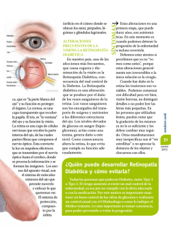
© Copyright 2026