
BrdU-HOECHST-ETHIDIUM BROMIDE (EB) QUENCHING
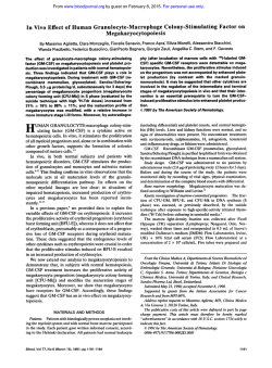
From www.bloodjournal.org by guest on February 6, 2015. For personal use only.
289
CORRESPONDENCE
BrdU-HOECHST-ETHIDIUM BROMIDE (EB) QUENCHING TECHNIQUE FOR
STUDYING KINETICS OF HEMATOPOIESIS
To the Editor:
In vitro stimulatory or inhibitory effects on hematopoietic bone
marrow (BM) progenitors are mostly assessed through evaluation
of the progeny at later stages (ie, colony scoring after 2 to 3 weeks).
The immediate kinetic effects are mostly ignored, although the
evaluation of the progenitor cell cycle status and kinetics of
response can be performed by a variety of methods such as
3H-thymidine incorporation and/or suicide 3H-thymidine technique, viable (Hoechst) or nonviable (EB, PI, etc) DNA staining.
These techniques have the disadvantage of detecting only the S
phase compartment at a given moment: a limitation shared by all
these techniques is indeed that continued growth of cells beyond a
single cell cycle and the kinetics of cell proliferation cannot be
further discriminated.
Therefore, we want to draw attention to the flow cytometric
BrdU/Hoechst propidium iodide (or EB) technique as a powerful
alternative for cell kinetic aaalysis, which is capable of not only
discriminating between cycling and noncycling cells but also
between cells that have replicated once, twice, or more times
within a given time interval: this novel analysis is capable of
showing the proliferative history of cells.
The technique was reviewed recently' and has already found
considerable interest in the immunological (mitogen response) and
pharmaceutical (drug effects) science.
From www.bloodjournal.org by guest on February 6, 2015. For personal use only.
290
CORRESPONDENCE
In our opinion, the wealth of cell kinetic information and the
ease by which this information can be obtained is left underestimated in the field of experimental hematology.
We have adapted some of the experimental conditions (which
were originally developed for mitogen-stimulated lymphocyte proliferation) to make the technique more suitable to analyze in
greater detail growth factor-induced proliferative response of
hematopoieticprogenitors (ie, by loweringthe BrdU concentration
of 5 kmol/L and using serum-deprived conditions). We here
present two examples that illustrate the power of this technique for
analysisof hematopoietic cell kinetics.
77te first eromple (Fig 1, top) represents the 72-hour culture
status of sorted CD34+ BM progenitors,grown in Ixove's modified
Dulbecco's medium (IMDM) supplemented with either granulocyte colony-stimulating factor (G-CSF) (Fig lA), granulocytemacrophage CSF (GM-CSF) (Fig le). or a combination of both
cytokines (Fig. IC). BrdU was added to the culture medium at
culture set-up and was present until harvest. The Row cytometric
BrdUlHoechst technique significantly discriminates between responders (R2 + R3 + R4). nonresponders (Go, RI), and dying
cells (R5). Within the responder compartment differentiation
between fast responders (R4. after 72 hours they have already
entered the third cell cycle), intermediate rate responders (R3.
second cell cycle), and slow responders (R2,still in the first cell
cycle) is possible. Because the sum of responders to G-CSF
stimulation (6296, Fig 1A) and to GM-CSF stimulation (50%. Fig
1B) is significantly higher (A of 47%) than the responders to
simultaneous G-and GM-CSF stimulation (65%. Fig lC), one can
conclude in a first approximation that the CD34+ compartment in
this particular experiment consists of target cells for both G-and
GM-CSF (47%), target cells for G-CSF alone (62-47 = lS%),
Eh: CD34+
bD: LIN-
+ G-CSF
1
%:
target cells for GM-CSF alone (50-47 = 3%). and cells unresponsive to either G- or GM-CSF (region R1 = 35%. Fig 1C). Comparing the number of cells in the individual cell cycles, it is evident that
the combination of both growth factors speeds up the cell cycle
traverse rate for a significant fraction of the target cells (cells in R4
of Fig 1, A through C).
In contrast to the first example where most BM progenitors start
from an almost synchronous CD34+ sorted fraction, the second
a m p l e (Fig 1. D through F) illustrates a total human BM culture
that is asynchronouslyproliferating after 1 week of culture using a
growth factor cocktail (GM-CSF. erythropoietin (Epo], interleukin-3 [IL-31). The data shown are restricted to all but the mature
cells (gatingon negative cells after CD3, CDl9, CDl lb, and CD14
MAB labeling), and show the cell kinetic status after 18 hours of
BrdU-supplemented culture. Although the analysisof the asynchronous BM culture is more complicated than in the previous
example, one can discriminate (Fig 1D) between GI nonresponders, and proliferating cells in the first (S,Gz/M) and second
cell cycle (Gi and an S' region). Further explanation on interpretation of asynchronousgrowing cultures can be found in references 2
and 3. Figure 1E displays the result after treatment of human BM
cells by cis-platinum. From this data it is evident that inhibition of
proliferation by this chemotherapeutic agent is accomplished by
the slowing down of GI and early and mid S-phase cells. Late S and
G2M cells divide; however, most of them die after division (see
nuclear decay line). However. addition of the putative chemoprotective agent diethyldithiocarbamate (DDTC) alone results in a
similar toxic response of the proliferating BM cells, except a
slightly higher cell viability of dividing late S and GzM cells, which
is shown by the presence of G i cells (Fig 1F).
CD34+ + GM-CSF
hE: LIN- + CIS-PLRTINUM
1
E]C: CD34+ + 0-CSF + GM-CSF
qF: LIN- + DDTC
czm
.i
:i
F
l
-
1
~
n.
1 ->
Fk 1. flow cytometrlc bivariate Hoechat versua EB fluorescema ytognnn from BM progsnlton (sod CD34+cella In A, B, and Cllinerg.
mathfafraction of total BM mononuclear population In D, E, and F) c u b d in the presence of G-CSF (A), GM-CSF (E), 0- GM-CSF (C), or
OM-CSF IL-3 + Epo (D through F). Cells in region Rl(38% in A, 50% in B, 35% in C), R2 (7% in A, 9% in B, 5% in C), R3 (28% in A, 15% in B, 28% in
C), R4 (14% in A 3% in B. 19%in C) and RS (14% in A 23% in B. 13% in C) represent cells in quiescence,in first, second, third cell cycle, or in decay,
+
rerpecttvely.
+
From www.bloodjournal.org by guest on February 6, 2015. For personal use only.
CORRESPONDENCE
Both examples illustrate the potential power of this novel flow
cytometric analysis in unraveling the complexity of the cell kinetic
response of hematopoietic BM cells under influence of a variety of
different agents (growth factors, growth inhibitors, drugs, toxic
compounds, etc): the wealth of early event information and its
refined biologic interpretation (detection and quantification of
subsets of target cells, cell cycle progression rate, cell-cycle
blockage position, cellular stability, etc) may be a valuable supplement to final outcome information (colony scoring). The mild
staining procedure results in little cell loss and allows for simultaneous analysis of intracellular and extracellular epitopes. We also
have proven that this technique can easily be performed, even at
higher resolution, using the relatively inexpensive He-Cd laser
instead of the expensive Argon la~er.4,~
We suggest that this
291
technique becomes more popular and available in in vitro research
of cell cycle kinetics of hematopoiesis.
DIRK R. VAN BOCKSTAELE
FILIP LARDON
HANS-W. SNOECK
MARC E. PEETERMANS
Experimental Hematology
University ofAntwerp (UZA)Edegem, Belgium
MANFRED KUBBIES
Cell Biology
Boehringer Mannheim
Penzberg, Germany
REFERENCES
1. Kubbies M, Hoehn H, Schindler D, Chen YC, Rabinovitch
cell populations by flow cytometry using bromodeoxyuridine label
PS: Cell cycle analysis via BrdU-Hoechst flow cytometryand Hoechst-Propidiumiodide stain. Cytometry 1992 (in press)
4. Van Bockstaele DR, Lardon F, Snoeck H-W, Peetermans
Principles and applications, in Yen A (ed): Flow Cytometry:
ME: BrdU-Hoechst flow cytometry using a 20 mW He-Cd laser as
Advanced Research and Clinical Applications,vol 11. Boca Raton,
UV light source: II/Evaluation of growth factor induced bone marrow
FL, CRC, 1989, p 5
progenitor (CD34+)proliferation. Cytometry Suppl5:54,1991(abstr)
2. Kubbies M, Ormerod MG: Cell cycle analysis of asynchronous cultures using the BrdU/Hoechst-PI flow cytometric method.
5. Kubbies M, Goller B, Van Bockstaele DR: Improved BRDUCytometry Suppl5:77,1991 (abstr)
Hoechst bivariate cell kinetic analysis by Helium-Cadmium single
laser excitation. Cytometry 1992 (in press)
3. Ormerod M, Kubbies M: Cell cycle analysis of asynchronous
From www.bloodjournal.org by guest on February 6, 2015. For personal use only.
1992 80: 289-291
BrdU-Hoechst-ethidium bromide (EB) quenching technique for
studying kinetics of hematopoiesis [letter]
DR Van Bockstaele, F Lardon, HW Snoeck, ME Peetermans and M Kubbies
Updated information and services can be found at:
http://www.bloodjournal.org/content/80/1/289.citation.full.html
Articles on similar topics can be found in the following Blood collections
Information about reproducing this article in parts or in its entirety may be found online at:
http://www.bloodjournal.org/site/misc/rights.xhtml#repub_requests
Information about ordering reprints may be found online at:
http://www.bloodjournal.org/site/misc/rights.xhtml#reprints
Information about subscriptions and ASH membership may be found online at:
http://www.bloodjournal.org/site/subscriptions/index.xhtml
Blood (print ISSN 0006-4971, online ISSN 1528-0020), is published weekly by the American
Society of Hematology, 2021 L St, NW, Suite 900, Washington DC 20036.
Copyright 2011 by The American Society of Hematology; all rights reserved.


© Copyright 2026