
Provisional PDF - BioMed Central
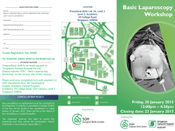
BMC Endocrine Disorders
This Provisional PDF corresponds to the article as it appeared upon acceptance. Fully formatted
PDF and full text (HTML) versions will be made available soon.
Transient hypercortisolism and symptomatic hyperthyroidism associated to
primary hyperparathyroidism in an elderly patient: case report and literature
review
BMC Endocrine Disorders 2015, 15:4
doi:10.1186/1472-6823-15-4
Chiara Sabbadin ([email protected])
Gabriella Donà ([email protected])
Luciana Bordin ([email protected])
Maurizio Iacobone ([email protected])
Valentina Camozzi ([email protected])
Caterina Mian ([email protected])
Decio Armanini ([email protected])
ISSN
Article type
1472-6823
Case report
Submission date
13 August 2014
Acceptance date
15 January 2015
Publication date
28 January 2015
Article URL
http://www.biomedcentral.com/1472-6823/15/4
Like all articles in BMC journals, this peer-reviewed article can be downloaded, printed and
distributed freely for any purposes (see copyright notice below).
Articles in BMC journals are listed in PubMed and archived at PubMed Central.
For information about publishing your research in BMC journals or any BioMed Central journal, go to
http://www.biomedcentral.com/info/authors/
© 2015 Sabbadin et al.
This is an Open Access article distributed under the terms of the Creative Commons Attribution License (http://creativecommons.org/licenses/by/4.0), which
permits unrestricted use, distribution, and reproduction in any medium, provided the original work is properly credited. The Creative Commons Public Domain
Dedication waiver (http://creativecommons.org/publicdomain/zero/1.0/) applies to the data made available in this article, unless otherwise stated.
Transient hypercortisolism and symptomatic
hyperthyroidism associated to primary
hyperparathyroidism in an elderly patient: case
report and literature review
Chiara Sabbadin1*
*
Corresponding author
Email: [email protected]
Gabriella Donà2
Email: [email protected]
Luciana Bordin2
Email: [email protected]
Maurizio Iacobone3
Email: [email protected]
Valentina Camozzi1
Email: [email protected]
Caterina Mian1
Email: [email protected]
Decio Armanini1
Email: [email protected]
1
Department of Medicine-Endocrinology, University of Padua, Via Ospedale
105, 35128 Padua, Italy
2
Department of Molecular Medicine-Biological Chemistry, University of Padua,
Padua, Italy
3
Minimally Invasive Endocrine Surgery Unit, Department of Surgery, Oncology
and Gastroenterology, University of Padua, Padua, Italy
Abstract
Background
Primary hyperparathyroidism (PHPT) is often found on routine blood tests, at a relatively
asymptomatic stage. However many studies suggest different systemic effects related to
PHPT, which could be enhanced by an abnormal cortisol release due to chronic stress of
hyperparathyroidism. Being PHPT frequently found in the 6th to 7th decade of life, a careful
and multifaceted approach should be taken.
Case presentation
We report the case of an elderly patient with symptomatic PHPT and incidental pulmonary
embolism. He was treated with hydration, zoledronic acid, cinacalcet and high-dose
unfractionated heparin. Parathyroid surgery was successfully performed, but patient’s
conditions suddenly worsened because of a transient thyrotoxicosis, probably induced by a
previous exposure to iodine load and/or thyroid surgical manipulation. A short-term treatment
with beta-blockers was introduced for symptomatic relief. The patient also presented a
transient hypercortisolism with elevated ACTH, likely due to stress related not only to aging
and hospitalization but also to PHPT, resolved only four months after parathyroid surgery.
Conclusion
Chronic hyperparathyroidism has been linked with increased all-cause mortality. A functional
chronic hypercortisolism could be established, enhancing PHPT related disorders. Only
parathyroid surgery has been demonstrated to cure PHPT and complications related, showing
similar outcome between older and younger patients. However, the management of postoperative period should be more careful in fragile patients. In particular, the early diagnosis
and treatment of a transient post-operative thyrotoxicosis could improve recovery. Due to the
increase in prevalence and the evidence of many related complications even in asymptomatic
PHPT, expert opinion-based guidelines for surgical treatment of PHPT should be developed
especially for elderly patients.
Keywords
Primary hyperparathyroidism, Elderly, Transient hyperthyroidism, Iodinated-contrast induced
thyrotoxicosis, Functional hypercortisolism
Background
Primary hyperparathyroidism (PHPT) is the third most common endocrinopathy seen today,
frequently found in the 6th to 7th decade of life. PHPT mainly is a sporadic disorder, caused in
85% of the cases by a single adenoma, in 15% by multi-gland disease and rarely by
parathyroid carcinoma. Less than 10% of cases are inheritable, often associated with multigland hyperplasia. The most common presentation of PHPT is an asymptomatic
hypercalcemia, incidentally found on routine blood tests. However, many patients may suffer
from minimal symptoms such as asthenia, constipation, polyuria, hypertension and neuropsychiatric complications [1]. In some cases a renal colic is the first presentation of the
disease. In older patients, concomitant morbidity and poly-pharmacotherapy may worsen
symptoms and complications, and impact the management of PHPT [2].
In this paper we report a complex case of an elderly patient with symptomatic PHPT
associated with a functional transient hypercortisolism, resolved only after parathyroid
surgery. The post-operative period was also characterized by a symptomatic transient
thyrotoxicosis, probably induced by previous exposure to iodine load and/or thyroid surgical
manipulation. The early diagnosis and treatment of this condition improved final outcome.
Case report
An 80-year old man was admitted to a general hospital for polyuria, vomiting, weight loss,
worsening asthenia, myalgia and progressive cognitive impairment. He had a personal history
of hypertension and type 2 diabetes, treated with losartan and metformin respectively. On
physical examination he was sleepy, apyretic, hypertensive (upright blood pressure 150/100
mmHg) and tachycardic (100 beats for minute). He did complain of dyspnea, dry skin and
mucosa and muscle weakness, without bone pain and neurological alterations. Biochemical
assays revealed hypernatremia (149 mmol/L), severe hypercalcemia (4.08 mmol/L),
hypophosfatemia (0.62 mmol/L), elevated levels of PTH (252 ng/L), reduced vitamin D (32
nmol/L) and slight renal failure (urea 8.7 mmol/L, creatinine 112 µmol/L). Blood count, liver
and thyroid function were normal (Table 1). The electrocardiogram did not show remarkable
signs of hypercalcemia. A cranial computed tomography (CT) scan excluded acute
cerebrovascular events. A CT pulmonary angiography detected partial thrombosis in three
segmental branches of the right upper lobe pulmonary artery. Doppler ultrasound (US)
revealed a deep vein thrombosis of the left posterior tibial vein. The patient was treated with
isotonic saline hydration, furosemide, supplementation of vitamin D and an injection of
zoledronate 4 mg, with a mild improvement of hypercalcemia and related symptoms. Daily
high-dose unfractionated heparin was also administered. The patient was then transferred to
our Endocrine Unit and treated with cinacalcet, with decrease of PTH, calcemia and calciuria
values. Amlodipine and insulin were also added for worsening hypertension and diabetes.
Neck US revealed an enlarged thyroid with normal vascular pattern and at the lower pole of
the left thyroid lobe a hypoechoic vascular nodule (14.7x10.5x9 mm), consistent with
enlarged parathyroid. A sesta-MIBI scintigraphy showed a homogeneous tracer uptake over
the thyroid in the early images and a remaining modest uptake in the lower left thyroid lobe
in the later images; no other abnormal or ectopic uptake was found. Dual-energy x-ray
absorptiometry revealed an osteopenia; all the radiological exams did not find brown tumors.
Because of multiple co-morbidities, further investigations were performed to exclude other
endocrine disorders: urinary metanephrine and normetanephrine values were normal, while
plasma morning ACTH and daily urinary free cortisol were increased with impaired circadian
cortisol rhythm, evaluated in two different measurements; serum cortisol after 1 mg overnight
dexamethasone administration was not suppressed (Table 1). Direct abdomen CT was
negative for adrenal diseases. Pituitary magnetic resonance imaging (MRI) evidenced a round
mass, about 4 mm, without enhancement after gadolinium injection, in the left lateral portion
of adenohypophysis, consistent with microadenoma. Screening for mutations of MEN genes
was negative. The other pituitary hormones were normal. Because of the patient’s general
conditions and a probably stress-induced hypercortisolism, no other investigations were
performed, giving priority to the surgical resolution of PHPT. Bilateral neck exploration was
performed with removal of the upper right parathyroid and both the lower and the upper left
parathyroid gland, with quick decrease of intra-operative PTH (from 1296 to 39 ng/L). The
histological diagnosis was consistent with multi-glandular hyperplasia. Laboratory tests
showed a decrease of calcium and PTH levels, treated with oral calcium and calcitriol.
However, four days after surgery the patient developed a sinusal tachycardia, mild heart
failure and agitation alternating with stupor, without evidence of infection nor of volemic
imbalance. Further investigation revealed suppressed TSH, elevated free T4 and T3 values
(Table 1), with undetectable anti-thyroid and TSH receptor antibodies. Thyroid palpation was
not painful. The post-operatory 99mTcO4 scintigraphy showed a reduced tracer uptake over
the thyroid, especially in the lower left thyroid lobe, consistent with an inflammatory area
(Figure 1). Suspecting interference by the iodinated contrast used for pulmonary angio-CT
about a month before, urinary iodine excretion was measured, resulting elevated (935 µg/L,
normal range 100–200). Considering a possible dual pathogenesis of thyrotoxicosis, both
destructive and iodine-induced, the patient was treated with atenolol for symptomatic relief.
Two months after surgery, thyroid function and ioduria were normal and beta-blocker was
progressively stopped; 1 mg overnight dexamethasone test (DST) was still pathological. After
other two months, calcium-phosphate balance was normal and serum PTH was near the lower
limit of normal, therefore calcium and calcitriol supplementation were continued. Adrenal
function was finally normalized with adequate cortisol suppression after 1 mg overnight DST
(Table 1). Pituitary MRI confirmed the presence of a microadenoma, compatible with a nonsecretory incidentaloma, in careful biochemical and radiological follow-up.
Table 1 Principal biochemical parameters of the patient before and after surgery for
primary hyperparathyroidism
Parameter
Normal
range
2,1-2,5
0,8-1,4
5-27
10-50
2,6-15,3
0,1-5,2
30-193
<50
Ca (mmol/L)
P (mmol/L)
PTH (ng/L)
ACTH (ng/L)
morning salivary cortisol (ng/mL)
late-night salivary cortisol (ng/mL)
daily urinary cortisol (nmol/24 h)
serum cortisol after 1 mg DST
(nmol/L)
TSH (mIU/L)
0,2-4
FT4 (pmol/L)
9-22
FT3 (pmol/L)
3,9-6,8
DST: dexamethasone soppression test.
1 month before
surgery
4,2
0,6
252
128
13,1
15,8
557
223
4 days after
surgery
2,5
0,7
<4
126
281
-
4 months after
surgery
2,2
1,1
12
38
12,2
3,4
80
30
1,04
19,9
3,9
0,03
26,9
10,4
1,17
18,7
4,1
Figure 1 99mTcO4 scintigraphy, performed 4 days after parathyroid surgery, evidences
a reduced uptake in total thyroid tissue, especially in the lower left lobe.
Discussion
We report an elderly patient presenting symptomatic hypercalcemia with moderate
hemodynamic and neuropsychiatric failure. The detection of hypercalcemia and elevated
PTH levels was diagnostic of PHPT. The biochemical/clinical presentation could also raise
the suspicion of parathyroid carcinoma, which was excluded by the histological
examinations. Rehydration was the first measure to take in this patient, not only to correct
dehydration and improve renal failure, but also to dilute calcium excretion. Bisphosphonate
administration was effective in reducing calcium levels and bone resorption, in particular
recent controlled trials demonstrated the superiority of zoledronate compared with previous
treatments [3]. The addition of cinacalcet may be useful in the elderly or in not-surgical
candidates, being well tolerated [4]. Parathyroid surgery is the only definite cure for PHPT,
but the risks and benefits of surgery should be extensively considered in the elderly, given
their more fragile state and co-morbidities [5]. Preoperative imaging with ultrasonography
and scintigraphy may be helpful before elective surgery and in suspected parathyroid
carcinoma, even if their sensitivity drops in detecting multi-glandular disease, as happened in
our case report.
Our finding of a transient thyrotoxicosis after parathyroid surgery could be due to a dual
pathogenesis: a destructive thyroiditis and/or an iodine-induced hyperthyroidism. The first
condition is fairly unknown and underestimated since the symptoms could be masked by
other postoperative events [6]. Thyrotoxicosis seems to be related to an increased release of
thyroid hormones and/or autoantigen during surgical manipulation, which could reactivate
underlying autoimmune thyroiditis [7]. It could be influenced by other mechanisms, like
preoperative hypercalcemic setting, pre-existing goiter and difficult parathyroid glands
identification during surgical exploration [8]. Our patient had an euthyroid goiter, without
abnormal MIBI-uptake in preoperative investigations nor previous or underlying autoimmune
thyroiditis. Retrospectively, the only apparent risk factors were the goiter, the previous
pronounced hypercalcemic condition and the occasional finding of a multi-gland disease.
The second possible cause of transient thyrotoxicosis could be related to the previous
iodinated contrast media exposure, leading to hypersecretion of thyroid hormones. This
phenomenon, known as the Jod-Basedow effect, usually develops over 2 to 12 weeks,
typically in old patients with underlying thyroid disease or living in areas of iodine
deficiency. Exposure to a large iodine load can also cause acute destructive thyroiditis in
people without pre-existing thyroid disorders [9]. TcO4-scintigraphy could not discriminate
the cause of hyperthyroidism, since pertechnetate is trapped by thyroid, but not organified
and the resulting tracer uptake may be reduced. As happened in our case, the assessment of
urinary iodine concentration may be helpful [10]. Some researchers have investigated the
incidence and the role of prophylactic measures to reduce the risk of iodine-induced
thyrotoxicosis, without conclusive findings [11]. In our case the concomitant neck surgery
could have been a precipitating factor in the pathogenesis of hyperthyroidism.
Both these conditions are usually self-limited and anti-thyroid drugs are not indicated.
However, a short-term treatment with beta-blockers could be required for symptomatic relief,
especially in fragile patients.
Since chronic hyperparathyroidism has been linked with increased all-cause mortality, in
asymptomatic and elderly patients the optimal timing for parathyroidectomy is controversial
[12]. Cardiovascular complications are the leading cause of this increased mortality [13],
linked not only to mineral homeostasis disruption but also to a direct effect of PTH on
cardiovascular structures [14]. The complexity of PTH functions is further highlighted by
data suggesting a bidirectional link between PTH and the renin-angiotensin-aldosteronesystem, playing a synergic role in enhancing metabolic and cardiovascular complications
[15]. Several studies have also evidenced an altered hypotalamic-pituitary-adrenal (HPA)
axis in PHPT, potentially contributing to cortical bone damage [16]. In vitro evidence
supports a stimulatory effect of PTH on cortisol secretion [17] and of calcemia on ACTH
release [18]. In vivo data show a hypercortisolism, loss of circadian rhythm and lack of
cortisol suppression after low-dose DST in PHPT [19], which are not always recovered after
surgical cure, as happened in our case.
Alteration of cortisol expression and its circadian variability are also typical of aging,
hospitalization, psychiatric and stress conditions [20]. False-positive results of the 1 mg DST
could be influenced by absorption, liver or renal alterations and the use of alcohol or drugs
inducing CYP3A4. Being PHPT a long-standing disease frequently affecting old patients, the
activation of adrenal function seems to recall a functional hypercortisolism due to chronic
stress, which could be preserved by aging and other co-morbidities, enhancing the damaging
effects of prolonged exposure to stress hormones [21].
In our case, HPA axis was evaluated because of multiple co-morbidities of the patient. The
data suggestive of Cushing’s syndrome were the incidental pulmonary thrombo-embolism,
the uncontrolled hypertension and diabetes and the evidence of a pituitary microadenoma;
however, the absence of typical stigmata and the presence of many confounding factors made
the diagnosis uncertain. After discharge, further investigations excluded a Cushing’s
syndrome, but the slow normalization of HPA axis only after four months seems to be related
to the resolution not only of the acute stressful condition but also of PHPT.
This complex picture suggests that parathyroid surgery may improve the prognosis,
normalizing also HPA axis, which could contribute to PHPT related pathologies, such as
bone metabolism, psychiatric, metabolic and cardiovascular disorders [22].
Conclusions
There is ample evidence that PHPT is associated with HPA axis alterations, which could be
involved in the increased metabolic, cardiovascular and neuropsychiatric complications. A
wide variety of medical therapies are available; however, only parathyroid surgery has been
demonstrated to cure PHPT and complications related, showing similar outcome between
older and younger patients. A transient thyrotoxicosis is a fairly underestimated condition,
which could be secondary to previous iodinated contrast media exposure or to thyroid
manipulation during parathyroid surgery. The early diagnosis and treatment of this
complication may increase a successful recovery, especially in fragile patients.
In conclusion, due to the increase in prevalence and the evidence of many related
complications even in asymptomatic PHPT, expert opinion-based guidelines for surgical
treatment of PHPT should be developed especially for elderly patients.
Consent
Written informed consent was obtained from the patient for publication of this Case report
and any accompanying images. A copy of the written consent is available for review by the
Editor of this journal.
Abbreviations
PHPT, Primary hyperparathyroidism; CT, Computed tomography; US, Ultrasound; MRI,
Magnetic resonance imaging; DST, Dexamethasone test; HPA, Hypotalamic-pituitaryadrenal
Competing interests
The authors declare that they have no competing interests.
Authors’ contributions
CS and DA treated the patient and drafted the article. GD and LB participated in the analysis
and interpretation of data. MI operated the patient and together with VC and CM critically
revised the manuscript. All authors read and approved the final manuscript.
Acknowledgements
We acknowledge Ms. Sophie Armanini for English revision.
References
1. Pyram R, Mahajan G, Gliwa A. Primary hyperparathyroidism: skeletal and non-skeletal
effects, diagnosis and management. Maturitas. 2011;70:246–55.
2. Conroy S, Moulias S, Wassif WS. Primary hyperparathyroidism in the older person. Age
Ageing. 2003;32:571–8.
3. Smith MR. Zoledronic acid to prevent skeletal complications in cancer: corroborating the
evidence. Cancer Treat Rev. 2005;31 Suppl 3:19–25.
4. Jacobs L, Samson MM, Verhaar HJ, Koek HL. Therapeutic challenges in elderly patients
with symptomatic hypercalcaemia caused by primary hyperparathyroidism. Neth J Med.
2012;70:35–8.
5. Jannasch O, Voigt C, Reschke K, Lippert H, Mroczkowski P. Comparison of outcome
between older and younger patients following surgery for primary hyperparathyroidism. Pol
Przegl Chir. 2013;85:598–604.
6. Rudofsky Jr G, Grafe IA, Metzner C, Leowardi C, Fohr B. Transient post-operative
thyrotoxicosis after parathyroidectomy. Med Sci Monit. 2009;15:CS41–3.
7. Walfish PG, Caplan D, Rosen B. Postparathyroidectomy transient thyrotoxicosis. J Clin
Endocrinol Metab. 1992;75:224–7.
8. Lindblom P, Valdemarsson S, Westerdahl J, Tennval J, Bergenfelz A. Hyperthyroidism
after surgery for primary hyperparathyroidism. Langenbeck’s Arch Surg. 1999;384:568–75.
9. Calvi L, Daniels GH. Acute thyrotoxicosis secondary to destructive thyroiditis associated
with cardiac catheterization contrast dye. Thyroid. 2011;21:443–9.
10. Bahn RS, Burch HB, Cooper DS, Garber JR, Greenlee MC, Klein I, et al.
Hyperthyroidism and other causes of thyrotoxicosis: management guidelines of the American
Thyroid Association and American Association of Clinical Endocrinologists. Endocr Pract.
2011;17:456–520. Erratum in: Endocr Pract. 2013;19:384.
11. Hintze G, Blombach O, Fink H, Burkhardt U, Kobberling J. Risk of iodine-induced
thyrotoxicosis after coronary angiography: an investigation in 788 unselected subjects. Eur J
Endocrinol. 1999;140:264–7.
12. Yu N, Donnan PT, Leese GP. A record linkage study outcomes in patients with mild
primary hyperparathyroidism: The Parathyroid Epidemiology and Audit Research Study
(PEARS). Clin Endocrinol (Oxf). 2011;75:169–76.
13. Pilz S, Tomaschitz A, Drechsler C, Ritz E, Boehm BO, Grammer TB, et al. Parathyroid
hormone level is associated with mortality and cardiovascular events in patients undergoing
coronary angiography. Eur Heart J. 2010;31:1591–8.
14. Osto E, Fallo F, Pelizzo MR, Maddalozzo A, Sorgato N, Corbetti F, et al. Coronary
microvascular dysfunction induced by primary hyperparathyroidism is restored after
parathyroidectomy. Circulation. 2012;126:1031–9.
15. Sabbadin C, Cavedon E, Zanon M, Iacobone M, Armanini D. Resolution of hypertension
and secondary aldosteronism after surgical treatment of primary hyperaldosteronism. J
Endocrinol Invest. 2013;36:665–6.
16. Gianotti L, Tassone F, Pia A, Bovio S, Reimondo G, Visconti G, et al. May an altered
hypotalamo-pituitary-adrenal axis contribute to cortical bone damage in primary
hyperparathyroidism? Calcif Tissue Int. 2009;84:425–9.
17. Mazzocchi G, Aragona F, Malendowicz LK, Nussdorfer GG. PTH and PTH-related
peptide enhance steroid secretion from human adrenocortical cells. Am J Physiol Endocrinol
Metab. 2001;280:E209–13.
18. Fuleihan GE-H, Brown EM, Gleason R, Scott J, Adler GK. Calcium modulation of
adrenocorticotropin levels in women – a clinical research study. J Clin Endocrinol Metab.
1996;81:932–6.
19. Rajput R, Bhansali A, Bhadada SK, Behera A, Mittal BR, Sialy R, et al. A pilot study on
hypothalamo-pituitary-adrenocortical axis in primary hyperparathyroidism. Indian J Med
Res. 2009;130:418–22.
20. Armanini D. Corticosteroid receptors in lymphocytes: a possible marker of brain
involution? J Steroid Biochem Mol Biol. 1994;49:429–34.
21. Aguilera G. HPA axis responsiveness to stress: implications for healthy aging. Exp
Gerontol. 2011;46:90–5.
22. Tirabassi G, Boscaro M, Arnaldi G. Harmful effects of functional hypercortisolism: a
working hypothesis. Endocrine. 2014;46:370–86.
Figure 1


© Copyright 2026